Myocardial and Atrial Strain Profiles in Pediatric Fontan Patients with Single Left Ventricle Using Two-Dimensional Speckle-Tracking Echocardiography: A Case–Control Study
Abstract
1. Introduction
Speckle-Tracking Echocardiography
2. Materials and Methods
2.1. Study Population
2.2. Echocardiography
2.2.1. Conventional 2D Echocardiographic Examination
2.2.2. Advanced 3D Echocardiographic Examination
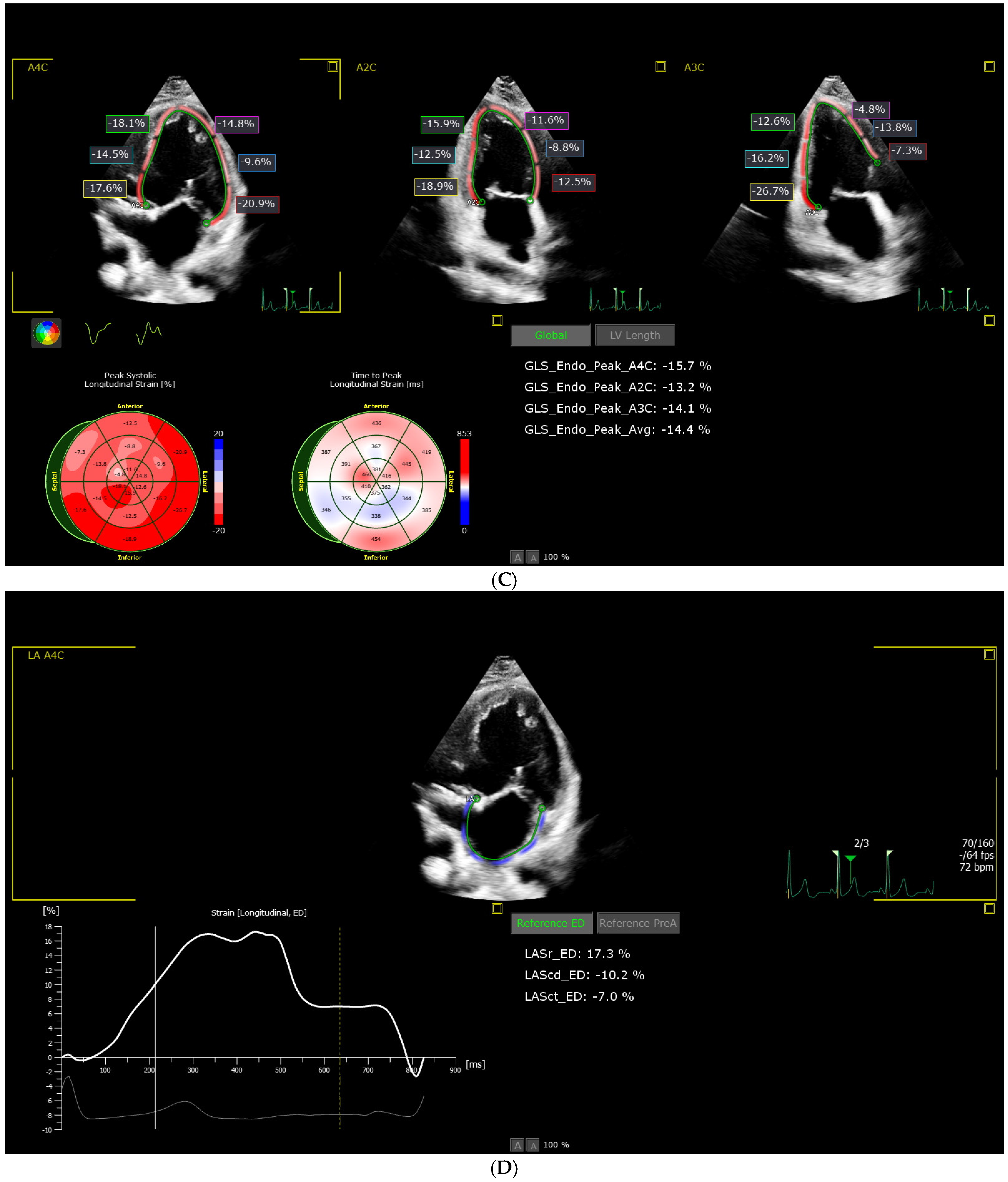
2.2.3. Speckle Tracking Acquisition and Analysis
2.3. Statistical Analysis
2.3.1. Intra-Observer Variability
2.3.2. Objectives
3. Results
3.1. Description of Studied Groups
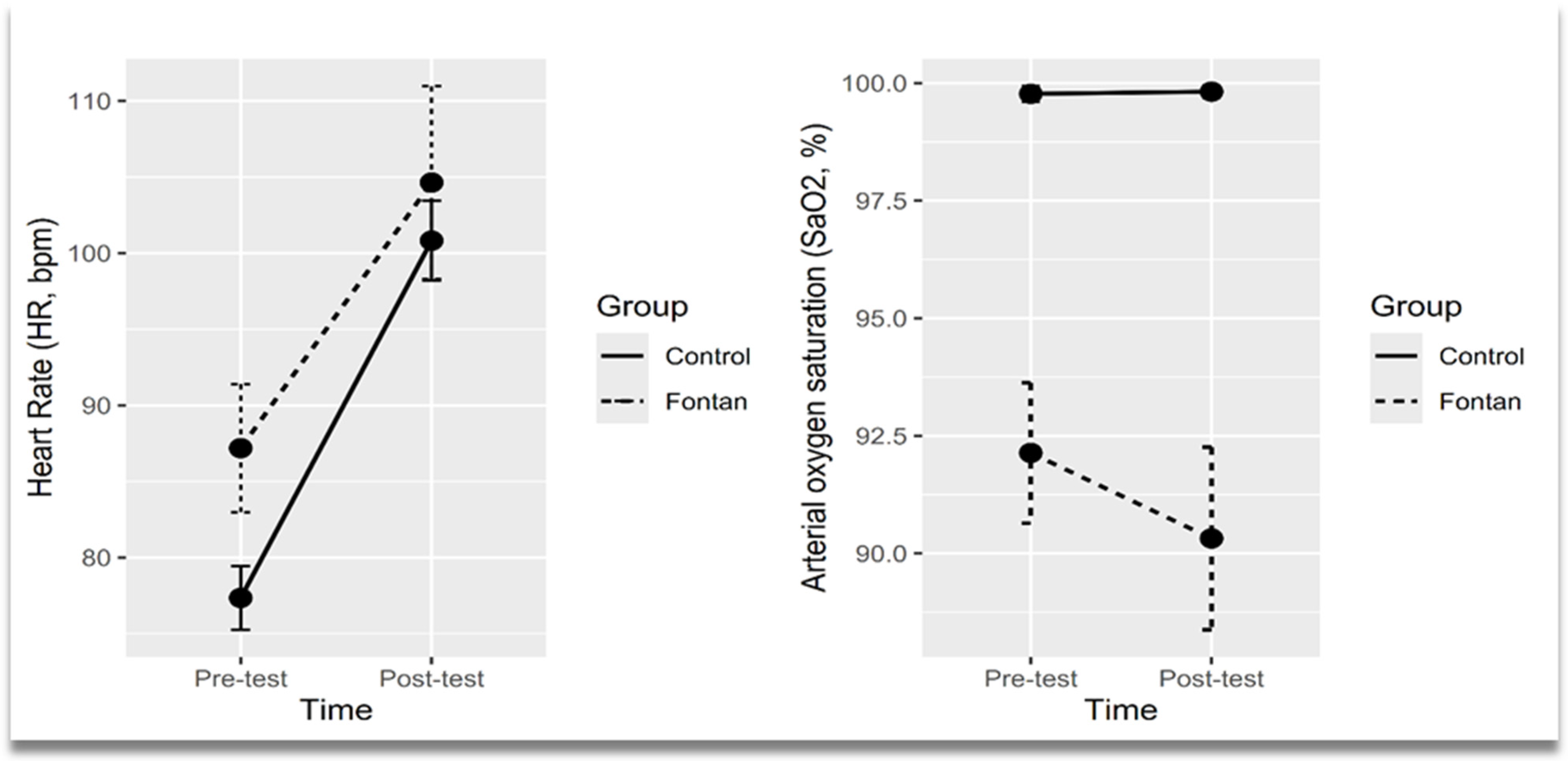
3.2. Evolution of Cardiorespiratory Parameters Assessed Using Six-Minute Walk Test (6 MWT) by Time and Group
3.3. Comparison of Conventional Left Ventricle 2D and 3D Echocardiographic Characteristics Between Fontan Group and Controls
3.4. Comparison of Two-Dimensional Echocardiographic Segmental and Global Left Ventricle Longitudinal Strain Indices in Controls and Fontan Patients
3.5. Corelations Between Conventional, Two-Dimensional Echocardiographic Global Left Ventricle Longitudinal and Left Atrium Strain Indices
4. Discussion
5. Conclusions
Author Contributions
Funding
Institutional Review Board Statement
Informed Consent Statement
Data Availability Statement
Conflicts of Interest
Abbreviations
| 6 MWT | Six-Minute Walk Test |
| A_mi | Peak mitral inflow velocity at atrial contraction (A wave) |
| A`_mi | Mitral annular late diastolic velocity (A` wave) |
| AA | Apical anterior segment (ASE model) |
| AAL | Apical anterolateral segment (ASE model) |
| AIS | Apical inferoseptal segment (ASE model) |
| AL | Apical lateral segment (ASE model) |
| BAL | Basal anterolateral segment (ASE model) |
| BAS | Basal anteroseptal segment (ASE model) |
| BA | Basal anterior segment (ASE model) |
| BI | Basal inferior segment (ASE model) |
| BIL | Basal inferolateral segment (ASE model) |
| BIS | Basal inferoseptal segment (ASE model) |
| MA | Mid anterior segment (ASE model) |
| MAL | Mid anterolateral segment (ASE model) |
| MAS | Mid anteroseptal segment (ASE model) |
| MI | Mid inferior segment (ASE model) |
| MIL | Mid inferolateral segment (ASE model) |
| ANCOVA | Analysis of Covariance |
| AVC | Atrioventricular Canal Defect |
| BSA | Body Surface Area |
| CHD | Congenital Heart Disease |
| CI | Confidence Interval |
| CoAo | Coarctation of the Aorta |
| CPET | Cardiopulmonary Exercise Testing |
| DBP | Diastolic Blood Pressure |
| DILV | Double Inlet Left Ventricle |
| E_mi | Peak mitral inflow velocity during early diastole (E wave) |
| E_mi/A_mi | Ratio between early and late mitral inflow velocities |
| E`_mi | Mitral annular early diastolic velocity (E` wave) |
| EDL | End-Diastolic Longitudinal Diameter |
| ED_mass | End-Diastolic Mass |
| EDV | End-Diastolic Volume |
| EF | Ejection Fraction |
| EMM | Estimated Marginal Mean |
| ESL | End-Systolic Longitudinal Diameter |
| ESV | End-Systolic Volume |
| FC | Functional Class (NYHA) |
| GAM | Generalized Additive Model |
| GSD | Geometric Standard Deviation |
| HR | Heart Rate |
| ICC | Intraclass Correlation Coefficient |
| IQR | Interquartile Range |
| IVCT_mi | Isovolumic Contraction Time |
| IVRT_mi | Isovolumic Relaxation Time |
| LAScd | Left Atrial Conduit Strain |
| LAScd_AC | Left Atrial Conduit Strain measured from P-wave onset |
| LAScd_ED | Left Atrial Conduit Strain measured from R-wave peak |
| LASct | Left Atrial Contractile Strain |
| LASct_AC | Left Atrial Contractile Strain measured from P-wave onset |
| LASct_ED | Left Atrial Contractile Strain measured from R-wave peak |
| LASr | Left Atrial Reservoir Strain |
| LASr_AC | Left Atrial Reservoir Strain measured from P-wave onset |
| LASr_ED | Left Atrial Reservoir Strain measured from R-wave peak |
| LPA | Left Pulmonary Artery |
| LS | Longitudinal Strain |
| LV | Left Ventricle |
| LV_ED mass | Left Ventricular End-Diastolic Mass |
| LV_EDV | Left Ventricular End-Diastolic Volume |
| LV_EF (3D) | Left Ventricular Ejection Fraction by 3D Echocardiography |
| LV_ESV | Left Ventricular End-Systolic Volume |
| LV_GLS | Left Ventricular Global Longitudinal Strain |
| LV_SV | Left Ventricular Stroke Volume |
| MAPSE | Mitral Annular Plane Systolic Excursion |
| NYHA | New York Heart Association |
| PS | Pulmonary Stenosis |
| RPA | Right Pulmonary Artery |
| RV | Right Ventricle |
| RVOT | Right Ventricular Outflow Tract |
| SaO2 | Arterial Oxygen Saturation |
| SBP | Systolic Blood Pressure |
| SD | Standard Deviation |
| S_mi | Mitral annular systolic velocity (S wave) |
| SLV | Single Left Ventricle |
| SRV | Single Right Ventricle |
| STE | Speckle-Tracking Echocardiography |
| SV | Single Ventricle |
| TA | Tricuspid Atresia |
| VSD | Ventricular Septal Defect |
| VO2 | Oxygen Consumption |
References
- Liao, M.; Pan, J.; Liao, T.; Liu, X.; Wang, L. Transthoracic echocardiographic assessment of ventricular function in functional single ventricle: A comprehensive review. Cardiovasc. Ultrasound 2025, 23, 9. [Google Scholar] [CrossRef]
- Kleitsioti, P.; Koulaouzidis, G.; Giannakopoulou, P.; Charisopoulou, D. The role of myocardial strain imaging in the pre and post-operative assessment of patients with single ventricle. Rev. Cardiovasc. Med. 2023, 24, 145. [Google Scholar] [CrossRef]
- Forsha, D.; Risum, N.; Barker, P. Activation delay-induced mechanical dyssynchrony in single-ventricle heart disease. Cardiol. Young 2017, 27, 1390–1391. [Google Scholar] [CrossRef] [PubMed]
- Kowalczyk, M.; Kordybach-Prokopiuk, M.; Marczak, M.; Hoffman, P.; Kowalski, M. The utility of novel STE parameters in echocardiographic assessment of single ventricle after Fontan palliation. Int. J. Cardiol. 2024, 412, 132286. [Google Scholar] [CrossRef] [PubMed]
- Davis, E.K.; Ginde, S.; Stelter, J.; Frommelt, P.; Hill, G.D. Echocardiographic assessment of single-ventricle diastolic function and its correlation to short-term outcomes after the Fontan operation. Congenit. Heart Dis. 2019, 14, 720–725. [Google Scholar] [CrossRef]
- Koopman, L.P.; Geerdink, L.M.; Bossers, S.S.M.; Duppen, N.; Kuipers, I.M.; Harkel, A.D.T.; van Iperen, G.; Weijers, G.; de Korte, C.; Helbing, W.A.; et al. Longitudinal Myocardial Deformation Does Not Predict Single Ventricle Ejection Fraction Assessed by Cardiac Magnetic Resonance Imaging in Children with a Total Cavopulmonary Connection. Pediatr. Cardiol. 2018, 39, 283–293. [Google Scholar] [CrossRef]
- Dorosz, J.L.; Lezotte, D.C.; Weitzenkamp, D.A.; Allen, L.A.; Salcedo, E.E. Performance of 3-dimensional echocardiography in measuring left ventricular volumes and ejection fraction: A systematic review and meta-analysis. J. Am. Coll. Cardiol. 2012, 59, 1799–1808. [Google Scholar] [CrossRef]
- Bunting, K.; Formisano, F.; Green, J.; Steeds, R.; Hudsmith, L.; Clift, P. Assessing Univentricular Function in Adult Fontan Using 3D Echocardiography. Congenit. Heart Dis. 2020, 15, 89–100. [Google Scholar] [CrossRef]
- Badano, L.P.; Dall’Armellina, E.; Monaghan, M.J.; Pepi, M.; Baldassi, M.; Cinello, M.; Fioretti, P.M. Real-time three-dimensional echocardiography: Technological gadget or clinical tool? J. Cardiovasc. Med. 2007, 8, 144–162. [Google Scholar] [CrossRef]
- Zhong, S.-W.; Zhang, Y.-Q.; Chen, L.-J.; Zhang, Z.-F.; Wu, L.-P.; Hong, W.-J. ventricular function and dyssynchrony in children with a functional single right ventricle using real time three-dimensional echocardiography after Fontan operation. Echocardiography 2021, 38, 1218–1227. [Google Scholar] [CrossRef]
- von Hehn, C.A.; Baron, R.; Woolf, C.J. Deconstructing the neuropathic pain phenotype to reveal neural mechanisms. Neuron 2012, 73, 638–652. [Google Scholar] [CrossRef]
- Lang, R.M.; Badano, L.P.; Tsang, W.; Adams, D.H.; Agricola, E.; Buck, T.; Faletra, F.F.; Franke, A.; Hung, J.; De Isla, L.P.; et al. EAE/ASE recommendations for image acquisition and display using three-dimensional echocardiography. Eur. Heart J. Cardiovasc. Imaging 2012, 13, 1–46. [Google Scholar] [CrossRef] [PubMed]
- Trzebiatowska-Krzynska, A.; Swahn, E.; Wallby, L.; Nielsen, N.E.; Carlhäll, C.J.; Engvall, J. Three-dimensional echocardiography to identify right ventricular dilatation in patients with corrected Fallot anomaly or pulmonary stenosis. Clin. Physiol. Funct. Imaging 2021, 41, 51–61. [Google Scholar] [CrossRef] [PubMed] [PubMed Central]
- Simpson, J.; Lopez, L.; Acar, P.; Friedberg, M.K.; Khoo, N.S.; Ko, H.H.; Marek, J.; Marx, G.; McGhie, J.S.; Meijboom, F.; et al. Three-dimensional Echocardiography in Congenital Heart Disease: An Expert Consensus Document from the European Association of Cardiovascular Imaging and the American Society of Echocardiography. J. Am. Soc. Echocardiogr. 2017, 30, 1–27. [Google Scholar] [CrossRef] [PubMed]
- Dragulescu, A.; Grosse-Wortmann, L.; Fackoury, C.; Mertens, L. Echocardiographic assessment of right ventricular volumes: A comparison of different techniques in children after surgical repair of tetralogy of Fallot. Eur. Heart J. Cardiovasc. Imaging 2012, 13, 596–604. [Google Scholar] [CrossRef] [PubMed]
- Gherbesi, E.; Gianstefani, S.; Angeli, F.; Ryabenko, K.; Bergamaschi, L.; Armillotta, M.; Guerra, E.; Tuttolomondo, D.; Gaibazzi, N.; Squeri, A.; et al. Myocardial strain of the left ventricle by speckle tracking echocardiography: From physics to clinical practice. Echocardiography 2024, 41, e15753. [Google Scholar] [CrossRef] [PubMed]
- Hakim, K.; Mekki, N.; Benothmen, R.; Malek, M.; Abdelkader, J.; Hela, M.; Mizouni, H.; Fatma, O. Assessment of ventricular function after total cavo-pulmonary derivation in adult patients: Interest of global longitudinal strain. J. Cardiovasc. Thorac. Res. 2023, 15, 262–268. [Google Scholar] [CrossRef]
- Steflik, D.; Butts, R.J.; Baker, G.H.; Bandisode, V.; Savage, A.; Atz, A.M.; Chowdhury, S.M. A preliminary comparison of two-dimensional speckle tracking echocardiography and pressure-volume loop analysis in patients with Fontan physiology: The role of ventricular morphology. Echocardiography 2017, 34, 1353–1359. [Google Scholar] [CrossRef] [PubMed]
- Singh, G.K.; Cupps, B.; Pasque, M.; Woodard, P.K.; Holland, M.R.; Ludomirsky, A. Accuracy and reproducibility of strain by speckle tracking in pediatric subjects with normal heart and single ventricular physiology: A two-dimensional speckle-tracking echocardiography and magnetic resonance imaging correlative study. J. Am. Soc. Echocardiogr. 2010, 23, 1143–1152. [Google Scholar] [CrossRef]
- Voigt, J.U.; Pedrizzetti, G.; Lysyansky, P.; Marwick, T.H.; Houle, H.; Baumann, R.; Pedri, S.; Ito, Y.; Abe, Y.; Metz, S.; et al. Definitions for a common standard for 2D speckle tracking echocardiography: Consensus document of the EACVI/ASE/industry task force to standardize deformation imaging. Eur. Heart J. Cardiovasc. Imaging 2015, 16, 1–11. [Google Scholar] [CrossRef]
- Toro-Salazar, O.H.; Ferranti, J.; Lorenzoni, R.; Walling, S.; Mazur, W.; Raman, S.V.; Davey, B.T.; Gillan, E.; O’loughlin, M.; Klas, B.; et al. Feasibility of Echocardiographic Techniques to Detect Subclinical Cancer Therapeutics-Related Cardiac Dysfunction among High-Dose Patients When Compared with Cardiac Magnetic Resonance Imaging. J. Am. Soc. Echocardiogr. 2016, 29, 119–131. [Google Scholar] [CrossRef]
- Shiraga, K.; Ozcelik, N.; Harris, M.A.; Whitehead, K.K.; Biko, D.M.; Partington, S.L.; Fogel, M.A. Imposition of Fontan physioloSgy: Effects on strain and global measures of ventricular function. J. Thorac. Cardiovasc. Surg. 2021, 162, 1813–1822.e3. [Google Scholar] [CrossRef] [PubMed]
- Rios, R.; Ginde, S.; Saudek, D.; Loomba, R.S.; Stelter, J.; Frommelt, P. Quantitative echocardiographic measures in the assessment of single ventricle function post-Fontan: Incorporation into routine clinical practice. Echocardiography 2017, 34, 108–115. [Google Scholar] [CrossRef]
- Abdulkarim, M.; Loomba, R.S.; Zaidi, S.J.; Li, Y.; Wilson, M.; Roberson, D.; Farias, J.S.; Flores, S.; Villarreal, E.G.; Husayni, T. Echocardiographic strain to predict need for transplant or mortality in Fontan patients with hypoplastic left heart syndrome. Pediatr. Cardiol. 2023, 45, 1475–1484. [Google Scholar] [CrossRef]
- Borrelli, N.; Di Salvo, G.; Sabatino, J.; Ibrahim, A.; Avesani, M.; Sirico, D.; Josen, M.; Penco, M.; Fraisse, A.; Michielon, G. Serial changes in longitudinal strain are associated with outcome in children with hypoplastic left heart syndrome. Int. J. Cardiol. 2020, 317, 56–62. [Google Scholar] [CrossRef] [PubMed]
- Lopez, C.; Mertens, L.; Dragulescu, A.; Friedberg, M.K.; Hunter, K.; Maria, M.V.D.; Landeck, B.; Younoszai, A. Strain and Rotational Mechanics in Children With Single Left Ventricles After Fontan. J. Am. Soc. Echocardiogr. 2018, 31, 1297–1306. [Google Scholar] [CrossRef] [PubMed]
- Khoo, N.S.; Smallhorn, J.F.; Kaneko, S.; Kutty, S.; Altamirano, L.; Tham, E.B. The assessment of atrial function in single ventricle hearts from birth to Fontan: A speckle-tracking study by using strain and strain rate. J. Am. Soc. Echocardiogr. 2013, 26, 756–764. [Google Scholar] [CrossRef]
- Li, S.J.; Wong, S.J.; Cheung, Y.F. Atrial and ventricular mechanics in patients after Fontan-type procedures: Atriopulmonary connection versus extracardiac conduit. J. Am. Soc. Echocardiogr. 2014, 27, 666–674. [Google Scholar] [CrossRef]
- Veldtman, G.; Possner, M.; Mohty, D.; Issa, Z.; Alsaleh, M.; AlMarzoog, A.T.; Emmanual, S.; Salam, Y.; AlHabdan, M.S.; Alsaied, T.; et al. Atrial function in the Fontan circulation: Comparison with invasively assessed systemic ventricular filling pressure. Int. J. Cardiovasc. Imaging 2021, 37, 2651–2660. [Google Scholar] [CrossRef]
- Peck, D.; Alsaied, T.; Pradhan, S.; Hill, G. Atrial Reservoir Strain is Associated with Decreased Cardiac Index and Adverse Outcomes Post Fontan Operation. Pediatr. Cardiol. 2021, 42, 307–314. [Google Scholar] [CrossRef]
- Rato, J.; Mendes, S.C.; Sousa, A.; Lemos, M.; Martins, D.S.; Anjos, R. The Influence of Atrial Strain on Functional Capacity in Patients with the Fontan Circulation. Pediatr. Cardiol. 2020, 41, 1730–1738. [Google Scholar] [CrossRef]
- Lai, W.W.; Geva, T.; Shirali, G.; Frommelt, P.C.; Humes, R.A.; Brook, M.M.; Pignatelli, R.H.; Rychik, J. Guidelines and standards for performance of a pediatric echocardiogram: A report from the task force of the Pediatric Council of the American Society of Echocardiography. J. Am. Soc. Echocardiogr. 2006, 19, 1413–1430. [Google Scholar] [CrossRef]
- Hove, D.T.; Jorgensen, T.D.; van der Ark, L.A. Updated guidelines on selecting an intraclass correlation coefficient for intra-observer reliability, with applications to incomplete observational designs. Psychol. Methods 2024, 29, 967–979. [Google Scholar] [CrossRef] [PubMed]
- Koo, T.K.; Li, M.Y. A Guideline of Selecting and Reporting Intraclass Correlation Coefficients for Reliability Research. J. Chiropr. Med. 2016, 15, 155–163. [Google Scholar] [CrossRef] [PubMed]
- Rychik, J.; Atz, A.M.; Celermajer, D.S.; Deal, B.J.; Gatzoulis, M.A.; Gewillig, M.H.; Hsia, T.-Y.; Hsu, D.T.; Kovacs, A.H.; McCrindle, B.W.; et al. Evaluation and management of the child and adult with Fontan circulation: A scientific statement from the American Heart Association. Circulation 2019, 140, e234–e284. [Google Scholar] [CrossRef]
- Gewillig, M.; Brown, S.C. The Fontan circulation after 45 years: Update in physiology. Heart 2016, 102, 1081–1086. [Google Scholar] [CrossRef]
- Meneguzzo, G.; Costola, G.; Constantine, A.; Ministeri, M.; Rafiq, I.; Pires, A.; Kempny, A.; Babu-Narayan, S.; Gatzoulis, M.; Dimopoulos, K. Peak oxygen uptake on cardio pulmonary exercise testing predicts mortality in adult Fontan patients. Eur. Heart J. 2020, 41 (Suppl. S2), ehaa946.2178. [Google Scholar] [CrossRef]
- Ohuchi, H.; Negishi, J.; Noritake, K.; Hayama, Y.; Sakaguchi, H.; Miyazaki, A.; Kagisaki, K.; Yamada, O. Prognostic value of exercise variables in 335 patients after the Fontan operation: A 23-year single-center experience of cardiopulmonary exercise testing. Congenit. Heart Dis. 2015, 10, 105–116. [Google Scholar] [CrossRef]
- Campbell, M.J.; Quartermain, M.D.; Cohen, M.S.; Faerber, J.; Okunowo, O.; Wang, Y.; Capone, V.; DiFrancesco, J.; Mercer-Rosa, L.; Goldberg, D.J. Longitudinal changes in echocardiographic measures of ventricular function after Fontan operation. Echocardiography 2020, 37, 1443–1448. [Google Scholar] [CrossRef]
- Mertens, L.; Friedberg, M.K. Imaging the single ventricle: Strategies for comprehensive assessment. Heart 2008, 94, 882–890. [Google Scholar]
- Moscatelli, S.; Borrelli, N.; Sabatino, J.; Leo, I.; Avesani, M.; Montanaro, C.; Di Salvo, G. Role of Cardiovascular Imaging in the Follow-Up of Patients with Fontan Circulation. Children 2022, 9, 1875. [Google Scholar] [CrossRef] [PubMed]
- Kiesewetter, C.H.; Sheron, N.; Vettukattill, J.J.; Hacking, N.; Stedman, B.; Millward-Sadler, H.; Haw, M.; Cope, R.; Salmon, A.P.; Sivaprakasam, M.C.; et al. Hepatic changes in the failing Fontan circulation. Heart 2007, 93, 579–584. [Google Scholar] [CrossRef] [PubMed]
- Fernandes, S.M.; Alexander, M.E.; Graham, D.A.; Rychik, J.; Khairy, P.; Clair, M.; Rodriguez, E.; Pearson, D.D.; Landzberg, M.J. Exercise testing identifies patients at increased risk for morbidity and mortality following Fontan surgery. Congenit. Heart Dis. 2023, 2, 294–303. [Google Scholar] [CrossRef] [PubMed]


| Variable | Fontan Group (n = 22) | Control Group (n = 44) | p-Value |
|---|---|---|---|
| Age at assessment (years) | 10.59 (3.39) | 10.48 (3.49) | 0.9042 (a) |
| Age at Fontan procedure (years) | 4 [4; 5] | NA | NA |
| Male sex (n, %) | 14 (63.6) | 28 (63.6) | 1.000 (c) |
| Heart rate (HR, bpm) | 85.45 (9.71) | 78.27 (6.82) | 0.0009 * (a) |
| Body mass index (BMI, kg/m2) | 16.53 (3.53) | 17.73 (2.30) | 0.0173 * (a) |
| BMI Z-scores | −0.91 (1.55) | 0.02 (0.90) | 0.0140 * (b) |
| Body surface area (BSA, m2) | 1.14 (0.35) | 1.25 (0.32) | 0.2319 (a) |
| BSA Z-scores | −0.81 (1.11) | −0.05 (0.65) | 0.0060 * (b) |
| SBP (mmHg) | 108.55 (8.91) | 109.02 (8.30) | 0.8305 (a) |
| SBP Z-score | 0.34 (0.67) | 0.45 (0.59) | 0.5078 (a) |
| Initial diagnosis: | |||
| DILV | 7 (31.82) | NA | NA |
| TA + PS + VSD | 9 (40.91) | NA | NA |
| Unbalanced AVC | 6 (27.27) | NA | NA |
| Surgical procedures: | |||
| Open fenestration | 13 (59.09) | NA | NA |
| LPA dilation/stent | 8 (36.36) | NA | NA |
| RPA dilation/stent | 6 (27.27) | NA | NA |
| Closure of fenestration | 2 (9.09) | NA | NA |
| Closure of veno-venous collaterals | 4 (18.18) | NA | NA |
| Closure of arterio-venous collaterals | 7 (31.82) | NA | NA |
| CoAo surgery | 3 (13.64) | NA | NA |
| Variable | Time | Fontan (n = 22) Mean (SD) | Control (n = 44) Mean (SD) | p-Value (b) Time ∗ Group | Fontan (n = 22) EMM (95% CI) | Control (n = 44) EMM (95% CI) | Partial η2 |
|---|---|---|---|---|---|---|---|
| 0.0229 * | 0.08 | ||||||
| HR (bpm) | Pre-test | 87.18 (9.49) | 77.34 (6.87) | 87.2 [83.2, 91.2] | 77.3 [74.5, 80.1] | ||
| Post-test | 104.64 (14.29) | 100.82 (8.60) | 104.6 [100.7, 108.6] | 100.8 [98.0, 103.6] | |||
| p-Value (a) | <0.0001 * | <0.0001 * | |||||
| 0.0012 * | 0.15 | ||||||
| SaO2 (%) | Pre-test | 92.14 (3.37) | 97.77 (0.52) | 92.1 [91.2, 93.1] | 99.8 [99.1, 100.5] | ||
| Post-test | 90.32 (4.38) | 99.82 (0.39) | 90.3 [89.4, 91.3] | 99.8 [99.1, 100.5] | |||
| p-Value (a) | 0.0299 * | 0.3229 |
| Variable | Fontan Group (n = 22) | Control Group (n = 44) | p-Value | Adjusted p-Value |
|---|---|---|---|---|
| Left ventricular measurements | ||||
| MAPSE (mm) | 12.38 (2.79) | 17.24 (2.14) | <0.0001 * (a) | <0.0001 * |
| EF (%) | 52.09 (9.23) | 59.75 (5.10) | 0.00115 * (b) | 0.00005 * |
| E_mi (m/s) | 0.79 (1.137) | 0.96 (1.15) | 0.01142 * (b) | 0.00145 * |
| A_mi (m/s) | 0.68 (0.20) | 0.62 (0.10) | 0.1752 (b) | 0.0925 |
| E_mi/A_mi | 1.23 (0.49) | 1.56 (0.18) | 0.0045 * (b) | 0.0001 * |
| E`_mi (cm/s) | 14.22 (1.41) | 24.03 (1.20) | <0.0001 * (b) | <0.0001 * |
| A`_mi (cm/s) | 8.21 (1.45) | 12.31 (1.28) | 0.00001 * (b) | 0.0004 * |
| S_mi (cm/s) | 7.33 (1.32) | 13.02 (1.19) | <0.0001 * (b) | <0.0001 * |
| E_mi/E`_mi | 0.05 [0.03, 0.05] | 0.03 [0.03, 0.04] | 0.0057 * (c) | 0.0246 * |
| IVCT_mi (ms) | 70.76 (1.25) | 56.92 (1.20) | 0.00006 * (b) | 0.00006 * |
| IVRT_mi (ms) | 76.32 (16.75) | 60.34 (10.26) | 0.0003 * (b) | 0.00001 * |
| EDV (mL) | 93.16 (1.40) | 82.50 (1.42) | 0.1800 (b) | 0.02754 * |
| ESV (mL) | 42.56 (1.50) | 35.00 (1.46) | 0.0564 (a) | 0.01246 * |
| LV_EF (3D) (%) | 52.50 [46.25, 60.00] | 58.5 [55.00, 62.50] | 0.0025 * (c) | 0.0020 * |
| EDL (mm) | 71.15 (1.17) | 73.94 (1.15) | 0.3077 (a) | 0.1401 |
| ESL (mm) | 62.24 (1.16) | 58.40 (1.14) | 0.0827 (a) | 0.0280 * |
| LV_SV (mL) | 50.18 (23.61) | 52.84 (18.37) | 0.6166 (a) | 0.4362 |
| LV_ED mass (g) | 77.75 (1.56) | 83.12 (1.41) | 0.5000 (a) | 0.2948 |
| Variable | Control Group (n = 44) | Fontan Group (n = 22) | p-Value | Adjusted p-Value |
|---|---|---|---|---|
| Left ventricular strain | ||||
| BIS (%) | −21.56 (5.22) | −14.13 (8.20) | 0.0005 * (b) | 0.0003 * |
| MIS (%) | −21.96 (5.49) | −11.24 (6.05) | <0.0001 * (a) | <0.0001 * |
| AIS (%) | −22.88 (5.80) | −11.65 (6.33) | <0.0001 * (a) | <0.0001 * |
| BAL (%) | −26.18 (7.73) | −17.33 (9.62) | 0.0001 * (a) | 0.0002 * |
| MAL (%) | −17.91 (6.84) | −16.10 (5.07) | 0.2772 (a) | 0.2790 |
| AAL (%) | −21.42 (6.34) | −15.79 (5.73) | 0.0008 * (a) | 0.0006 * |
| BI (%) | −24.93 (8.24) | −21.50 (8.48) | 0.1190 (a) | 0.1023 |
| MI (%) | −18.15 (3.92) | −13.60 (5.24) | 0.0002 * (a) | 0.0002 * |
| AI (%) | −21.06 (5.22) | −15.98 (8.04) | 0.0114 * (b) | 0.0082 * |
| BA (%) | −24.00 (7.25) | −13.98 (8.67) | <0.0001 * (a) | <0.0001 * |
| MA (%) | −20.83 (5.13) | −10.83 (5.87) | <0.0001 * (a) | <0.0001 * |
| AA (%) | −19.73 (5.59) | −10.08 (6.24) | <0.0001 * (a) | 0.0005 * |
| BIL (%) | −28.11 (7.45) | −20.61 (9.36) | 0.0008 * (a) | 0.0004 * |
| MIL (%) | −18.31 (4.25) | −12.93 (7.71) | 0.0005 * (b) | 0.0047 * |
| AL (%) | −18.31 (4.25) | −12.93 (7.71) | 0.0001 * (b) | 0.0001 * |
| BAS (%) | −22.07 (6.73) | −9.10 (6.31) | <0.0001 * (a) | <0.0001 * |
| MAS (%) | −20.72 (4.58) | −8.26 (5.16) | <0.0001 * (a) | <0.0001 * |
| AA (%) | −19.74 (5.59) | −10.08 (6.24) | <0.0001 * (a) | <0.0001 * |
| LV_GLS | −22.36 (1.42) | −13.80 (2.48) | <0.0001 * (b) | <0.0001 * |
| Left atrium strain | ||||
| LASr_ED (%) | 49.26 (16.26) | 26.01 (9.19) | <0.0001 * (b) | <0.0001 * |
| LAScd_ED (%) | −36.93 (12.85) | −17.09 (8.74) | <0.0001 * (a) | <0.0001 * |
| LASct_ED (%) | −12.21(6.47) | −10.50 (8.65) | 0.3685 (a) | 0.3856 |
| LASr_AC (%) | 43.85 (12.46) | 24.09 (8.46) | <0.0001 * (a) | <0.0001 * |
| LAScd_AC (%) | −32.95 (10.52) | −15.95 (8.43) | <0.0001 * (a) | <0.0001 * |
| LASct_AC (%) | −10.65 (5.05) | −8.21 (4.50) | 0.0598 (a) | 0.0618 |
| Fontan Group (n = 22) | Control Group (n = 44) | |
|---|---|---|
| Correlation with LV_GLS | Correlation Estimate (p-Value) | Correlation Estimate (p-Value) |
| MAPSE (mm) | −0.11 (0.6258) | −0.33 (0.0300 *) |
| EF (%) | −0.27 (0.2324) | −0.42 (0.0044 *) |
| Log E_mi (m/s) | 0.03 (0.9047) | −0.10 (0.5135) |
| A_mi (m/s) | 0.08 (0.7353) | −0.06 (0.6761) |
| E_mi/A_mi | 0.11 (0.6274) | −0.03 (0.8253) |
| Log E`_mi (cm/s) | −0.10 (0.6603) | 0.15 (0.3192) |
| Log A`_mi (cm/s) | 0.13 (0.5661) | 0.08 (0.6005) |
| Log S_mi (cm/s) | 0.08 (0.7356) | −0.27 (0.0801) |
| E_mi/E`_mi | 0.12 (0.5907) | −0.09 (0.5615) |
| Log IVCT_mi (ms) | −0.50 (0.0175 *) | −0.07 (0.6550) |
| IVRT_mi (ms) | 0.03 (0.8895) | 0.23 (0.1314) |
| Log EDV (mL) | −0.12 (0.6027) | 0.10 (0.5297) |
| Log ESV (mL) | −0.14 (0.5454) | 0.35 (0.0201 *) |
| Log EDL (mm) | 0.05 (0.8374) | 0.11 (0.4612) |
| Log ESL (mm) | 0.06 (0.7972) | 0.08 (0.6085) |
| Log LV_SV (mL) | −0.10 (0.6719) | 0.001 (0.9982) |
| Log LV_ED mass (g) | 0.001 (0.9957) | 0.14 (0.3754) |
| Fontan Group (n = 22) | Control Group (n = 44) | |
|---|---|---|
| Correlation Estimate (p-Value) | Correlation Estimate (p-Value) | |
| Correlation with LASr_ED | ||
| MAPSE (mm) | 0.40 (0.0634) | 0.10 (0.5152) |
| EF (%) | −0.30 (0.1722) | 0.17 (0.2628) |
| Log E_mi (m/s) | −0.04 (0.8545) | 0.33 (0.0271 *) |
| A_mi (m/s) | 0.43 (0.0441 *) | 0.28 (0.0609) |
| E_mi/A_mi | −0.39 (0.0722) | 0.03 (0.8591) |
| Log E`_mi (cm/s) | −0.15 (0.5090) | −0.07 (0.6682) |
| Log A`_mi (cm/s) | −0.06 (0.7973) | 0.02 (0.8851) |
| Log S_mi (cm/s) | −0.13 (0.5791) | 0.23 (0.1305) |
| E_mi/E`_mi | −0.12 (0.5843) | 0.23 (0.1328) |
| Log IVCT_mi (ms) | 0.16 (0.4782) | −0.01 (0.9322) |
| IVRT_mi (ms) | −0.19 (0.3983) | −0.15 (0.3371) |
| Log EDV (mL) | 0.01 (0.9726) | 0.07 (0.6713) |
| Log ESV (mL) | 0.16 (0.4781) | −0.08 (0.6236) |
| Log EDL (mm) | 0.05 (0.8361) | 0.04 (0.7832) |
| Log ESL (mm) | 0.26 (0.2474) | 0.08 (0.6136) |
| Log LV_SV (mL) | −0.17 (0.4536) | 0.15 (0.3233) |
| Log LV_ED mass (g) | 0.09 (0.6880) | 0.07 (0.6439) |
| Correlation with LAScd_ED | ||
| MAPSE (mm) | −0.38 (0.0784) | −0.07 (0.6677) |
| EF (%) | 0.27 (0.2268) | −0.15 (0.3298) |
| Log E_mi (m/s) | −0.18 (0.4189) | −0.35 (0.0189 *) |
| A_mi (m/s) | −0.21 (0.3373) | −0.25 (0.0964) |
| E_mi/A_mi | 0.06 (0.7822) | −0.11 (0.4782) |
| Log E`_mi (cm/s) | 0.16 (0.4748) | 0.07 (0.6641) |
| Log A`_mi (cm/s) | 0.30 (0.1683) | −0.04 (0.8095) |
| Log S_mi (cm/s) | 0.24 (0.2853) | −0.12 (0.2621) |
| E_mi/E`_mi | 0.02 (0.9454) | −0.14 (0.3561) |
| Log IVCT_mi (ms) | −0.20 (0.3643) | 0.04 (0.8042) |
| IVRT_mi (ms) | 0.13 (0.5534) | 0.20 (0.1853) |
| Log EDV (mL) | −0.24 (0.2819) | −0.09 (0.5573) |
| Log ESV (mL) | −0.34 (0.1184) | 0.09 (0.5723) |
| Log EDL (mm) | −0.21 (0.3453) | −0.06 (0.6908) |
| Log ESL (mm) | −0.37 (0.1144) | −0.09 (0.5799) |
| Log LV_SV (mL) | 0.04 (0.8535) | −0.15 (0.3408) |
| Log LV_ED mass (g) | −0.02 (0.9317) | −0.03 (0.8327) |
| Correlation with LASct_ED | ||
| MAPSE (mm) | 0.19 (0.3920) | −0.12 (0.4532) |
| EF (%) | 0.07 (0.7625) | −0.18 (0.2383) |
| Log E_mi (m/s) | 0.32 (0.1468) | −0.17 (0.2625) |
| A_mi (m/s) | 0.07 (0.7530) | −0.23 (0.1379) |
| E_mi/A_mi | 0.12 (0.5922) | 0.14 (0.3629) |
| Log E`_mi (cm/s) | 0.07 (0.6795) | 0.07 (0.6368) |
| Log A`_mi (cm/s) | −0.09 (0.6795) | −0.03 (0.8487) |
| Log S_mi (cm/s) | 0.04 (0.8767) | −0.27 (0.0740) |
| E_mi/E`_mi | 0.15 (0.5065) | −0.32 (0.0334 *) |
| Log IVCT_mi (ms) | 0.21 (0.3590) | −0.04 (0.7822) |
| IVRT_mi (ms) | −0.01 (0.9757) | −0.001 (0.9843) |
| Log EDV (mL) | −0.09 (0.7069) | 0.05 (0.7415) |
| Log ESV (mL) | −0.18 (0.4194) | 0.05 (0.7568) |
| Log EDL (mm) | −0.07 (0.7452) | 0.08 (0.6150) |
| Log ESL (mm) | 0.01 (0.9807) | 0.02 (0.9120) |
| Log LV_SV (mL) | −0.06 (0.7989) | −0.06 (0.6875) |
| Log LV_ED mass (g) | −0.11 (0.6189) | −0.09 (0.5546) |
| Correlation with LASr_AC | ||
| MAPSE (mm) | 0.43 (0.0461 *) | 0.12 (0.4468) |
| EF (%) | −0.33 (0.1337) | 0.18 (0.2345) |
| Log E_mi (m/s) | 0.03 (0.8899) | 0.35 (0.0201 *) |
| A_mi (m/s) | 0.41 (0.0598) | 0.27 (0.0811) |
| E_mi/A_mi | −0.32 (0.1424) | 0.08 (0.6182) |
| Log E`_mi (cm/s) | −0.15 (0.5004) | −0.07 (0.6743) |
| Log A`_mi (cm/s) | −0.11 (0.6174) | 0.01 (0.9305) |
| Log S_mi (cm/s) | −0.17 (0.4549) | 0.20 (0.1857) |
| E_mi/E`_mi | −0.10 (0.6578) | 0.21 (0.1686) |
| Log IVCT_mi (ms) | 0.22 (0.3226) | −0.001 (0.9995) |
| IVRT_mi (ms) | −0.13 (0.5716) | −0.18 (0.2360) |
| Log EDV (mL) | 0.04 (0.8468) | 0.06 (0.6817) |
| Log ESV (mL) | 0.20 (0.3637) | −0.10 (0.5108) |
| Log EDL (mm) | 0.07 (0.7410) | 0.04 (0.8009) |
| Log ESL (mm) | 0.30 (0.1762) | 0.06 (0.6819) |
| Log LV_SV (mL) | −0.16 (0.4709) | 0.14 (0.3780) |
| Log LV_ED mass (g) | 0.06 (0.8055) | 0.03 (0.8291) |
| Correlation with LAScd_AC | ||
| MAPSE (mm) | −0.38 (0.0784) | −0.07 (0.6680) |
| EF (%) | 0.27 (0.2268) | −0.15 (0.32000 |
| Log E_mi (m/s) | −0.22 (0.3257) | −0.35 (0.0185 *) |
| A_mi (m/s) | −0.20 (0.3844) | −0.22 (0.1503) |
| E_mi/A_mi | 0.02 (0.9394) | −0.16 (0.2939) |
| Log E`_mi (cm/s) | 0.15 (0.5057) | 0.07 (0.6736) |
| Log A`_mi (cm/s) | 0.32 (0.1427) | −0.03 (0.8264) |
| Log S_mi (cm/s) | 0.25 (0.2651) | −0.14 (0.3552) |
| E_mi/E`_mi | 0.003 (0.9888) | −0.11 (0.4928) |
| Log IVCT_mi (ms) | −0.23 (0.2983) | 0.03 (0.8484) |
| IVRT_mi (ms) | 0.10 (0.6560) | 0.23 (0.1315) |
| Log EDV (mL) | −0.26 (0.2474) | −0.09 (0.5773) |
| Log ESV (mL) | −0.36 (0.1025) | 0.11 (0.4821) |
| Log EDL (mm) | −0.23 (0.3136) | −0.05 (0.7360) |
| Log ESL (mm) | −0.36 (0.1029) | −0.07 (0.6638) |
| Log LV_SV (mL) | 0.03 (0.8914) | −0.13 (0.4121) |
| Log LV_ED mass (g) | −0.002 (0.9924) | 0.01 (0.9732) |
| Correlation with LASct_AC | ||
| MAPSE (mm) | −0.07 (0.7584) | −0.15 (0.3349) |
| EF (%) | 0.11 (0.6321) | −0.19 (0.2237) |
| Log E_mi (m/s) | 0.36 (0.0970) | −0.18 (0.2409) |
| A_mi (m/s) | −0.37 (0.0940) | −0.22 (0.1484) |
| E_mi/A_mi | 0.55 (0.0082 *) | 0.12 (0.4195) |
| Log E`_mi (cm/s) | −0.02 (0.9405) | 0.06 (0.7151) |
| Log A`_mi (cm/s) | −0.40 (0.0640) | −0.03 (0.8362) |
| Log S_mi (cm/s) | −0.17 (0.4545) | −0.25 (0.0969) |
| E_mi/E`_mi | 0.19 (0.3982) | −0.32 (0.0315 *) |
| Log IVCT_mi (ms) | 0.05 (0.8298) | −0.06 (0.7226) |
| IVRT_mi (ms) | 0.05 (0.8305) | 0.01 (0.9483) |
| Log EDV (mL) | 0.39 (0.0708) | 0.06 (0.6823) |
| Log ESV (mL) | 0.28 (0.2055) | 0.07 (0.6772) |
| Log EDL (mm) | 0.28 (0.2053) | 0.09 (0.5680) |
| Log ESL (mm) | 0.11 (0.6257) | 0.04 (0.8166) |
| Log LV_SV (mL) | 0.24 (0.2887) | −0.04 (0.7810) |
| Log LV_ED mass (g) | −0.09 (0.6851) | −0.08 (0.6252) |
Disclaimer/Publisher’s Note: The statements, opinions and data contained in all publications are solely those of the individual author(s) and contributor(s) and not of MDPI and/or the editor(s). MDPI and/or the editor(s) disclaim responsibility for any injury to people or property resulting from any ideas, methods, instructions or products referred to in the content. |
© 2025 by the authors. Licensee MDPI, Basel, Switzerland. This article is an open access article distributed under the terms and conditions of the Creative Commons Attribution (CC BY) license (https://creativecommons.org/licenses/by/4.0/).
Share and Cite
Șuteu, C.C.; Cerghit-Paler, A.; Gozar, L.; Fagarasan, A.; Suteu, N.; Iancu, M. Myocardial and Atrial Strain Profiles in Pediatric Fontan Patients with Single Left Ventricle Using Two-Dimensional Speckle-Tracking Echocardiography: A Case–Control Study. J. Clin. Med. 2025, 14, 8134. https://doi.org/10.3390/jcm14228134
Șuteu CC, Cerghit-Paler A, Gozar L, Fagarasan A, Suteu N, Iancu M. Myocardial and Atrial Strain Profiles in Pediatric Fontan Patients with Single Left Ventricle Using Two-Dimensional Speckle-Tracking Echocardiography: A Case–Control Study. Journal of Clinical Medicine. 2025; 14(22):8134. https://doi.org/10.3390/jcm14228134
Chicago/Turabian StyleȘuteu, Carmen Corina, Andreea Cerghit-Paler, Liliana Gozar, Amalia Fagarasan, Nicola Suteu, and Mihaela Iancu. 2025. "Myocardial and Atrial Strain Profiles in Pediatric Fontan Patients with Single Left Ventricle Using Two-Dimensional Speckle-Tracking Echocardiography: A Case–Control Study" Journal of Clinical Medicine 14, no. 22: 8134. https://doi.org/10.3390/jcm14228134
APA StyleȘuteu, C. C., Cerghit-Paler, A., Gozar, L., Fagarasan, A., Suteu, N., & Iancu, M. (2025). Myocardial and Atrial Strain Profiles in Pediatric Fontan Patients with Single Left Ventricle Using Two-Dimensional Speckle-Tracking Echocardiography: A Case–Control Study. Journal of Clinical Medicine, 14(22), 8134. https://doi.org/10.3390/jcm14228134


