Abstract
Background: Dry eye disease is a multifactorial ocular surface disorder characterized by tear film instability, inflammation, and neurosensory abnormalities that can lead to corneal pain and discomfort. In this study, we evaluated the effects of specific eye drops for dry eyes on neuronal pain receptors to gain insight into the mechanisms underlying corneal nerve pain in patients with dry eyes using a primary cell culture model of murine trigeminal ganglion cells. Methods: Trigeminal ganglia were obtained from wild-type postnatal day 7–10 mice. Primary cultures were prepared using the cell suspension method. After culturing for one week, the cells were stained with neuron-specific anti-neuronal nuclei, polymodal nociceptor, and transient receptor potential vanilloid 1 (TRPV1) antibodies. The calcium ion probe Fura2-AM® was added to cultured cells after 2 weeks of incubation. The effects of capsaicin alone, in combination with the TRPV1 antagonist AMG9810, and in the presence of components of commercially available eye drops (cyclosporine, diquafosol tetrasodium, or rebamipide) were evaluated by monitoring calcium signals. Results: Neural excitation and capsaicin-induced increase in fluorescence intensity ratio were suppressed by AMG9810, cyclosporine, and diquafosol tetrasodium, but not by rebamipide. Conclusions: Inhibition of cellular excitation by cyclosporine and diquafosol tetrasodium may underlie their clinical pain suppressive effects. The primary culture model described here may serve as a useful tool for future studies on corneal perception.
1. Introduction
Dry eye is a multifactorial disease affecting the ocular surface, characterized by loss of tear film homeostasis, with ocular symptoms in which tear film instability and hyperosmolarity, ocular surface inflammation and damage, and neurosensory abnormalities play etiological roles [].
However, clinical aspects of the condition—including diagnosis of a corneal epithelial disorder or low lacrimal fluid volume—do not always correlate with subjective symptoms such as pain and foreign body sensation. Additionally, after mechanical, thermal, or chemical stimulation, patients with dry eyes exhibit corneal paresthesia, which is potentially associated with corneal nerve damage [].
The ophthalmic nerve is the first division of the trigeminal nerve that supplies sensory innervation to the cornea, which has a high density of sensory nerves and is 300–400 times more sensitive than the fingers or tooth pulp. Therefore, even slight corneal damage can cause a foreign body sensation and induce severe pain []. The corneal nerve has polymodal nociceptors and mechanoreceptors that perceive mechanical stimulation, as well as cold nociceptors that sense cold temperature []. Nociceptive pain is perceived by a sensory nerve following substantial injury involving inflammation in non-neural tissues.
Evaluation of nociceptor sensitivity can identify the mechanisms underlying pain perception by the corneal nerve; however, mouse models to evaluate nociceptor sensitivity are currently lacking [,,,,,,,]. Therefore, in this study, we evaluated the sensitivity of polymodal corneal nociceptors using cultured trigeminal ganglion-derived cells (TRGCs) from mice and examined the nociceptor-specific transient receptor potential vanilloid 1 (TRPV1) responses. Furthermore, we investigated whether current clinical medications exert excitatory or inhibitory effects on these cells.
2. Materials and Methods
2.1. Animals
Pregnant C57BL/6J mice were purchased from Charles River Laboratories Japan, Inc. (Tokyo, Japan). Young mice aged 7–10 days were used for experiments. Animals were treated in accordance with the animal experiment protocol approved by the Animal Experiment Committee of Nihon University School of Medicine (approval number: AP20MED040-1). All methods were performed in accordance with relevant guidelines and regulations. Furthermore, this study was conducted in accordance with the ARRIVE guidelines (https://arriveguidelines.org). Pregnant mice were housed together with their offspring in the same cage until used for experiments. The group allocation was managed by SY.
2.2. Primary Culture of Trigeminal Ganglion Neurons
Mice were cervically dislocated and decapitated under hypothermia, without anesthesia, by skilled technicians. After sterilization with 0.5% povidone-iodine, the scalp was removed. The tissue was rinsed three times with Hank’s Balanced Salt Solution (HBSS; FUJIFILM Wako Pure Chemical Corp., Osaka, Japan). The trigeminal ganglia—visible as white, straight-line tissue on the cranial base after opening the skull—were removed from the exposed brain tissue. All procedures were performed on a clean bench. Each trigeminal ganglion was placed in a non-coated 35 mm dish containing 2 mL HBSS and minced using spring-handle scissors. The tissue was transferred into STEMFULL® centrifuge tubes (Sumitomo Bakelite Co., Ltd., Tokyo, Japan), which reduce cell adhesion to tube walls, and centrifuged for 5 min at 800–1000× g. Neural cells were dispersed using a neural cell dispersion solution set (DS Pharma Biomedical Co., Ltd., Osaka, Japan Cat. No. MB-X9901D), which includes enzyme, dispersion, and removal solution. The supernatant was removed, enzyme solution was added, and the samples were incubated at 37 °C for 30 min. After gentle pipetting, samples were centrifuged again for 5 min at 800–1000× g. The supernatant was removed, dispersion solution was added, and samples were pipetted again. Removal solution was then slowly added from the bottom of the centrifuge tube to form two layers with the dispersion solution. The volume of each solution was adjusted according to the number of mice and the amount of tissue collected.
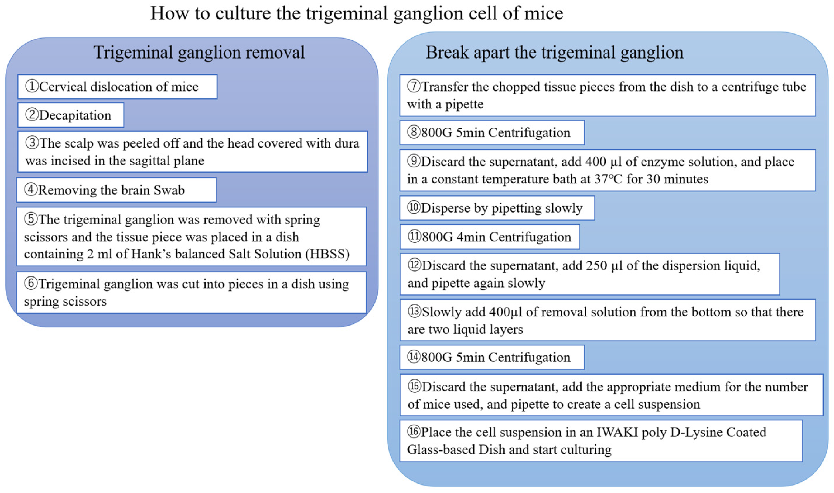
Following a final centrifugation (5 min at 800–1000× g), the supernatant was removed, and neural cell culture medium (FUJIFILM Wako Pure Chemical Corp., Osaka, Japan; Cat. No. 148-09671) was added to prepare the neural cell suspension, which was then seeded into wells for culture. The suspension was filtered through pluriStrainer Mini 70 μm filters (pluriSelect Life Science, Leipzig, Germany) and distributed into poly-D-lysine–coated plastic cell culture dishes (Iwaki, ACG Techno Glass Co., Ltd., Shizuoka, Japan). The culture medium was replaced every other day. From the second exchange (day 4), 0.1% antibiotic–antimycotic solution (Gibco, Thermo Fisher Scientific, Waltham, MA, USA) was added. To prevent cell aggregation and promote neurite growth, the cell suspension was filtered through a 70-µm strainer. Medium changes were performed gently to avoid cell detachment, as the cells adhered directly to the dish surface. The cells were non-proliferating and therefore could not be sub-cultured. A flowchart of the culture process is shown in Figure 1.
Figure 1.
Culturing murine trigeminal ganglion cells. This flowchart shows the process of culturing murine trigeminal ganglion cells in two sections.
2.3. Immunofluorescence Staining
Primary trigeminal ganglion cell cultures (PTGCs) were fixed in 4% paraformaldehyde/phosphate-buffered saline (PBS) for 30 min at room temperature in a 35 mm dish. After washing twice with Dulbecco’s PBS (D-PBS; FUJIFILM Wako Pure Chemical Corp., Osaka, Japan), the cultures were blocked in 10% goat serum for 30 min at room temperature. PTGCs were incubated overnight at 4 °C in the dark with rabbit anti-TRPV1 (1:250; ab3487, polyclonal; Abcam, Cambridge, UK) and rabbit anti-neuronal nuclei (NeuN) (1:500; ab177487, monoclonal; Abcam, Cambridge, UK) antibodies [,]. After three washes with D-PBS, the cultures were incubated with Alexa FluorTM 488 goat anti-rabbit IgG (H + L) (1:200; Thermo Fisher Scientific K.K., Tokyo, Japan) for 90 min at room temperature in the dark. Finally, cultures were counterstained with 4′,6-diamidino-2-phenylindole (DAPI Fluoromount-G, SouthernBiotech, Birmingham, AL, USA), and staining was observed under a fluorescence microscope (BZ-X Serises, Keyence Corp., Osaka, Japan).
2.4. Calcium Imaging of Primary Cultures
To evaluate cell excitation, primary cultures were incubated with 5 μM of Fura-2AM® (Invitrogen, Carlsbad, CA, USA), a calcium ion probe [,], in 0.04% Pluronic F-127 (COSMO BIO Co., Ltd., Tokyo, Japan) [,] at 37 °C under 6% CO2 for 60 min. Cultures were then washed twice with HBSS. HBSS, cyclosporine [,,] (10 and 100 µM), rebamipide [,,,] (10 and 100 µM), diquafosol tetrasodium (1, 10, and 100 µM), or AMG9810 [,,,,,] (1, 10, and 100 µM; a TRPV1 antagonist) were added to each dish. Continuous images were acquired, and fluorescence intensity was measured with a microplate reader (Infinite® 200 PRO; Tecan Group Ltd., Männedorf, Switzerland) before and after stimulation with 10 µM of capsaicin. The excitation wavelengths were 340 nm and 380 nm, and the emission wavelength was 510 nm. Capsaicin acts on TRPV1, allowing calcium ions to enter the cell, which subsequently activates anoctamin 1. Because sensory nerves have high intracellular chloride concentrations, the cells release chloride ions, generating an action potential [].
2.5. Cell Viability
The viability of TRGCs was assessed using the Cell Counting Kit-8 (CCK-8; Dojindo Laboratories, Kumamoto, Japan) according to the manufacturer’s instructions. Cells were seeded into 24-well plates, and 400 μL of culture medium containing 40 μL of CCK-8 solution was added to each well, followed by incubation for 24 h. Absorbance was measured at 450 nm using a microplate reader (Infinite® 200 PRO; Tecan Group Ltd., Männedorf, Switzerland).
2.6. Statistical Analysis
Statistical analyses were performed using GraphPad Prism version 9.1.2 (GraphPad Software, San Diego, CA, USA). Dunnett’s multiple comparisons were performed following one-way analysis of variance, with the Brown–Forsythe applied to account for groups with different sample sizes. Multiple culture experiments were conducted to ensure each group had at least n = 3 or more replicates. The sample size was minimized according to the principles of the 3Rs.
3. Results
3.1. Establishment of PTGCs
We evaluated the following factors to improve cell growth: (1) the age of the mice, (2) contamination prevention methods, and (3) the coating applied to culture dishes and wells.
Although previous studies used mice that were several weeks or months old, they failed to obtain robust cultures of neuronal cells. In contrast, cells isolated from postnatal day 7 mice yielded optimal TRGCs. Povidone-iodine during tissue removal was essential for contamination prevention during culture. Among the various coating materials available to culture neural cells, we selected poly-D-lysine-coated dishes or wells based on previous findings [,,].
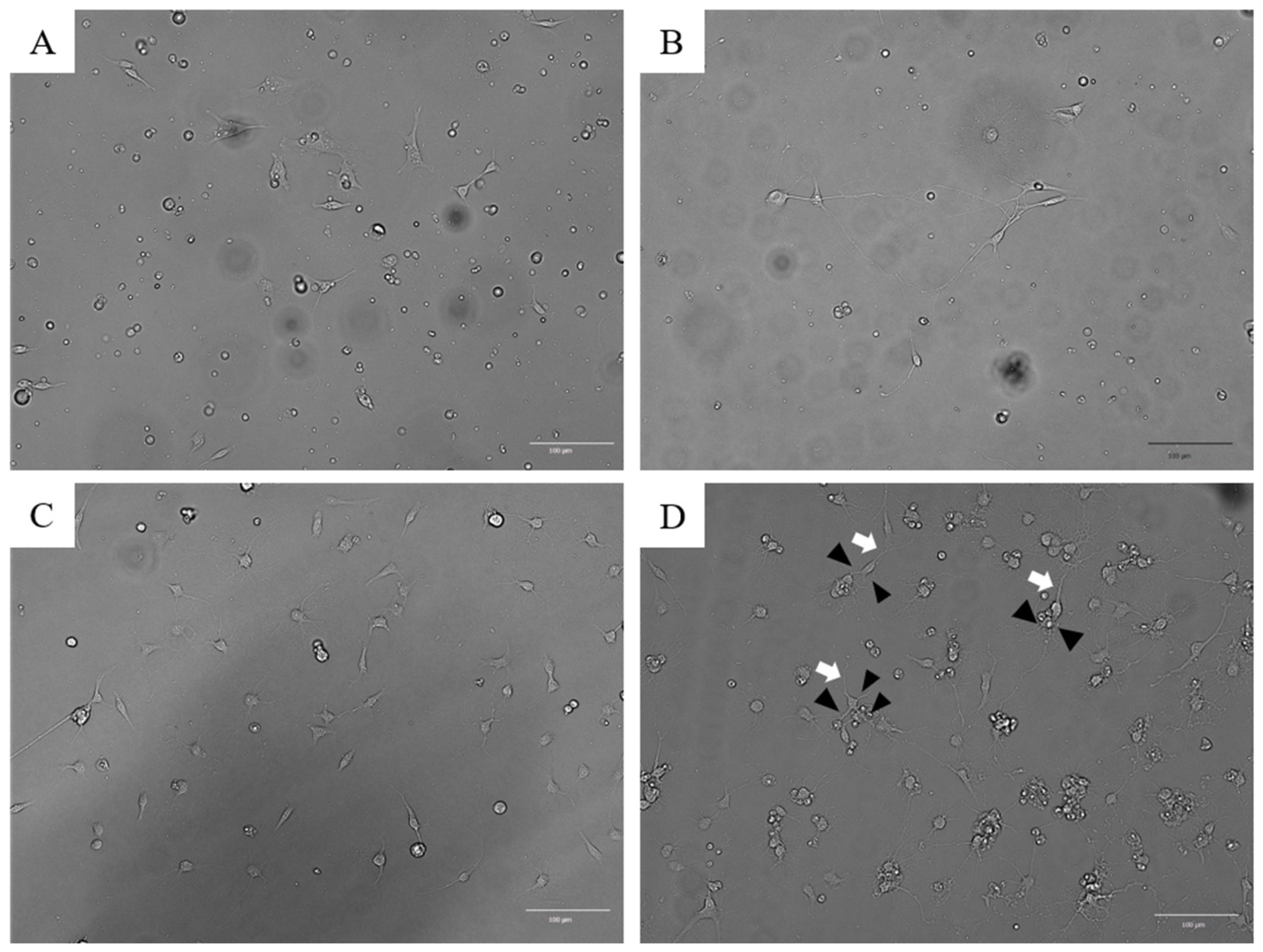
Cell growth was monitored using microscopy. TRGCs remained attached to the bottom of the dishes on days 1 and 2 after dispersion (Figure 2A,B). However, cells that remained in suspension during this period rarely adhered later. The adherent cells extended dendrites and axons (Figure 2B), although they did not undergo cell division. After 1–2 weeks of culture, dendrites formed a mesh-like network (Figure 2C). The degree of cell adhesion observed within 2–3 days after plating was predictive of subsequent growth. Approximately 80% of adherent TRGCs developed neurites by day 7. After 1–2 weeks of culture, these cells differentiated into neuron-like cells, each bearing a single long axon and multiple dendrites (Figure 2D).
Figure 2.
Microscopic images of the developmental process in primary cultures of mouse trigeminal ganglion cells. (A) Attached cells on day 1. Attached cells are observed throughout the culture dish, but dendrites and axons are absent; (B) Neurites from attached cells on day 2. Some of the cells had neurites, whereas most did not; (C) Cell culture on day 7. The dendrites are stretched out like a mesh (white arrow); (D) Cell culture on day 11. One long axon and many short dendritic spines were found in neurons (the white arrow and black arrowheads indicate the axon and dendrites, respectively).
3.2. Immunofluorescence Staining with Anti-NeuN and Anti-TRPV1 Antibodies
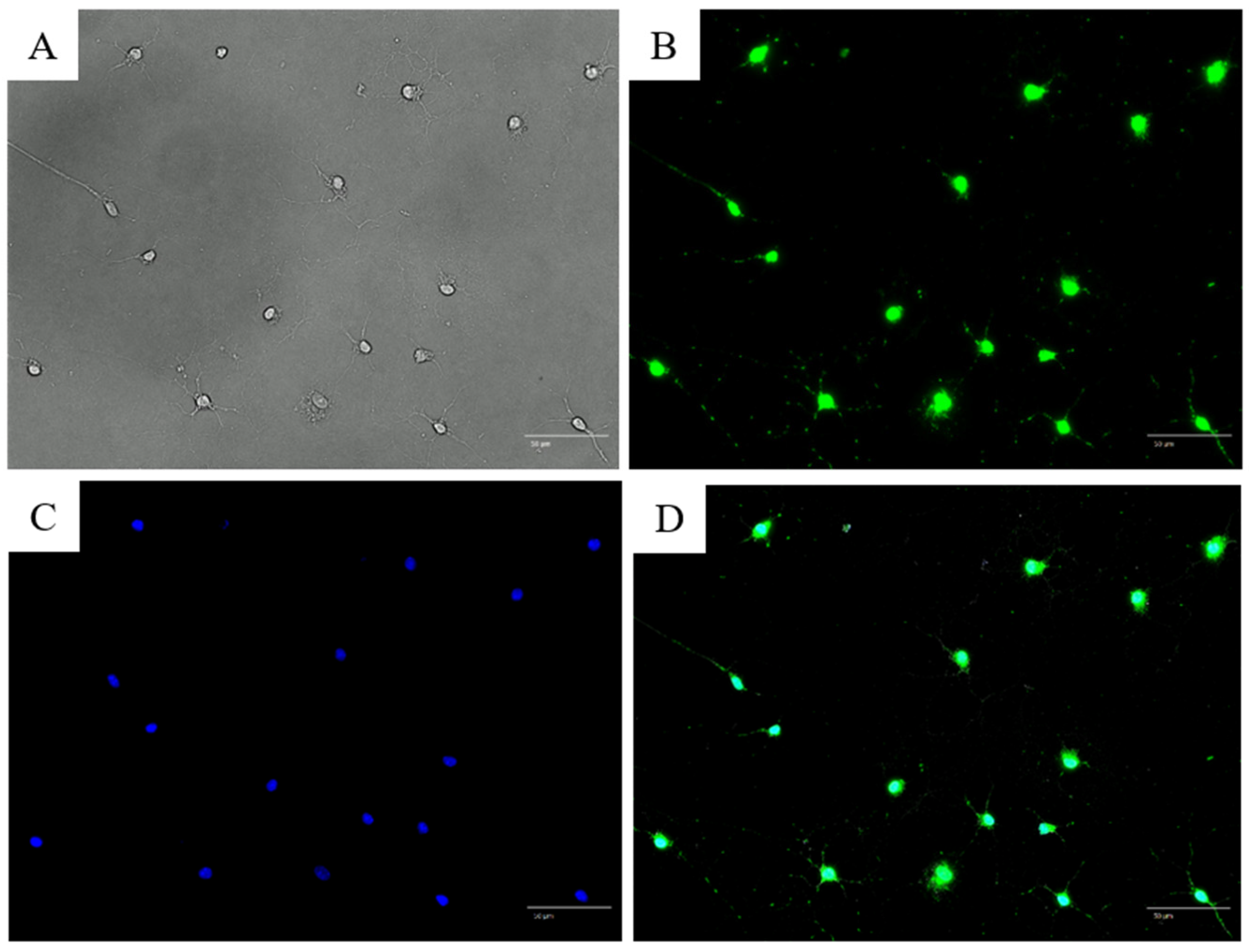
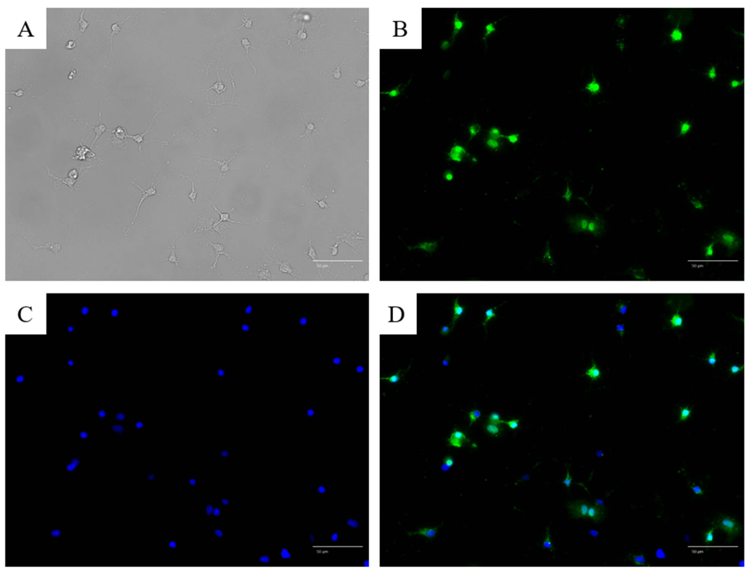
NeuN is a marker of postmitotic neurons. NeuN antibody staining revealed green nuclei, indicating NeuN positivity and confirming that the cultured cells were neurons (Figure 3A–D). Further, anti-TRPV1 antibody staining revealed that a subset of cultured cells expressed the nociceptor-specific TRPV1 (Figure 4).
Figure 3.
Microscopic images of trigeminal ganglion cells stained with anti-neuronal nuclei antibody after 1 week of culture. (A) Bright-field image. One long, thick axon and several dendritic spines are shown; (B) Anti-neuronal nuclei (NeuN) antibody staining; (C) 4′,6-diamidino-2-phenylindole (DAPI) nuclear staining; (D) Merged image of (B,C). All cells stained positive with the anti-NeuN antibody.
Figure 4.
Microscopic images of trigeminal ganglion cells stained with anti-TRPV1 (monolabel). (A) Bright-field image showing neuron-like morphology with one thick axon and several dendritic branches. (B) TRPV1 immunofluorescence signal (green) showing cytoplasmic localization in a subset of neurons. (C) Nuclear counterstaining with DAPI (blue). (D) Merged image of (B,C).
Although double immunostaining with NeuN and TRPV1 was not performed in this experiment, the neuronal identity of cultured cells was confirmed separately by NeuN staining (Figure 3), and the TRPV1 expression pattern was consistent with previous reports in trigeminal neurons.
3.3. Calcium Imaging of Primary Trigeminal Ganglion-Derived Cultured Cells (TRGCs)
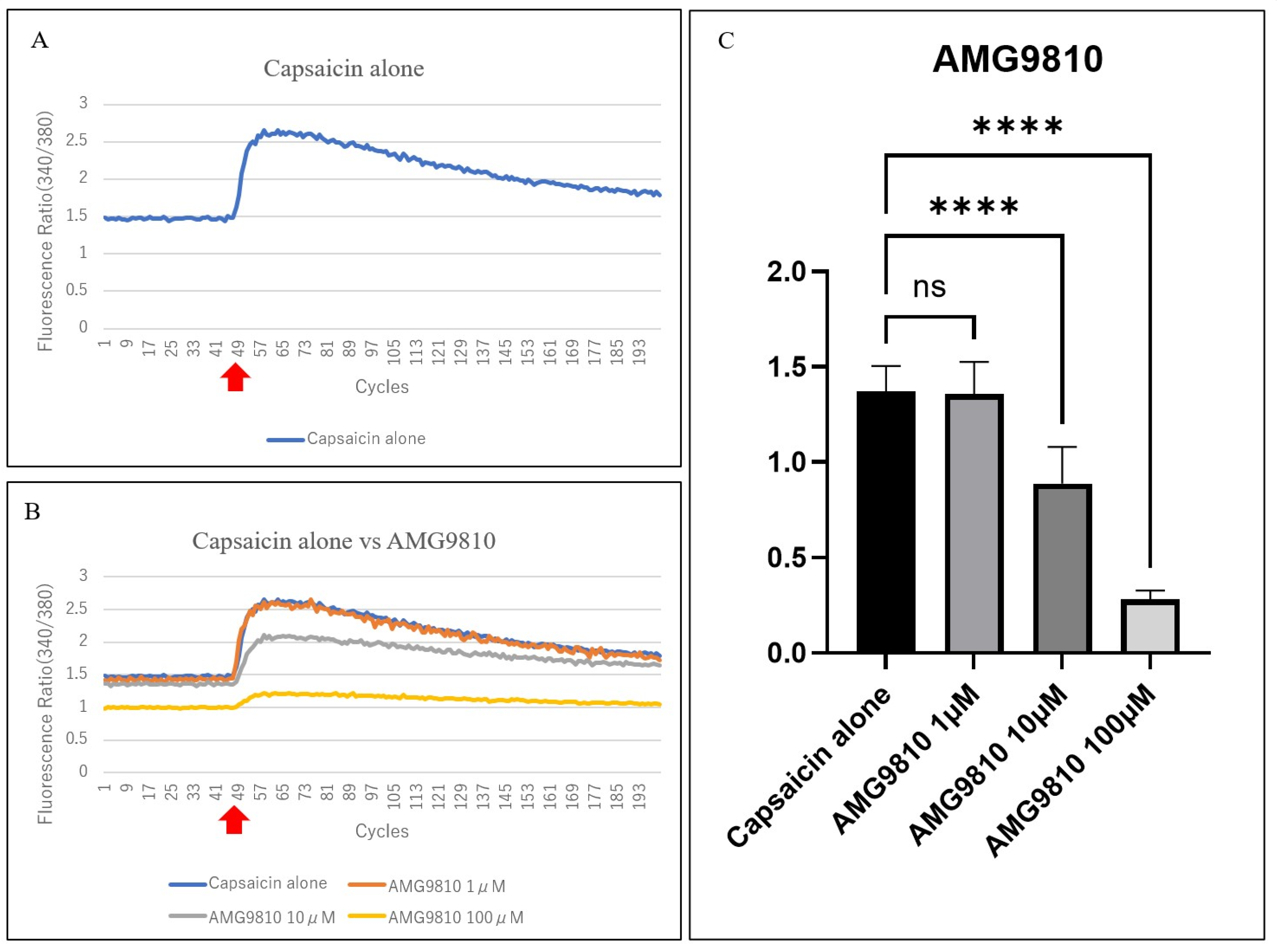
We found that TRGCs could be excited after capsaicin stimulation (Figure 5A,B red arrow). Capsaicin induced a rapid increase in the 340/380 fluorescence intensity ratio, confirming TRPV1-mediated neuronal excitation (Figure 5A). The fluorescence intensity at 340 nm was also elevated because of the increase in Fura2-AM® bound to calcium. In contrast, the fluorescence intensity of Fura2-AM® alone decreased at 380 nm, causing an increase in the 340 nm/380 nm fluorescence intensity ratio. AMG9810 inhibited this response in a concentration-dependent manner, with significant suppression at 10 and 100 µM (both p < 0.0001), whereas 1 µM showed minimal effects (Figure 5A–C).
Figure 5.
Comparison of the effects of capsaicin and the TRPV1 antagonist AMG9810 on TRGCs. The vertical and horizontal axes of the line graph show the 340 nm/380 nm fluorescence intensity ratio and the number of measurements (cycles), respectively. Cycles 1–47 were the baseline, and capsaicin was injected at cycle 48. Additionally, Δ340 nm/380 nm was defined as the maximum value of the 340 nm/380 nm fluorescence intensity ratio minus the mean baseline value. (A) Change in the fluorescence intensity ratio (340 nm/380 nm) over time before and after the addition of capsaicin. Capsaicin (10 μM) was added to stimulate cells approximately 1 min after the start of measurement (cycle 48, red arrow). The 340 nm/380 nm fluorescence intensity ratio increased when cellular excitation occurred. The mean 340 nm/380 nm ratio before capsaicin stimulation (red arrow) was used as the baseline, and the maximum 340 nm/380 nm ratio after capsaicin stimulation in cycle 48 was recorded. The difference between the maximum and baseline values was defined as Δ340 nm/380 nm; (B) Comparison of the change in the fluorescence intensity ratio (340 nm/380 nm) over time before and after the addition of capsaicin or AMG9810 (1 µM (n = 6), 10 µM (n = 16), and 100 µM (n = 14)) and capsaicin. Capsaicin alone (blue line (n = 17)) served as the no-addition group, and the other lines show the 340 nm/380 nm ratio before and after capsaicin stimulation for each AMG9810 concentration. The ratio increased rapidly after capsaicin stimulation (red arrow) in all groups. Compared with capsaicin alone, 1 µM AMG9810 showed little change; however, 10 µM and 100 µM of AMG9810 suppressed Δ340 nm/380 nm; (C) Δ340 nm/380 nm was significantly inhibited at 10 µM and 100 µM of AMG9810 compared with capsaicin alone. p > 0.999, p < 0.0001, and p < 0.0001 for 1 µM, 10 µM, and 100 µM of AMG9810, respectively; Dunnett’s multiple comparisons test; ns: not significant. Error bars indicate standard deviation (SD). ns, not significant; **** p < 0.0001.
3.4. Evaluation of the Suppression of Capsaicin-Related Excitation in Primary TRGCs with Commercially Available Dry Eye Medications
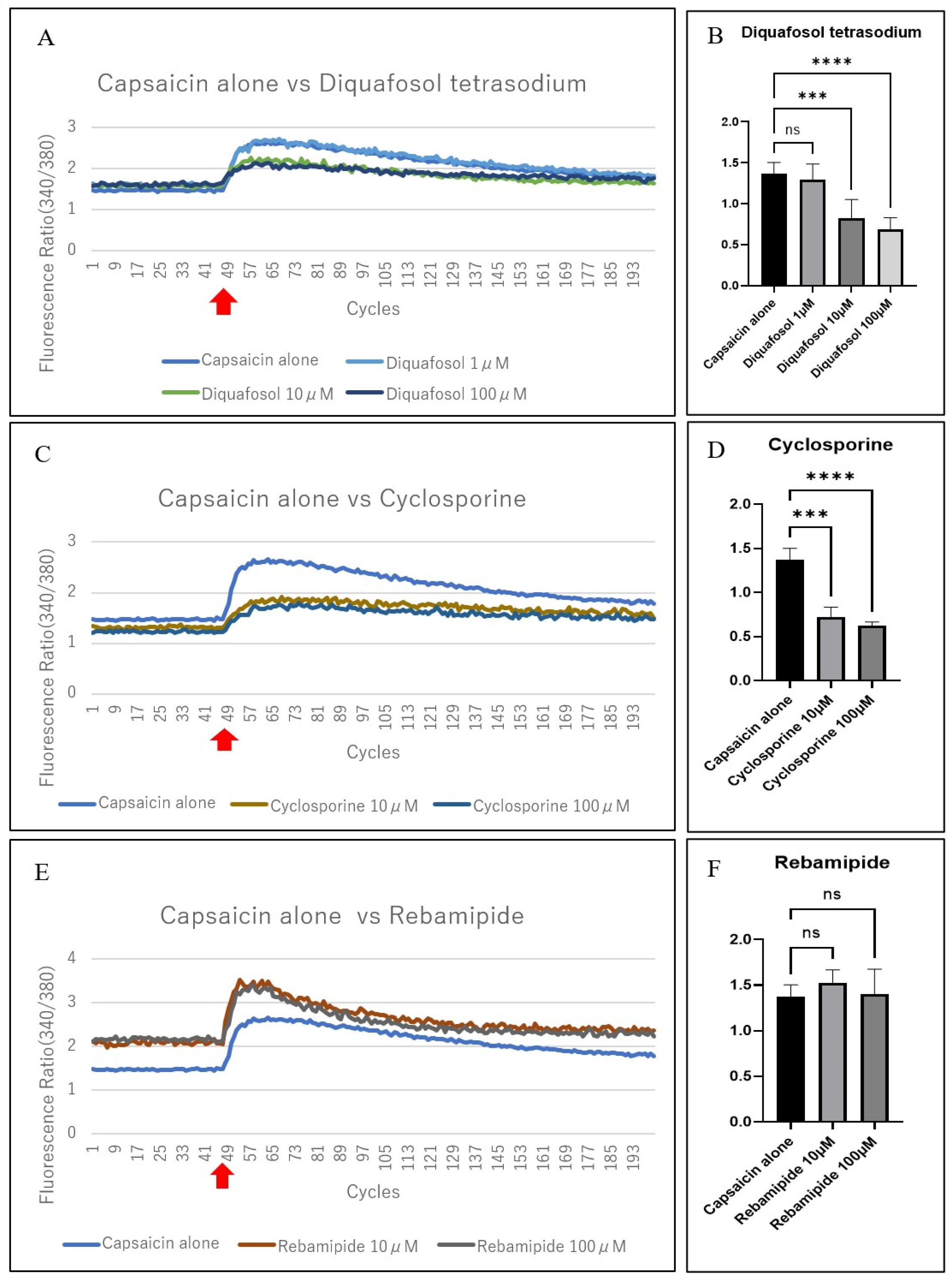
We investigated whether commercially available dry eye medications containing diquafosol tetrasodium, cyclosporine, or rebamipide, suppressed the capsaicin-induced excitation of TRGCs. Diquafosol tetrasodium and cyclosporine significantly reduced capsaicin-induced Δ340/380 at concentrations ≥10 µM (p < 0.001), while rebamipide showed no notable inhibitory effect at either tested concentration (Figure 6A–F). Diquafosol tetrasodium (10 and 100 µM) suppressed neural excitation following capsaicin stimulation (Figure 6A); however, no suppression was observed with 1 µM diquafosol tetrasodium (Figure 6B). Capsaicin-induced excitation was significantly suppressed by the addition of 10 or 100 µM cyclosporine (Figure 6C,D). In contrast, 10 or 100 µM of rebamipide did not influence capsaicin-induced excitation (Figure 6E), with no significant differences at either concentration (Figure 6F). A summary of these results is presented in Supplementary Table S1 online.
Figure 6.
Comparison of the effects of capsaicin and commercially available eye drops on TRGCs. (A) Comparison of the change over time before and after the addition of capsaicin alone or diquafosol tetrasodium (1 µM (n = 8), 10 µM (n = 8), and 100 µM (n = 8)) plus capsaicin. Little difference was observed with 1 µM diquafosol tetrasodium compared with capsaicin alone; however, 10 and 100 µM diquafosol tetrasodium suppressed Δ340 nm/380 nm; (B) Δ340 nm/380 nm was substantially inhibited with 10 and 100 µM diquafosol tetrasodium compared with capsaicin alone. p = 0.67, p < 0.001, and p < 0.0001 for 1, 10, and 100 µM, respectively; Dunnett’s T3 multiple comparisons test; (C) Comparison of the change over time after the addition of capsaicin alone or cyclosporine (10 µM (n = 4) and 100 µM (n = 4)) plus capsaicin. Compared with capsaicin alone, 10 and 100 µM cyclosporine suppressed Δ340 nm/380 nm; (D) Δ340 nm/380 nm was substantially inhibited by 10 and 100 µM cyclosporine compared with capsaicin alone. p < 0.001 and p < 0.0001, respectively; Dunnett’s multiple comparisons test; (E) Comparison of the change over time before and after the addition of capsaicin alone or rebamipide (10 µM (n = 4) and 100 µM (n = 4)) plus capsaicin. Both concentrations of rebamipide showed little difference compared with capsaicin alone; (F) No substantial difference from capsaicin alone was observed with 10 or 100 µM rebamipide. p = 0.20 and p = 0.97, respectively; Dunnett’s multiple comparisons test. ns: not significant; Error bars indicate standard deviation (SD). Δ340/380 was calculated as the maximum fluorescence ratio minus the mean baseline value for each individual sample (B,D,F). ns, not significant; *** p < 0.001; **** p < 0.0001. Red arrows indicate capsaicin stimulation.
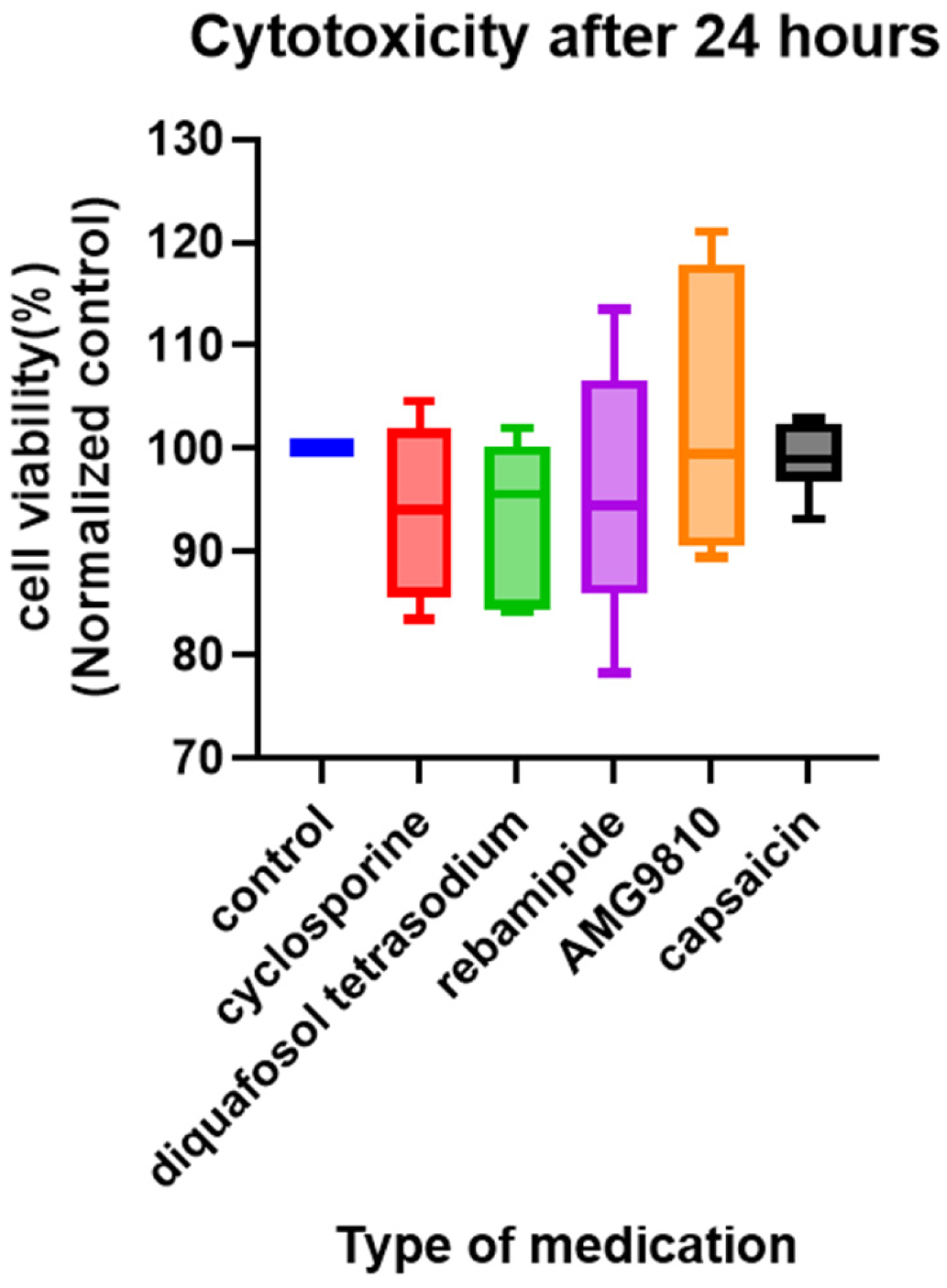
No cytotoxicity was observed with any medication under the tested conditions (Figure 7).
Figure 7.
Viability of TRGCs exposed to various compounds. None of the compounds exhibited cytotoxicity against TRGCs. The control group consisted of untreated cells. All groups were cultured for 24 h. The sample size for each group was n = 6.
4. Discussion
In this study, we successfully used primary neuronal cell cultures from TRPV1-expressing corneal polymodal nociceptors obtained from postnatal day 7 mice to demonstrate increased intracellular calcium ions following capsaicin (a TRPV1 agonist) treatment. To our knowledge, this is the first study to evaluate mouse TRGC responses using a calcium ion probe after stimulation with clinically used medications to treat dry eyes. We found that diquafosol tetrasodium or cyclosporine, but not rebamipide, suppressed cellular excitation in TRGCs. Therefore, our findings suggest that the inhibition of cellular excitation by clinical agents may be associated with clinical pain suppression as patients with dry eyes demonstrate corneal pain sensitivity [] and foreign body sensation and/or pain on the ocular surface.
Using bright-field microscopy, we observed that the TRGC cultures from postnatal day 7 mice showed a neuron-like morphology, including dendrites and axons. The neuronal identity of these cultures was further confirmed by NeuN immunostaining, as shown in Figure 3. To our knowledge, few studies have shown clear pictures of cultured mouse neurons using brightfield microscopy and positive staining for the neuronal marker NeuN. However, despite repeated attempts, we could not culture primary sensory neurons from TRGCs derived from postnatal day 1 mice or those older than postnatal day 12, suggesting technical difficulties in the culture of TRGCs derived from mice. Therefore, immortalized cells with gene transfer would contribute to further development of research using this experimental system.
Rebamipide and diquafosol tetrasodium have mucin-inducing and secretion-promoting effects []. However, our results demonstrated differential mechanisms of these products in relieving pain in patients with dry eye. Diquafosol tetrasodium and cyclosporine suppressed the capsaicin-induced excitation of TRGCs, while rebamipide did not. Nevertheless, the exact suppressive mechanisms of diquafosol tetrasodium and cyclosporine remain unclear.
Diquafosol tetrasodium is an agonist of the P2Y2 receptor that promotes corneal epithelial damage repair [] and is expressed in various parts of the eye. P2Y2 agonists, including diquafosol tetrasodium, have also been reported to affect tumor necrosis factor receptor 1 (TNFR1) []. These results suggest that diquafosol tetrasodium binds to the P2Y2 receptor, causing TNFR1 release into the extracellular space, thus suppressing TNF-α signaling that may be involved in suppressing neural excitement through capsaicin stimulation.
Cyclosporine is a calcineurin inhibitor and an immunosuppressive agent, suppressing production of cytokines such as interleukin 2 []. Furthermore, cyclosporine may have an indirect suppressive effect on the production of cytokines involved in the cellular excitation of cultured mouse TRGCs. Further studies are required to identify the mechanisms underlying cyclosporine suppression of capsaicin-induced excitation in cultured TRGCs.
Dry eye disease is associated with peripheral sensitization of corneal nociceptors, partly mediated by TRPV1 activation through tear hyperosmolarity and inflammatory cytokines []. Sensitized TRPV1 triggers the release of neuropeptides such as substance P and calcitonin gene-related peptide (CGRP) [], which may amplify ocular discomfort via neurogenic inflammation. Our finding that cyclosporine and diquafosol suppress TRPV1-dependent neuronal excitation suggests a plausible mechanism for their reported improvement in ocular pain in clinical settings. However, our in vitro experimental system did not incorporate corneal epithelial and immune cell interactions or tear components, which are key modulators of sensory function in vivo.
This study has some limitations. Although we investigated the effects of diquafosol tetrasodium and cyclosporine on neuronal excitation in mouse TRGCs, the underlying molecular mechanisms remain unresolved. In vivo studies involving specific receptors and dynamic changes in cytokines using knockout mice or a dry eye mouse model are required to validate our in vitro findings []. Clinical studies using eye drops and a Cochet–Bonnet esthesiometer [] may also contribute to a better understanding of the mechanisms of pain development and reduction in pain in patients with dry eyes by commercially available eye drops. Furthermore, it remains unclear whether the suppression of cellular excitation was due to changes in calcium kinetics or the toxicity of the drugs added to the cultured cells. Furthermore, this study is limited by the absence of physiological microenvironmental factors, including epithelial–neuronal–immune cell crosstalk and tear film components. Future work involving quantification of neuropeptide release (e.g., CGRP, substance P) and correlating TRPV1 sensitivity with clinical corneal pain phenotypes using esthesiometry and in vivo confocal microscopy will help elucidate underlying mechanisms.
In conclusion, we used a primary cell culture model of TRGCs derived from postnatal day 7 mice that allowed the screening of medications that may affect nociceptive and neuropathic pain in the cornea. Our findings indicate that certain components of dry eye medications may act via inhibition of neuronal excitation. This model may serve as a powerful tool for future studies on corneal perception.
Supplementary Materials
The following supporting information can be downloaded at: https://www.mdpi.com/article/10.3390/jcm14228038/s1, Table S1: Summary of Results.
Author Contributions
Conceptualization, T.S.; Formal analysis, C.S. and T.H.; Project administration, T.S.; Supervision, T.S.; Validation, T.S.; Writing—Original Draft Preparation, C.S.; Writing—Review and Editing, C.S., T.H. and S.Y. All authors have read and agreed to the published version of the manuscript.
Funding
This work was supported by JSPS KAKENHI [Grant Number JP20K09812].
Institutional Review Board Statement
The animal study protocol was approved by the Institutional Review Board of the Animal Experiment Committee of Nihon University School of Medicine (approval number: AP18MED066-2, approved on 1 March 2019).
Informed Consent Statement
Not applicable.
Data Availability Statement
All data generated or analyzed during this study are included in this published article and in its Supplementary Information Files.
Acknowledgments
We thank Akiko Tomioka for her excellent technical support.
Conflicts of Interest
The authors declare no competing interest.
Abbreviations
The following abbreviations are used in this manuscript:
| PTGCs | Primary trigeminal ganglion cell cultures |
| TRGCs | Trigeminal ganglion-derived cells |
| TRPV1 | transient receptor potential vanilloid 1 |
References
- Nelson, J.D.; Craig, J.P.; Akpek, E.K.; Azar, D.T.; Belmonte, C.; Bron, A.J.; Clayton, J.A.; Dogru, M.; Dua, H.S.; Foulks, G.N.; et al. TFOS DEWS II introduction. Ocul. Surf. 2017, 15, 269–275. [Google Scholar] [CrossRef] [PubMed]
- Bourcier, T.; Acosta, M.C.; Borderie, V.; Borrás, F.; Gallar, J.; Bury, T.; Laroche, L.; Belmonte, C. Decreased corneal sensitivity in patients with dry eye. Investig. Ophthalmol. Vis. Sci. 2005, 46, 2341–2345. [Google Scholar] [CrossRef]
- Yang, A.Y.; Chow, J.; Liu, J. Corneal innervation and sensation: The eye and beyond. Yale J. Biol. Med. 2018, 91, 13–21. [Google Scholar]
- Belmonte, C.; Viana, F. Molecular and cellular limits to somatosensory specificity. Mol. Pain 2008, 4, 1744–8069. [Google Scholar] [CrossRef]
- Schöbel, N.; Radtke, D.; Lübbert, M.; Gisselmann, G.; Lehmann, R.; Cichy, A.; Schreiner, B.S.P.; Altmüller, J.; Spector, A.C.; Spehr, J.; et al. Trigeminal ganglion neurons of mice show intracellular chloride accumulation and chloride-dependent amplification of capsaicin-induced responses. PLoS ONE 2012, 7, e48005. [Google Scholar] [CrossRef]
- Rossi, H.L.; Broadhurst, K.A.; Luu, A.S.K.; Lara, O.; Kothari, S.D.; Mohapatra, D.P.; Recober, A. Abnormal trigeminal sensory processing in obese mice. Pain 2016, 157, 235–246. [Google Scholar] [CrossRef]
- Launay, P.S.; Reboussin, E.; Liang, H.; Kessal, K.; Godefroy, D.; Rostene, W.; Sahel, J.A.; Baudouin, C.; Melik Parsadaniantz, S.; Le Goazigo, A.R. Ocular inflammation induces trigeminal pain, peripheral and central neuroinflammatory mechanisms. Neurobiol. Dis. 2016, 88, 16–28. [Google Scholar] [CrossRef]
- Di, G.; Qi, X.; Zhao, X.; Zhang, S.; Danielson, P.; Zhou, Q. Corneal epithelium-derived neurotrophic factors promote nerve regeneration. Investig. Ophthalmol. Vis. Sci. 2017, 58, 4695–4702. [Google Scholar] [CrossRef]
- Hu, J.; Hu, X.; Kan, T. MiR-34c participates in diabetic corneal neuropathy via regulation of autophagy. Investig. Ophthalmol. Vis. Sci. 2019, 60, 16–25. [Google Scholar] [CrossRef] [PubMed]
- Im, S.T.; Jo, Y.Y.; Han, G.; Jo, H.J.; Kim, Y.H.; Park, C.K. Dexmedetomidine inhibits voltage-gated sodium channels via α2-adrenoceptors in trigeminal ganglion neurons. Mediat. Inflamm. 2018, 2018, 1782719. [Google Scholar] [CrossRef] [PubMed]
- Poulsen, J.N.; Warwick, R.; Duroux, M.; Hanani, M.; Gazerani, P. Oxaliplatin enhances gap junction-mediated coupling in cell cultures of mouse trigeminal ganglia. Exp. Cell Res. 2015, 336, 94–99. [Google Scholar] [CrossRef]
- Kayama, Y.; Shibata, M.; Takizawa, T.; Ibata, K.; Shimizu, T.; Ebine, T.; Toriumi, H.; Yuzaki, M.; Suzuki, N. Functional interactions between transient receptor potential M8 and transient receptor potential V1 in the trigeminal system: Relevance to migraine pathophysiology. Cephalalgia 2018, 38, 833–845. [Google Scholar] [CrossRef]
- Duan, W.; Zhang, Y.P.; Hou, Z.; Huang, C.; Zhu, H.; Zhang, C.Q.; Yin, Q. Novel insights into NeuN: From neuronal marker to splicing regulator. Mol. Neurobiol. 2016, 53, 1637–1647. [Google Scholar] [CrossRef]
- Dredge, B.K.; Jensen, K.B. NeuN/Rbfox3 nuclear and cytoplasmic isoforms differentially regulate alternative splicing and nonsense-mediated decay of Rbfox2. PLoS ONE 2011, 6, e21585. [Google Scholar] [CrossRef]
- Toivari, E.; Manninen, T.; Nahata, A.K.; Jalonen, T.O.; Linne, M.L. Effects of transmitters and amyloid-beta peptide on calcium signals in rat cortical astrocytes: Fura-2AM measurements and stochastic model simulations. PLoS ONE 2011, 6, e17914. [Google Scholar] [CrossRef]
- Takeuchi, K.; Sato, S.I.; Abe, K.; Kimura, M.; Abe, T.A.; Yoshinaga, K.; Inaba, H. Intracellular compartmentalization of fura-2 dye demonstrated by laser-excitation fluorescence microscopy: A problem in measuring cytosolic free calcium concentration using fura-2 fluorescence in vascular smooth muscle cells. Tohoku J. Exp. Med. 1989, 159, 23–35. [Google Scholar] [CrossRef]
- Drummond, I.A.; Lee, A.S.; Resendez, E., Jr.; Steinhardt, R.A. Depletion of intracellular calcium stores by calcium ionophore A23187 induces the genes for glucose-regulated proteins in hamster fibroblasts. J. Biol. Chem. 1987, 262, 12801–12805. [Google Scholar] [CrossRef] [PubMed]
- Poenie, M.; Alderton, J.; Steinhardt, R.; Tsien, R. Calcium rises abruptly and briefly throughout the cell at the onset of anaphase. Science 1986, 233, 886–889. [Google Scholar] [CrossRef] [PubMed]
- Cortina, P.; Navea, A.; Terencio, M.C.; Diaz-Llopis, M.; Gómez-Lechón, M.J.; Menezo, J.L. Diclofenac sodium and cyclosporin A inhibit human lens epithelial cell proliferation in culture. Graefe’s Arch. Clin. Exp. Ophthalmol. 1997, 235, 180–185. [Google Scholar] [CrossRef] [PubMed]
- Duncan, J.I. Differential inhibition of cutaneous T-cell-mediated reactions and epidermal cell proliferation by cyclosporin A, FK-506, and rapamycin. J. Investig. Dermatol. 1994, 102, 84–88. [Google Scholar] [CrossRef]
- Kim, S.Y.; Shim, M.S.; Kim, K.Y.; Weinreb, R.N.; Wheeler, L.A.; Ju, W.K. Inhibition of cyclophilin D by cyclosporin A promotes retinal ganglion cell survival by preventing mitochondrial alteration in ischemic injury. Cell Death Dis. 2014, 5, e1105. [Google Scholar] [CrossRef]
- Ríos, J.D.; Shatos, M.; Urashima, H.; Tran, H.; Dartt, D.A. OPC-12759 increases proliferation of cultured rat conjunctival goblet cells. Cornea 2006, 25, 573–581. [Google Scholar] [CrossRef]
- Suzuki, M.; Miura, S.; Mori, M.; Kai, A.; Suzuki, H.; Fukumura, D.; Suematsu, M.; Tsuchiya, M. Rebamipide, a novel antiulcer agent, attenuates Helicobacter pylori induced gastric mucosal cell injury associated with neutrophil derived oxidants. Gut 1994, 35, 1375–1378. [Google Scholar] [CrossRef] [PubMed]
- Ríos, J.D.; Shatos, M.A.; Urashima, H.; Dartt, D.A. Effect of OPC-12759 on EGF receptor activation, p44/p42 MAPK activity, and secretion in conjunctival goblet cells. Exp. Eye Res. 2008, 86, 629–636. [Google Scholar] [CrossRef]
- Kim, C.D.; Kim, H.H.; Hong, K.W. Inhibitory effect of rebamipide on the neutrophil adherence stimulated by conditioned media from Helicobacter pylori-infected gastric epithelial cells. J. Pharmacol. Exp. Ther. 1999, 288, 133–138. [Google Scholar] [CrossRef]
- Kwon, S.G.; Roh, D.H.; Yoon, S.Y.; Moon, J.Y.; Choi, S.R.; Choi, H.S.; Kang, S.Y.; Han, H.J.; Beitz, A.J.; Oh, S.B.; et al. Acid evoked thermal hyperalgesia involves peripheral P2Y1 receptor mediated TRPV1 phosphorylation in a rodent model of thrombus induced ischemic pain. Mol. Pain 2014, 10, 2. [Google Scholar] [CrossRef]
- Huang, R.; Wang, F.; Yang, Y.; Ma, W.; Lin, Z.; Cheng, N.; Long, Y.; Deng, S.; Li, Z. Recurrent activations of transient receptor potential vanilloid-1 and vanilloid-4 promote cellular proliferation and migration in esophageal squamous cell carcinoma cells. FEBS Open Bio 2019, 9, 206–225. [Google Scholar] [CrossRef] [PubMed]
- Jian, T.; Yang, N.; Yang, Y.; Zhu, C.; Yuan, X.; Yu, G.; Wang, C.; Wang, Z.; Shi, H.; Tang, M.; et al. TRPV1 and PLC participate in histamine H4 receptor-induced itch. Neural Plast. 2016, 2016, 1682972. [Google Scholar] [CrossRef] [PubMed]
- Saloman, J.L.; Chung, M.K.; Ro, J.Y. P2X3 and TRPV1 functionally interact and mediate sensitization of trigeminal sensory neurons. Neuroscience 2013, 232, 226–238. [Google Scholar] [CrossRef]
- Wang, S.; Brigoli, B.; Lim, J.; Karley, A.; Chung, M.K. Roles of TRPV1 and TRPA1 in spontaneous pain from inflamed masseter Muscle. Neuroscience 2018, 384, 290–299. [Google Scholar] [CrossRef]
- Obi, S.; Nakajima, T.; Hasegawa, T.; Kikuchi, H.; Oguri, G.; Takahashi, M.; Nakamura, F.; Yamasoba, T.; Sakuma, M.; Toyoda, S.; et al. Heat induces interleukin-6 in skeletal muscle cells via TRPV1/PKC/CREB pathways. J. Appl. Physiol. 2017, 122, 683–694. [Google Scholar] [CrossRef]
- Takayama, Y.; Uta, D.; Furue, H.; Tominaga, M. Pain-enhancing mechanism through interaction between TRPV1 and anoctamin 1 in sensory neurons. Proc. Natl. Acad. Sci. USA 2015, 112, 5213–5218. [Google Scholar] [CrossRef]
- Kaido, M.; Kawashima, M.; Ishida, R.; Tsubota, K. Relationship of corneal pain sensitivity with dry eye symptoms in dry eye with short tear break-up time. Investig. Ophthalmol. Vis. Sci. 2016, 57, 914–919. [Google Scholar] [CrossRef]
- Yamane, M.; Ogawa, Y.; Fukui, M.; Kamoi, M.; Saijo-Ban, Y.; Yaguchi, S.; Mukai, S.; Kawakita, T.; Simmura, S.; Tsubota, K. Long-term rebamipide and diquafosol in two cases of immune-mediated dry eye. Optom. Vis. Sci. 2015, 92 (Suppl. S1), S25–S32. [Google Scholar] [CrossRef]
- Byun, Y.S.; Yoo, Y.S.; Kwon, J.Y.; Joo, J.S.; Lim, S.A.; Whang, W.J.; Mok, J.W.; Choi, J.S.; Joo, C.K. Diquafosol promotes corneal epithelial healing via intracellular calcium-mediated ERK activation. Exp. Eye Res. 2016, 143, 89–97. [Google Scholar] [CrossRef] [PubMed]
- Sakimoto, T.; Ohnishi, T.; Ishimori, A. Significance of ectodomain shedding of TNF receptor 1 in ocular surface. Investig. Ophthalmol. Vis. Sci. 2014, 55, 2419–2423. [Google Scholar] [CrossRef] [PubMed]
- Camlar, S.A.; Soylu, A.; Kavukçu, S. Cyclosporine in pediatric nephrology. Iran J. Kidney Dis. 2018, 12, 319–330. [Google Scholar]
- Fakih, D.; Migeon, T.; Moreau, N.; Baudouin, C.; Goazigo, A.R.-L.; Parsadaniantz, S.M. Transient receptor potential channels: Important players in ocular pain and dry eye disease. Pharmaceutics 2022, 14, 1859. [Google Scholar] [CrossRef]
- Ma, L.; Yang, L.; Wang, X.; Zhao, L.; Bai, X.; Qi, X.; Chen, Q.; Li, Y.; Zhou, Q. CGRP released by corneal sensory nerve maintains tear secretion of the lacrimal gland. Investig. Ophthalmol. Vis. Sci. 2024, 65, 30. [Google Scholar] [CrossRef]
- Shinomiya, K.; Ueta, M.; Kinoshita, S. A new dry eye mouse model produced by exorbital and intraorbital lacrimal gland excision. Sci. Rep. 2018, 8, 1483. [Google Scholar] [CrossRef] [PubMed]
- Kaido, M.; Inoue, S.; Kawashima, M.; Ishida, R.; Nakamura, S.; Tsubota, K. Capsaicin-induced pain sensitivity in short tear break-up time dry eye. Ocul. Surf. 2020, 18, 620–626. [Google Scholar] [CrossRef] [PubMed]
Disclaimer/Publisher’s Note: The statements, opinions and data contained in all publications are solely those of the individual author(s) and contributor(s) and not of MDPI and/or the editor(s). MDPI and/or the editor(s) disclaim responsibility for any injury to people or property resulting from any ideas, methods, instructions or products referred to in the content. |
© 2025 by the authors. Licensee MDPI, Basel, Switzerland. This article is an open access article distributed under the terms and conditions of the Creative Commons Attribution (CC BY) license (https://creativecommons.org/licenses/by/4.0/).