Systematic Review

Is Ti-Coated PEEK Superior to PEEK for Lumbar and Cervical Fusion Procedures? A Systematic Review and Meta-Analysis

1
Department of Neurosurgery, Perelman School of Medicine, University of Pennsylvania, Philadelphia, PA 19104, USA
2
Department of Neurosurgery, Mass General Brigham, Harvard Medical School, Boston, MA 02114, USA
3
School of Medicine, University of Virginia Health, Charlottesville, VA 22908, USA
4
Department of Neurosurgery, University of California San Francisco, San Francisco, CA 94143, USA
*
Author to whom correspondence should be addressed.
J. Clin. Med. 2025, 14(21), 7696; https://doi.org/10.3390/jcm14217696
Submission received: 3 September 2025 / Revised: 26 October 2025 / Accepted: 26 October 2025 / Published: 30 October 2025
(This article belongs to the Special Issue Clinical Advances in Spinal Neurosurgery)

Abstract

Background/Objectives: Utilization of polyetheretherketone (PEEK) cages for spinal fusion has surged in the U.S., yet comprehensive comparisons evaluating its postoperative effectiveness with alternative materials remain limited. This systematic review investigates the efficacy of PEEK cages against traditional fusion materials across various surgery types, elucidating PEEK’s impact on fusion rates, postoperative outcomes, and long-term success. Methods: A systematic search of PubMed, CINAHL, Scopus, Embase, and Web of Science was conducted through 14 October 2024. Included studies were randomized controlled trials (RCTs) comparing PEEK cages with titanium, silicon nitride, and metal-coated PEEK cages for anterior cervical discectomy and fusion (ACDF), posterior lumbar interbody fusion (PLIF), and transforaminal lumbar interbody fusion (TLIF). Article quality was assessed using GRADE criteria. Results: From 288 initially screened articles, 25 RCTs involving 2046 patients (mean follow-up 23.1 ± 18.2 months) met inclusion criteria and were determined as moderate (n = 21) or high (n = 4) quality. Fusion rates by cage material for PEEK (n = 1041), Ti-PEEK (n = 291), and titanium (n = 53) were 85.63 ± 18.00%, 80.05 ± 19.9%, and 92.75 ± 11.31%, respectively. In ACDF, titanium cages achieved higher fusion rates than PEEK (100% vs. 94%). In PLIF and TLIF, coated PEEK outperformed uncoated PEEK (75% vs. 71% and 94% vs. 84%, respectively). Uncoated PEEK achieved fusion rates of 94.04 ± 5.04% for ACDF, 71.21 ± 21.93% for PLIF, and 83.50 ± 24.66% for TLIF, with titanium outperforming PEEK in early fusion outcomes. Coated PEEK demonstrated potential improvements in fusion rates over uncoated PEEK in PLIFs and TLIFs. Conclusions: Selection of cage material for spinal fusions should be tailored to surgical requirements and patient needs. While titanium and PEEK are effective, their performance varies across contexts. New materials and surface modifications may enhance these outcomes further, warranting future research in long-term studies and development of novel materials. These findings can help surgeons choose cage materials according to procedure type, patient characteristics, and imaging needs.

1. Introduction

Spinal fusion is a widely accepted surgical approach essential for treating pathologies such as scoliosis, degenerative spondylolisthesis, and lumbar and cervical stenosis [1,2,3]. In recent decades, the global incidence of spinal fusion surgeries has risen alongside an aging population. In the United States, the rate of cervical and lumbar fusions has increased by 89% and 134%, respectively, reflecting the large demand for treatment of debilitating spinal pathologies [4,5,6,7].
Currently, anterior cervical discectomy and fusion (ACDF), transforaminal lumbar interbody fusion (TLIF), and posterior lumbar interbody fusion (PLIF) are among the most common techniques for spinal fusion [1,7,8]. Although each procedure involves distinct features, the adoption of interbody cages has revolutionized these approaches, leading to reduced postoperative pain, fewer complications, and increased fusion rates compared to traditional bone grafts [9,10,11,12].
Various materials for interbody cages exist, with polyetheretherketone (PEEK) and titanium alloy being the most frequently used [12,13,14]. In the 1990s, PEEK arose as a commonly used graft due to its unique characteristics which mimic the biomechanical and biological properties of natural bone [14,15,16,17]. PEEK possesses an elastic modulus similar to cortical bone, enabling the reduction of stress-shielding effects. Another advantage of PEEK is its radiolucency which allows for enhanced assessment of fusion imaging [15,16,18,19,20,21].
The literature currently lacks comprehensive, controlled studies that assess the postoperative efficacy of PEEK cages against other fusion materials. Earlier reviews were either not restricted to randomized controlled trials or limited to a single procedure, which reduced the strength and generalizability of their conclusions. While there are reports on specific comparisons, a broader analysis across surgery types is missing. This gap underscores the critical need for detailed comparative research on fusion rates, post-surgical outcomes, and long-term effectiveness of various cage materials. Accordingly, this systematic review, limited exclusively to randomized controlled trials across ACDF, PLIF, and TLIF, aims to compare fusion rates and peri-operative outcomes of PEEK to other commonly used fusion cage materials, thereby guiding cage selection and operative decision-making across surgical approaches.

2. Materials and Methods

2.1. Information Sources and Search Strategy

This systematic review was designed in accordance with Preferred Reporting Items for Systematic Reviews and Meta-Analyses (PRISMA) guidelines (Supplementary Materials). A search for the variable uses and applications of PEEK for spinal surgery was performed using PubMed, CINAHL, Scopus, Embase, and Web of Science from database inception through 14 October 2024. Our search terms are as follows: (“Cage” OR “Cages”) AND ((“Titanium Alloy (TiAl6V4)” OR “Porous Titanium” OR TiAl6V4) OR “Carbon Fiber Reinforced Polymer” OR (“Polyetheretherketone” OR “PEEK”) OR (3D-printed)) AND (“Fusion” OR “Spine Fusion” OR “Spinal Fusion” OR “Lumbar “OR “Lumbar Fusion” OR “Lumbar Interbody Fusion”).

2.2. Inclusion and Exclusion Criteria

For this systematic review, all randomized controlled trials (RCTs) comparing PEEK and other fusion materials such as titanium and metal-coated PEEK cages for ACDF, PLIF, and TLIF were included. The primary outcome of analysis was postoperative fusion rates. The following exclusion criteria were applied:
  • Non-English studies;
  • Non-original articles such as commentaries, systematic reviews, or meta-analysis;
  • Studies lacking fusion rates following surgical intervention;
  • Study designs other than RCTs;
  • Studies lacking PEEK comparison.

2.3. Article Selection Process

The software Covidence (Veritas Health Innovation Ltd., Melbourne, Australia) was used for the review organization. Articles were screened by title and abstract followed by full-text analysis applying the inclusion and exclusion criteria. At each step, two independent reviewers (JK, AV) screened all articles. If a consensus could not be reached, a third reviewer (RK) adjudicated, resolving any conflicts and making a final decision to prevent bias in the article selection process. No authors with conflicts of interest participated in data extraction or quality grading.

2.4. Data Extraction

The primary outcome measure was postoperative fusion rate, quantified by the proportion of patients that were deemed “fused” by the study. Other recorded data included study demographics (author, year of publication, article type, surgery type, number of patients, patient sex, and average patient age), objective outcomes (blood loss, hospital stay length, operative time, and follow-up time), and subjective outcomes (Visual Analog Scale [VAS] scores and Oswestry Disability Index [ODI] scores). Studies that provided both preoperative and postoperative values for patient-reported outcome measures (PROMs) were included in the analysis. Studies that did not separate VAS reporting into VAS Neck, Arm, Leg, or Back were excluded from the VAS analysis. To ensure consistency, standardized definitions for case and control groups were established and applied uniformly across all included studies. The case group was defined as the uncoated PEEK cage, and the control group was defined as the coated PEEK and non-PEEK cages. Values were reported as frequency-weighted means (FWM). For consistency, we extracted outcomes at the latest reported follow-up time as definitions of early vs. long-term fusion varied across studies.

2.5. Meta-Analysis

Random-effects meta-analyses for (i) fusion rate (proportion fusion) and (ii) patient-reported outcomes (VAS and ODI) were performed. For fusion, study-level proportions were pooled using inverse-variance random effects after variance-stabilizing transfomrion; for PROMs, we pooled mean change from baseline (or post-op means when change was unavailable) using random-effects models. Heterogeneity was summarized by I2 and τ2, and subgroup analyses were prespecified by procedure (ACDF, PLIF, TLIF) and cage material (uncoated PEEK, coated PEEK, titanium, other).

2.6. Article Quality Grading

Article quality grading for this systemic review was conducted using the Grading of Recommendations Assessment, Development, and Evaluation (GRADE) criteria [22]. GRADE incorporates considerations of inconsistency, risk of bias, imprecision, and publication bias. The studies began with a high-quality grade and remained high unless there was a risk of bias, inconsistency, imprecision, or publication bias, in which case they were downgraded. This review was not registered in PROSPERO due to different registration standards at the time of data collection, posing a limitation in methodology.

3. Results

Our search resulted in 288 articles. After removing duplicates, 196 articles remained. Title and abstract screening narrowed this to 68. After a full-text search, 25 articles met the inclusion criteria and were included in the study, reporting data from 11 countries. All included studies (n = 25) were RCTs and were designated as moderate or high-quality according to the GRADE criteria (Table 1, Figure 1) [23,24,25,26,27,28,29,30,31,32,33,34,35,36,37,38,39,40,41,42,43,44,45,46,47].

3.1. Study Demographics

All included patients (n = 2046) had a frequency-weighted mean age of 54.90 ± 7.01 years. Females represented 51.32% of the total cohort. The frequency-weighted postoperative follow-up time for all patients was 23.11 ± 18.15 months. There were three reported categories of surgery types, including ACDF (n = 11), PLIF (n = 6), and TLIF (n = 7) studies for a total of 24 studies. One remaining study compared both ACDF and TLIF. A focused breakdown of procedure characteristics such as blood loss and hospital stay is found in Table 2.

3.2. Postoperative Fusion Outcomes in ACDF

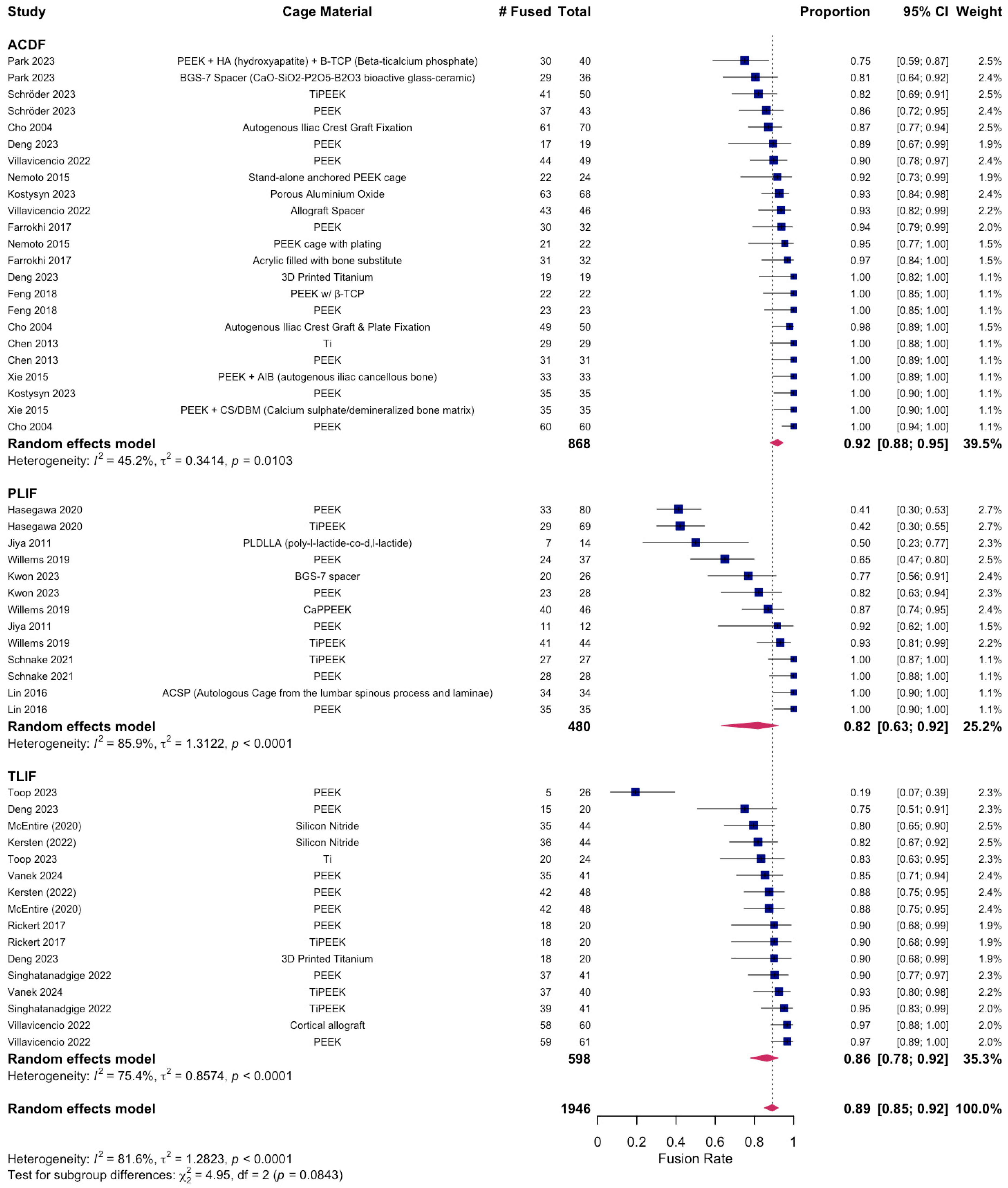
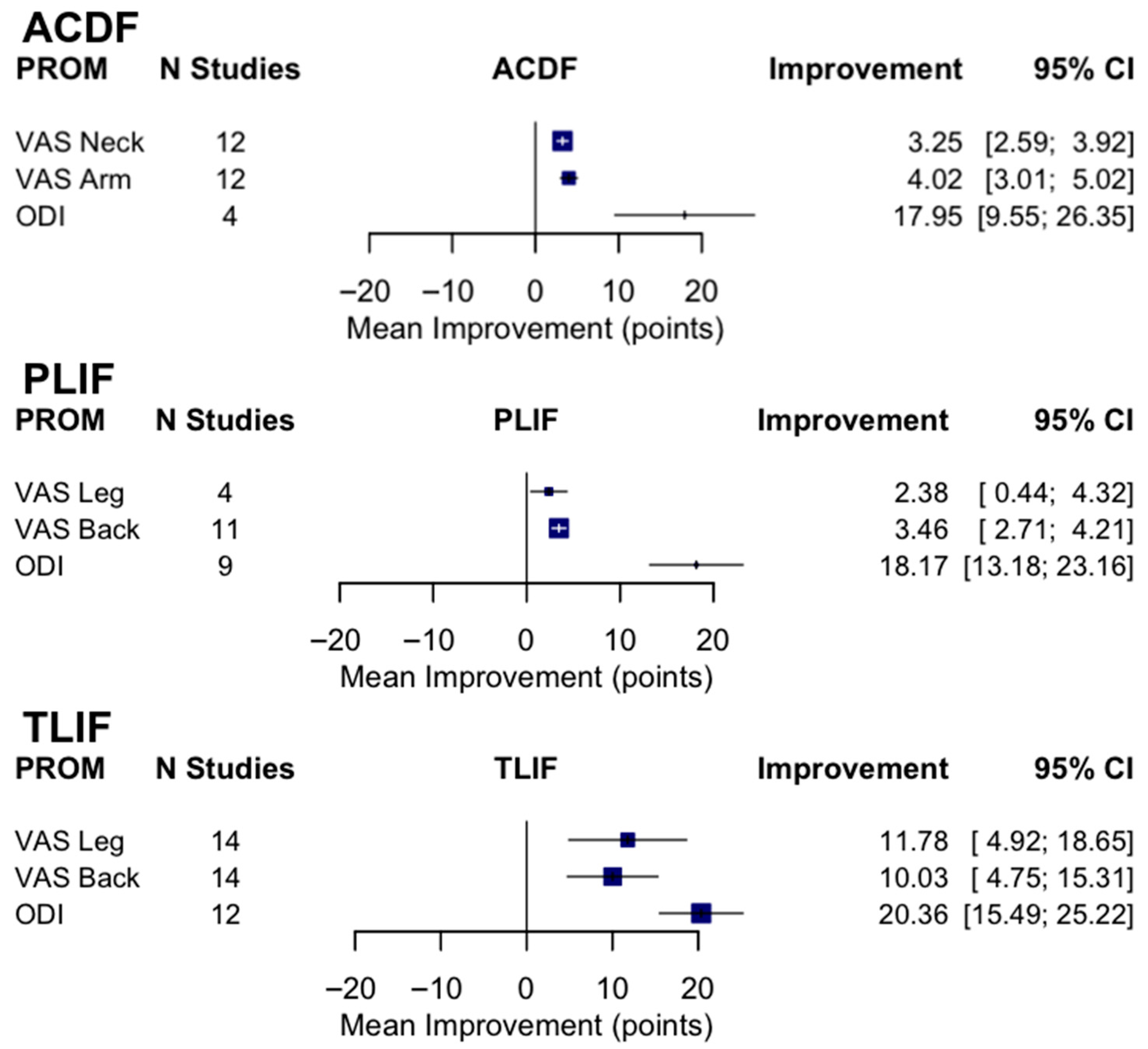
A total of 12 RCTs with a total of 968 patients assessed the efficacy of PEEK against other spinal fusion materials in ACDF procedures. Between uncoated PEEK and titanium cages, Chen et al. found no significant differences in clinical assessment for spinal fusion [25]. Across studies that compared PEEK cages that differed in stabilization properties, coating material, or bone substitutes, no significant differences were observed in fusion rates, subsidence rates, or ODI and VAS Arm/Leg scores between the comparison groups [26,27,32,45]. Notably, 24 months after surgery, the standalone anchored PEEK cage showed lower incidence of adjacent-level ossification than PEEK cages with plating (12.5% vs. 63.6%, p = 0.001) [26]. Six studies compared PEEK cages against varying fusion materials in ACDF patients [23,29,30,39,42,44]. There were no statistical differences in clinical outcomes, fusion and recovery rates, and VAS Arm/Neck between PEEK cages vs. porous silicon nitride spacers, allograft spacers, and 3D-printed titanium cages [27,32,33]. Similarly, no differences were observed between PEEK cages filled with a mixture of hydroxyapatite (HA) and β-tricalcium phosphate (β-TCP) and CaO-SiO2-P2O5-B2O3 glass–ceramic (BGS-7) spacers [23,29,39,41]. In contrast, Kostysyn et al. found that at 18 months, PEEK cages had higher fusion rates (100% vs. 92.6%) than the porous aluminum oxide cages and reported enhanced grade I fusion in the PEEK group over the aluminum oxide group (77.1% vs. 39.7%, p < 0.001) at the final follow-up (Table 3). Additionally, the pooled fusion rate was 0.92 (95% CI 0.88–0.95; I2 = 45.2%, τ2 = 0.34, p = 0.01), indicating moderate heterogeneity (Figure 2). Subgroup analyses demonstrated significant differences by cage material (χ2 = 25.30, df = 3, p < 0.0001). Pooled fusion rates were 0.93 [0.85–0.97] for PEEK, 0.91 [0.76–0.97] for coated PEEK, 0.98 [0.78–1.00] for titanium, and 0.91 [0.81–0.96] for other cage types (Figure 3). All materials performed well, with titanium and uncoated PEEK trending toward the highest estimates. Additionally, pooling across studies, ACDF improved VAS-Neck by +3.25 points [2.59–3.92] and VAS-Arm by +4.02 points [3.01–5.02]; ODI decreased by −17.95 points [−26.35 to −9.55] (Figure 4). Disability reduction was substantial, though ODI estimates are based on fewer studies.

3.3. Postoperative Fusion Outcomes in PLIF

A total of 480 patients were analyzed across six RCTs that assessed PEEK’s recovery trends against other fusion materials in PLIF procedures. Three studies compared the outcomes of uncoated PEEK cages with those of TiPEEK and CaP-PEEK cages and did not find significant differences in improvements in ODI and VAS Leg/Back following surgery [33,34,36]. However, Hasegawa et al. found that the bone fusion rates were significantly higher at 6 months after surgery in the TiPEEK group than in the PEEK group in the unadjusted modified intention-to-treat analysis (p = 0.03). Willems et al. found that nanocoated PEEK cages had significantly increased fusion rates as compared to PEEK alone after one year (PEEK: 65.6% vs. TiPEEK: 93.9%, p = 0.0034, CaP-PEEK: 88.0%, p = 0.032). Similarly, at 24 months there were no differences in fusion rates between PEEK and autologous cages using the lumbar spinous process and laminae (ACSP) [28]. However, the subsidence around the BGS-7 cages was significantly less than PEEK (p < 0.05) (Figure 3) [43]. The pooled fusion rate was 0.82 (95% CI 0.63–0.92; I2 = 85.9%, τ2 = 1.31, p < 0.0001), reflecting substantial heterogeneity (Figure 2). Subgroup analyses by cage material yielded wide confidence intervals due to the limited number of studies: 0.82 [0.43–0.97] for PEEK, 0.86 [0.27–0.99] for coated PEEK, and 0.79 [0.03–1.00] for other cage types (Figure 3). Because of heterogeneity and the wide confidence intervals, no clear differences could be determined between cage types in PLIF. In PLIF, VAS-Leg improved by +2.38 points [0.44–4.32] and VAS-Back by +3.46 points [2.71–4.21]. ODI decreased by −18.17 points [−23.16 to −13.18] (Figure 4).

3.4. Postoperative Fusion Outcomes in TLIF

A total of 598 patients were analyzed across eight RCTs that assessed PEEK against other fusion materials in TLIF procedures (Table 3, Figure 2). Toop Et Al. compared PEEK cages with activated titanium, a form of titanium cage that has received micro- and nanoscale internal and topographic cage modifications. In contrast to PEEK at 6 months, titanium cages optimized postoperative fusion (84.0% vs. 20.6%, p < 0.001) and had lower rates of subsidence (20.8% vs. 41.4%, p < 0.001) [46]. Three studies explored the clinical effects of utilizing titanium-coated PEEK (TiPEEK) over uncoated PEEK [31,38,47]. Out of the three studies, Vanek et al. found subsidence rates higher in the PEEK group (26.8%) than the TiPEEK group (5%) (p = 0.007), and Singhatanadgige et al. noted that the TiPEEK cohort demonstrated a higher fusion rate than PEEK at 6 months postoperation (91.8% vs. 76%; p = 0.03), with no difference in fusion rate and cage subsidence at 12 months. Two studies comparing Si3N4 with PEEK as the fusion cage material found no significant difference in fusion rates and VAS Leg/Back [35,37]. Villavicencio et al. reported comparable fusion rates for PEEK interbody lordotic spacers to cortical allograft spacers at 24 months (97.5% vs. 97.5%), and while all other clinical outcomes for pain and radiographic outcome data were equally positive with significant improvements in all measured outcomes (p < 0.0001), no differences were detected between the groups at any of the follow-up times [40]. Deng et al. observed statistically similar fusion rates between 3D-printed titanium cages (3DPT) and PEEK at 6 months (92.3% vs. 75%, p = 0.225) but superior bone–cage interface contact (15.4% vs. 75%, p < 0.001) and lower subsidence (0.7 ± 0.3 mm vs. 1.5 ± 0.8 mm, p < 0.001) for the 3DPT (Table 4, Figure 3) [41]. The pooled fusion rate was 0.86 (95% CI 0.78–0.92; I2 = 75.4%, τ2 = 0.86, p < 0.0001) (Figure 2). Subgroup analyses by cage material showed pooled fusion of 0.83 [0.61–0.94] for PEEK, 0.93 [0.84–0.97] for coated PEEK, 0.86 [0.15–1.00] for titanium, and 0.87 [0.35–0.99] for other cage types (Figure 3). Although confidence intervals overlapped, coated PEEK tended to outperform uncoated PEEK in TLIF. TLIF demonstrated greater absolute improvements than the other procedures, with VAS-Leg improving by +11.78 points [4.92–18.65], VAS-Back by +10.03 points [4.75–15.31], and ODI decreasing by −20.36 [−25.22 to −15.49] (Figure 4).
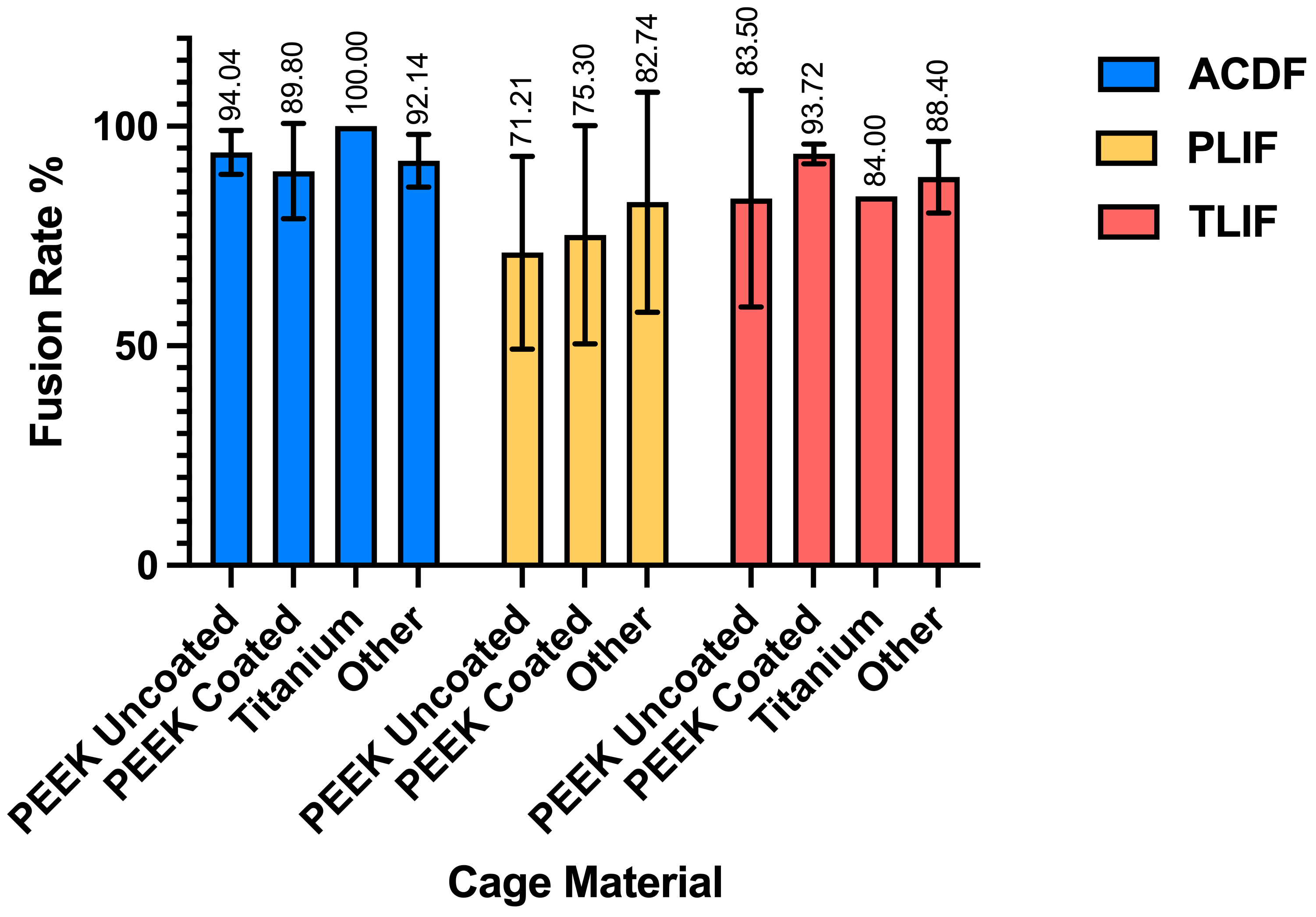
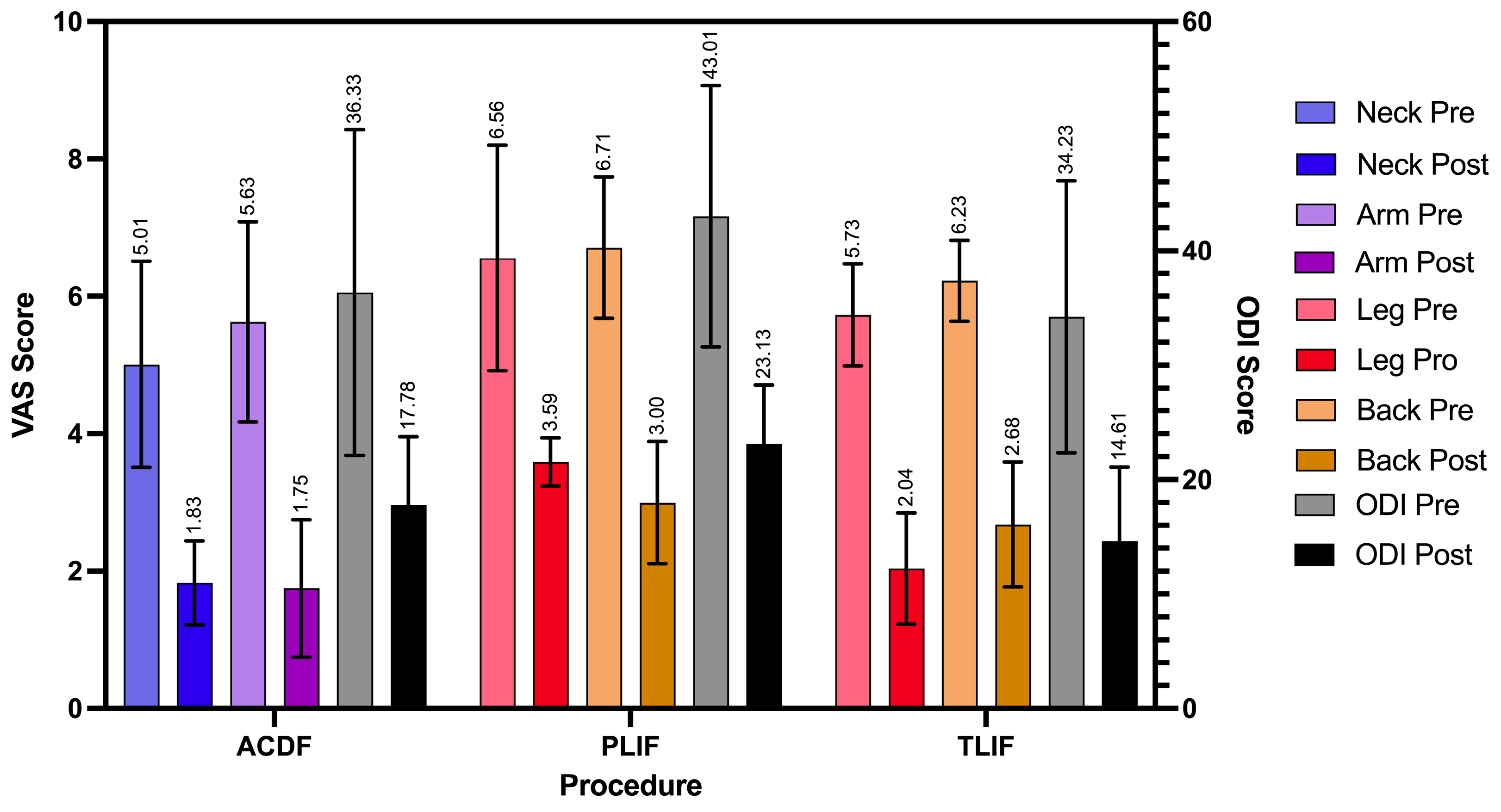
Taken together, we found high fusion rates overall but with differences by procedure and cage type: across 1946 patients, the pooled fusion rate was 89% [95% CI: 85–92%], though heterogeneity was substantial (I2 = 81.6%) (Figure 5). These analyses demonstrate consistently high fusion rates after ACDF, more variable rates after PLIF and TLIF, and procedure-specific trends suggesting that cage material may influence outcomes (Figure 2, Figure 3, Figure 4, Figure 5 and Figure 6).

4. Discussion

4.1. Study Trends

This systematic review compares the postoperative fusion rates and perioperative outcomes of PEEK with other fusion materials after ACDF, TLIF, and PLIF. After reviewing 25 RCTs, we observed varying outcomes regarding the efficacy of PEEK, coated PEEK, titanium, and other materials across procedures. For ACDF, 12 included studies often demonstrated high fusion rates for titanium, with all included subgroups of the material achieving 100% fusion. While some patient groups treated with PEEK or coated PEEK achieved 100% fusion, the average fusion rates were slightly below 100%. Uncoated PEEK performed slightly better than coated PEEK in ACDF procedures. Contrarily, the results from the six included PLIF studies show that coated PEEK outperforms uncoated PEEK in achieving higher fusion rates. Similarly, in the included studies for TLIF, coated PEEK reported higher fusion rates compared to uncoated PEEK, with titanium reporting fusion rates between the two. However, this was not uniformly the case, as some studies for both PLIF and TLIF reported no significant differences between titanium-coated and uncoated PEEK. Additionally, the heterogeneity of included trials and small sample sizes serves as a limitation for making definitive conclusions. This review demonstrates that the performance of PEEK, coated PEEK, and titanium is context-dependent, and there is no clear difference in fusion rates between the titanium and titanium-coated PEEK interbodies for achieving successful fusion. These results also highlight the advantages of material and design-specific modifications to titanium and PEEK cages in PLIFs and TLIFs.

4.2. Historical Trends

The historical evolution of spinal fusion materials provides critical context to this review’s findings. When spinal fusions via interbody cages first became popularized, titanium was among the earliest materials used due to its strength, corrosion resistance, biocompatibility, and bone ingrowth promotion [18,48,49,50]. However, titanium possesses a mismatch in the elastic modulus of the vertebrae and cortical bone, which can lead to stress shielding and subsequent graft subsidence [18,48,49,50]. Additionally, the high radiodensity of titanium causes imaging artifacts that make postoperative imaging difficult to assess fusion status [18,50]. These challenges align with this review’s findings, where titanium showed strong early fusion outcomes but was occasionally associated with higher subsidence rates compared to bioactive-coated PEEK.
In response to limitations of titanium interbody cages, PEEK emerged in the early 1990s in the hopes of increasing fusion rates [49]. Unlike titanium, PEEK has a modulus of elasticity similar to vertebrae and cortical bone, causing less stress shielding and subsidence [18,48,49,50]. Additionally, PEEK displays no radiographical artifacts, contributing to greater accuracy with postoperative imaging [18]. Due to these benefits, PEEK has become the most commonly used interbody material since the late 1990s [49]. Evidenced in this review, this trend is for good reason: At 18 months, the uncoated PEEK cage group had higher fusion rates and grade I fusion than the porous aluminum oxide cage group in ACDF [42]. Additionally, the majority of ACDF studies reported no significant differences in clinical assessment between PEEK and other cage materials. In two studies, PEEK also demonstrated lower complication rates or incidence of adjacent-level ossification, proving the reliability of PEEK cages. However, because PEEK is bioinert and hydrophobic, it has a limited osteointegration capacity which impedes fusion capability [18,48,49,50]. Additionally, potential biofilm formation of PEEK cage surfaces can disrupt binding to the host bone which can impede solid fusion [48]. Thus, continued innovation has introduced coated PEEK cages to preserve the modulus of elasticity of PEEK while promoting bioactivity.
Combining the benefits of titanium and PEEK interbody cages, TiPEEK has become a popular material for spinal fusions. TiPEEK, a version of titanium-coated PEEK, was created to increase the osteointegration of uncoated PEEK, one of PEEK’s drawbacks [51,52]. Other bioactive materials, such as hydroxyapatite and calcium, have also been used to coat PEEK in hopes of promoting its bioactivity [33,53].
Likewise, this study also analyzes postoperative outcomes for PEEK and coated PEEK, allowing for comparisons in fusion rates and clinical outcomes. While PEEK shows clinically acceptable fusion rates for ACDF, PLIF, and TLIF, its ability to be modified by coatings allows for enhanced bioactivity which can lead to greater fusion rates compared to titanium and uncoated PEEK. These significant improvements in fusion outcomes observed with studies’ modifications to PEEK, such as TiPEEK and CaP-PEEK, suggest closed performance gaps specifically in PLIF and TLIF, supporting their clinical relevance. Amidst these advancements, the landscape of spinal fusion materials has continued to diversify. Materials such as bioactive ceramics, 3D-printed titanium, and acrylic cages are being explored for their potential to improve fusion rates and biocompatibility [18,29,30]. In this systematic review, advancements in titanium, such as activated and 3D-printed titanium cages, demonstrate advantages in fusion rates, subsidence, and bone–cage interface contact compared to traditional PEEK cages in TLIFs. In addition, bioactive ceramics such as silicon nitride demonstrated comparable fusion outcomes to PEEK with potential advantages in reducing imaging artifacts. These developments underscore a broader trend in biomedical engineering towards materials that can actively contribute to biological processes, potentially leading to faster healing and better long-term outcomes.

4.3. Upcoming Materials

Although progress has been made with spinal fusion materials, challenges remain in achieving optimal fusion, particularly in complex cases or in patients with underlying health. As the field progresses, new materials are being explored in the hopes of improving long-term outcomes, including improvements in 3D printing. Recent technical improvements have created the opportunity for 3D printing with PEEK, in which a nano-rough surface with antibacterial characteristics can promote fusion similarly to TiPEEK [18]. Another future direction includes bioactive glass which aims to provide a balance of strength, flexibility, and bioactivity that could potentially outperform current materials [44,54]. Additionally, more recent studies have shown that 3D-printed tantalum might be a future direction for cage materials, as early results show that tantalum promotes osseointegration and excellent intervertebral fusion [55]. The integration of biologically active agents directly into the cage materials, such as TiPEEK and CaP-PEEK, also possesses the potential for future improvement in fusion surgery outcomes.

4.4. Clinical Implications and Decision-Making Framework

Given the comparable fusion rates and clinical outcomes associated with PEEK, coated PEEK, and titanium cages, material selection should be guided by procedure type, patient characteristics, and imaging needs. For ACDF procedures, uncoated PEEK may be beneficial due to its radiolucency and reduced imaging artifact, therefore facilitating postoperative assessment of fusion status. Conversely, coated PEEK and titanium cages may offer improved early fusion rates in PLIF and TLIF procedures and may be advantageous in surgical cases where enhanced osteointegration is desired. Furthermore, surgeon familiarity, cost factors, and institutional availability remain key aspects for consideration in cage selection. Recent studies have shown that overall hospital costs for spinal fusion performed with PEEK cages have been slightly lower than those of titanium cages, and cost may be a deciding factor for some patients and surgeons [56]. Additionally, some studies have shown that titanium coatings may shear off coated PEEK cages, posing a possible risk to patients, which might also impact surgical decisions of which interbody cages to use [57]. This individualized, context-dependent approach may enhance surgical planning and optimize patient outcomes across spinal fusion procedures. However, with continuous improvements to PEEK technology, PEEK possesses the ability to become an even more effective material for spinal fusion surgeries, possibly overtaking other cage materials.

4.5. Limitations

Although our study synthesizes findings from multiple RCTs assessing the efficacy of various spinal fusion materials, there are limitations that should be acknowledged. The selected RCTs contain variance in patient populations, surgical techniques, and follow-up duration, complicating the generalizability of the results. Outcome measures like blood loss and operative time are also heavily influenced by the complexity of the surgical case, introducing additional variability to the RCTs. Additionally, some studies lack detailed demographic data which limits our understanding of results across various populations, thus limiting our ability to create patient-specific treatment strategies. Furthermore, many trials have relatively small sample sizes and most RCTs have follow-up periods shorter than 24 months, limiting our ability to draw definitive conclusions about the long-term effectiveness and safety of the materials studied. Additionally, we were unable to stratify by early vs. long-term follow-up because studies used inconsistent timepoints and definitions of fusion; instead, we prioritized each trial’s final reported follow-up to maximize comparability. Heterogeneity is high in lumbar procedures (PLIF AND TLIF), which reflects variability in study design, patient populations, and outcome reporting. Furthermore, the assessment methods for bone fusion vary among the included studies, adding to the heterogeneity in these studies. As such, while our pooled estimates provide an overall synthesis, they should be considered in the context of underlying heterogeneity across studies. In addition, not all included trials reported measures of variance or subgroup sample sizes for key perioperative outcomes (e.g., operative time, hospital stay, blood loss in PLIF). Lastly, some studies might be subject to publication bias, as studies with positive outcomes are more likely to be published, skewing the data towards more favorable results.

5. Conclusions

The findings of this systematic review illustrate the postoperative efficacy of PEEK cages and other commonly used fusion materials across a wide array of surgical approaches. The analysis of 25 RCT studies revealed that both uncoated and coated PEEK and titanium show clinically acceptable fusion rates in specific scenarios, but their efficacy can vary depending on the surgical context and material properties. While titanium cages demonstrate the highest fusion rates for ACDF procedures, there is no clear difference in fusion rates between titanium and coated PEEK for both TLIF and PLIF procedures, additionally confirmed by meta-analytic pooling. This study demonstrates that PEEK has acceptable baseline fusion rates for all procedures, and the introduction of coatings to PEEK can further increase fusion rates, principally in PLIF and TLIF. Thus, as improvements are made to PEEK technology, PEEK possesses the ability to become an even more effective material for spinal fusion surgeries, overtaking previously used cage materials. However, given the variability observed across different types of procedures and materials, it is imperative that future research continues to explore and define the optimal conditions under which each material can achieve the best outcomes. More detailed studies are required to substantiate these findings and help refine surgical practices, ultimately enhancing patient recovery and long-term success in spinal fusion surgeries.

Supplementary Materials

The following supporting information can be downloaded at: https://www.mdpi.com/article/10.3390/jcm14217696/s1, PRISMA 2020 Checklist [58].

Author Contributions

Conceptualization/methodology, J.K., R.J.K., A.V., R.W.T., and J.W.Y.; validation, D.D.L., D.C., M.M.D., R.J.C., H.S.A., Y.G., and B.G.; formal analysis, J.K., R.J.K., A.V., R.W.T., and D.D.L.; investigation, J.K., R.J.K., A.V., and R.W.T.; data curation, J.K., R.J.K., A.V., and R.W.T.; writing—original draft preparation, J.K., R.J.K., and A.V.; writing—review and editing, D.C., M.M.D., R.J.C., H.S.A., Y.G., B.G., and J.W.Y.; visualization, J.K. and R.J.K.; supervision, R.W.T., D.C., and J.W.Y.; project administration, R.W.T., D.C., and J.W.Y.; All authors have read and agreed to the published version of the manuscript.

Funding

This research did not receive any specific grant from funding agencies in the public, commercial, or not-for-profit sectors.

Data Availability Statement

The original data presented in the study are openly available in PubMed, CINAHL, Scopus, Embase, and Web of Science.

Conflicts of Interest

Dr. Yoon is a consultant for DePuy Synthes, Pacira, and Biedermann Motech, with direct stock ownership in Kinesiometrics Inc. and Medcyclops LLC. For the remaining authors, none were declared.

Abbreviations

The following abbreviations are used in this manuscript:
ACDFAnterior Cervical Discectomy and Fusion
PLIFPosterior Lumbar Interbody Fusion
TLIFTransforaminal Lumbar Interbody Fusion
PEEKPolyetheretherketone
TiPEEKTitanium-coated Polyetheretherketone
HAHydroxyapatite
β-TCPBeta-Tricalcium Phosphate
BGS-7CaO-SiO2-P2O5-B2O3 Glass-Ceramic Spacer
ODIOswestry Disability Index
VASVisual Analog Scale
RCTRandomized Controlled Trial
GRADEGrading of Recommendations, Assessment, Development, and Evaluation
3DPT3D-Printed Titanium
ACSPAutologous Cage using Spinous Process/Laminae
Si3N4Silicon Nitride

References

  1. Mobbs, R.J.; Phan, K.; Malham, G.; Seex, K.; Rao, P.J. Lumbar interbody fusion: Techniques, indications and comparison of interbody fusion options including PLIF, TLIF, MI-TLIF, OLIF/ATP, LLIF and ALIF. J. Spine Surg. 2015, 1, 2–18. [Google Scholar] [CrossRef]
  2. Reid, P.C.; Morr, S.; Kaiser, M.G. State of the union: A review of lumbar fusion indications and techniques for degenerative spine disease. J. Neurosurg. Spine 2019, 31, 1–14. [Google Scholar] [CrossRef]
  3. Viola, A., III; Appiah, J.; Donnally, C.J., III; Kim, Y.H.; Shenoy, K. Bone Graft Options in Spinal Fusion: A Review of Current Options and the Use of Mesenchymal Cellular Bone Matrices. World Neurosurg. 2022, 158, 182–188. [Google Scholar] [CrossRef]
  4. Cowan, J.A., Jr.; Dimick, J.B.; Wainess, R.; Upchurch, G.R., Jr.; Chandler, W.F.; La Marca, F. Changes in the utilization of spinal fusion in the United States. Neurosurgery 2006, 59, 15–20; discussion 15–20. [Google Scholar] [CrossRef]
  5. Cruz, A.; Ropper, A.E.; Xu, D.S.; Bohl, M.; Reece, E.M.; Winocour, S.J.; Buchanan, E.; Kaung, G. Failure in Lumbar Spinal Fusion and Current Management Modalities. Semin. Plast. Surg. 2021, 35, 54–62. [Google Scholar] [CrossRef] [PubMed]
  6. Martin, B.I.; Mirza, S.K.; Spina, N.; Spiker, W.R.; Lawrence, B.; Brodke, D.S. Trends in Lumbar Fusion Procedure Rates and Associated Hospital Costs for Degenerative Spinal Diseases in the United States, 2004 to 2015. Spine 2019, 44, 369–376. [Google Scholar] [CrossRef] [PubMed]
  7. Reisener, M.J.; Pumberger, M.; Shue, J.; Girardi, F.P.; Hughes, A.P. Trends in lumbar spinal fusion-a literature review. J. Spine Surg. 2020, 6, 752–761. [Google Scholar] [CrossRef] [PubMed]
  8. Riew, K.D.; Ecker, E.; Dettori, J.R. Anterior cervical discectomy and fusion for the management of axial neck pain in the absence of radiculopathy or myelopathy. Evid.-Based Spine-Care J. 2010, 1, 45–50. [Google Scholar] [CrossRef]
  9. Chong, E.; Pelletier, M.H.; Mobbs, R.J.; Walsh, W.R. The design evolution of interbody cages in anterior cervical discectomy and fusion: A systematic review. BMC Musculoskelet. Disord. 2015, 16, 99. [Google Scholar] [CrossRef]
  10. Jain, S.; Eltorai, A.E.; Ruttiman, R.; Daniels, A.H. Advances in Spinal Interbody Cages. Orthop. Surg. 2016, 8, 278–284. [Google Scholar] [CrossRef]
  11. Johnson, S.E.; Michalopoulos, G.D.; Flanigan, P.M.; Katsos, K.; Ibrahim, S.; Freedman, B.A.; Bydon, M. Interbody cages versus structural bone grafts in lumbar arthrodesis: A systematic review and meta-analysis. J. Neurosurg. Spine 2024, 41, 188–198. [Google Scholar] [CrossRef]
  12. Patel, D.V.; Yoo, J.S.; Karmarkar, S.S.; Lamoutte, E.H.; Singh, K. Interbody options in lumbar fusion. J. Spine Surg. 2019, 5 (Suppl. S1), S19–S24. [Google Scholar] [CrossRef] [PubMed]
  13. Fogel, G.; Martin, N.; Williams, G.M.; Unger, J.; Yee-Yanagishita, C.; Pelletier, M.; Walsh, W.; Peng, Y.; Jekir, M. Choice of Spinal Interbody Fusion Cage Material and Design Influences Subsidence and Osseointegration Performance. World Neurosurg. 2022, 162, e626–e634. [Google Scholar] [CrossRef]
  14. Kim, D.Y.; Kwon, O.H.; Park, J.Y. Comparison Between 3-Dimensional-Printed Titanium and Polyetheretherketone Cages: 1-Year Outcome After Minimally Invasive Transforaminal Interbody Fusion. Neurospine 2022, 19, 524–532. [Google Scholar] [CrossRef]
  15. Ahmed, A.F.; Al Dosari, M.A.A.; Al Kuwari, A.; Khan, N.M. The outcomes of stand alone polyetheretherketone cages in anterior cervical discectomy and fusion. Int. Orthop. 2021, 45, 173–180. [Google Scholar] [CrossRef]
  16. Li, G.; Yang, L.; Wu, G.; Qian, Z.; Li, H. An update of interbody cages for spine fusion surgeries: From shape design to materials. Expert Rev. Med. Devices 2022, 19, 977–989. [Google Scholar] [CrossRef]
  17. Seaman, S.; Kerezoudis, P.; Bydon, M.; Torner, J.C.; Hitchon, P.W. Titanium vs. polyetheretherketone (PEEK) interbody fusion: Meta-analysis and review of the literature. J. Clin. Neurosci. 2017, 44, 23–29. [Google Scholar] [CrossRef]
  18. Laubach, M.; Kobbe, P.; Hutmacher, D.W. Biodegradable interbody cages for lumbar spine fusion: Current concepts and future directions. Biomaterials 2022, 288, 121699. [Google Scholar] [CrossRef]
  19. Muthiah, N.; Yolcu, Y.U.; Alan, N.; Agarwal, N.; Hamilton, D.K.; Ozpinar, A. Evolution of polyetheretherketone (PEEK) and titanium interbody devices for spinal procedures: A comprehensive review of the literature. Eur. Spine J. 2022, 31, 2547–2556. [Google Scholar] [CrossRef] [PubMed]
  20. Panayotov, I.V.; Orti, V.; Cuisinier, F.; Yachouh, J. Polyetheretherketone (PEEK) for medical applications. J. Mater. Sci. Mater. Med. 2016, 27, 118. [Google Scholar] [CrossRef] [PubMed]
  21. Yao, Y.C.; Chou, P.H.; Lin, H.H.; Wang, S.T.; Chang, M.C. Outcome of Ti/PEEK Versus PEEK Cages in Minimally Invasive Transforaminal Lumbar Interbody Fusion. Glob. Spine J. 2023, 13, 472–478. [Google Scholar] [CrossRef]
  22. Guyatt, G.; Oxman, A.D.; Akl, E.A.; Kunz, R.; Vist, G.; Brozek, J.; Norris, S.; Falck-Ytter, Y.; Glasziou, P.; DeBeer, H.; et al. GRADE guidelines: 1. Introduction-GRADE evidence profiles and summary of findings tables. J. Clin. Epidemiol. 2011, 64, 383–394. [Google Scholar] [CrossRef]
  23. Cho, D.Y.; Lee, W.Y.; Sheu, P.C. Treatment of multilevel cervical fusion with cages. Surg. Neurol. 2004, 62, 378–385; discussion 385. [Google Scholar] [CrossRef] [PubMed]
  24. Jiya, T.U.; Smit, T.; van Royen, B.J.; Mullender, M. Posterior lumbar interbody fusion using non resorbable poly-ether-ether-ketone versus resorbable poly-L-lactide-co-D,L-lactide fusion devices. Clinical outcome at a minimum of 2-year follow-up. Eur. Spine J. 2011, 20, 618–622. [Google Scholar] [CrossRef] [PubMed]
  25. Chen, Y.; Wang, X.; Lu, X.; Yang, L.; Yang, H.; Yuan, W.; Chen, D. Comparison of titanium and polyetheretherketone (PEEK) cages in the surgical treatment of multilevel cervical spondylotic myelopathy: A prospective, randomized, control study with over 7-year follow-up. Eur. Spine J. 2013, 22, 1539–1546. [Google Scholar] [CrossRef]
  26. Nemoto, O.; Kitada, A.; Naitou, S.; Tachibana, A.; Ito, Y.; Fujikawa, A. Stand-alone anchored cage versus cage with plating for single-level anterior cervical discectomy and fusion: A prospective, randomized, controlled study with a 2-year follow-up. Eur. J. Orthop. Surg. Traumatol. 2015, 25, 127–134. [Google Scholar] [CrossRef] [PubMed]
  27. Xie, Y.; Li, H.; Yuan, J.; Fu, L.; Yang, J.; Zhang, P. A prospective randomized comparison of PEEK cage containing calcium sulphate or demineralized bone matrix with autograft in anterior cervical interbody fusion. Int. Orthop. 2015, 39, 1129–1136. [Google Scholar] [CrossRef]
  28. Lin, B.; Yu, H.; Chen, Z.; Huang, Z.; Zhang, W. Comparison of the PEEK cage and an autologous cage made from the lumbar spinous process and laminae in posterior lumbar interbody fusion. BMC Musculoskelet. Disord. 2016, 17, 374. [Google Scholar] [CrossRef]
  29. Arts, M.P.; Wolfs, J.F.C.; Corbin, T.P. Porous silicon nitride spacers versus PEEK cages for anterior cervical discectomy and fusion: Clinical and radiological results of a single-blinded randomized controlled trial. Eur. Spine J. 2017, 26, 2372–2379. [Google Scholar] [CrossRef]
  30. Farrokhi, M.R.; Nikoo, Z.; Gholami, M.; Hosseini, K. Comparison Between Acrylic Cage and Polyetheretherketone (PEEK) Cage in Single-level Anterior Cervical Discectomy and Fusion: A Randomized Clinical Trial. Clin. Spine Surg. 2017, 30, 38–46. [Google Scholar] [CrossRef]
  31. Rickert, M.; Fleege, C.; Tarhan, T.; Schreiner, S.; Makowski, M.R.; Rauschmann, M.; Arabmotlagh, M. Transforaminal lumbar interbody fusion using polyetheretherketone oblique cages with and without a titanium coating. Bone Jt. J. 2017, 99-B, 1366–1372. [Google Scholar] [CrossRef] [PubMed]
  32. Feng, S.W.; Chang, M.C.; Chou, P.H.; Lin, H.; Wang, S.T.; Liu, C.L. Implantation of an empty polyetheretherketone cage in anterior cervical discectomy and fusion: A prospective randomised controlled study with 2 years follow-up. Eur. Spine J. 2018, 27, 1358–1364. [Google Scholar] [CrossRef]
  33. Willems, K.; Lauweryns, P.; Verleye, G.; Van Goethem, J. Randomized controlled trial of posterior lumbar interbody fusion with Ti- and cap-nanocoated polyetheretherketone cages: Comparative study of the 1-year radiological and clinical outcome. Int. J. Spine Surg. 2019, 13, 575–587. [Google Scholar] [CrossRef] [PubMed]
  34. Hasegawa, T.; Ushirozako, H.; Shigeto, E.; Ohba, T.; Oba, H.; Mukaiyama, K.; Shimizu, S.; Yamato, Y.; Ide, K.; Shibata, S.; et al. The Titanium-coated PEEK Cage Maintains Better Bone Fusion with the Endplate Than the PEEK Cage 6 Months After PLIF Surgery: A Multicenter, Prospective, Randomized Study. Spine 2020, 45, E892–E902. [Google Scholar] [CrossRef]
  35. McEntire, B.J.; Maslin, G.; Sonny Bal, B. Two-year results of a double-blind multicenter randomized controlled non-inferiority trial of polyetheretherketone (Peek) versus silicon nitride spinal fusion cages in patients with symptomatic degenerative lumbar disc disorders. J. Spine Surg. 2020, 6, 523–540. [Google Scholar] [CrossRef] [PubMed]
  36. Schnake, K.J.; Fleiter, N.; Hoffmann, C.; Pingel, A.; Scholz, M.; Langheinrich, A.; Kandziora, F. PLIF surgery with titanium-coated PEEK or uncoated PEEK cages: A prospective randomised clinical and radiological study. Eur. Spine J. 2021, 30, 114–121. [Google Scholar] [CrossRef]
  37. Kersten, R.F.; van Gaalen, S.M.; Arts, M.P.; Roes, K.C.; de Gast, A.; Corbin, T.P.; Oner, F. The SNAP trial: A double blind multi-center randomized controlled trial of a silicon nitride versus a PEEK cage in transforaminal lumbar interbody fusion in patients with symptomatic degenerative lumbar disc disorders: Study protocol. BMC Musculoskelet. Disord. 2014, 15, 57. [Google Scholar] [CrossRef]
  38. Singhatanadgige, W.; Tangchitcharoen, N.; Kerr, S.J.; Tanasansomboon, T.; Yingsakmongkol, W.; Kotheeranurak, V.; Limthongkul, W. A Comparison of Polyetheretherketone and Titanium-Coated Polyetheretherketone in Minimally Invasive Transforaminal Lumbar Interbody Fusion: A Randomized Clinical Trial. World Neurosurg. 2022, 168, e471–e479. [Google Scholar] [CrossRef]
  39. Villavicencio, A.T.; Nelson, E.L.; Rajpal, S.; Beasley, K.; Burneikiene, S. Prospective, Randomized, Blinded Clinical Trial Comparing PEEK and Allograft Spacers in Patients Undergoing Anterior Cervical Discectomy and Fusion Surgeries. Spine 2022, 47, 1043–1054. [Google Scholar] [CrossRef]
  40. Villavicencio, A.T.; Nelson, E.L.; Rajpal, S.; Beasley, K.; Burneikiene, S. Prospective, randomized, double-blinded clinical trial comparing PEEK and allograft spacers in patients undergoing transforaminal lumbar interbody fusion surgeries. Spine J. 2022, 22, 84–94. [Google Scholar] [CrossRef]
  41. Deng, Z.; Zou, Q.; Wang, L.; Wang, L.; Xiu, P.; Feng, G.; Song, Y.; Yang, X. Comparison between Three-Dimensional Printed Titanium and PEEK Cages for Cervical and Lumbar Interbody Fusion: A Prospective Controlled Trial. Orthop. Surg. 2023, 15, 2889–2900. [Google Scholar] [CrossRef]
  42. Kostysyn, R.; Ryska, P.; Jandura, J.; Selke-Krulichova, I.; Poczos, P.; Hosszu, T.; Cesak, T. Speed and quality of interbody fusion in porous bioceramic Al2O3 and polyetheretherketone cages for anterior cervical discectomy and fusion: A comparative study. J. Orthop. Surg. Res. 2023, 18, 165. [Google Scholar] [CrossRef] [PubMed]
  43. Kwon, B.T.; Kim, H.J.; Lee, S.; Park, S.M.; Ham, D.W.; Park, H.J.; Kwon, O.; Yeom, J.S. Feasibility and safety of a CaO-SiO2-P2O5-B2O3 bioactive glass ceramic spacer in posterior lumbar interbody fusion compared with polyetheretherketone cage: A prospective randomized controlled trial. Acta Neurochir. 2023, 165, 135–144. [Google Scholar] [CrossRef]
  44. Park, J.; Park, S.M.; Ham, D.W.; Hong, J.Y.; Kim, H.J.; Yeom, J.S. Anterior Cervical Discectomy and Fusion Performed Using a CaO-SiO2-P2O5-B2O3 Bioactive Glass Ceramic or Polyetheretherketone Cage Filled with Hydroxyapatite/β-Tricalcium Phosphate: A Prospective Randomized Controlled Trial. J. Clin. Med. 2023, 12, 4069. [Google Scholar] [CrossRef]
  45. Schröder, J.; Kampulz, T.; Bajaj, S.K.; Hellwig, G.; Winking, M. PEEK Cages versus Titanium-Coated PEEK Cages in Single-Level Anterior Cervical Fusion: A Randomized Controlled Study. J. Neurol. Surg. Part A Cent. Eur. Neurosurg. 2023, 85, 262–268. [Google Scholar] [CrossRef]
  46. Toop, N.; Dhaliwal, J.; Gifford, C.S.; Gibbs, D.; Keister, A.; Miracle, S.; Forghani, R.; Grossbach, A.; Farhadi, F. Promotion of higher rates of early fusion using activated titanium versus polyetheretherketone cages in adults undergoing 1- and 2-level transforaminal lumbar interbody fusion procedures: A randomized controlled trial. J. Neurosurg. Spine 2023, 39, 709–718. [Google Scholar] [CrossRef]
  47. Vanek, P.; Svoboda, N.; Bradac, O.; Malik, J.; Kaiser, R.; Netuka, D. Clinical and radiological results of TLIF surgery with titanium-coated PEEK or uncoated PEEK cages: A prospective single-centre randomised study. Eur. Spine J. 2024, 33, 332–338. [Google Scholar] [CrossRef]
  48. Chang, S.Y.; Kang, D.-H.; Cho, S.K. Innovative Developments in Lumbar Interbody Cage Materials and Design: A Comprehensive Narrative Review. Asian Spine J. 2024, 18, 444–457. [Google Scholar] [CrossRef]
  49. Gelfand, B.W.; Paek, S.; Zelenty, W.D.; Paek, S.; Zelenty, W.D.; Girardi, F.P. History and current state of interbody fusion device material science. Semin. Spine Surg. 2022, 34, 100972. [Google Scholar] [CrossRef]
  50. Phan, K.; Mobbs, R.J. Evolution of Design of Interbody Cages for Anterior Lumbar Interbody Fusion. Orthop. Surg. 2016, 8, 270–277. [Google Scholar] [CrossRef] [PubMed]
  51. Han, C.M.; Lee, E.J.; Kim, H.E.; Koh, Y.H.; Kim, K.N.; Ha, Y.; Kuh, S. The electron beam deposition of titanium on polyetheretherketone (PEEK) and the resulting enhanced biological properties. Biomaterials 2010, 31, 3465–3470. [Google Scholar] [CrossRef] [PubMed]
  52. Wu, X.; Liu, X.; Wei, J.; Ma, J.; Deng, F.; Wei, S. Nano-TiO2/PEEK bioactive composite as a bone substitute material: In vitro and in vivo studies. Int. J. Nanomed. 2012, 7, 1215–1225. [Google Scholar] [CrossRef]
  53. Litak, J.; Czyzewski, W.; Szymoniuk, M.; Pastuszak, B.; Litak, J.; Litak, G.; Grochowski, C.; Rahnama-Hezavah, M.; Kamieniak, P. Hydroxyapatite Use in Spine Surgery-Molecular and Clinical Aspect. Materials 2022, 15, 2906. [Google Scholar] [CrossRef]
  54. Cottrill, E.; Pennington, Z.; Lankipalle, N.; Ehresman, J.; Valencia, C.; Schilling, A.; Feghali, J.; Perdomo-Pantoja, A.; Theodore, N.; Sciubba, D.; et al. The effect of bioactive glasses on spinal fusion: A cross-disciplinary systematic review and meta-analysis of the preclinical and clinical data. J. Clin. Neurosci. 2020, 78, 34–46. [Google Scholar] [CrossRef]
  55. Liang, H.; Tu, J.; Wang, B.; Song, Y.; Wang, K.; Zhao, K.; Hua, W.; Li, S.; Tan, L.; Feng, X.; et al. Innovative 3D-printed porous tantalum cage with non-window design to accelerate spinal fusion: A proof-of-concept study. Mater. Today Bio. 2025, 31, 101576. [Google Scholar] [CrossRef]
  56. Corso, K.A.; Teferra, A.A.; Michielli, A.; Corrado, K.; Marcini, A.; Lotito, M.; Smith, C.; Costa, M.; Ruppenkamp, J.; Wallace, A. Evaluation of Healthcare Outcomes of Patients Treated with 3D-Printed-Titanium and PEEK Cages During Fusion Procedures in the Lumbar Spine. Med. Devices Evid. Res. 2025, 18, 37–51. [Google Scholar] [CrossRef]
  57. Kienle, A.; Graf, N.; Wilke, H.J. Does impaction of titanium-coated interbody fusion cages into the disc space cause wear debris or delamination? Spine J. 2016, 16, 235–242. [Google Scholar] [CrossRef]
  58. Page, M.J.; McKenzie, J.E.; Bossuyt, P.M.; Boutron, I.; Hoffmann, T.C.; Mulrow, C.D.; Shamseer, L.; Tetzlaff, J.M.; Akl, E.A.; Brennan, S.E.; et al. The PRISMA 2020 statement: An updated guideline for reporting systematic reviews. BMJ 2021, 372, n71. [Google Scholar] [CrossRef] [PubMed]
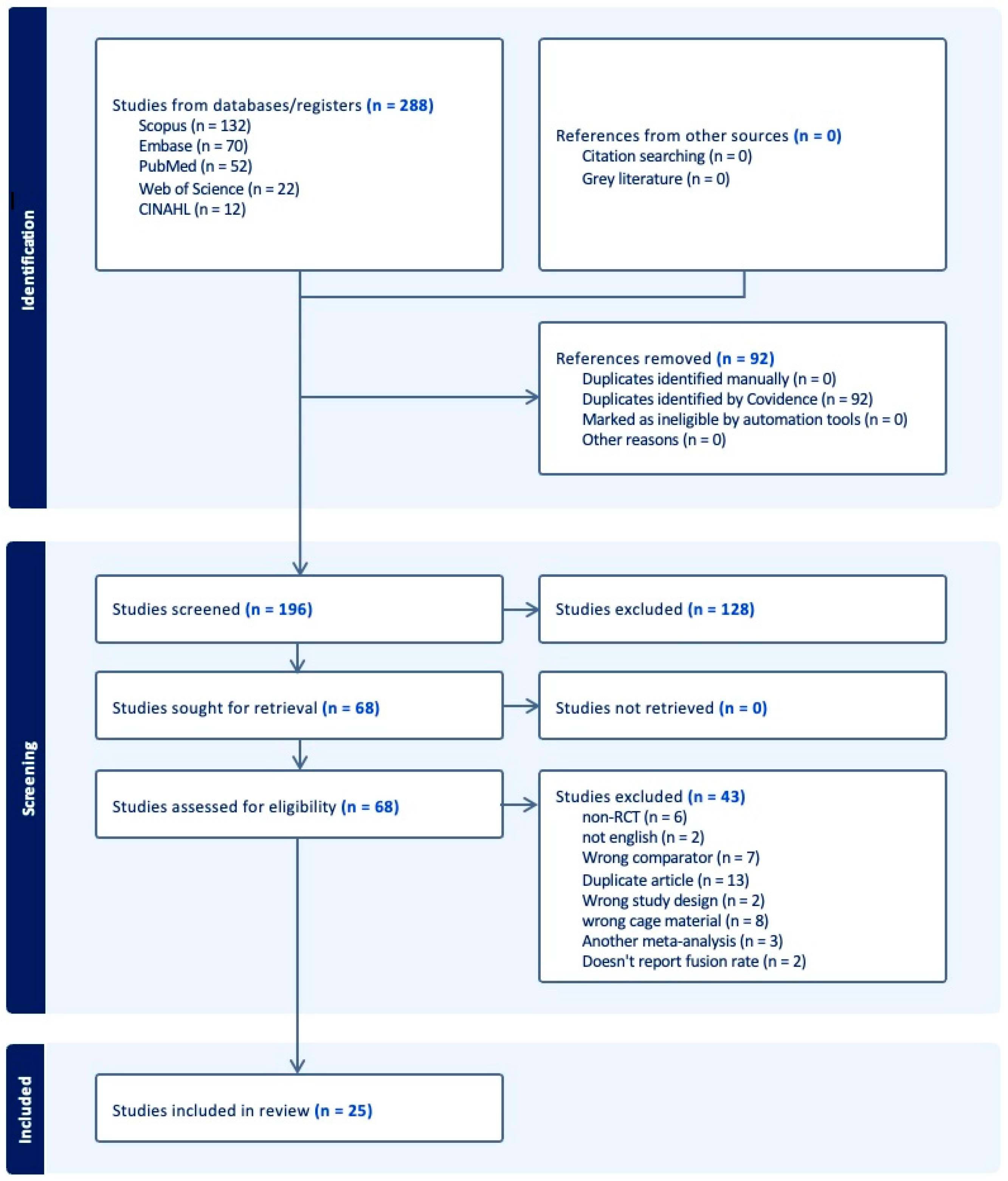
Figure 1. PRISMA diagram.
Jcm 14 07696 g001
Figure 2. Overall pooled fusion rates stratified by procedure and by material [23,24,25,26,27,28,30,31,32,33,34,35,36,37,38,39,40,41,42,43,44,45,46,47].
Jcm 14 07696 g002
Figure 3. Random-effects meta-analysis of fusion rates by cage material within each procedure (ACDF, PLIF, TLIF) [23,24,25,26,27,28,30,31,32,33,34,35,36,37,38,39,40,41,42,43,44,45,46,47].
Jcm 14 07696 g003
Figure 4. Random-effects pooled mean change in patient-reported outcomes (PROMs) by procedure.
Jcm 14 07696 g004
Figure 5. Bar graph of fusion rates by surgery type and fusion cage material.
Jcm 14 07696 g005
Figure 6. Bar graph of VAS (Neck, Arm, Leg, Back) and ODI scores pre- and postoperation. Studies assessing ACDF typically reported ODI scores. However, three reported Neck Disability Index (NDI) and were included in the analysis.
Jcm 14 07696 g006
Table 1. Baseline characteristics of included studies.
Author(s), YearProcedureControl GroupControl (N)Case GroupCase (N)CountryFinal Quality Grade
Cho et al., 2004 [23]ACDF(1) AICG fixation
(2) AICG and plate fixation
(1) 70
(2) 50
PEEK cage60ChinaModerate
Jiya et al., 2011 [24]PLIFPLDLLA cage14PEEK cage12The NetherlandsModerate
Chen et al., 2013 [25]ACDFTitanium cage29PEEK cage31ChinaHigh
Nemoto et al., 2015 [26]ACDFPEEK cage with plating22Standalone anchored PEEK cage24JapanModerate
Xie et al., 2015 [27]ACDFPEEK cage containing IAB33PEEK cage containing CS/DBM35ChinaHigh
Lin et al., 2016 [28]PLIFACSP34PEEK cage35ChinaModerate
Arts et al., 2017 [29]ACDFPorous silicon nitride spacer52PEEK cage containing autograft from osteophyte48The NetherlandsModerate
Farrokhi et al., 2017 [30]ACDFAcrylic cage filled with bone substitute32PEEK cage32IranModerate
Rickert et al., 2017 [31]TLIFTiPEEK cage20PEEK cage20GermanyModerate
Feng et al., 2018 [32]ACDFPEEK cage containing β-TCP22PEEK cage 23TaiwanModerate
Willems et al., 2019 [33]PLIF(1) TiPEEK cage
(2) CaP-PEEK cage
(1) 44
(2) 46
PEEK cage37BelgiumModerate
Hasegawa et al., 2020 [34]PLIFTiPEEK cage69PEEK cage80JapanModerate
McEntire et al., 2020 [35]TLIFSilicon nitride cage44PEEK cage48USAModerate
Schnake et al., 2021 [36]PLIFTiPEEK cage27PEEK cage28GermanyModerate
Kersten et al., 2014 [37]TLIFSilicon nitride cage44PEEK cage48The NetherlandsModerate
Singhatanadgige et al., 2022 [38]TLIFTiPEEK cage41PEEK cage41ThailandModerate
Villavicencio et al., 2022 [39]TLIFCortical allograft60PEEK cage61USAHigh
Villavicencio et al., 2022 [40]ACDFCortical allograft spacer46PEEK lordotic spacer49USAModerate
Deng et al., 2023 [41](1) ACDF
(2) TLIF
3DPT cage(1) 19
(2) 20
PEEK cage(1) 19
(2) 20
ChinaModerate
Kostysyn et al., 2023 [42]ACDFPorous aluminum oxide cage68PEEK cage35Czech RepublicModerate
Kwon et al., 2023 [43]PLIFBGS-7 spacer26PEEK cage28South KoreaModerate
Park et al., 2023 [44]ACDFBGS-7 spacer36PEEK cage containing HA and β-TCP40South KoreaModerate
Schröder et al., 2023 [45]ACDFTiPEEK cage50PEEK cage43GermanyModerate
Toop et al., 2023 [46]TLIFTitanium cage24PEEK cage26USAModerate
Vanek et al., 2024 [47]TLIFTiPEEK cage40PEEK cage41Czech RepublicHigh
Abbreviations: ACDF, anterior cervical discectomy and fusion; PLIF, posterior lumbar interbody fusion; TLIF, transforaminal lumbar interbody fusion; PEEK, polyetheretherketone; TiPEEK, pitanium-coated polyetheretherketone; BGS-7, CaO-SiO2-P2O5-B2O3 glass–ceramic spacer; ACSP, Autologous Cage using Spinous Process/Laminae.
Table 2. Focused table of patient demographics and objective outcomes segmented by procedure type.
Surgery TypePatients (N)MalesFemalesFWM Age (Years) (N = 25)FWM Follow-Up Time (Months) (N = 25)FWM Operative Time (Minutes) (N = 12)FWM Hospital Stay (Days) (N = 9)FWM Blood Loss (mL) (N = 12)FWM Postop Fusion Rate (%) (N = 25)
ACDF96853043851.64 ± 5.6830.08 ± 24.01155.05 ± 99.703.17 ± 2.9785.91 ± 34.4292.60 ± 6.88
PLIF48022825256.88 ± 8.6214.38 ± 5.7673.55 ± 22.45
TLIF59823836058.58 ± 5.0018.84 ± 8.05178.28 ± 38.0813.46 ± 17.15291.78 ± 129.9686.62 ± 18.16
Abbreviations: FWM, frequency-weighted mean; postop, postoperative.
Table 3. Frequency-weighted mean data values for different cage materials for ACDF, PLIF, and TLIF.
Patients (N)Age (Years)Follow-Up (Months)Blood Loss (mL)Operative Time (Minutes)Hospital Stay (Days)Fusion Rate (%)
ACDF
PEEK Uncoated36451.25 +/− 6.9024.62 +/− 27.8970.47 +/− 27.94152.95 +/− 109.0912.25 +/− 3.3694.04 +/− 5.04
PEEK Coated20251.53 +/− 6.1521.62 +/− 4.9077.37 +/− 26.4179.32 +/− 26.666.74 +/− 0.3589.80 +/− 10.83
Titanium2945.7 +/− 7.297.2 +/− 0100 +/− 0
Other (3D-Printed, Acrylic, Porous Aluminum, BGS-7, Autogenous Iliac, Allograft Spacer, Porous Silicon Nitride)37352.1 +/− 3.9327.52 +/− 16.04102.52 +/− 46.28188.91 +/− 107.782 +/− 3.0392.14 +/− 5.99
PLIF
PEEK Uncoated22058.11 +/− 10.2414.56 +/− 6.2071.21 +/− 21.93
PEEK Coated18656.60 +/− 8.5612 +/− 075.30 +/− 24.82
Other (Autologous, PLDLLA, BGS-7)7453.91 +/− 8.5519.78 +/− 6.9382.74 +/− 25.05
TLIF
PEEK Uncoated30559.35 +/− 5.8718.89 +/− 8.33260.56 +/− 73.00171.38 +/− 59.4513.89 +/− 18.5883.50 +/− 24.66
PEEK Coated10163.71 +/− 016.75 +/− 6.9393.72 +/− 2.23
Titanium2461 +/− 06 +/− 0227.9 +/− 03.9 +/− 084 +/− 0
Other (Silicon Nitride, 3D-Printed, Cortical Allograft)16856.99 +/− 2.2421.86 +/− 9.0341.37 +/− 150.85169.5 +/− 31.5614.30 +/− 19.7788.40 +/− 8.15
Abbreviations: PEEK, polyetheretherketone; BGS-7, CaO-SiO2-P2O5-B2O3 glass–ceramic spacer.
Table 4. Focused table of frequency-weighted mean reported outcomes segmented by procedure type.
Surgery TypeVAS Neck Pre/Postop (N = 6)VAS Arm Pre/Postop (N = 6)VAS Leg Pre/Postop (N = 9)VAS Back Pre/Postop (N = 12)ODI Preop (N = 12)ODI Postop (N = 12)
ACDF5.01 ± 1.50,
1.83 ± 0.61
5.63 ± 1.46,
1.75 ± 1.00
36.33 ± 14.2317.78 ± 5.96
PLIF6.56 ± 1.64, 3.59 ± 0.356.71 ± 1.03, 3.00 ± 0.8943.01 ± 11.4223.13 ± 5.15
TLIF5.73 ± 0.74, 2.04 ± 0.816.23 ± 0.59, 2.68 ± 0.9134.23 ± 11.8814.61 ± 6.48
Abbreviations: VAS, Visual Analog Scale; ODI, Oswestry Disability Index; preop, preoperative; postop, postoperative.
Disclaimer/Publisher’s Note: The statements, opinions and data contained in all publications are solely those of the individual author(s) and contributor(s) and not of MDPI and/or the editor(s). MDPI and/or the editor(s) disclaim responsibility for any injury to people or property resulting from any ideas, methods, instructions or products referred to in the content.