Article

Gastric Cancer Risk in Patients with Intestinal Metaplasia: Long-Term Outcomes from a Large Single-Center Cohort in Türkiye

by
Veysel Baran Tomar
1,*,
Ali Karataş
2,
Azar Abiyev
3,
Haluk Cihad Albayrak
4,
Serkan Dumanlı
5,
Serhat Haliloğlu
6,
Efkan Öz
6,
Mehmet Arda İnan
7,
Mehmet Cindoruk
2,
Tarkan Karakan
2 and
Murat Kekilli
2
1
Division of Nephrology, Department of Internal Medicine, Gazi University Faculty of Medicine, 06500 Ankara, Türkiye
2
Division of Gastroenterology, Department of Internal Medicine, Gazi University Faculty of Medicine, 06500 Ankara, Türkiye
3
Department of Gastroenterology, Lokman Hekim University, 06510 Ankara, Türkiye
4
Department of Gastroenterology, Atatürk Sanatoryum Training and Research Hospital, 06280 Ankara, Türkiye
5
Department of Gastroenterology, Hatay Training and Research Hospital, 31060 Hatay, Türkiye
6
Department of Internal Medicine, Gazi University Faculty of Medicine, 06500 Ankara, Türkiye
7
Department of Pathology, Acıbadem University, 34752 İstanbul, Türkiye
*
Author to whom correspondence should be addressed.
J. Clin. Med. 2025, 14(21), 7662; https://doi.org/10.3390/jcm14217662
Submission received: 27 August 2025 / Revised: 26 October 2025 / Accepted: 27 October 2025 / Published: 28 October 2025
(This article belongs to the Section Gastroenterology & Hepatopancreatobiliary Medicine)

Abstract

Background/Objectives: Gastric intestinal metaplasia (GIM) is a recognized premalignant condition for gastric cancer (GC), but long-term outcomes and predictors of progression remain incompletely understood. This study aimed to evaluate the progression of GIM and identify factors associated with malignant transformation. Methods: In this retrospective single-center study, 1204 adult patients with histologically confirmed GIM and at least 12 months of follow-up after esophagogastroduodenoscopy (EGD) were analyzed. Clinical and pathological variables, including GIM extent, Helicobacter pylori status, family history of GC, demographic factors, and residence in endemic regions, were assessed. Patients were stratified into high- and low-risk groups according to established criteria, and progression to GC or other neoplasms was recorded. Results: During a mean follow-up of 38.6 months, 49.1% of patients had no detectable GIM at the end of follow-up, 48.7% remained unchanged, and 2.2% showed disease progression. Among progressed cases, adenocarcinoma accounted for 66.7%, dysplasia for 29.6%, and SCC for 3.7%. Progression was significantly more common among males, older patients, and those with antrum + corpus involvement. The overall progression rate from GIM to adenocarcinoma was 1.5% (approximately 0.45% per patient-year). No significant difference in progression or survival was observed between high- and low-risk groups. Conclusions: The long-term malignant transformation rate of GIM is low. Male sex and extensive gastric involvement were associated with higher progression rates, while H. pylori was not predictive of malignant transformation. These findings support individualized surveillance strategies for patients with GIM, while routine surveillance of antrum-limited GIM may provide minimal benefit but increase healthcare burden.

1. Introduction

Gastric cancer (GC) remains a major global health challenge, ranking as the fourth leading cause of cancer-related mortality worldwide [1]. Its incidence exhibits pronounced geographic variability, with the highest burdens observed in East Asia, Eastern Europe, and parts of Latin America [2]. This heterogeneity reflects complex interactions between genetic susceptibility, environmental exposures, and infectious agents [3].
Most gastric adenocarcinomas evolve through a multistep histopathological sequence that is often clinically silent in its early stages, offering a critical window for surveillance. This process, classically described as the Correa cascade, involves the transition from normal gastric mucosa to chronic gastritis, atrophic gastritis (AG), multifocal AG (MAG), gastric intestinal metaplasia (GIM), low-grade or high-grade dysplasia (LGD/HGD), and ultimately invasive adenocarcinoma [3]. Among known risk factors, H. pylori infection is predominant, accounting for approximately 75–89% of non-cardia gastric adenocarcinomas [4].
AG, GIM, and dysplasia are recognized as gastric premalignant conditions (GPMCs). GIM is histologically defined as the replacement of oxyntic or antral gastric mucosa by an intestinal-type epithelium containing goblet cells, absorptive cells, and Paneth cells [5]. The likelihood of GIM development and its progression to GC is influenced by multiple determinants, including H. pylori exposure history, ethnicity, migration status, age, family history, and environmental cofactors [6]. Reported annual progression rates to GC vary considerably: approximately 0.25% in Western European cohorts compared with up to 10% in certain East Asian populations [7,8].
In high-incidence countries, particularly across Asia, secondary prevention strategies combining endoscopic screening and targeted surveillance of premalignant lesions have been implemented to enable earlier detection and improve prognosis [3]. Nevertheless, the absence of harmonized international guidelines, coupled with the scarcity of large-scale longitudinal studies, limits the global generalizability of these approaches. Existing research on surveillance intervals for GIM has often involved small patient cohorts, and the optimal duration and intensity of follow-up remain a matter of debate.
The present study aims to evaluate the long-term outcomes of GIM and to identify factors that may facilitate its progression to gastric cancer. By considering variables such as GIM extent, presence of additional risk factors, age, and sex, we sought to provide a comprehensive risk assessment for malignant transformation. To our knowledge, this is the largest single-center cohort from Türkiye evaluating GIM progression, thereby offering novel insights from a region with an intermediate incidence of gastric cancer.

2. Materials and Methods

2.1. Study Design and Setting

This single-center, multidisciplinary, retrospective archival study was conducted at the Division of Gastroenterology, Department of Internal Medicine, and the Department of Pathology, Gazi University Faculty of Medicine, Ankara, Türkiye. The primary objective was to assess the risk of gastric cancer progression among patients with GIM based on lesion distribution, presence of additional risk factors, age, and sex. A secondary objective was to determine the prevalence of H. pylori, GIM, gastric adenocarcinoma, gastrointestinal stromal tumors (GIST), and neuroendocrine tumors (NET) among patients undergoing esophagogastroduodenoscopy (EGD) with biopsy.

2.2. Study Population and Data Sources

All patients aged ≥18 years who underwent EGD for any indication at Gazi University Hospital between January 2013 and December 2021 were screened. Endoscopic and histopathological records were retrieved from the institutional electronic medical record system and pathology archives.
A total of 25,130 patients underwent EGD during the study period. The Sydney protocol for gastric endoscopic biopsies was routinely followed and reported for all cases in the study. Of these, 3368 patients were histologically diagnosed with GIM based on gastric biopsy specimens. The diagnosis of GIM was confirmed by experienced gastrointestinal pathologists using hematoxylin-eosin staining, with ancillary stains (e.g., Alcian blue, PAS) applied when necessary.

2.3. Inclusion and Exclusion Criteria

For the longitudinal risk assessment, only patients with histologically confirmed GIM who had at least one follow-up EGD with biopsy performed ≥12 months after the index procedure were eligible. Patients with incomplete records, prior gastric cancer diagnosis, autoimmune gastritis, or follow-up shorter than one year were excluded. Patients who did not attend scheduled follow-up endoscopies or whose follow-up intervals were inconsistent were also excluded to ensure standardized longitudinal assessment.

2.4. Primary and Secondary Endpoints

The primary endpoint of this study was the development of gastric adenocarcinoma during the follow-up period. A secondary, composite endpoint was defined as any histological progression within the Correa cascade (including transition from non-atrophic gastritis to atrophy, intestinal metaplasia, dysplasia, or carcinoma). These definitions were applied consistently across all analyses.

2.5. Final Cohort

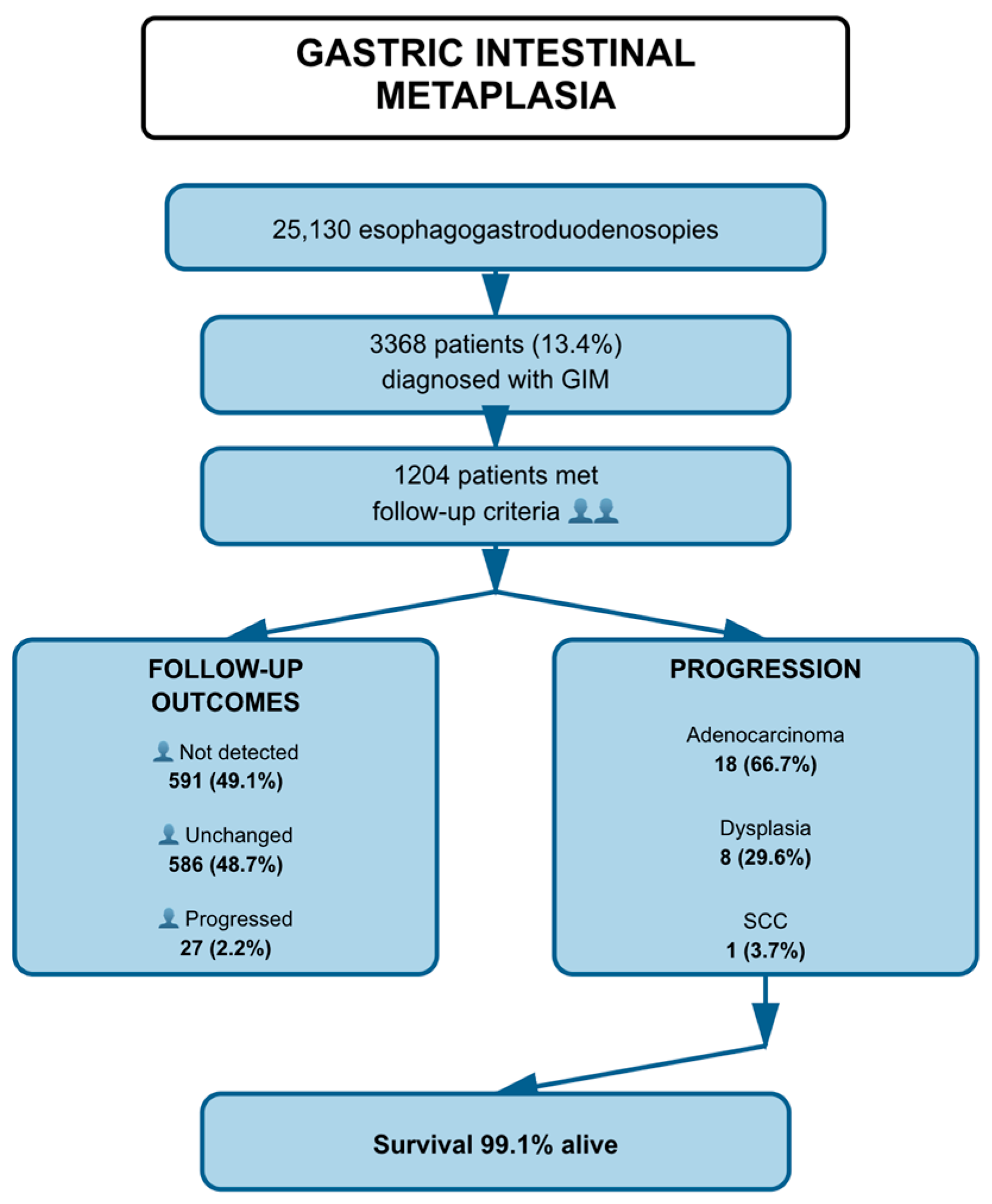
Out of 3368 patients diagnosed with GIM between 2013 and 2021, 1204 (35.7%) fulfilled the eligibility criteria and constituted the final study cohort (Figure 1). At baseline, the median age was 57 years and 56.9% were female. Limited GIM, confined to the antrum/incisura, was observed in 87.6% of patients, while 12.4% had extensive involvement including the corpus. H. pylori infection was detected in 23.9% of cases, and 22.3% reported a family history of gastric cancer. The overall loss to follow-up rate was therefore approximately 64.3%. A detailed breakdown of baseline characteristics is presented in Section 3.

2.6. Variables and Definitions

  • GIM extent: classified as confined to the antrum, confined to the corpus, or involving both antrum and corpus.
  • Additional risk factors: presence of H. pylori infection at baseline (determined histologically), family history of gastric cancer in a first-degree relative, residence in a region of Türkiye endemic for gastric cancer (Eastern Anatolian region) [9], demographic characteristics (age, sex), and other documented risk modifiers.
  • İntestinal metaplasia risk groups: as outlined in the AJG guidelines, patients with GIM were stratified into two risk groups: the low-risk group, comprising individuals with GIM confined to the antrum; and the high-risk group, which included patients with GIM involving both the antrum and corpus, those with a first-degree relative diagnosed with gastric cancer, and individuals residing in endemic regions [3].
  • Outcome: development of histologically confirmed gastric adenocarcinoma during follow-up. Follow-up outcomes of GIM were categorized into three groups: Regression, defined as the histological absence of GIM in follow-up biopsies. Unchanged, defined as the continued presence of GIM without histological progression. Progressed, defined as the development of dysplasia, adenocarcinoma, or other neoplastic lesions during follow-up.

2.7. Ethical Approval

The study was conducted in accordance with the principles of the Declaration of Helsinki and was approved by the Gazi University Faculty of Medicine Medical Ethics Committee (Decision No. 343, dated 24 April 2023).

2.8. Statistical Analysis

All statistical analyses were performed using SPSS software, version 22.0 (IBM Corp., Armonk, NY, USA). Descriptive statistics were expressed as frequencies and percentages for categorical variables, and as means ± standard deviation or medians (minimum–maximum) for continuous variables, as appropriate. The normality of variable distributions was assessed both visually (histograms and probability plots) and analytically (Kolmogorov–Smirnov/Shapiro–Wilk tests). Comparisons between independent groups were performed using Pearson’s chi-square and Fisher’s exact tests for categorical variables, and one-way ANOVA or Student’s t-test for continuous variables. When significant differences were detected among groups, post hoc analyses were conducted using the Bonferroni correction. A two-tailed p-value < 0.05 was considered statistically significant.

3. Results

3.1. Patient Flow and Baseline Characteristics

A total of 25,130 patients aged ≥18 years underwent esophagogastroduodenoscopy (EGD) between January 2013 and December 2021. GIM was histologically diagnosed in 3368 patients (13.4%). Among these, 1541 patients met the inclusion criteria for follow-up analysis, defined as having at least 12 months between the index and control EGD with biopsy. Of these, 337 patients were excluded due to a diagnosis of autoimmune gastritis. Consequently, 1204 patients were included in the study. Among them, 489 patients belonged to the high-risk GIM group, while 715 were classified as low-risk GIM.
The median age of the study cohort was 57 years (range: 18–91), and 56.9% were female. Limited GIM, defined as GIM confined to the antrum and incisura, was present in 87.6% of patients, whereas 12.4% had anatomically extensive GIM involving the corpus. H. pylori infection was detected in 23.9% of patients at baseline. Additional risk factors included a family history of gastric cancer in a first-degree relative (22.3%) and residence in a region of Türkiye endemic for gastric cancer (11.6%).

3.2. Follow-Up Outcomes of GIM Patients

The mean follow-up period of patients followed with GIM was 38.6 ± 26.7 months. Among 1204 patients, 49.1% (n = 591) had no detectable GIM at the end of follow-up, 48.7% (n = 586) showed no change, and 2.2% (n = 27) demonstrated disease progression.
Among the 27 patients with documented progression, the most common pathological outcome was adenocarcinoma (66.7%, n = 18), followed by dysplasia (29.6%, n = 8), and SCC (3.7%, n = 1) (Figure 2).
The progression rate from GIM to adenocarcinoma was 18 out of 1204 patients (1.5%) over a mean follow-up of 38.6 months, corresponding to an annual incidence of approximately 0.45% per patient-year.
Regarding survival status, 99.1% (n = 1193) of patients were alive at the last follow-up, while 0.9% (n = 11) had died (Table 1).
Nearly half of the patients showed regression of GIM, while only 2.9% progressed during follow-up.
Table 2 summarizes the distribution of follow-up outcomes according to clinical and pathological factors in 1204 patients with GIM. Statistically significant associations were observed between follow-up status and gender, location of involvement and age (all p < 0.05). Disease progression was significantly more frequent among males, older age and antrum + corpus location.
Progression was significantly more common among males, older patients, and those with antrum + corpus involvement.
A significant association was found between follow-up outcome and survival in patients with GIM (p = 0.001). Mortality was significantly higher in patients who showed disease progression during follow-up (9.8%) compared to those with unchanged or no detectable GIM (0.4% and 0.9%, respectively) (Table 3).
Among 262 patients diagnosed with adenocarcinoma, 83.2% had adenocarcinoma detected at the initial EGD. Prior GIM diagnosis was noted in 8.1% of these patients before progression to adenocarcinoma, while 8.7% had no premalignant or malignant lesions identified in previous endoscopies.
Mortality was substantially higher in the progression group compared to patients without progression.

3.3. Disease Progression and Survival Outcomes by Risk Group

Among 1204 patients categorized into high- and low-risk groups, significant association was found between risk level and disease progression (p = 0.004). Progression rates were 2.7% in the low-risk group and 1.6% in the high-risk group (Table 4).
Progression rates were significantly different between high- and low-risk groups.
Among the 36 patients who exhibited disease progression during follow-up, the distribution of pathological diagnoses was assessed according to risk group. In both the high-risk (n = 12) and low-risk (n = 24) groups, adenocarcinoma was the most common diagnosis (31.6% vs. 68.4%), followed by neuroendocrine tumors (NET) and dysplasia. Less frequent diagnoses included gastrointestinal stromal tumor (GIST) and lymphoma, both occurring exclusively in the low-risk group.
Statistical analysis revealed no significant difference in the distribution of pathological diagnoses between high- and low-risk groups (p = 0.901), indicating that risk classification was not associated with a specific pathological outcome among patients who progressed (Table 5).
Adenocarcinoma was the predominant progression outcome in both risk groups.
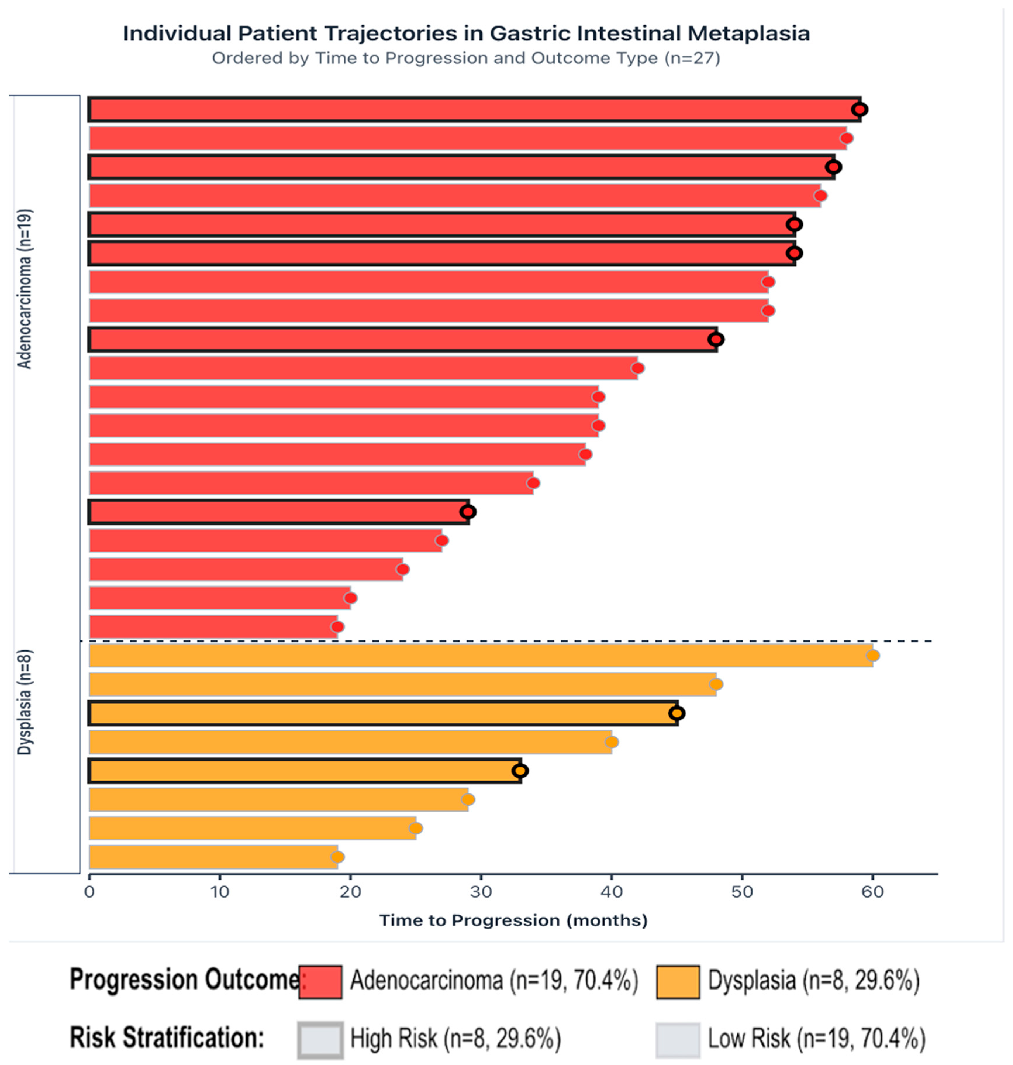
Swimmer plot illustrating individual patient trajectories from baseline endoscopy to disease progression among 27 patients with GIM (Figure 3).
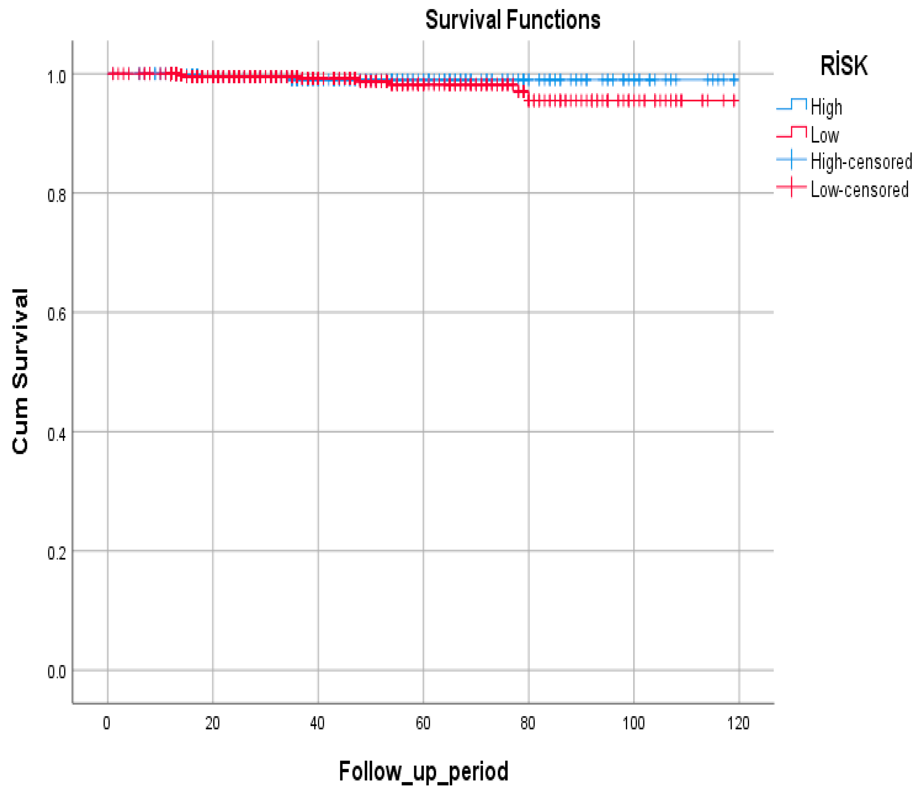
Survival analysis showed no statistically significant difference between high- and low-risk groups (p = 0.386). Mortality rates were low in both groups, with 0.6% in the high-risk and 1.1% in the low-risk groups (Table 6).
Survival rates were similarly high in both high- and low-risk groups (Figure 4).

4. Discussion

The prevalence of GIM in a given population is largely influenced by the rate of H. pylori infection. A Northern European study reported an GIM prevalence of 19% [10]. In our cohort of 25,130 individuals undergoing EGD, GIM was detected in 13.4%, a figure consistent with a study from Van, Turkey (2010–2014), which reported a prevalence of 13.8% among 4050 patients. Determining the true prevalence remains challenging due to the asymptomatic nature of GIM [11].
In large-scale cohort studies, such as those from the Netherlands (1991–2004), the annual progression rate of GIM to adenocarcinoma ranged from 0.1% to 0.9% [12]. In our cohort, the progression rate from GIM to adenocarcinoma was 1.5% over a mean follow-up of 38.6 months (annual incidence ~0.45% per patient-year). This rate is consistent with low-risk Western cohorts but remains lower than those reported from high-incidence Asian populations [13].
Gender differences play a role in gastric cancer pathogenesis. Data from the United States indicate that men are more likely to develop proximal gastric cancer [14], and global age-standardized rates are approximately twice as high in men as in women (12.8% vs. 5.7%) [15]. In the United Kingdom, the male-to-female ratio declines after age 50–55, likely due to reduced protective hormonal effects in postmenopausal women [16]. Long-term estrogen exposure has been associated with a lower gastric cancer risk [17]. In line with previous studies, we observed higher malignant progression rates in men with GIM (3.3%) compared to women (2.6%).
H. pylori infection, a key initiator in the Correa cascade, affects an estimated 4.4 billion people worldwide [18]. In the United Kingdom, 32% of gastric cancers are attributable to H. pylori [19]. The World Health Organization (WHO) classifies H. pylori as a class I carcinogen [20,21]. However, in advanced gastritis and GIM, mucosal changes may render the gastric environment inhospitable to H. pylori, leading to spontaneous bacterial clearance [22]. In our study, H. pylori was detected in 23.9% of GIM patients, and progression was more common in H. pylori-negative individuals (3.2%) compared to H. pylori-positive individuals (1.9%). Whether this reflects prior eradication therapy could not be determined.
Histologically, GIM can be classified into three subtypes (Types 1–3) according to mucin staining [23,24,25], with incomplete Type 3 GIM showing the strongest association with gastric cancer [26,27]. Due to technical challenges and the use of toxic reagents, such staining is now largely restricted to research settings [28], and our study did not perform histologic subtyping.
ECL cell hyperplasia, often associated with autoimmune gastritis, was present in 5.6% of our GIM patients. Progression occurred in 6.1% of these cases compared to 3.4% without ECL hyperplasia, though no adenocarcinoma developed in the hyperplasia group. The lack of statistical association between ECL hyperplasia and adenocarcinoma aligns with its stronger link to type I neuroendocrine tumors [29,30,31,32,33,34].
Anatomically, GIM is categorized as limited (antrum or incisura only) or extensive (involving corpus and additional sites) [35,36,37,38]. Prior studies suggest that extent of involvement may be more prognostically relevant than histological subtype [39]. In our series, progression rates were highest for extensive involvement (5.5%), followed by limited antrum involvement only (3.2%). Adenocarcinoma was significantly more common in cases involving the corpus or both corpus and antrum (p < 0.05). While our findings suggest that routine surveillance may not be necessary for antrum-limited GIM, this conclusion should be interpreted with caution given the low number of progression events and limited statistical power.
Globally, there is no uniform consensus on GIM surveillance. High-incidence countries such as Japan and South Korea recommend population-based endoscopic screening, with national programs in place since the early 2000s [40,41]. In low-incidence European countries, the European Society of Gastrointestinal Endoscopy (ESGE) advises three-year follow-up for patients with GIM in both antrum and corpus, but no surveillance for antrum-only disease [42].
According to the latest ACG guidelines, patients with GIM are categorized into two groups for surveillance: high-risk and low-risk [3]. The high-risk group includes patients with extensive involvement, those with a first-degree relative with gastric cancer, and individuals from regions endemic for gastric cancer. In our study, patients were similarly stratified. Patients born in Eastern Anatolia, a gastric cancer endemic region in our country, were included in the high-risk group [9]. Interestingly, in our cohort the low-risk group demonstrated a slightly higher progression rate compared to the high-risk group (2.7% vs. 1.6%, p = 0.004), although survival outcomes did not differ. This paradoxical finding may partly reflect the absence of histological subtyping (complete vs. incomplete GIM), since incomplete GIM is known to carry a higher malignant potential and its distribution between groups was not captured. In addition, the use of geographic endemicity as a risk criterion may have led to misclassification in a population with frequent historical internal migration, thereby obscuring true risk patterns.
Several limitations should be acknowledged. First, this study was retrospective, which may introduce selection and information biases. In particular, heterogeneous follow-up intervals and variability in biopsy sampling protocols could have influenced the observed outcomes. Second, histological subtyping of GIM (Types 1–3) was not performed, limiting the assessment of subtype-specific risk. Incomplete subtyping of GIM was not performed, which is an important limitation since incomplete GIM is associated with a higher risk of malignant transformation. Third, H. pylori eradication history and post-treatment confirmation were not systematically available. Therefore, interpretation of H. pylori negativity should be made with caution, as some cases may represent prior successful eradication or spontaneous clearance in advanced atrophic gastritis. This limitation introduces the potential for misclassification bias and restricts firm conclusions regarding the role of H. pylori status in progression. Fourth, although follow-up data were comprehensive, the relatively low number of progression events limits the statistical power for certain subgroup analyses. Additionally, the relatively high regression rate observed in our cohort should be interpreted with caution, as it may reflect both true histological reversal and potential biopsy sampling variability. This possibility has been reported in prior studies and represents an inherent limitation of observational GIM cohorts. Fifth, patients with autoimmune gastritis were excluded, which might have led to underestimation of the true incidence of neuroendocrine tumors in this population. Sixth, loss to follow-up and exclusion of patients without adequate surveillance endoscopies may have introduced a selection bias, potentially underestimating progression risk. The small number of events and incomplete survival curves precluded estimation of median survival or RMST. Therefore, mean survival times were reported, which more appropriately represent the available follow-up information in this cohort. Finally, as a single-country study, our findings may not be generalizable to populations with different H. pylori prevalence, genetic backgrounds, or gastric cancer incidence.

5. Conclusions

In our large cohort study, GIM was detected in 13.4% of patients undergoing esophagogastroduodenoscopy, with an overall cumulative progression rate to adenocarcinoma of 1.5% during a mean follow-up of 38.6 months (annual incidence ~0.45% per patient-year). Male sex and extensive gastric involvement were associated with higher progression rates. H. pylori infection, while important in the pathogenesis of gastric carcinogenesis, was not predictive of malignant transformation in our cohort, possibly due to prior eradication therapy or spontaneous clearance in advanced lesions. Our findings also suggest that heterogeneous risk profiles, including birth in gastric cancer endemic regions, family history, and extent of GIM, may obscure clear stratification between high- and low-risk groups. These results support the need for individualized surveillance strategies based on anatomical extent of GIM and patient-specific risk factors rather than H. pylori status alone. Routine surveillance of patients with GIM limited to the antrum appears to provide minimal clinical benefit while imposing unnecessary procedural burden and healthcare costs.

Author Contributions

Conceptualization, V.B.T. and A.K.; methodology, V.B.T., A.K., and A.A.; validation, M.C., T.K., and M.K.; formal analysis, V.B.T. and A.K.; investigation, V.B.T., A.K., A.A., H.C.A., and S.D.; resources, M.C., T.K., and M.K.; data curation, V.B.T., A.K., and A.A.; writing—original draft preparation, V.B.T.; writing—review and editing, V.B.T., A.K., A.A., H.C.A., S.D., M.C., T.K., M.K., S.H., E.Ö., and M.A.İ.; visualization, V.B.T.; supervision, M.C., T.K., and M.K.; project administration, V.B.T. All authors have read and agreed to the published version of the manuscript.

Funding

This research received no external funding.

Institutional Review Board Statement

The study was conducted in accordance with the Declaration of Helsinki, and approved by the Gazi University Faculty of Medicine Medical Ethics Committee (Decision No. 343, dated 24 April 2023).

Informed Consent Statement

Patient consent was waived due to the retrospective design of the study and anonymization of all data.

Data Availability Statement

The data that support the findings of this study are available from the corresponding author upon reasonable request.

Acknowledgments

The authors thank the staff of the Departments of Gastroenterology, Pathology, and Internal Medicine for their support during data collection and patient follow-up.

Conflicts of Interest

The authors declare no conflicts of interest.

References

  1. Sung, H.; Ferlay, J.; Siegel, R.L.; Laversanne, M.; Soerjomataram, I.; Jemal, A.; Bray, F. Global Cancer Statistics 2020: GLOBOCAN Estimates of Incidence and Mortality Worldwide for 36 Cancers in 185 Countries. CA Cancer J. Clin. 2021, 71, 209–249. [Google Scholar] [CrossRef] [PubMed]
  2. Siegel, R.L.; Miller, K.D.; Jemal, A. Cancer Statistics, 2020. CA Cancer J. Clin. 2020, 70, 7–30. [Google Scholar] [CrossRef]
  3. Morgan, D.R.; Corral, J.E.; Li, D.; Montgomery, E.A.; Riquelme, A.; Kim, J.J.; Sauer, B.; Shah, S.C. ACG Clinical Guideline: Diagnosis and Management of Gastric Premalignant Conditions. Am. J. Gastroenterol. 2025, 120, 709–737. [Google Scholar] [CrossRef]
  4. Plummer, M.; Franceschi, S.; Vignat, J.; Forman, D.; de Martel, C. Global Burden of Gastric Cancer Attributable to Helicobacter pylori. Int. J. Cancer 2015, 136, 487–490. [Google Scholar] [CrossRef]
  5. Park, Y.H.; Kim, N. Review of Atrophic Gastritis and Intestinal Metaplasia as a Premalignant Lesion of Gastric Cancer. J. Cancer Prev. 2015, 20, 25–40. [Google Scholar] [CrossRef] [PubMed]
  6. Huang, R.J.; Choi, A.Y.; Truong, C.D.; Yeh, M.M.; Hwang, J.H. Diagnosis and Management of Gastric Intestinal Metaplasia: Current Status and Future Directions. Gut Liver 2019, 13, 596–603. [Google Scholar] [CrossRef]
  7. Song, H.; Ekheden, I.G.; Zheng, Z.; Ericsson, J.; Nyrén, O.; Ye, W. Incidence of Gastric Cancer among Patients with Gastric Precancerous Lesions: Observational Cohort Study in a Low-Risk Western Population. BMJ 2015, 351, h3867. [Google Scholar] [CrossRef] [PubMed]
  8. Torre, L.A.; Bray, F.; Siegel, R.L.; Ferlay, J.; Lortet-Tieulent, J.; Jemal, A. Global Cancer Statistics, 2012. CA Cancer J. Clin. 2015, 65, 87–108. [Google Scholar] [CrossRef]
  9. Yalcin, S. Gastric Cancer in Turkey—A Bridge between West and East. Gastrointest. Cancer Res. 2009, 3, 29–32. [Google Scholar] [PubMed]
  10. Eriksson, N.K.; Kärkkäinen, P.A.; Färkkilä, M.A.; Arkkila, P.E. Prevalence and Distribution of Gastric Intestinal Metaplasia and Its Subtypes. Dig. Liver Dis. 2008, 40, 355–360. [Google Scholar] [CrossRef]
  11. Olmez, S.; Aslan, M.; Erten, R.; Sayar, S.; Bayram, I. The Prevalence of Gastric Intestinal Metaplasia and Distribution of Helicobacter pylori Infection, Atrophy, Dysplasia, and Cancer in Its Subtypes. Gastroenterol. Res. Pract. 2015, 2015, 434039. [Google Scholar] [CrossRef]
  12. O’Connor, A.; McNamara, D.; O’Moráin, C.A. Surveillance of Gastric Intestinal Metaplasia for the Prevention of Gastric Cancer. Cochrane Database Syst. Rev. 2013, 9, CD009322. [Google Scholar] [CrossRef]
  13. Li, Y.; Jiang, F.; Wu, C.Y.; Leung, W.K. Prevalence and temporal trend of gastric preneoplastic lesions in Asia: A systematic review with meta-analysis. United Eur. Gastroenterol. J. 2024, 12, 139–151. [Google Scholar] [CrossRef] [PubMed] [PubMed Central]
  14. Yao, J.C.; Schnirer, I.I.; Reddy, S.; Chiang, S.; Najam, A.; Yu, C.; Giacco, G.; Hess, K.; Rashid, A.; Xie, K.; et al. Effects of sex and racial/ethnic group on the pattern of gastric cancer localization. Gastric Cancer 2002, 5, 208–212. [Google Scholar] [CrossRef] [PubMed]
  15. Ferlay, J.; Soerjomataram, I.; Dikshit, R.; Eser, S.; Mathers, C.; Rebelo, M.; Parkin, D.M.; Forman, D.; Bray, F. Cancer Incidence and Mortality Worldwide: Sources, Methods and Major Patterns in GLOBOCAN 2012. Int. J. Cancer 2015, 136, E359–E386. [Google Scholar] [CrossRef] [PubMed]
  16. Derakhshan, M.H.; Liptrot, S.; Paul, J.; Brown, I.L.; Morrison, D.; McColl, K.E. Oesophageal and gastric intestinal-type adenocarcinomas show the same male predominance due to a 17 year delayed development in females. Gut 2009, 58, 16–23. [Google Scholar] [CrossRef] [PubMed]
  17. Camargo, M.C.; Goto, Y.; Zabaleta, J.; Morgan, D.R.; Correa, P.; Rabkin, C.S. Sex Hormones, Hormonal Interventions, and Gastric Cancer Risk: A Meta-Analysis. Cancer Epidemiol. Biomark. Prev. 2012, 21, 20–38. [Google Scholar] [CrossRef] [PubMed]
  18. He, J.; Hu, W.; Ouyang, Q.; Zhang, S.; He, L.; Chen, W.; Li, X.; Hu, C. Helicobacter pylori Infection Induces Stem Cell-Like Properties in Correa Cascade of Gastric Cancer. Cancer Lett. 2022, 542, 215764. [Google Scholar] [CrossRef]
  19. Parkin, D.M.; Boyd, L.; Walker, L.C. The Fraction of Cancer Attributable to Lifestyle and Environmental Factors in the UK in 2010. Br. J. Cancer 2011, 105 (Suppl. 2), S77–S81. [Google Scholar] [CrossRef]
  20. Fan, C.; Xu, K.; Huang, Y.; Liu, S.; Wang, T.; Wang, W.; Hu, W.; Liu, L.; Xing, M.; Yang, S. Viscosity and Degradation Controlled Injectable Hydrogel for Esophageal Endoscopic Submucosal Dissection. Bioact. Mater. 2021, 6, 1150–1162, Correction in Bioact. Mater. 2021, 6, 4161–4162. [Google Scholar] [CrossRef]
  21. Tokat, M.; van Tilburg, L.; Koch, A.D.; Spaander, M.C.W. Artificial Intelligence in Upper Gastrointestinal Endoscopy. Dig. Dis. 2022, 40, 395–408. [Google Scholar] [CrossRef]
  22. González, C.A.; Pardo, M.L.; Ruiz Liso, J.M.; Alonso, P.; Bonet, C.; Garcia, R.M.; Sala, N.; Capella, G.; Sanz-Anquela, J.M. Gastric Cancer Occurrence in Preneoplastic Lesions: A Long-Term Follow-Up in a High-Risk Area in Spain. Int. J. Cancer 2010, 127, 2654–2660. [Google Scholar] [CrossRef]
  23. Spicer, S.S. Diamine Methods for Differentiating Mucosubstances Histochemically. J. Histochem. Cytochem. 1965, 13, 211–234. [Google Scholar] [CrossRef] [PubMed]
  24. Filipe, M.I.; Muñoz, N.; Matko, I.; Kato, I.; Pompe-Kirn, V.; Jutersek, A.; Teuchmann, S.; Benz, M.; Prijon, T. Intestinal Metaplasia Types and the Risk of Gastric Cancer: A Cohort Study in Slovenia. Int. J. Cancer 1994, 57, 324–329. [Google Scholar] [CrossRef] [PubMed]
  25. Jass, J.R.; Filipe, M.I. The Mucin Profiles of Normal Gastric Mucosa, Intestinal Metaplasia and Its Variants and Gastric Carcinoma. Histochem. J. 1981, 13, 931–939. [Google Scholar] [CrossRef] [PubMed]
  26. Filipe, M.I.; Potet, F.; Bogomoletz, W.V.; Dawson, P.A.; Fabiani, B.; Chauveinc, P.; Fenzy, A.; Gazzard, B.; Goldfain, D.; Zeegen, R. Incomplete Sulphomucin-Secreting Intestinal Metaplasia for Gastric Cancer: Preliminary Data from a Prospective Study from Three Centres. Gut 1985, 26, 1319–1326. [Google Scholar] [CrossRef]
  27. Conchillo, J.M.; Houben, G.; de Bruïne, A.; Stockbrügger, R.W. Is Type III Intestinal Metaplasia an Obligatory Precancerous Lesion in Intestinal-Type Gastric Carcinoma? Eur. J. Cancer Prev. 2001, 10, 307–312. [Google Scholar] [CrossRef]
  28. Shah, S.C.; Gawron, A.J.; Mustafa, R.A.; Piazuelo, M.B. Histologic Subtyping of Gastric Intestinal Metaplasia: Overview and Considerations for Clinical Practice. Gastroenterology 2020, 158, 745–750. [Google Scholar] [CrossRef] [PubMed] [PubMed Central]
  29. Choudhuri, J.; Hall, S.; Castrodad-Rodriguez, C.A.; Westerhoff, M.; El Jabbour, T.; Jain, S.; Panarelli, N.C. Features That Aid Identification of Autoimmune Gastritis in a Background of Active Helicobacter pylori Infection. Arch. Pathol. Lab. Med. 2021, 145, 1536–1543. [Google Scholar] [CrossRef]
  30. Park, J.Y.; Cornish, T.C.; Lam-Himlin, D.; Shi, C.; Montgomery, E. Gastric Lesions in Patients with Autoimmune Metaplastic Atrophic Gastritis (AMAG) in a Tertiary Care Setting. Am. J. Surg. Pathol. 2010, 34, 1591–1598. [Google Scholar] [CrossRef]
  31. Coati, I.; Fassan, M.; Farinati, F.; Graham, D.Y.; Genta, R.M.; Rugge, M. Autoimmune Gastritis: Pathologist’s Viewpoint. World J. Gastroenterol. 2015, 21, 12179–12189. [Google Scholar] [CrossRef]
  32. Borch, K.; Ahrén, B.; Ahlman, H.; Falkmer, S.; Granérus, G.; Grimelius, L. Gastric Carcinoids: Biologic Behavior and Prognosis after Differentiated Treatment in Relation to Type. Ann. Surg. 2005, 242, 64–73. [Google Scholar] [CrossRef]
  33. Vannella, L.; Lahner, E.; Annibale, B. Risk for Gastric Neoplasias in Patients with Chronic Atrophic Gastritis: A Critical Reappraisal. World J. Gastroenterol. 2012, 18, 1279–1285. [Google Scholar] [CrossRef]
  34. Murphy, G.; Dawsey, S.M.; Engels, E.A.; Ricker, W.; Parsons, R.; Etemadi, A.; Lin, S.-W.; Abnet, C.C.; Freedman, N.D. Cancer Risk After Pernicious Anemia in the US Elderly Population. Clin. Gastroenterol. Hepatol. 2015, 13, 2282–2289.e4. [Google Scholar] [CrossRef]
  35. Dixon, M.F.; Genta, R.M.; Yardley, J.H.; Correa, P. Classification and Grading of Gastritis: The Updated Sydney System. Am. J. Surg. Pathol. 1996, 20, 1161–1181. [Google Scholar] [CrossRef] [PubMed]
  36. Correa, P.; Piazuelo, M.B.; Wilson, K.T. Pathology of Gastric Intestinal Metaplasia: Clinical Implications. Am. J. Gastroenterol. 2010, 105, 493–498. [Google Scholar] [CrossRef] [PubMed]
  37. de Vries, A.C.; Haringsma, J.; de Vries, R.A.; ter Borg, F.; Nagtzaam, N.M.; Steyerberg, E.W.; van Dekken, H.; Kuipers, E.J. The use of clinical, histologic, and serologic parameters to predict the intragastric extent of intestinal metaplasia: A recommendation for routine practice. Gastrointest. Endosc. 2009, 70, 18–25. [Google Scholar] [CrossRef] [PubMed]
  38. Capelle, L.G.; de Vries, A.C.; Haringsma, J.; Ter Borg, F.; de Vries, R.A.; Bruno, M.J.; van Dekken, H.; Meijer, J.; van Grieken, N.C.; Kuipers, E.J. The Staging of Gastritis with the OLGA System by Using Intestinal Metaplasia as an Accurate Alternative for Atrophic Gastritis. Gastrointest. Endosc. 2010, 71, 1150–1158. [Google Scholar] [CrossRef]
  39. Cassaro, M.; Rugge, M.; Gutierrez, O.; Leandro, G.; Graham, D.Y.; Genta, R.M. Topographic Patterns of Intestinal Metaplasia and Gastric Cancer. Am. J. Gastroenterol. 2000, 95, 1431–1438. [Google Scholar] [CrossRef] [PubMed]
  40. Leung, W.K.; Wu, M.-S.; Kakugawa, Y.; Kim, J.J.; Yeoh, K.-G.; Goh, K.L.; Wu, K.-C.; Wu, D.-C.; Sollano, J.; Kachintorn, U.; et al. Screening for Gastric Cancer in Asia: Current Evidence and Practice. Lancet Oncol. 2008, 9, 279–287. [Google Scholar] [CrossRef]
  41. Hamashima, C.; Goto, R. Potential Capacity of Endoscopic Screening for Gastric Cancer in Japan. Cancer Sci. 2017, 108, 101–107. [Google Scholar] [CrossRef] [PubMed]
  42. Dinis-Ribeiro, M.; Areia, M.; de Vries, A.C.; Marcos-Pinto, R.; Monteiro-Soares, M.; O’connor, A.; Pereira, C.; Pimentel-Nunes, P.; Correia, R.; Ensari, A.; et al. Management of Precancerous Conditions and Lesions in the Stomach (MAPS): Guideline from the European Society of Gastrointestinal Endoscopy (ESGE), European Helicobacter Study Group (EHSG), European Society of Pathology (ESP), and the Sociedade Portuguesa de Endoscopia Digestiva (SPED). Virchows Arch. 2012, 460, 19–46. [Google Scholar] [CrossRef] [PubMed]
Figure 1. Flowchart of study population.
Jcm 14 07662 g001
Figure 2. Follow-up outcomes and progression patterns in patients with GIM.
Jcm 14 07662 g002
Figure 3. Individual patient trajectories in GIM. Each horizontal bar represents one patient, with bar length corresponding to the time (in months) from the initial diagnosis to the first documented histological progression. Color coding indicates progression type: red bars represent progression to adenocarcinoma (n = 19, 70.4%), and orange bars represent progression to dysplasia (n = 8, 29.6%). Border thickness denotes risk stratification: thick black borders indicate high-risk patients (n = 8, 29.6%), and thin gray borders indicate low-risk patients (n = 19, 70.4%).
Jcm 14 07662 g003
Figure 4. Kaplan–Meier survival curves of patients with GIM according to risk stratification (high-risk vs. low-risk groups). Time axis represents follow-up duration in months. No significant difference in overall survival was observed between groups (log-rank p = 0.386).
Jcm 14 07662 g004
Table 1. Outcomes and Survival Status of Patients Followed with GIM.
CategoryVariablen%
Follow-up Outcome (n = 1204)Not detected59149.1
Unchanged58648.7
Progressed272.2
Pathology Results in Progressed Cases
(n = 27)
Adenocarcinoma1866.7
Dysplasia829.6
SCC13.7
Survival Status (n = 1204)Alive119399.1
Deceased110.9
Note: Percentages are reported as column percentages.
Table 2. Factors Associated with Follow-up Outcomes in Patients with GIM (n = 1204).
VariableCategoryNot Detected n (%)/Mean ± SDUnchanged n (%)/Mean ± SDProgressed n (%)/Mean ± SDp
GenderFemale372 (54.0)299 (43.4)18 (2.6)<0.001
Male219 (42.5)279 (54.2)17 (3.3)
Location of InvolvementAntrum540 (52.8)455 (44.5)28 (2.7)<0.001
Antrum + Corpus51 (28.2)123 (68.0)7 (3.9)
ECL HyperplasiaPresent12 (44.4)15 (55.6)0 (0.0)0.747
Absent579 (49.2)563 (47.8)35 (3.0)
H. pylori PresencePresent162 (52.6)110 (45.5)6 (1.9)0.233
Absent429 (47.9)438 (48.9)29 (3.2)
Age (years)54.8 ± 12.458.3 ± 11.060.5 ± 11.6<0.001
Follow-up Duration (months)39.3 ± 26.238.3 ± 27.231.2 ± 24.90.111
Table 3. Survival Outcomes by Follow-up Status and Initial EGD Findings in Patients with GIM.
VariableCategoryAlive n (%)Deceased n (%)p-Value
Survival by Follow-up Status (n = 1204)Not Detected587 (99.3)4 (0.7)<0.001
Unchanged583 (99.5)3 (0.5)
Progressed23 (85.2)4 (14.8)
Table 4. Progression outcomes of GIM risk groups.
Risk GroupNot DetectedUnchangedProgressedTotalp Value
High215 (44.0%)266 (54.4%)8 (1.6%)489 (40.6%)0.004
Low376 (52.6%)320 (44.8%)19 (2.7%)715 (59.4%)
Total591 (49.1%)586 (48.7%)27 (2.2%)1204 (100.0%)
Table 5. Summary of adverse outcomes during follow-up.
Pathology DiagnosisTotal n (%)High Risk n (%)Low Risk n (%)
Adenocarcinoma19 (52.8%)6 (31.6%)13 (68.4%)
NET (Neuroendocrine Tumor)5 (13.9%)4 (80.0%)1 (20.0%)
Dysplasia8 (22.2%)2 (25.0%)6 (75.0%)
GIST (Gastrointestinal Stromal Tumor)3 (8.3%)0 (0.0%)3 (100.0%)
Lymphoma1 (2.8%)0 (0%)1 (100.0%)
Total36 (100%)12 (100%)24 (100%)
Table 6. Survival outcomes of GIM risk groups.
Risk GroupAliveDeceasedTotal
High486 (99.4%)3 (0.6%)489
Low707 (98.9%)8 (1.1%)715
Total1193 (99.1%)11 (0.9%)1204
Disclaimer/Publisher’s Note: The statements, opinions and data contained in all publications are solely those of the individual author(s) and contributor(s) and not of MDPI and/or the editor(s). MDPI and/or the editor(s) disclaim responsibility for any injury to people or property resulting from any ideas, methods, instructions or products referred to in the content.