Article

Effect of Leukocyte- and Platelet-Rich Fibrin on Peri-Implant Mucosal Thickness in Edentulous Patients Treated with Mandibular Implant-Retained Overdentures: A Randomized Controlled Trial

1
San Camilo Hospital, San Felipe 8940000, Chile
2
Faculty of Dentistry, Universidad de Chile, Santiago 8320000, Chile
3
Clinic of Reconstructive Dentistry, Center of Dental Medicine, University of Zurich, 8006 Zurich, Switzerland
4
Health Science Faculty, Universidad Autonoma de Chile, Santiago 8940000, Chile
5
Department of Periodontology, Medical University of Vienna, 1090 Vienna, Austria
6
School of Dentistry, Universidad Andres Bello, Santiago 7591538, Chile
*
Author to whom correspondence should be addressed.
J. Clin. Med. 2025, 14(19), 6917; https://doi.org/10.3390/jcm14196917
Submission received: 1 September 2025 / Revised: 24 September 2025 / Accepted: 28 September 2025 / Published: 29 September 2025
(This article belongs to the Special Issue Advances in Periodontitis and Other Periodontal Diseases)

Abstract

Background/Objectives: The maintenance of peri-implant soft tissue health is critical for the long-term success of implant therapy, particularly in edentulous patients rehabilitated with mandibular overdentures. Leukocyte- and platelet-rich fibrin (L-PRF) has been proposed as an autologous biomaterial to enhance peri-implant tissue quality. This randomized controlled clinical trial evaluated the effect of L-PRF on peri-implant mucosal thickness in edentulous patients treated with mandibular implant-retained overdentures. Methods: Edentulous patients received two interforaminal implants to retain a mandibular overdenture and were randomly assigned to a test group (L-PRF applied during surgery) or a control group (standard protocol without L-PRF). Clinical measurements of keratinized mucosal thickness and width were recorded at baseline, 12 weeks, and 24 weeks. Volumetric analyses of soft and hard tissue changes were performed using digital superimposition of STL models. The trial was conducted in accordance with the Declaration of Helsinki and approved by the Scientific Ethics Committee of the Aconcagua Health Service. All participants provided written informed consent. Results: A significant increase in keratinized mucosal thickness was observed in the L-PRF group at 12 and 24 weeks compared with baseline (p < 0.01). No significant differences were detected between the groups in soft tissue volume (p = 0.12) or bone volume (p = 0.45). Mucosal width remained stable in both groups throughout follow-up. Conclusions: The application of L-PRF at implant placement resulted in a significant gain in peri-implant mucosal thickness, suggesting a soft tissue modulating effect. Enhancing keratinized mucosal thickness during implant surgery may improve peri-implant tissue quality and support long-term stability of mandibular overdentures.

1. Introduction

Tooth loss leads to alveolar bone resorption and disruption of the alveolar process architecture. In the anterior mandible, this resorption pattern typically involves both vertical and horizontal bone loss, while in the posterior region, bone loss is predominantly vertical [1,2]. Atrophy in the anterior mandible often results in knife-edge ridges characterized by preserved height but severely reduced width [3], creating suboptimal conditions for oral rehabilitation.
Although conventional removable prostheses remain a reliable and effective solution for edentulous patients, unfavorable residual bone conditions often lead to poor stability, reduced masticatory function, and patient dissatisfaction [4]. Approximately two-thirds of patients wearing conventional mandibular prostheses report problems with retention and stability [5]. A well-established alternative is the implant-retained overdenture, which consists of a removable acrylic complete denture retained by two interforaminal implants. This approach provides improved retention, leading to enhanced stability, function, aesthetics, and quality of life in patients with atrophic ridges [4]. Studies have shown significantly greater masticatory efficiency and bite force in patients using mandibular implant-retained overdentures compared with conventional prostheses. Consequently, these patients also demonstrate better nutritional status, with notable improvements in biochemical and anthropometric nutritional parameters [6].
Advanced age has not been shown to compromise implant survival rates [7]; however, individuals over the age of 65 are more susceptible to developing peri-implant mucositis and peri-implantitis [8]. Therefore, while geriatric patients are generally suitable candidates for implant therapy, it is essential to identify and manage local factors that may increase the risk of peri-implant inflammation [8,9].
Leukocyte- and platelet-rich fibrin (L-PRF) is an autologous biomaterial prepared by centrifugation of the patient’s own blood [10]. Its use is based on the premise that supraphysiological concentrations of growth factors may enhance the early stages of wound healing and tissue regeneration beyond what is achieved under physiological conditions [11]. L-PRF has been associated with improved implant primary stability and enhanced early healing in some clinical models [10,12].
The application of L-PRF has also been proposed to promote the development of a keratinized mucosal band around the implant neck, potentially contributing to improved peri-implant soft tissue quality and long-term implant success [13].
Both the width and thickness of the keratinized mucosa are important parameters in maintaining peri-implant tissue health. A minimum band of keratinized mucosa facilitates oral hygiene, reduces mucosal inflammation, and maintains soft tissue integrity around implants, particularly in removable overdenture protocols [14]. Inadequate keratinized width may result in mucosal mobility, discomfort during function, and increased plaque accumulation—factors that can compromise long-term implant success [14,15]. Similarly, greater mucosal thickness has been associated with enhanced resistance to mechanical and microbial challenges and may help preserve marginal bone levels and reduce the risk of mucosal recession [14]. These considerations are especially relevant in edentulous patients rehabilitated with mandibular overdentures, where mucosal health is critical for prosthesis retention, hygiene maintenance, and patient comfort [16].
In this context, leukocyte- and platelet-rich fibrin (L-PRF) has been proposed as a soft tissue augmentation option. Clinical evidence indicates that its use can increase both the width of keratinized mucosa and the soft tissue thickness, while reducing operative time and postoperative morbidity compared with connective tissue grafts [17]. In alveolar ridge preservation procedures, L-PRF has been associated with greater gains in soft tissue thickness after 6 months [16]. Additionally, systematic reviews suggest that PRF-based interventions may offer clinical outcomes comparable to autogenous grafts in terms of keratinized mucosa width and tissue thickness [14].
The objective of this split-mouth randomized clinical trial was to evaluate the effect of L-PRF on implant stability, peri-implant clinical parameters, keratinized mucosal thickness, and patient-reported post-surgical pain in edentulous elderly patients receiving implant-retained mandibular overdentures.
Null hypothesis: The adjunctive use of L-PRF during implant placement in edentulous elderly patients receiving implant-retained overdentures does not significantly affect implant stability, peri-implant clinical parameters, keratinized mucosa thickness, or patient-reported outcomes measures compared with the control treatment.

2. Materials and Methods

2.1. Study Design

This split-mouth randomized clinical trial was conducted at the Periodontics Clinic of San Camilo Hospital, San Felipe, Chile. A total of 40 edentulous patients over the age of 60 were screened. Eligible participants had a history of failed treatment with conventional removable mandibular prostheses. Exclusion criteria included systemic or psychological conditions contraindicating implant placement, mandibular extractions within the previous 6 months, heavy smoking, oral anticoagulant therapy, and hemodialysis.

2.2. Screening and Consenting

All participants and outcome assessors were blinded to group allocation. The clinical examiner was a calibrated periodontist (X.M.). Randomization was performed using a computer-generated sequence to assign the experimental (L-PRF) side, and sealed envelopes were used to conceal allocation. Ethical approval was obtained from the Scientific Ethics Committee of the Aconcagua Health Service (CEC09/2020), and all participants signed informed consent prior to enrollment. The study was conducted in accordance with the Declaration of Helsinki and was registered at ClinicalTrials.gov (ID: NCT04429373; 9 June 2020).

2.3. Outcome Measures

Measured outcomes included implant stability, assessed by Implant Stability Quotient (ISQ) via resonance frequency analysis (Osstell, W&H, Vienna, Austria); keratinized tissue width (KTW), measured as the vertical distance from the mucosal margin to the mucogingival junction; and probing depth (PD). Mucosal thickness (MT) was measured at the mid-buccal point of each implant site using a standardized transgingival probing technique. A sterile #15 endodontic K-file equipped with a silicone stopper was employed as the measuring instrument. After applying a topical anesthetic gel to minimize discomfort, the file was inserted perpendicularly through the mucosa until firm contact with the underlying bone was achieved. The silicone stopper was then positioned flush with the external mucosal surface, and the distance from the file tip to the stopper was measured with a digital caliper to the nearest 0.1 mm. Each site was measured twice, and the mean value was recorded. To ensure reproducibility, all measurements were performed under standardized conditions of lighting and 2.5× magnification. Examiner calibration was conducted prior to the study on five non-study patients, resulting in an intraclass correlation coefficient (ICC) > 0.90. This approach provides a direct, minimally invasive, and reproducible method for quantifying mucosal thickness.
Cone-beam computed tomography (CBCT) scans (PLANMECA ProMax, Helsinki, Finland) were obtained preoperatively and 6 months post implant installation. Images were analyzed using Blue Sky Plan 4 (BlueSky Bio, Grayslake, IL, USA) by a researcher blinded to the treatment allocation. Three-dimensional models were generated via semi-automatic segmentation and aligned using six anatomical landmarks, following a validated protocol [18,19]. STL surface models were exported and analyzed using MeshMixer 3.5.474 (Autodesk, San Francisco, CA, USA). Additionally, conventional silicon impressions were obtained and later scanned using a Medit T710 scanner (Medit, Seoul, Republic of Korea) to acquire STL models at baseline and at 6 months post implant placement.
Patient-reported postoperative pain was evaluated using a visual analog scale (VAS, 0–10), where 0 indicated “no pain” and 10 indicated “worst pain imaginable.” Each participant received standardized written and verbal instructions on how to complete the VAS. Assessments were performed at 7 and 14 days post-surgery during follow-up appointments by a blinded examiner. Patients marked their pain intensity on a 10 cm horizontal line, and the score was measured in millimeters from the left end of the scale to the patient’s mark. These values were recorded and included in the statistical analysis as continuous variables.

2.4. Surgical Protocol

L-PRF was prepared following established protocols [11,20]. Briefly, two 10 mL sterile plastic tubes made of polyethylene terephthalate (PET) with siliconized inner walls (16 mm × 100 mm; red cap, BD Vacutainer) were filled with peripheral blood collected from the median cubital vein and centrifuged at 400× g (≈2700 rpm, IntraSpin™, Boca Raton, FL, USA, centrifuge, Intra-Lock) for 12 min at room temperature (Intra-Lock, Vista, CA, USA). The resulting fibrin clots were carefully separated from red blood cell fractions by gently scraping off the red portion and compressed using an L-PRF box (Xpression, IntraLock, Vista, CA, USA) for 5 min to obtain membranes. All surgical procedures were performed by experienced periodontists (P.N., F.C.). A supracrestal full-thickness flap with a periosteal releasing incision was elevated to expose the underlying bone. Then, an osteotomy to place two interforaminal implants following the manufacturer’s instructions was performed (Tapered Internal, BioHorizons, Birmingham, AL, USA). The implants were installed at the paramedian mandibular edentulous ridge, at both sides of the central line, spaced ≤ 15 mm apart. ISQ values were measured immediately after implant placement. Two L-PRF membranes were placed near the implant platform over the buccal bone at the experimental site before wound closure using simple sutures (Monosyn 5/0, B. Braun, Melsungen, Germany). A healing cap was installed on every implant, and follow-up ISQ measurements were taken at 8, 12, and 24 weeks.

2.5. Statistical Analysis

Statistical analysis was performed by a blinded independent researcher. Quantitative variables were expressed as means and standard deviations. Data distribution was assessed by visual inspection and the Shapiro–Wilk test. Paired comparisons were conducted using Student’s t-test or the Wilcoxon signed-rank test for non-parametric data. Statistical significance was set at p < 0.05. Analyses were performed using Stata v14 (StataCorp, College Station, TX, USA).

3. Results

A total of 48 patients were screened for eligibility. Eight were excluded; three declined to participate, and five did not meet the inclusion criteria. Thus, 40 patients (26 women; mean age 68.4 ± 4.95 years) were enrolled, and 36 completed the 6-month follow-up (Figure 1). In total, 80 implants were placed (2 per patient): 50 with a diameter of 3.4 mm and 30 with a diameter of 3.8 mm. Implant lengths were 10.5 mm (n = 50) and 12 mm (n = 30), selected according to the anatomical characteristics of the recipient sites.
Of the participants, 18 (45%) were classified as ASA I and 22 (55%) as ASA II. Thirty-one patients (77.5%) were non-smokers, while nine reported smoking fewer than ten cigarettes per day. Regarding prosthesis history, 33 participants (83%) previously used mandibular removable dentures; however, 63% reported dissatisfaction, mainly due to functional limitations. Masticatory performance, assessed with the Leake Index, was rated as low or moderate in all patients.
Most implants (88%) achieved optimal primary stability (insertion torque > 35 Ncm; ISQ > 60). Initial ISQ values averaged 73.1 ± 5.8, with comparable baseline values between the experimental and control sides (72.4 ± 9.1 vs. 73.4 ± 7.3). At week 8 (T1), mean ISQ values were 75.1 ± 7.0 (experimental) and 76.3 ± 6.5 (control). At week 12 (T2), a significant difference was observed (83.5 ± 9.4 vs. 86.7 ± 7.8; p = 0.04). By week 24 (T3), ISQ values were 84.0 ± 8.1 and 84.6 ± 7.7, with no significant differences (Figure 2).
Sixteen postoperative complications were recorded, mainly hematomas (n = 15). One patient developed gingival overgrowth at 8 weeks (control side), which required surgical excision. Overall, complication rates did not differ significantly between the groups (p = 0.08).
PD was measured at T2 and T3 and KTW at T0, T2, and T3. No significant between-group differences were detected at any time point. At T2, PD was slightly lower in the experimental group (1.61 ± 0.5 mm) than in controls (1.76 ± 0.6 mm; p = 0.6). The difference was not significant and was not maintained at T3 (Table 1).
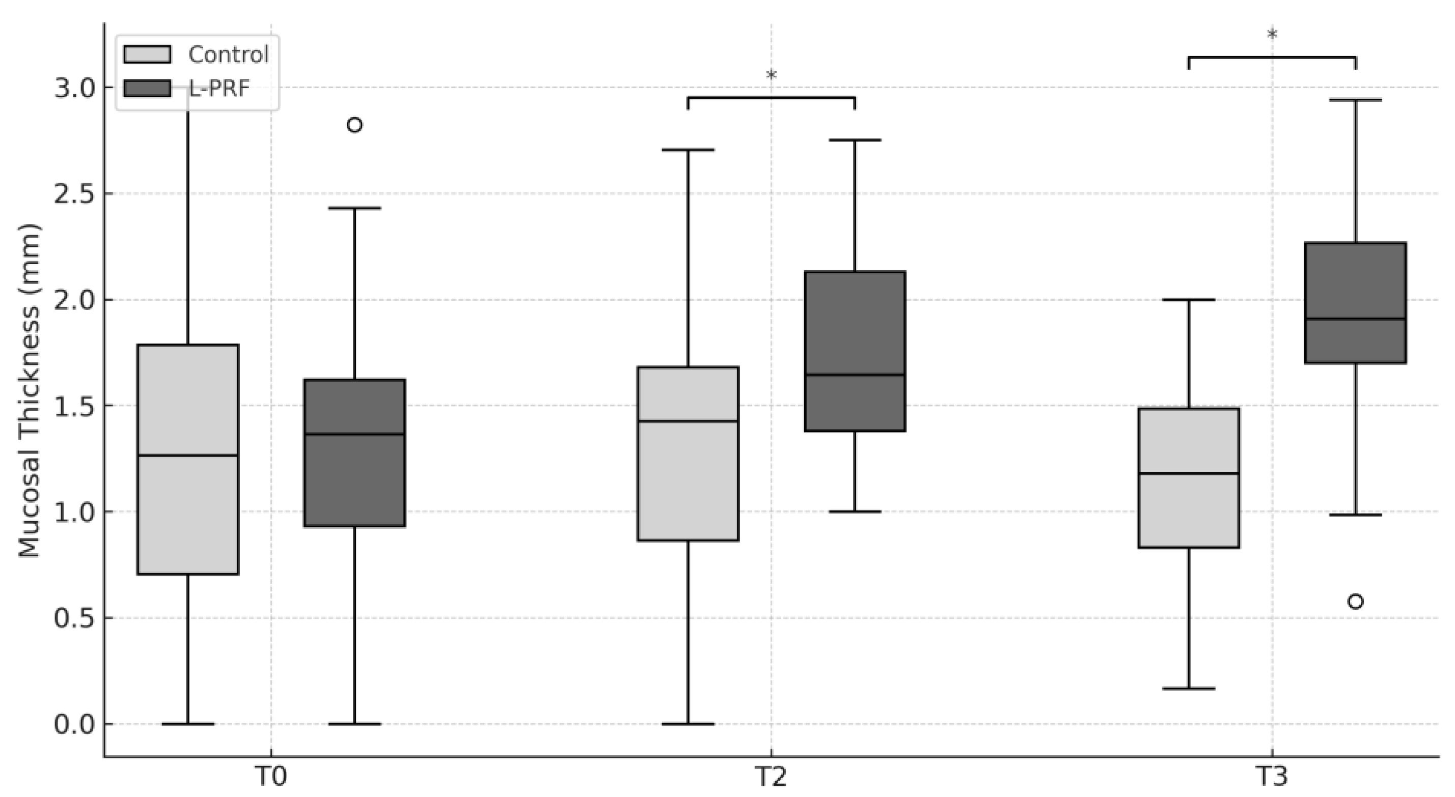
Baseline MT values were comparable between groups. A significant increase in MT was observed in the experimental group at both T2 and T3, indicating a favorable effect of L-PRF on soft tissue thickness (Figure 3). The mean difference between control and L-PRF was 0.26 mm at T2 (p = 0.012) and 0.71 mm at T3 (p < 0.001).
Volumetric analysis using CBCT reconstructions was performed in the 36 patients who completed follow-up. No significant differences were observed in bone volume between baseline (6533.35 ± 1825.8 mm3) and T3 (6425.90 ± 1603.25 mm3; p = 0.45). Similarly, soft tissue volume showed minimal variation (366.68 ± 30.03 mm3 at T0 vs. 362.89 ± 26.97 mm3 at T3; p = 0.12).
Postoperative pain assessed with the VAS was generally low. At day 7, mean scores were 1.6 ± 2.3 in the experimental group and 1.2 ± 2.0 in the control group. On day 14, scores decreased to 0.5 ± 1.2 and 0.1 ± 0.4, respectively, with no significant differences between groups (p = 0.12).

4. Discussion

This study demonstrates that the application of L-PRF significantly increased mucosal thickness around interforaminal implants after 12 and 24 weeks. Consequently, the null hypothesis was partially rejected. This effect was consistent and represents the most clinically relevant findings of our study. An increase in MT may improve the quality of the peri-implant mucosal seal [21,22], thereby promoting peri-implant health [23] and reducing long-term biological complications associated with plaque accumulation, inflammation, and trauma from overdenture movement.
In contrast, no clinically significant differences were found in implant stability between the experimental and control groups at any of the time points, except at 12 weeks, where a modest but statistically significant difference was observed. These results are consistent with previous studies indicating that ISQ values increase progressively over time as a marker of successful osseointegration [24]. Recent studies have demonstrated that PRF applications may enhance early implant stability, with significantly higher ISQ values at 1 week and 1 month post implant placement compared with controls [25]. In a study by Guan et al., implants treated with PRF showed higher stability at 12 weeks (mean ISQ 74.5) than controls (mean ISQ 70.8), which aligns with our findings of transient ISQ differences at 12 weeks [26].
The greater MT observed in the experimental group supports prior evidence on the positive effects of L-PRF in maintaining soft tissue architecture. A recent systematic review of injectable PRF (i-PRF) for non-surgical gingival augmentation concluded that i-PRF can significantly increase gingival thickness and, in some cases, keratinized tissue width, while also providing superior patient-reported comfort and aesthetic outcomes compared with traditional grafting techniques [27]. However, protocols for i-PRF application are not yet standardized. While some RCTs used one, others applied three or four sessions at 7–10-day intervals, complicating direct comparisons of outcomes and highlighting the need for protocol optimization in future studies.
By contrast, a recent study reported decreased mucosal thickness following the use of L-PRF [28,29]. This discrepancy may be related to differences in healing mode, as all patients in that study experienced soft tissue dehiscence and healed by second intention, whereas in our study, primary wound closure was achieved in all cases. Moreover, another clinical study [30] showed that combining L-PRF with subepithelial connective tissue grafts resulted in greater gains in keratinized tissue thickness compared with connective tissue grafting alone, suggesting a potential synergistic effect.
Other clinical parameters such as probing depth (PD) and keratinized tissue width (KTW) did not differ significantly between the groups, in agreement with previously published data [31]. Likewise, volumetric analyses revealed no significant differences in hard or soft tissue volumes between the groups at 6 months. The analysis of soft tissue volume using STL files also showed no significant changes, which may reflect methodological limitations inherent to digital subtraction techniques [32,33,34].
Another important consideration is the lack of standardization across clinical protocols involving PRF. Recent trials have differed substantially in terms of the type of PRF used (L-PRF, A-PRF, i-PRF, or T-PRF), centrifugation settings, and the number of applications. For instance, in non-surgical gingival augmentation protocols, some randomized trials have applied a single session of i-PRF, while others used up to three or four sessions at weekly intervals [27,35]. Despite these variations, the clinical use of L-PRF offers several practical advantages. Its preparation is straightforward, cost-effective, and free of additives such as anticoagulants or chemical manipulation [36]. This facilitates its integration into routine practice without additional burden for clinicians or patients.
From a biological perspective, the positive effect of L-PRF on mucosal thickness may be explained by its gradual release of growth factors such as PDGF, VEGF, and TGF-β1, which enhance angiogenesis, fibroblast proliferation, and collagen deposition [37,38]. The fibrin matrix also serves as a scaffold that supports cellular migration and neovascularization. Previous in vitro and in vivo studies [38] have shown that L-PRF membranes sustain growth factor release for up to 7–14 days, which may explain the enhanced tissue maturation observed clinically. These biological mechanisms provide a plausible rationale for the consistent increase in mucosal thickness observed in the present study [39,40].
Future studies should evaluate the long-term stability of the increased mucosal thickness observed with L-PRF and its impact on peri-implant health and prosthetic outcomes. Research in different clinical scenarios and patient populations, combined with improved digital measurement methods, is needed to confirm these findings. Comparative trials with other platelet concentrates and mechanistic investigations may also help clarify the biological basis and relative effectiveness of L-PRF in enhancing peri-implant soft tissue outcomes.

5. Limitations

This study has some limitations that should be acknowledged. First, although the split-mouth design was chosen to minimize inter-individual variability and to increase statistical power with a limited number of participants, this design inherently carries the risk of carry-over effects between experimental and control sites. Local biological responses to L-PRF may influence adjacent tissues, thereby potentially attenuating the true magnitude of the differences between groups.
Second, the study sample consisted exclusively of elderly edentulous patients rehabilitated with mandibular overdentures. While this represents a highly relevant clinical population, the results cannot be directly extrapolated to younger individuals, partially edentulous patients, or cases involving different implant sites and prosthetic designs. Moreover, the relatively small sample size limits the ability to detect subtle differences in some clinical outcomes and increases the risk of type II error.
Third, the follow-up period of 24 weeks, although sufficient to capture short- and medium-term soft tissue changes, does not provide information about the long-term stability of the observed mucosal thickness gain or its influence on peri-implant health and prosthetic survival. Longer observational periods are needed to determine whether the soft tissue benefits of L-PRF translate into improved biological and prosthetic outcomes over time.
Finally, the study did not assess biological markers or histological outcomes that could elucidate the underlying mechanisms of the observed clinical changes. Such data would help clarify the pathways through which L-PRF contributes to mucosal thickening and whether these effects are primarily related to angiogenesis, fibroblast activation, or collagen maturation. Taken together, these limitations indicate that while our findings support the potential of L-PRF to enhance peri-implant mucosal thickness, further research with larger, more diverse populations, longer follow-up, and complementary biological analyses is required to fully establish its clinical effectiveness.

6. Conclusions

L-PRF use during implant placement was associated with increased peri-implant mucosal thickness, which may improve soft tissue outcomes in the rehabilitation of edentulous patients with mandibular overdentures. Further studies are needed to confirm these findings and to clarify their long-term clinical implications.

Author Contributions

X.M.: investigation; data curation; writing—original draft. P.N.: methodology; investigation; writing—original draft. F.J.S.: formal analysis; visualization; writing—review and editing. M.I.M.: formal analysis; visualization; writing—review and editing. R.G.: conceptualization; supervision; writing—review and editing. F.C.: conceptualization; methodology; project administration; writing—review and editing. All authors have read and agreed to the published version of the manuscript.

Funding

This research was funded by the Oral Reconstruction Foundation (grant ORF-42004) and UNAB regular research grant (DI03_2024). Ximena Moreno received a master’s degree scholarship from ANID (2021–22210987).

Institutional Review Board Statement

The study was conducted according to the guidelines of the Declaration of Helsinki, and approved by the Ethics Committee of Aconcagua Health Service (CEC09/2020).

Informed Consent Statement

Informed consent was obtained from all subjects involved in the study.

Data Availability Statement

The data supporting the findings of this study are not publicly available due to patient confidentiality and legal and ethical restrictions. However, the datasets generated and analyzed during the current study are available from the corresponding author upon reasonable request.

Conflicts of Interest

The authors declare no conflicts of interest.

References

  1. Enlow, D.H.; Bianco, H.J.; Eklund, S. The remodeling of the edentulous mandible. J. Prosthet. Dent. 1976, 36, 685–693. [Google Scholar] [CrossRef] [PubMed]
  2. Berkovitz, B.K.; Heap, P.F. The effect of fluoride on iron, calcium and phosphorus distribution in the rat incisor. Caries Res. 1976, 10, 337–351. [Google Scholar] [CrossRef] [PubMed]
  3. Cawood, J.I.; Howell, R.A. A classification of the edentulous jaws. Int. J. Oral Maxillofac. Surg. 1988, 17, 232–236. [Google Scholar] [CrossRef] [PubMed]
  4. Sanchez-Siles, M.; Ballester-Ferrandis, J.F.; Salazar-Sanchez, N.; Gomez-Garcia, F.J.; Moraleja-Ruiz, R.; Camacho-Alonso, F. Long-term evaluation of quality of life and satisfaction between implant bar overdentures and conventional complete dentures: A 23 years retrospective study. Clin. Implant Dent. Relat. Res. 2018, 20, 208–214. [Google Scholar] [CrossRef]
  5. Zhang, L.; Lyu, C.; Shang, Z.; Niu, A.; Liang, X. Quality of Life of Implant-Supported Overdenture and Conventional Complete Denture in Restoring the Edentulous Mandible: A Systematic Review. Implant Dent. 2017, 26, 945–950. [Google Scholar] [CrossRef]
  6. Morais, J.A.; Heydecke, G.; Pawliuk, J.; Lund, J.P.; Feine, J.S. The effects of mandibular two-implant overdentures on nutrition in elderly edentulous individuals. J. Dent. Res. 2003, 82, 53–58. [Google Scholar] [CrossRef]
  7. Becker, S.T.; Beck-Broichsitter, B.E.; Rossmann, C.M.; Behrens, E.; Jochens, A.; Wiltfang, J. Long-term survival of Straumann dental implants with TPS surfaces: A retrospective study with a follow-up of 12 to 23 years. Clin. Implant Dent. Relat. Res. 2015, 18, 480–488. [Google Scholar] [CrossRef]
  8. Rakic, M.; Galindo-Moreno, P.; Monje, A.; Radovanovic, S.; Wang, H.L.; Cochran, D.; Sculean, A.; Canullo, L. How frequent does peri-implantitis occur? A systematic review and meta-analysis. Clin. Oral Investig. 2018, 22, 1805–1816. [Google Scholar] [CrossRef]
  9. Diaz, P.; Gonzalo, E.; Villagra, L.J.G.; Miegimolle, B.; Suarez, M.J. What is the prevalence of peri-implantitis? A systematic review and meta-analysis. BMC Oral Health 2022, 22, 449. [Google Scholar] [CrossRef]
  10. Nevins, M.; Kao, R.T.; McGuire, M.K.; McClain, P.K.; Hinrichs, J.E.; McAllister, B.S.; Reddy, M.S.; Nevins, M.L.; Genco, R.J.; Lynch, S.E.; et al. Platelet-derived growth factor promotes periodontal regeneration in localized osseous defects: 36-month extension results from a randomized, controlled, double-masked clinical trial. J. Periodontol. 2013, 84, 456–464. [Google Scholar] [CrossRef]
  11. Choukroun, J.; Adda, F.; Schoeffler, C.; Vervelle, A. Une opportunité en paro-implantologie: Le PRF. Implantodontie 2001, 42, 55–62. [Google Scholar]
  12. Öncü, E.; Alaaddinoglu, E.E. The effect of platelet-rich fibrin on implant stability. Int. J. Oral Maxillofac. Implants 2015, 30, 578–582. [Google Scholar] [CrossRef] [PubMed]
  13. Hauser, F.; Gaydarov, N.; Badoud, I.; Vazquez, L.; Bernard, J.P.; Ammann, P. Clinical and histological evaluation of postextraction platelet-rich fibrin socket filling: A prospective randomized controlled study. Implant Dent. 2013, 22, 295–303. [Google Scholar] [CrossRef] [PubMed]
  14. Giammarinaro, E.; Baldini, N.; Covani, U.; Menini, M.; Pesce, P.; Marconcini, S. Does platelet-rich fibrin enhance the outcomes of peri-implant soft tissues? A systematic review. BMC Oral Health 2025, 25, 615. [Google Scholar] [CrossRef] [PubMed]
  15. Patnaik, B.B.; Penmetsa, G.S.; Raju, M.S.; Haripriya, N.; Gera, D.; Ramesh, K. Peri-implant mucosal enhancement using leukocyte platelet rich fibrin under Sohn’s poncho technique: A randomized controlled clinical trial. Clin. Adv. Periodontics 2024, 14, 134–141. [Google Scholar] [CrossRef]
  16. Razi, M.A.; Mahajan, A.; Zarrin, R.; Roy, S.; Singh, M.K.; Kumari, S. Impact of Platelet-Rich Fibrin (PRF) Versus Freeze-Dried Bone Allograft (FDBA) on Peri-Implant Soft and Hard Tissue in Alveolar Ridge Preservation. J. Pharm. Bioallied Sci. 2024, 16 (Suppl. S4), S3550–S3552. [Google Scholar] [CrossRef]
  17. Temmerman, A.; Cleeren, G.J.; Castro, A.B.; Teughels, W.; Quirynen, M. L-PRF for increasing the width of keratinized mucosa around implants: A split-mouth, randomized, controlled pilot clinical trial. J. Periodontal Res. 2018, 53, 793–800. [Google Scholar] [CrossRef]
  18. Al-Ekrish, A.A. Comparative study of the accuracy of CBCT implant site measurements using different software programs. Saudi Dent. J. 2021, 33, 355–361. [Google Scholar] [CrossRef]
  19. Swennen, G.R.; Mollemans, W.; De Clercq, C.; Abeloos, J.; Lamoral, P.; Lippens, F.; Neyt, N.; Casselman, J.; Schutyser, F. A cone-beam computed tomography triple scan procedure to obtain a three-dimensional augmented virtual skull model appropriate for orthognathic surgery planning. J. Craniofacial Surg. 2009, 20, 297–307. [Google Scholar] [CrossRef]
  20. Strauss, F.J.; Stähli, A.; Gruber, R. The use of platelet-rich fibrin to enhance the outcomes of implant therapy: A systematic review. Clin. Oral Implants Res. 2018, 29 (Suppl. S18), 6–19. [Google Scholar] [CrossRef]
  21. Tomasi, C.; Tessarolo, F.; Caola, I.; Wennström, J.; Nollo, G.; Berglundh, T. Morphogenesis of peri-implant mucosa revisited: An experimental study in humans. Clin. Oral Implants Res. 2014, 25, 997–1003. [Google Scholar] [CrossRef]
  22. Berglundh, T.; Lindhe, J.; Ericsson, I.; Marinello, C.P.; Liljenberg, B.; Thomsen, P. The soft tissue barrier at implants and teeth. Clin. Oral Implants Res. 1991, 2, 81–90. [Google Scholar] [CrossRef] [PubMed]
  23. Thoma, D.S.; Naenni, N.; Figuero, E.; Hämmerle, C.H.F.; Schwarz, F.; Jung, R.E.; Sanz-Sánchez, I. Effects of soft tissue augmentation procedures on peri-implant health or disease: A systematic review and meta-analysis. Clin. Oral Implants Res. 2018, 29 (Suppl. S15), 32–49. [Google Scholar] [CrossRef] [PubMed]
  24. Nedir, R.; Bischof, M.; Szmukler-Moncler, S.; Bernard, J.P.; Samson, J. Predicting osseointegration by means of implant primary stability. Clin. Oral Implants Res. 2004, 15, 520–528. [Google Scholar] [CrossRef] [PubMed]
  25. Kapoor, A.; Ali, A.R.; Saini, N.; Gautam, K.; Goyal, A.; Prakash, V. Comparative evaluation of implant stability with and without autologous platelet-rich fibrin prior to prosthetic loading—A split-mouth randomized clinical trial. J. Indian Soc. Periodontol. 2022, 26, 137–142. [Google Scholar]
  26. Guan, S.; Xiao, T.; Bai, J.; Ning, C.; Zhang, X.; Yang, L.; Li, X. Clinical application of platelet-rich fibrin to enhance dental implant stability: A systematic review and meta-analysis. Heliyon 2023, 9, e13196. [Google Scholar] [CrossRef]
  27. Żurek, J.; Niemczyk, W.; Dominiak, M.; Niemczyk, S.; Wiench, R.; Skaba, D. Gingival Augmentation Using Injectable Platelet-Rich Fibrin (i-PRF)-A Systematic Review of Randomized Controlled Trials. J. Clin. Med. 2024, 13, 5591. [Google Scholar] [CrossRef]
  28. Boora, P.; Rathee, M.; Bhoria, M. Effect of platelet rich fibrin (PRF) on peri-implant soft tissue and crestal bone in one-stage implant placement: A randomized controlled trial. J. Clin. Diagn. Res. 2015, 9, ZC18–ZC21. [Google Scholar] [CrossRef]
  29. Zhan, L.; Yang, Y.; Zhao, X.; Wang, C.; Man, Y.; Qu, Y. Clinical outcomes of a peri-implant keratinized mucosa augmentation procedure using concentrated growth factor: A prospective case series. BMC Oral Health 2025, 25, 1064. [Google Scholar] [CrossRef]
  30. Prakash, P.; Rath, S.K.; Mukherjee, M. Clinical efficacy of periosteal pedicle graft with subepithelial connective tissue graft in gingival recession coverage. J. Indian Soc. Periodontol. 2019, 23, 442–447. [Google Scholar] [CrossRef]
  31. Aldommari, E.A.; Omair, A.; Qasem, T. Titanium-prepared platelet-rich fibrin enhances alveolar ridge preservation: A randomized controlled clinical and radiographic study. Sci. Rep. 2025, 15, 2551. [Google Scholar] [CrossRef] [PubMed]
  32. Hosseini, M.; Worsaae, N.; Gotfredsen, K. Tissue changes at implant sites in the anterior maxilla with and without connective tissue grafting: A five-year prospective study. Clin. Oral Implants Res. 2020, 31, 18–28. [Google Scholar] [CrossRef] [PubMed]
  33. Huber, S.; Zeltner, M.; Hammerle, C.H.F.; Jung, R.E.; Thoma, D.S. Non-interventional 1-year follow-up study of peri-implant soft tissues following previous soft tissue augmentation and crown insertion in single-tooth gaps. J. Clin. Periodontol. 2018, 45, 504–512. [Google Scholar] [CrossRef] [PubMed]
  34. Tavelli, L.; Barootchi, S.; Majzoub, J.; Siqueira, R.; Mendonça, G.; Wang, H.L. Volumetric changes at implant sites: A systematic appraisal of traditional methods and optical scanning-based digital technologies. J. Clin. Periodontol. 2021, 48, 315–334. [Google Scholar] [CrossRef]
  35. Ramos, E.U.; Bizelli, V.F.; Pereira Baggio, A.M.; Ferriolli, S.C.; Silva Prado, G.A.; Farnezi Bassi, A.P. Do the New Protocols of Platelet-Rich Fibrin Centrifugation Allow Better Control of Postoperative Complications and Healing After Surgery of Impacted Lower Third Molar? A Systematic Review and Meta-Analysis. J. Oral Maxillofac. Surg. 2022, 80, 1238–1253. [Google Scholar] [CrossRef]
  36. Dohan, M.; Bielecki, T.; Jimbo, R.; Barbé, G.; Del Corso, M.; Inchingolo, F.; Sammartino, G. Do the Fibrin Architecture and Leukocyte Content Influence the Growth Factor Release of Platelet Concentrates? An Evidence-based Answer Comparing a Pure Platelet-Rich Plasma (P-PRP) Gel and a Leukocyte- and Platelet-Rich Fibrin (LPRF). Curr. Pharmacol. Biotechnol. 2012, 13, 1145–1152. [Google Scholar]
  37. Di Summa, F.; Kargarpour, Z.; Nasirzade, J.; Stähli, A.; Mitulović, G.; Panić-Janković, T.; Koller, V.; Kaltenbach, C.; Müller, H.; Panahipour, L.; et al. TGFβ activity released from platelet-rich fibrin adsorbs to titanium surface and collagen membranes. Sci. Rep. 2020, 10, 10203. [Google Scholar] [CrossRef]
  38. Strauss, F.J.; Nasirzade, J.; Kargarpoor, Z.; Stähli, A.; Gruber, R. Effect of platelet-rich fibrin on cell proliferation, migration, differentiation, inflammation, and osteoclastogenesis: A systematic review of in vitro studies. Clin. Oral Investig. 2020, 24, 569–584. [Google Scholar] [CrossRef]
  39. Wang, X.; Fok, M.R.; Pelekos, G.; Jin, L.; Tonetti, M.S. In Vitro and Ex Vivo Kinetic Release Profile of Growth Factors and Cytokines from Leucocyte- and Platelet-Rich Fibrin (L-PRF) Preparations. Cells 2022, 11, 2089. [Google Scholar] [CrossRef]
  40. Coucke, B.; Dilissen, E.; Cremer, J.; Schrijvers, R.; Theys, T.; Van Gerven, L. Leukocyte-and Platelet-Rich Fibrin for enhanced tissue repair: An in vitro study characterizing cellular composition, growth factor kinetics and transcriptomic insights. Mol. Biol. Rep. 2024, 51, 954. [Google Scholar] [CrossRef]
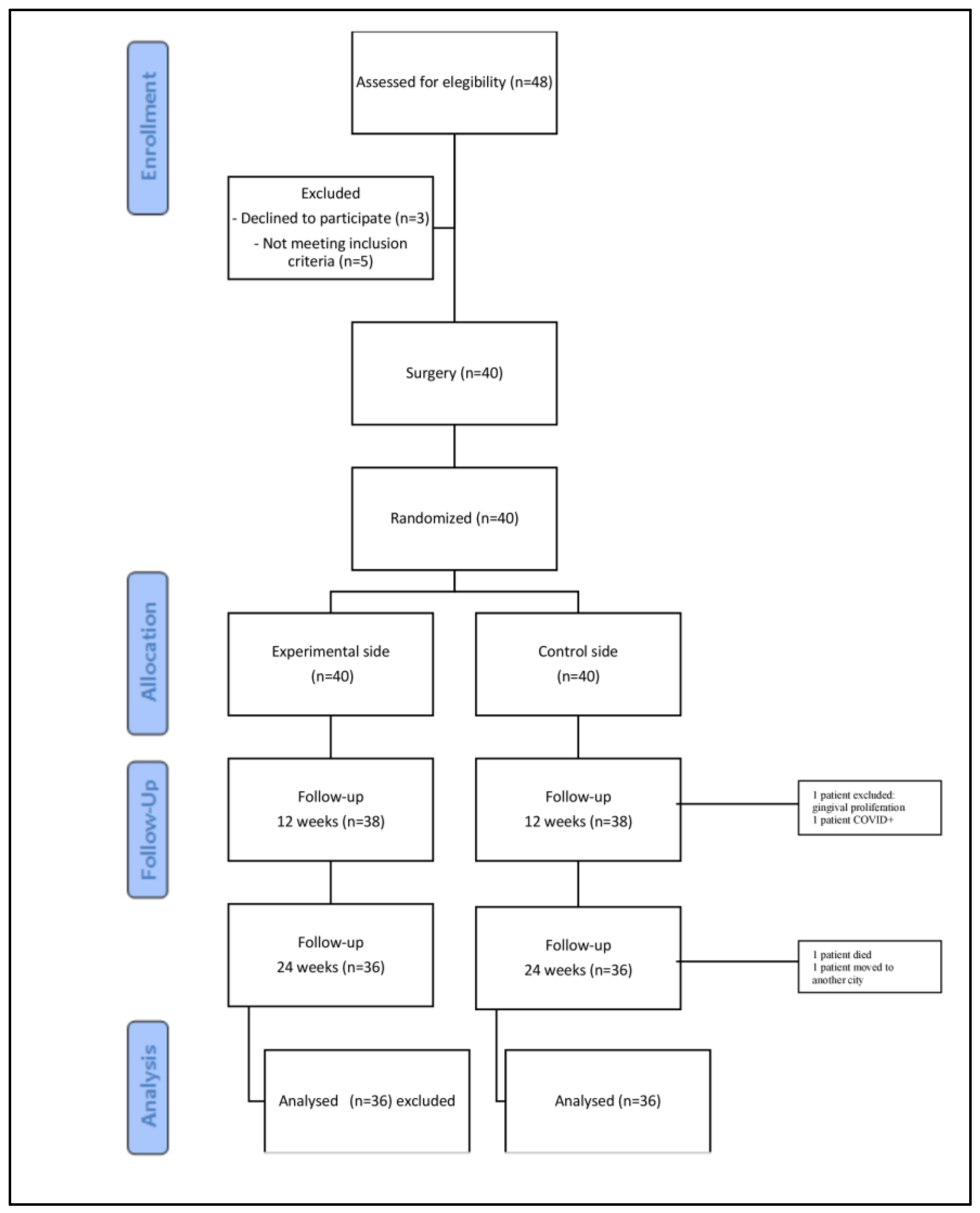
Figure 1. CONSORT flow diagram.
Jcm 14 06917 g001
Figure 2. Implant stability at baseline (T0), 8 weeks (T1), 12 weeks (T2), and 24 weeks (T3). * = p < 0.05. ISQ: Implant Stability Quotient; L-PRF: leukocyte- and platelet-rich fibrin.
Jcm 14 06917 g002
Figure 3. Mucosal thickness (MT) at baseline (T0), 8 weeks (T1), 12 weeks (T2), and 24 weeks (T3). * = p < 0.05. L-PRF: leukocyte- and platelet-rich fibrin.
Jcm 14 06917 g003
Table 1. Clinical parameters of probing depth and keratinized tissue width. T0 = baseline, T2 = 12 weeks post-surgery, T3 = 24 weeks post-surgery.
L-PRFControlp-Value
T0T2T3T0T2T3T0T2T3
PD (mm) 1.6 (±0.5)1.4 (±0.5) 1.8 (±0.6)1.4 (±0.5) 0.600.46
KTW (mm)2.7 (±1.6)2.5 (±1.8)2.4 (±0.9)2.5 (±1.8)2.4 (±0.9)2.7 (±0.9)0.530.680.79
Disclaimer/Publisher’s Note: The statements, opinions and data contained in all publications are solely those of the individual author(s) and contributor(s) and not of MDPI and/or the editor(s). MDPI and/or the editor(s) disclaim responsibility for any injury to people or property resulting from any ideas, methods, instructions or products referred to in the content.