Case Report

Late Recurrence of High-Grade Vulvar Leiomyosarcoma After 5 Years: A Rare Case Report and Expanded Review of Reported Cases

1
Department of Gynaecological Oncology, Medical University Pleven, 5800 Pleven, Bulgaria
2
Department of General and Clinical Pathology, University Hospital “Dr. Georgi Stranski”, 5800 Pleven, Bulgaria
3
Department of Gynaecology, St. Anna University Hospital, 9002 Varna, Bulgaria
4
Research Institute, Medical University Pleven, 5800 Pleven, Bulgaria
5
Department of Obstetrics and Gynecology, University Hospital “Sofiamed”, 1000 Sofia, Bulgaria
6
Faculty of Medicine, Medical University Pleven, 5800 Pleven, Bulgaria
*
Authors to whom correspondence should be addressed.
These authors contributed equally to this work.
J. Clin. Med. 2025, 14(17), 6032; https://doi.org/10.3390/jcm14176032
Submission received: 8 July 2025 / Revised: 11 August 2025 / Accepted: 22 August 2025 / Published: 26 August 2025
(This article belongs to the Section Oncology)

Abstract

Background: Vulvar leiomyosarcoma (VLMS) is a rare and aggressive soft tissue malignancy arising from smooth muscle cells, comprising less than 3% of vulvar cancers. Its clinical resemblance to benign vulvar lesions often leads to delayed diagnosis. Despite surgical resection and adjuvant therapy, VLMS is associated with high recurrence rates and a poor prognosis, and due to its rarity, there is no standardized management or surveillance protocol. Case Report: We present a case of high-grade VLMS in a postmenopausal woman, initially diagnosed in 2020 and managed with surgical excision and adjuvant radiotherapy. The primary tumor was a 10 cm solid, lobulated mass involving the mons pubis, with histology confirming high-grade leiomyosarcoma based on marked cellular atypia, high mitotic activity, and smooth muscle differentiation. Immunohistochemistry was positive for SMA, vimentin, and CD34, and negative for S100 and MyoD1. Five years later, the patient developed a local recurrence with an enlarged inguinal lymph node. She underwent complete tumor resection and bilateral inguinal lymphadenectomy. Histology of the recurrent lesion mirrored the initial findings, with no lymph node metastases. This case highlights the aggressive nature and potential for late recurrence in vulvar leiomyosarcoma, underscoring the importance of long-term surveillance. Conclusions: High-grade VLMS is a rare malignancy with a high recurrence risk. This case highlights the importance of early diagnosis, radical surgical treatment, and long-term surveillance. Although recurrence occurred five years after the initial treatment, timely surgical intervention led to a favorable postoperative course. Multidisciplinary management and individualized follow-up strategies remain key to improving outcomes in these rare gynecologic sarcomas.

1. Introduction

Vulvar leiomyosarcoma (VLMS) is an exceptionally rare and aggressive malignancy derived from smooth muscle cells, accounting for less than 3% of vulvar cancers and an even smaller proportion of all gynecologic malignancies [1]. While the majority of vulvar tumors are benign (e.g., Bartholin gland cysts, fibromas, or lipomas), LMS can clinically mimic benign lesions due to its slow, painless growth in the early stages, resulting in frequent delays in diagnosis and treatment. Histologically, it is characterized by spindle-shaped cells with eosinophilic cytoplasm and pleomorphic nuclei, arranged in fascicles, often demonstrating high mitotic activity and areas of necrosis [1].
Despite complete surgical excision and adjuvant therapy, vulvar LMS is notorious for local recurrence, potential for late metastasis, and poor overall prognosis [2]. The rarity of the disease results in a lack of standardized treatment protocols and limited consensus on optimal surveillance strategies [2]. Therefore, each reported case adds meaningful data to the sparse literature, helping shape future clinical decision-making.
This case report describes a rare instance of recurrent high-grade VLMS occurring five years after the initial curative surgery and adjuvant radiotherapy in 2020 in a postmenopausal woman, as reported by Yordanov et al. [3]. It analyzes the diagnostic challenges, histopathologic features, and immunohistochemical profile of both the primary and recurrent tumor, evaluating any phenotypic changes over time. Furthermore, it presents a comprehensive review of published VLMS cases from the past four decades, examining recurrence patterns, treatment approaches and prognostic factors, with the aim of offering practical recommendations for clinical management and long-term follow-up.

2. Case Report

Initial diagnosis and treatment in 2020, a 74-year-old postmenopausal white woman, with no significant comorbidities other than arterial hypertension and a prior cholecystectomy (2006), presented with a painful, enlarging mass of the mons pubis, measuring 10 cm in diameter (Figure 1A) [3]. She reported severe pain, partially relieved by analgesics, but denied vaginal bleeding or discharge [3].
Upon examination, a lobulated, mobile, subcutaneous tumor was noted, infiltrating the overlying skin, but not fixed to deep structures. The rest of the gynecological exam was unremarkable.
She underwent wide local excision. Intraoperatively, a solid, lobulated tumor measuring approximately 10 × 10 cm was excised from the symphysis with macroscopically and microscopically clear margins. The wound was repaired in two layers, and the patient had an uncomplicated recovery.
Postoperative recovery was uneventful and the patient began adjuvant radiotherapy five weeks after surgery. Following surgery, the patient received adjuvant radiotherapy consisting of two courses of deep X-ray therapy. The first course delivered 30 Gy as five fractions of 400 R, and the second course delivered 20 Gy as four fractions of 400 R. She completed radiotherapy without complications and remained clinically disease-free for five years.
Five years post-surgery, the patient presented with a recurrent tumor in the mons pubis area, accompanied by an enlarged left inguinal lymph node. A clinical exam confirmed a solid, lobulated formation measuring ~15 × 10 cm and a palpable left inguinal mass (Figure 1B–D). Preoperative ultrasound, Doppler ultrasound, and PET-CT were performed.
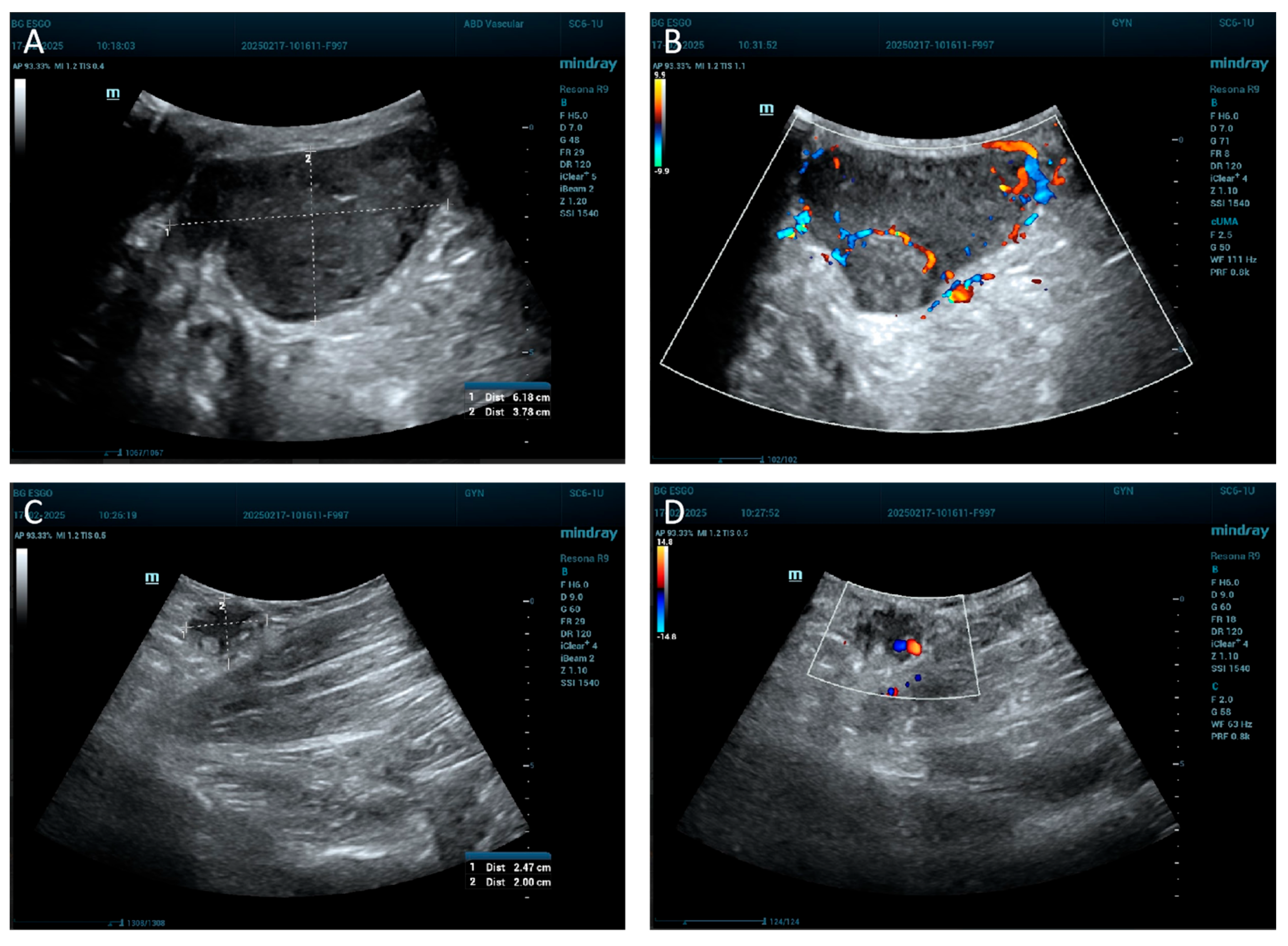
The ultrasound image shows a well-defined structure with dimensions of approximately 6.18 cm in length and 3.78 cm in width. It appears as a relatively hypoechoic area, darker than the surrounding tissue (Figure 2A). The lymph node ultrasound image reveals a small, well-defined structure measuring approximately 2.47 cm in length and 2.00 cm in width, consistent with an enlarged lymph node. The node is relatively hypoechoic and oval in shape, suggesting a reactive or possibly pathological nature (Figure 2C). A Doppler ultrasound was also performed to further assess vascular characteristics of the tumor formation (Figure 2B) and the lymph node (Figure 2D).
PET-CT shows increased uptake in the symphysis and left pelvic regions, consistent with pathological lymphadenopathy, likely involving the inguinal lymph nodes. No other abnormal metabolic activity is noted (Figure 3).
She underwent surgical excision of the recurrent vulvar tumor (complete extirpation of the mons pubis tumor with a 2 cm safety margin) and bilateral inguinal lymphadenectomy (Figure 4A–C). The tumor was solid, lobulated, and yellow-gray on the cut section with a lipomatous appearance (Figure 4C). The overlying skin was not involved, and both the deep and lateral resection margins were free of tumor infiltration. No intraoperative complications; the defect was closed primarily using layered sutures without the use of a flap, wound closure and drainage were completed successfully.
The patient recovered well postoperatively, with no fever, no wound complications, and spontaneous micturition and defecation restored.
Histologically, the morphology of the recurrent tumor closely resembled the primary lesion from 2020, confirming the diagnosis of recurrent high-grade leiomyosarcoma [3]. The recurrent surgical specimen, consisted of a wide local resection encompassing the entire mons pubis and surrounding soft tissues, measuring 150 × 150 mm in total area. Multiple tumor nodules were identified within the subcutaneous tissue, the largest measuring approximately 50–60 mm. On the cut surface, the tumor was solid, lobulated, and yellow-gray in appearance with a fatty texture. There was no involvement of the overlying skin grossly. The deep and lateral margins appeared uninvolved by tumor upon gross examination.
Also included in the resection were bilateral inguinal lymph node packages. The left inguinal region contained a notably enlarged lymph node measuring 25 × 20 mm, while other nodes appeared unremarkable.
Microscopically, the morphology of the recurrent tumor closely resembled the primary leiomyosarcoma excised in 2020, as presented in Figure 5A–D [3]. The tumor consisted of densely cellular, intersecting fascicles composed of spindle-shaped cells with marked nuclear pleomorphism, coarse and irregular chromatin, and prominent nucleoli. The cytoplasm was eosinophilic and moderate in volume, consistent with smooth muscle differentiation. The tumor exhibited high mitotic activity, with numerous mitoses per high-power field, including atypical forms. Scattered areas of coagulative tumor necrosis were present, as well as foci of peritumoral fibrosis. No lymphovascular invasion was identified in the histological specimen.
The resection margins—both lateral and deep—were histologically free of tumor cells. There was no direct extension or presence of tumor infiltration observed in the resection margins. The resection line was at least 20 mm away from the tumor infiltration front. The dermis and epidermis overlying the tumor were not involved, consistent with the absence of skin infiltration observed grossly.
A total of five inguinal lymph nodes were examined. All demonstrated reactive follicular hyperplasia and sinus histiocytosis, with no evidence of metastatic tumor involvement in either the left or right inguinal regions.
Immunohistochemical staining of the recurrent lesion confirmed a consistent profile with the primary tumor from 2020 [3]. In both specimens, tumor cells showed: Smooth Muscle Actin (SMA)–strong, diffuse positivity, confirming smooth muscle differentiation; Vimentin (+) positive, indicating mesenchymal origin; CD34 local positivity; while not specific, this marker is occasionally expressed in soft tissue sarcomas; S100 Protein (-) negative; effectively excluding tumors of neural crest origin such as malignant peripheral nerve sheath tumors or melanoma; MyoD1 (−) negative; ruling out rhabdomyoblastic differentiation. In the 2020 specimen, CD68 demonstrated focal positivity, likely indicative of background histiocytic activity rather than neoplastic expression [3]. This was not a significant feature in the 2025 recurrence.
The immunophenotype of the recurrent lesion also demonstrated somewhat partial loss of specific smooth muscle differentiation, with evident but aberrant positivity for alpha-SMA (Figure 6A) and a current lack of Desmin expression (Figure 6B). MyoD1 remained negative (Figure 6C). The current appearance and differentiation closely resemble that of undifferentiated pleomorphic sarcoma.
The immunophenotype remained stable over time, with no significant phenotypic shifts observed between the initial tumor and the recurrence. These findings support the diagnosis of recurrent high-grade leiomyosarcoma with smooth muscle differentiation.
Adjuvant therapy was reinitiated in 2025, but treatment was interrupted after one month due to the development of radiodermatitis and mucoid reactions, limiting the tolerability of further radiotherapy at that time.

3. Discussion

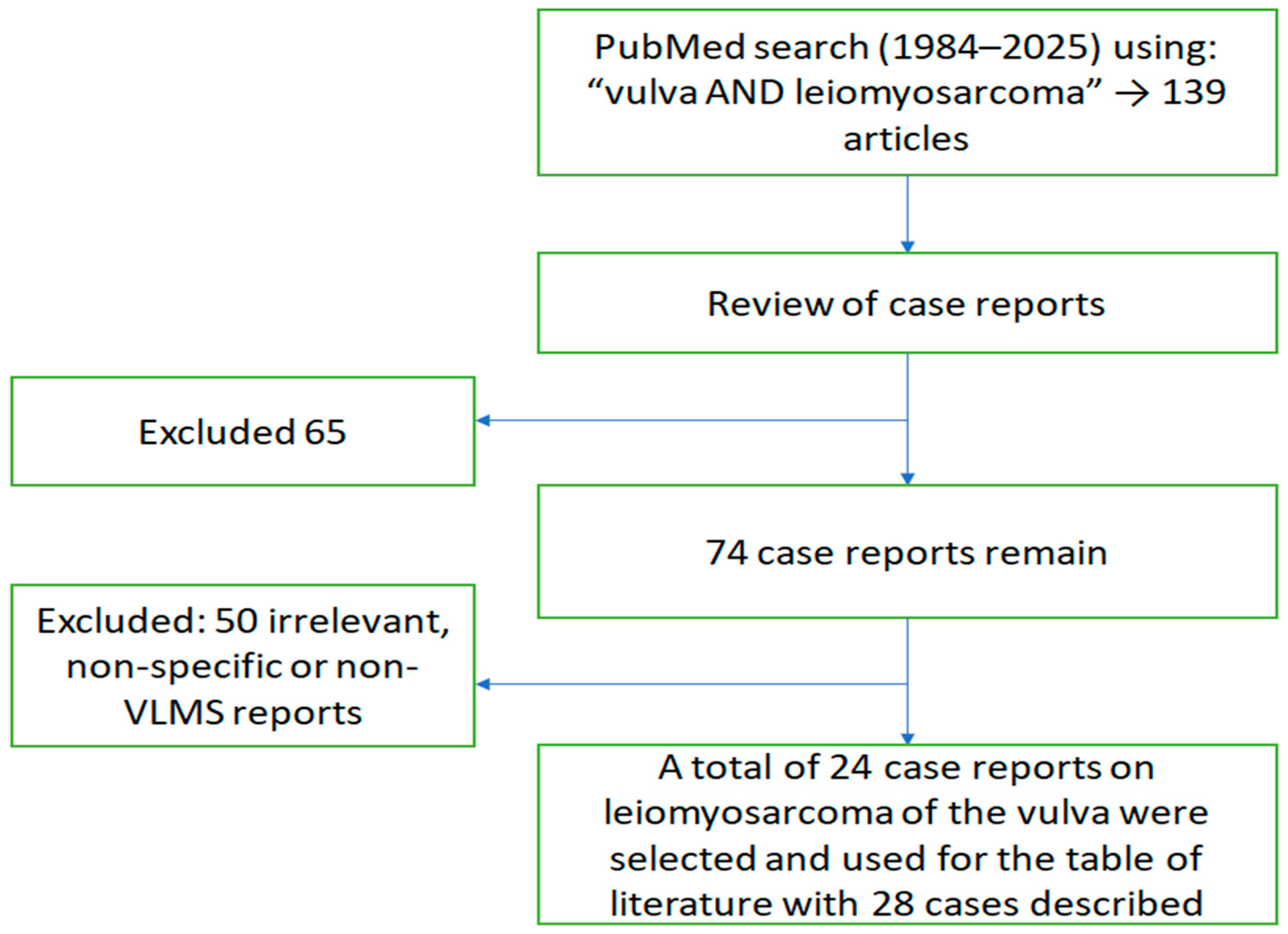
A literature search was performed to provide an overview of all the cases and case characteristics of VLMS described in the literature. We conducted a search in the PubMed® database from the years 1984 to 2025 for the terms “vulva AND leiomyosarcoma”. This yielded a total of 139 citations. Based on the article type filter-case report, we excluded 65 of them. Of the remaining 74, only 24 meeting the criteria. Thus, a total of 24 publications on LMS of the vulva were taken into account to use for the discussion and review of literature, including the table of literature (Figure 7).
The clinical data and characteristics of the VLMS database are summarized in Table 1 [3,4,5,6,7,8,9,10,11,12,13,14,15,16,17,18,19,20,21,22,23,24,25].
VLMS is a rare malignancy, with only 28 well-documented cases published in the literature between 1980 and 2025. An analysis of these cases reveals several clinical patterns, particularly concerning recurrence risk, tumor behavior, and management outcomes.
Our current case represents a recurrence of the tumor first, described by Yordanov et al. in 2020, five years after the original diagnosis [3]. This recurrence highlights the latent and unpredictable nature of VLMS. While the majority of documented recurrences occur within the first 2–3 years post-treatment, a few cases—including ours—demonstrate that VLMS can recur after prolonged disease-free intervals, underscoring the importance of long-term surveillance.
From the 28 reviewed cases, recurrence was noted in nine patients. Among those, five experienced multiple recurrences, with at least three ultimately succumbing to the disease. Recurrence was often associated with larger tumors (≥8 cm). The mean size of tumors that recurred was approximately 8.5 cm, while non-recurrent tumors averaged closer to 5.2 cm, suggesting a correlation between tumor size and recurrence risk.
Metastasis was less common, reported in 6 out of the 28 cases, typically involving the lungs. Interestingly, even large tumors did not universally metastasize, suggesting variable biological behavior. Notably, the current case presented a substantial recurrence (15 × 10 cm), yet without nodal or distant metastasis, aligning with prior reports that VLMS tends to recur locally before systemic spread.
Treatment strategies varied widely. Most patients underwent surgical excision, ranging from simple tumorectomy (TE) to radical vulvectomy with bilateral lymph node dissection (BILND). Unfortunately, most authors have described their surgical interventions without specifying the safety margins, which are crucial to assess given the rarity of the disease and their importance in predicting patient outcomes. Adjuvant radiotherapy and chemotherapy were employed inconsistently, with no consensus on effectiveness. Cases with radical excision appeared to have better outcomes. In our case, both the primary and recurrent tumors were managed surgically with macroscopically complete resection and histologically negative margins.
Histologically, the recurrent tumor mirrored the initial lesion, demonstrating consistent morphology and immunophenotype across the five-year interval. This lack of dedifferentiation suggests a stable tumor biology in this case. The immunophenotype, although somewhat discrepant with the commonly expected for leiomyosarcoma is consistent with the dedifferentiation observed in leiomyosarcoma of the uterine corpus in the context of tumor progression [26]. According to the present case, dedifferentiation during tumor progression can be observed in the context of leiomyosarcoma of the vulva. Such phenotypic stability, though not universal, may correlate with lower metastatic potential.
Demographically, most affected individuals were postmenopausal women aged 45–74. The most common presenting symptoms included a painless or painful vulvar mass, with occasional lymphadenopathy or skin involvement. However, the clinical resemblance to benign lesions often delayed diagnosis.
Overall, this case contributes to the increasing amount of data indicating that, even in cases that were first thought to be cured, VLMS has a significant risk of late recurrence. It also supports the idea that aggressive surgical management can achieve long-term control, especially when combined with vigilant long-term follow-up.
There is not a systematic approach for the management and follow-up of VLMS because of the small number of patients and the variation in presentation and prognosis. Further multicenter collaboration and centralized case registries may improve our understanding of VLMS and help develop evidence-based guidelines for optimal care.

4. Conclusions

High-grade VLMS remains a rare and aggressive malignancy with a notable risk for delayed recurrence. This case, representing a five-year recurrence of a previously reported tumor, demonstrates the necessity for extended surveillance even in the absence of early relapse. The consistent histopathologic features over time, absence of metastasis, and successful surgical re-excision suggest that timely detection and multidisciplinary management can still yield favorable outcomes. This case reinforces the importance of individualized follow-up strategies in VLMS and contributes to the sparse long-term data available for this disease.

Author Contributions

Conceptualization, A.Y. and V.D.; methodology, I.H. and S.K.; formal analysis, I.I.; investigation, V.D. and S.K.; resources, V.D. and I.I.; data curation: I.H.; writing—original and draft preparation: V.D.; writing—review and editing, A.Y.; visualization, I.I. and I.H.; supervision, A.Y. All authors have read and agreed to the published version of the manuscript.

Funding

This research received no external funding.

Institutional Review Board Statement

This study was conducted in accordance with the Declaration of Helsinki. The ethical approval has been waived by the Institutional Review Board of Medical University Pleven for reporting individual cases or case series when informed consent is provided by the patient.

Informed Consent Statement

Written informed consent has been obtained from the patient to publish this paper.

Data Availability Statement

Data presented in this study are available upon request from the corresponding author due to privacy concerns.

Conflicts of Interest

The authors declare no conflicts of interest.

References

  1. Chokoeva, A.A.; Tchernev, G.; Cardoso, J.C.; Patterson, J.W.; Dechev, I.; Valkanov, S.; Zanardelli, M.; Lotti, T.; Wollina, U. Vulvar sarcomas: Short guideline for histopathological recognition and clinical management. Part 1. Int. J. Immunopathol. Pharmacol. 2015, 28, 168–177. [Google Scholar] [CrossRef] [PubMed]
  2. Korkmaz, V.; Kurdoğlu, Z.; Kardag, B.; Arslanca, T.; Caydere, M.; Ergun, Y. A rare case of leiomyosarcoma localized in the Bartholin’s gland area and review of the literature. J. Obstet. Gynaecol. Res. 2016, 42, 589–592. [Google Scholar] [CrossRef]
  3. Yordanov, A.; Slavchev, S.; Kostov, S.; Strashilov, S.; Ivanov, I.; Nikolova, M. Leiomyosarcoma of the vulva: A case report. Prz Menopauzalny 2020, 19, 184–187. [Google Scholar] [CrossRef] [PubMed] [PubMed Central]
  4. Audet-Lapointe, P.; Paquin, F.; Guerard, M.J.; Charbonneau, A.; Methot, F.; Morand, G. Leiomyosarcoma of the vulva. Gynecol. Oncol. 1980, 10, 350–355. [Google Scholar] [CrossRef] [PubMed]
  5. Smit, W.L.; Knobel, J.; van der Merwe, J.V. Leiomioom en leiomiosarkoom van die vulva [Leiomyoma and leiomyosarcoma of the vulva]. S Afr. Med. J. 1984, 66, 961–962. (In Afrikaans) [Google Scholar] [PubMed]
  6. Krag Møller, L.B.; Nygaard Nielsen, M.; Trolle, C. Leiomyosarcoma vulvae. Acta Obstet. Gynecol. Scand. 1990, 69, 187–189. [Google Scholar] [CrossRef] [PubMed]
  7. Kuller, J.A.; Zucker, P.K.; Peng, T.C. Vulvar leiomyosarcoma in pregnancy. Am. J. Obstet. Gynecol. 1990, 162, 164–166. [Google Scholar] [CrossRef] [PubMed]
  8. Bakri, Y.N.; Akhtar, M.; el-Senoussi, M.; Wierzbicki, R. Vulvar sarcoma: A report of four cases. Gynecol. Oncol. 1992, 46, 384–390. [Google Scholar] [CrossRef] [PubMed]
  9. Patel, S.; Kapadia, A.; Desai, A.; Dave, K.S. Leiomyosarcoma of the vulva. Eur. J. Gynaecol. Oncol. 1993, 14, 406–407. [Google Scholar] [PubMed]
  10. Dhar, K.K.; Sau, A.K.; Dey, P.; Vasishta, K. Leiomyosarcoma of the labium minus. Int. J. Gynaecol. Obstet. 1994, 44, 166–167. [Google Scholar] [CrossRef] [PubMed]
  11. Tawfik, O.; Huntrakoon, M.; Collins, J.; Owiety, T.; Seoud, M.A.; Weed, J., Jr. Leiomyosarcoma of the vulva: Report of a case. Gynecol. Oncol. 1994, 54, 242–249. [Google Scholar] [CrossRef] [PubMed]
  12. Lösch, A.; Joura, E.A.; Stani, J.; Breitenecker, G.; Lahodny, J. Leiomyosarcoma of the vulva, A case report. J. Reprod. Med. 2001, 46, 609–612. [Google Scholar] [PubMed]
  13. Rawal, N.; Saridogan, E.; Khan, N.; Weekes, A. Leiomyosarcoma of the vulva in association with lichen sclerosus. J. Obstet. Gynaecol. 2005, 25, 87–88. [Google Scholar] [CrossRef] [PubMed]
  14. Androutsopoulos, G.; Adonakis, G.; Ravazoula, P.; Tsapanos, V.; Kourounis, G. Leiomyosarcoma of the vulva: A case report. Eur. J. Gynaecol. Oncol. 2005, 26, 577–578. [Google Scholar] [PubMed]
  15. Dewdney, S.; Kennedy, C.M.; Galask, R.P. Leiomyosarcoma of the vulva: A case report. J. Reprod. Med. 2005, 50, 630–632. [Google Scholar] [PubMed]
  16. Shankar, S.; Todd, P.M.; Rytina, E.; Crawford, R.A. Leiomyosarcoma of the vulva. J. Eur. Acad. Dermatol. Venereol. 2006, 20, 116–117. [Google Scholar] [CrossRef] [PubMed]
  17. González-Bugatto, F.; Añón-Requena, M.J.; López-Guerrero, M.A.; Báez-Perea, J.M.; Bartha, J.L.; Hervías-Vivancos, B. Vulvar leiomyosarcoma in Bartholin’s gland area: A case report and literature review. Arch. Gynecol. Obstet. 2009, 279, 171–174. [Google Scholar] [CrossRef] [PubMed]
  18. McKenzie, M.; Pintilie, H.; Wilkinson, N.; Lane, G.; Orton, J.; El-Ghobashy, A. A rare case of vulval leiomyosarcoma: Management and an updated review of the literature. J. Obstet. Gynaecol. 2011, 31, 675–676. [Google Scholar] [CrossRef] [PubMed]
  19. Levy, R.A.; Winham, W.M.; Bryant, C.S.; Quick, C.M. Smooth muscle neoplasms of the vulva masquerading as Bartholin gland duct cysts. Proc. (Bayl. Univ. Med. Cent.) 2014, 27, 25–27. [Google Scholar] [CrossRef] [PubMed] [PubMed Central]
  20. Alnafisah, F.; Alfieri, J. Lung Metastasis in a Case of Recurrent Poorly Differentiated Leiomyosarcoma of the Bartholin Gland: A Case Report and Review of the Literature. Cureus 2016, 8, e550. [Google Scholar] [CrossRef] [PubMed] [PubMed Central]
  21. Saquib, S.; Keloth, T.R. Letter in Reply: Leiomyosarcoma of the Vulva Mimicking as Chronic Bartholin Cyst: A Case Report. Oman Med. J. 2021, 36, e258. [Google Scholar] [CrossRef] [PubMed] [PubMed Central]
  22. Aljehani, A.M.; Quatei, A.; Qattea, L.; Aljohani, R.M.; Alkushi, A. Vulvar Leiomyosarcoma in Pregnancy. Cureus 2021, 13, e18772. [Google Scholar] [CrossRef] [PubMed] [PubMed Central]
  23. Reinicke, T.; Anderson, D.J.; Kumar, D.; Griggs, C. Vulvar Leiomyosarcoma Masquerading as a Bartholin’s Gland Cyst in an Adolescent. Cureus 2022, 14, e21674. [Google Scholar] [CrossRef] [PubMed] [PubMed Central]
  24. Capalbo, G.; Logoteta, A.; Gallo, R.; Cuccu, I.; Gentile, G.; Arienzo, F.; Musella, A.; Pernazza, A.; Perniola, G.; Di Donato, V.; et al. Chemo-surgical approach in vulvar leiomyosarcoma: A case report. Tumori 2022, 108, NP26–NP29. [Google Scholar] [CrossRef] [PubMed]
  25. Rathore, R.; Singh, A.; Bhatla, N.; Mathur, S. Primary leiomyosarcoma of the vulva—A rare occurrence. Pol. J. Pathol. 2023, 74, 56–58. [Google Scholar] [CrossRef] [PubMed]
  26. Kim, S.; Bae, H.; Kim, H.-S. Dedifferentiation in uterine leiomyosarcoma: Diagnostic pitfalls and emerging molecular insights. Diagnostics 2024, 14, 160. [Google Scholar] [CrossRef]
Figure 1. Macroscopic view of the tumor: (A) 2020; (B) 2025; (C) 2025; (D) 2025.
Jcm 14 06032 g001
Figure 2. Ultrasound of the tumor formation and lymph node. (A) Ultrasound of the tumor formation. (B) Doppler ultrasound of the tumor formation. (C) Ultrasound of the lymph node. (D) Doppler ultrasound of the lymph node.
Jcm 14 06032 g002
Figure 3. (A) PET-CT axial section. (B) PET-CT coronal section.
Jcm 14 06032 g003
Figure 4. (A). Intraoperative view after complete excision and bilateral lymphatic dissection. (B) Macroscopic view of the extirpated tumor. (C) Macroscopic view of the dissected tumor. (D) The 15th postoperative day.
Jcm 14 06032 g004
Figure 5. (A) HE, 4×—Cellular tumor with evidence of tumor necrosis and cellular pleomorphism. (B) HE, 10×—Cellular tumor with evidence of tumor necrosis and cellular pleomorphism, scant lymphocytic infiltrate can be noticed. (C) HE 40×—Significant pleomorphism and cellular atypia can be noted. (D) HE 40×—Significant pleomorphism and cellular atypia are observed, single mitoses are evident.
Jcm 14 06032 g005
Figure 6. (A) Alpha SMA, 100×. (B) Desmin, 100×. (C) Myo-D1, 100×.
Jcm 14 06032 g006
Figure 7. Flowchart of the literature search and selection process for the literature used in the review and discussion.
Jcm 14 06032 g007
Table 1. Clinical and morphological features of patients diagnosed with VLMS.
Author/YearAgeLocationSymptomsInitial Tumor
Size (cm)
Recurrence
Size (cm)
Primary
Treatment
Recurrence
Treatment
Recurrence
(Yes/No)
Follow-Up
(months)
Metastasis
1Audet-Lapointe
et al., 1980 [4]
48Left labium
majus
Painless growing
nodule
2 × 47 × 2 → 3TETE, Radical
Vulvectomy + BILND
Yes (2×)24No
2Smith et al.,
1984 [5]
49n.a.n.a.7n.a.WLE + RTNot specifiedYes18No
3KragMoller et al.,
1990 [6]
54Left labium
majus
Growing tender
swelling
8 × 7 × 4-TE, Radical
Vulvectomy + BILND
-No30No
4Kuller et al.,
1990 [7]
17Hymeneal ring,
left
No4 × 4 × 4-TE, WLE, BLINK-No20No
5Bakri et al.,
1992 Case 1 [8]
29Right labium
majus
No9MultipleWLE4 × WLEYes (4×)120Lung
6Bakri et al.,
1992 Case 2 [8]
41Left labium
majus
Painless growing
nodule
10 × 8MultipleWLE3 × WLEYes (3×)90Lung
7Bakri et al.,
1992 Case 3 [8]
17Left labium
majus
Lump10 × 15MultipleWLE2 × WLEYes (2×)108- deathLung
8Bakri et al.,
1992 Case 4 [8]
20Left vulva/
groin
Pain/swelling8 × 10;
5 × 3 (ILN)
n.a.WLE, Laparotomyn.a.n.a.n.a.Internal
iliac nodes
9Patel et al.,
1993 [9]
n.a.n.a.n.a.n.a.n.a.TERadical Vulvectomy,
BLINK, WLE
Yes (1×)n.a.No
10Dhar et al.,
1994 [10]
45Left labium
minus
Itching, pain4 × 6 × 3-FNA ILN,
Simple Vulvectomy
-No12No
11Tawfik et al.,
1994 [11]
52Right labia
majora/minora
Pain, swelling12 × 15-TE-No14No
12Lösch et al.,
2001 [12]
38VulvaSlowly growing
mass
n.a.-TE-No24No
13Rawal et al.,
2005 [13]
81Right vulvaLichen sclerosus over 30 yr, 1 yr postmenopausal,
bleeding
5×2-WLE-No9No
14Androutsopoulos et al.,
2005 [14]
55Right labia
majora
5 yr
enlargement
8×9n.a.n.a.n.a.n.a.6 (Death)Multiple
15Dewdney et al.,
2005 [15]
36VulvaSlowly growing painless massn.a.-Modified
Radical Vulvectomy
-No13No
16Shankar et al.,
2006 [16]
58Right vulvaEnlarging asymptomatic
lump
3-TE-No42No
17Gonzalez-Bugatto et
al., 2009 [17]
52Left Bartholin
gland area
Painless nodule
rapid growth
63–4Excision → Hemivulvectomy+ ILND
→ Adjuvant RT + Chemo
WLEYes48No
18McKenzie et al.,
2011 [18]
45Right labium
majus
n.a.n.a.-TE-No24No
19Levy et al.,
2014 Case 1 [19]
50Left labia inBartholin
area
No4×6n.a.TE-n.a.n.a.n.a.
20Levy et al.,
2014 Case 2 [19]
57Bartholin
area
No4×2n.a.TE-n.a.n.a.n.a.
21Alnafisah et al.,
2016 [20]
37Left vulvaRapid enlarging
mass
5.0 × 3.9 × 2.9LungWLE → Radical Vulvectomy + ILND
→ Chemo → Brachytherapy + EBRT
Lung lobectomy +
Chemo
Yes24+Lung
22Korkmaz et al.,
2016 [2]
65Left vulvar Bartholin
area
Enlarging vulvar
mass
5 × 6-TE-No6No
23Saquib et al.,
2020 [21]
63Left labia
majora
Painless
swelling
2.8 × 2.4× 1.4-TE-No6No
24Yordanov et al., 2020
[3]/Present Case 2025
73/78SymphysisPain, rapid
growth
7 × 515×10WLERe-excision + LNDYes60No
25Aljehani et al.,
2021 [22]
38
(pregnant)
Left labia
majora/minora
Vulvar mass15 × 10n.a.TE-No12No
26Reinicke et al.,
2022 [23]
14Left labia
majora
Growing mass6 × 6n.a.TEn.a.n.a.n.a.n.a.
27Capalbo et al.,
2022 [24]
74Vulvan.a.12.5n.a.Neoadjuvant Chemo →
Radical Vulvectomy → Chemo
-No12No
28Rathore et al.,
2023 [25]
47Left lower of the vulvaVulvar swelling8 × 8n.a.Radical Vulvectomy-No8No
Abbreviations: FU = follow-up; TE = tumorectomy; WLE = wide local excision; BILND = bilateral inguinal lymph node dissection; RT = radiotherapy; ILND = inguinal lymph node dissection n.a. = not available.
Disclaimer/Publisher’s Note: The statements, opinions and data contained in all publications are solely those of the individual author(s) and contributor(s) and not of MDPI and/or the editor(s). MDPI and/or the editor(s) disclaim responsibility for any injury to people or property resulting from any ideas, methods, instructions or products referred to in the content.