

Predictors and Outcomes of Right Ventricular Dysfunction in Patients Admitted to the Medical Intensive Care Unit for Sepsis—A Retrospective Cohort Study

 ,
,  , ,
, ,
Abstract
1. Introduction
2. Methods
2.1. Study Population and Design
2.2. Data Collection
2.3. Transthoracic Echocardiography
2.4. Statistical Analysis
3. Results
4. Discussion
5. Limitations and Future Perspectives
6. Conclusions
Author Contributions
Funding
Institutional Review Board Statement
Informed Consent Statement
Data Availability Statement
Conflicts of Interest
References
- Singer, M.; Deutschman, C.S.; Seymour, C.W.; Shankar-Hari, M.; Annane, D.; Bauer, M.; Bellomo, R.; Bernard, G.R.; Chiche, J.-D.; Coopersmith, C.M.; et al. The Third International Consensus Definitions for Sepsis and Septic Shock (Sepsis-3). JAMA 2016, 315, 801–810. [Google Scholar] [CrossRef]
- Ehrman, R.R.; Sullivan, A.N.; Favot, M.J.; Sherwin, R.L.; Reynolds, C.A.; Abidov, A.; Levy, P.D. Pathophysiology, Echocardiographic Evaluation, Biomarker Findings, and Prognostic Implications of Septic Cardiomyopathy: A Review of the Literature. Crit. Care 2018, 22, 112. [Google Scholar] [CrossRef] [PubMed]
- L’Heureux, M.; Sternberg, M.; Brath, L.; Turlington, J.; Kashiouris, M.G. Sepsis-Induced Cardiomyopathy: A Comprehensive Review. Curr. Cardiol. Rep. 2020, 22, 35. [Google Scholar] [CrossRef] [PubMed]
- Naksuk, N.; Tan, N.; Padmanabhan, D.; Kancharla, K.; Makkar, N.; Yogeswaran, V.; Gaba, P.; Kaginele, P.; Riley, D.C.; Sugrue, A.M.; et al. Right Ventricular Dysfunction and Long-Term Risk of Sudden Cardiac Death in Patients With and Without Severe Left Ventricular Dysfunction. Circ. Arrhythm. Electrophysiol. 2018, 11, e006091. [Google Scholar] [CrossRef]
- Sanders, J.L.; Koestenberger, M.; Rosenkranz, S.; Maron, B.A. Right Ventricular Dysfunction and Long-Term Risk of Death. Cardiovasc. Diagn. Ther. 2020, 10, 1646–1658. [Google Scholar] [CrossRef]
- Bansal, M.; Mehta, A.; Machanahalli Balakrishna, A.; Kalyan Sundaram, A.; Kanwar, A.; Singh, M.; Vallabhajosyula, S. Right Ventricular Dysfunction in Sepsis: An Updated Narrative Review. Shock 2023, 59, 829–837. [Google Scholar] [CrossRef] [PubMed]
- Mukherjee, M.; Rudski, L.G.; Addetia, K.; Afilalo, J.; D’Alto, M.; Freed, B.H.; Friend, L.B.; Gargani, L.; Grapsa, J.; Hassoun, P.M.; et al. Guidelines for the Echocardiographic Assessment of the Right Heart in Adults and Special Considerations in Pulmonary Hypertension: Recommendations from the American Society of Echocardiography. J. Am. Soc. Echocardiogr. 2025, 38, 141–186. [Google Scholar] [CrossRef]
- Lanspa, M.J.; Cirulis, M.M.; Wiley, B.M.; Olsen, T.D.; Wilson, E.L.; Beesley, S.J.; Brown, S.M.; Hirshberg, E.L.; Grissom, C.K. Right Ventricular Dysfunction in Early Sepsis and Septic Shock. Chest 2021, 159, 1055–1063. [Google Scholar] [CrossRef]
- Agarwal, R.; Krishnanda, S.I.; Yausep, O.E.; Nugraha, R.A.; Priyonugroho, G.; Hertine, S.; Wicaksono, S.H.; Almazini, P.; Zamroni, D.; Muliawan, H.S. The Role of Neutrophil-to-Lymphocyte Ratio and Right Ventricular Dysfunction in Indonesian Patients with COVID-19: A Retrospective Cohort Study. J. Clin. Med. 2025, 14, 2051. [Google Scholar] [CrossRef]
- Kim, J.-S.; Kim, Y.-J.; Kim, M.; Ryoo, S.M.; Kim, W.Y. Association between Right Ventricle Dysfunction and Poor Outcome in Patients with Septic Shock. Heart 2020, 106, 1665–1671. [Google Scholar] [CrossRef]
- Konstam, M.A.; Kiernan, M.S.; Bernstein, D.; Bozkurt, B.; Jacob, M.; Kapur, N.K.; Kociol, R.D.; Lewis, E.F.; Mehra, M.R.; Pagani, F.D.; et al. Evaluation and Management of Right-Sided Heart Failure: A Scientific Statement From the American Heart Association. Circulation 2018, 137, e578–e622. [Google Scholar] [CrossRef]
- Padang, R.; Chandrashekar, N.; Indrabhinduwat, M.; Scott, C.G.; Luis, S.A.; Chandrasekaran, K.; Michelena, H.I.; Nkomo, V.T.; Pislaru, S.V.; Pellikka, P.A.; et al. Aetiology and Outcomes of Severe Right Ventricular Dysfunction. Eur. Heart J. 2020, 41, 1273–1282. [Google Scholar] [CrossRef]
- Vallabhajosyula, S.; Shankar, A.; Vojjini, R.; Cheungpasitporn, W.; Sundaragiri, P.R.; DuBrock, H.M.; Sekiguchi, H.; Frantz, R.P.; Cajigas, H.R.; Kane, G.C.; et al. Impact of Right Ventricular Dysfunction on Short-Term and Long-Term Mortality in Sepsis: A Meta-Analysis of 1,373 Patients. Chest 2021, 159, 2254–2263. [Google Scholar] [CrossRef]
- Butrous, G. Human Immunodeficiency Virus-Associated Pulmonary Arterial Hypertension: Considerations for Pulmonary Vascular Diseases in the Developing World. Circulation 2015, 131, 1361–1370. [Google Scholar] [CrossRef] [PubMed]
- Doria de Vasconcellos, H.; Post, W.S.; Ervin, A.-M.; Haberlen, S.A.; Budoff, M.; Malvestutto, C.; Magnani, J.W.; Feinstein, M.J.; Brown, T.T.; Lima, J.A.C.; et al. Associations Between HIV Serostatus and Cardiac Structure and Function Evaluated by 2-Dimensional Echocardiography in the Multicenter AIDS Cohort Study. J. Am. Heart Assoc. 2021, 10, e019709. [Google Scholar] [CrossRef] [PubMed]
- Çetin Güvenç, R.; Ceran, N.; Güvenç, T.S.; Tokgöz, H.C.; Velibey, Y. Right Ventricular Hypertrophy and Dilation in Patients With Human Immunodeficiency Virus in the Absence of Clinical or Echocardiographic Pulmonary Hypertension. J. Card. Fail. 2018, 24, 583–593. [Google Scholar] [CrossRef]
- US Preventive Services Task Force; Barry, M.J.; Nicholson, W.K.; Silverstein, M.; Chelmow, D.; Coker, T.R.; Davis, E.M.; Donahue, K.E.; Jaén, C.R.; Kubik, M.; et al. Preexposure Prophylaxis to Prevent Acquisition of HIV: US Preventive Services Task Force Recommendation Statement. JAMA 2023, 330, 736–745. [Google Scholar] [CrossRef]
- Hiraiwa, H.; Kasugai, D.; Ozaki, M.; Goto, Y.; Jingushi, N.; Higashi, M.; Nishida, K.; Kondo, T.; Furusawa, K.; Morimoto, R.; et al. Clinical Impact of Visually Assessed Right Ventricular Dysfunction in Patients with Septic Shock. Sci. Rep. 2021, 11, 18823. [Google Scholar] [CrossRef]
- Bruno, R.R.; Wernly, B.; Mamandipoor, B.; Rezar, R.; Binnebössel, S.; Baldia, P.H.; Wolff, G.; Kelm, M.; Guidet, B.; De Lange, D.W.; et al. ICU-Mortality in Old and Very Old Patients Suffering From Sepsis and Septic Shock. Front. Med. 2021, 8, 697884. [Google Scholar] [CrossRef] [PubMed]
- Xu, C.; Lv, L.; Hu, W.; Yu, Z.; Qian, H.; Chen, F. Long-Term Outcomes in Older Patients with Sepsis in the ICU: A Retrospective Study. Altern. Ther. Health Med. 2024, 30, 124–130. [Google Scholar]
- Kotfis, K.; Wittebole, X.; Jaschinski, U.; Solé-Violán, J.; Kashyap, R.; Leone, M.; Nanchal, R.; Fontes, L.E.; Sakr, Y.; Vincent, J.-L.; et al. A Worldwide Perspective of Sepsis Epidemiology and Survival According to Age: Observational Data from the ICON Audit. J. Crit. Care 2019, 51, 122–132. [Google Scholar] [CrossRef] [PubMed]
- Michels, E.H.A.; Butler, J.M.; Reijnders, T.D.Y.; Cremer, O.L.; Scicluna, B.P.; Uhel, F.; Peters-Sengers, H.; Schultz, M.J.; Knight, J.C.; van Vught, L.A.; et al. Association between Age and the Host Response in Critically Ill Patients with Sepsis. Crit. Care 2022, 26, 385. [Google Scholar] [CrossRef] [PubMed]
- Casserly, B.; Phillips, G.S.; Schorr, C.; Dellinger, R.P.; Townsend, S.R.; Osborn, T.M.; Reinhart, K.; Selvakumar, N.; Levy, M.M. Lactate Measurements in Sepsis-Induced Tissue Hypoperfusion: Results from the Surviving Sepsis Campaign Database. Crit. Care Med. 2015, 43, 567–573. [Google Scholar] [CrossRef]
- Evans, L.; Rhodes, A.; Alhazzani, W.; Antonelli, M.; Coopersmith, C.M.; French, C.; Machado, F.R.; Mcintyre, L.; Ostermann, M.; Prescott, H.C.; et al. Surviving Sepsis Campaign: International Guidelines for Management of Sepsis and Septic Shock 2021. Crit. Care Med. 2021, 49, e1063–e1143. [Google Scholar] [CrossRef]
- Hameed, A.; Condliffe, R.; Swift, A.J.; Alabed, S.; Kiely, D.G.; Charalampopoulos, A. Assessment of Right Ventricular Function-a State of the Art. Curr. Heart Fail. Rep. 2023, 20, 194–207. [Google Scholar] [CrossRef] [PubMed]

| Entire Cohort N = 361 | No RV Dysfunction N = 314 | RV Dysfunction N = 47 | p-Value | ||||
|---|---|---|---|---|---|---|---|
| Demographics, Social, and Past Medical History | |||||||
| Age, mean (SD) | 66.79 | 16.51 | 66.25 | 16.51 | 70.38 | 16.20 | 0.083 † |
| Females, N (%) | 197 | 54.6% | 175 | 55.7% | 22 | 46.8% | 0.252 |
| BMI, mean (SD) | 29.11 | 15.85 | 29.23 | 16.57 | 28.28 | 9.81 | 0.919 † |
| Hypertension, N (%) | 236 | 65.6% | 201 | 64.2% | 35 | 74.5% | 0.168 |
| Diabetes, N (%) | 148 | 41.2% | 125 | 40.1% | 23 | 48.9% | 0.249 |
| Hyperlipidemia, N (%) | 125 | 34.8% | 108 | 34.6% | 17 | 36.2% | 0.835 |
| Current smoking, N (%) | 77 | 22.1% | 68 | 22.4% | 9 | 19.6% | 0.661 |
| Use of cocaine, N (%) | 21 | 6.1% | 17 | 5.7% | 4 | 8.7% | 0.502 ‡ |
| Use of alcohol, N (%) | 58 | 16.6% | 53 | 17.5% | 5 | 10.9% | 0.261 |
| History of CVA, N (%) | 81 | 22.5% | 74 | 23.6% | 7 | 14.9% | 0.180 |
| Cirrhosis, N (%) | 33 | 9.2% | 29 | 9.3% | 4 | 8.5% | 1.000 ‡ |
| ESRD, N (%) | 29 | 8.1% | 22 | 7.1% | 7 | 14.9% | 0.082 ‡ |
| HIV infection, N (%) | 22 | 6.2% | 16 | 5.1% | 6 | 13.0% | 0.049 ‡ |
| COPD, N (%) | 78 | 21.7% | 66 | 21.1% | 12 | 25.5% | 0.490 |
| CHF, N (%) | 66 | 18.4% | 52 | 16.7% | 14 | 29.8% | 0.030 |
| Coronary artery disease, N (%) | 62 | 17.2% | 53 | 16.9% | 9 | 19.1% | 0.708 |
| Peripheral artery disease, N (%) | 26 | 7.2% | 24 | 7.7% | 2 | 4.3% | 0.553 ‡ |
| VTE, N (%) | 72 | 19.9% | 62 | 19.7% | 10 | 21.3% | 0.806 |
| Atrial fibrillation, N (%) | 65 | 18.1% | 47 | 15.1% | 18 | 38.3% | <0.001 |
| Data on Admission | |||||||
| Mean arterial pressure, mean (SD) | 84.82 | 23.19 | 86.11 | 23.26 | 76.24 | 20.92 | 0.008 † |
| Shock index at admission, mean (SD) | 0.97 | 0.41 | 0.95 | 0.39 | 1.10 | 0.52 | 0.099 † |
| AKI at admission, N (%) | 254 | 70.4% | 216 | 68.8% | 38 | 80.9% | 0.091 |
| White blood cell count, mean (SD) | 14.26 | 16.39 | 14.38 | 17.41 | 13.49 | 6.44 | 0.612 † |
| HbA1c (%), mean (SD) | 6.57 | 2.44 | 6.59 | 2.58 | 6.50 | 1.10 | 0.467 † |
| APACHE-2, mean (SD) | 18.99 | 8.25 | 18.61 | 8.25 | 21.49 | 7.87 | 0.037 † |
| SOFA, mean (SD) | 6.14 | 3.70 | 5.93 | 3.68 | 7.55 | 3.58 | 0.002 † |
| LV systolic dysfunction, N (%) | 43 | 11.9% | 23 | 7.3% | 20 | 42.6% | <0.01 |
| LV ejection fraction, mean (SD) | 59.88 | 14.24 | 61.77 | 11.84 | 47.32 | 21.08 | <0.001 † |
| Source of Sepsis by Culture Positivity | |||||||
| Positive blood culture | 89 | 24.7% | 73 | 23.2% | 16 | 34.0% | 0.109 |
| Positive urine culture | 57 | 15.8% | 50 | 15.9% | 7 | 14.9% | 0.857 |
| Positive sputum culture | 61 | 16.9% | 51 | 16.2% | 10 | 21.3% | 0.390 |
| Admission Outcomes | |||||||
| Peak lactate (mmol/L), mean (SD) | 4.99 | 4.61 | 4.83 | 4.53 | 5.96 | 5.03 | 0.074 † |
| Hemodialysis required, N (%) | 47 | 13.2% | 42 | 13.5% | 5 | 10.6% | 0.582 |
| Mechanical ventilation required, N (%) | 168 | 46.5% | 138 | 43.9% | 30 | 63.8% | 0.011 |
| Vasopressors required, N (%) | 144 | 39.9% | 115 | 36.6% | 29 | 61.7% | 0.001 |
| Developed ARDS, N (%) | 20 | 5.6% | 15 | 4.8% | 5 | 10.6% | 0.160 ‡ |
| Developed PE, N (%) | 21 | 5.9% | 15 | 4.8% | 6 | 13.0% | 0.040 |
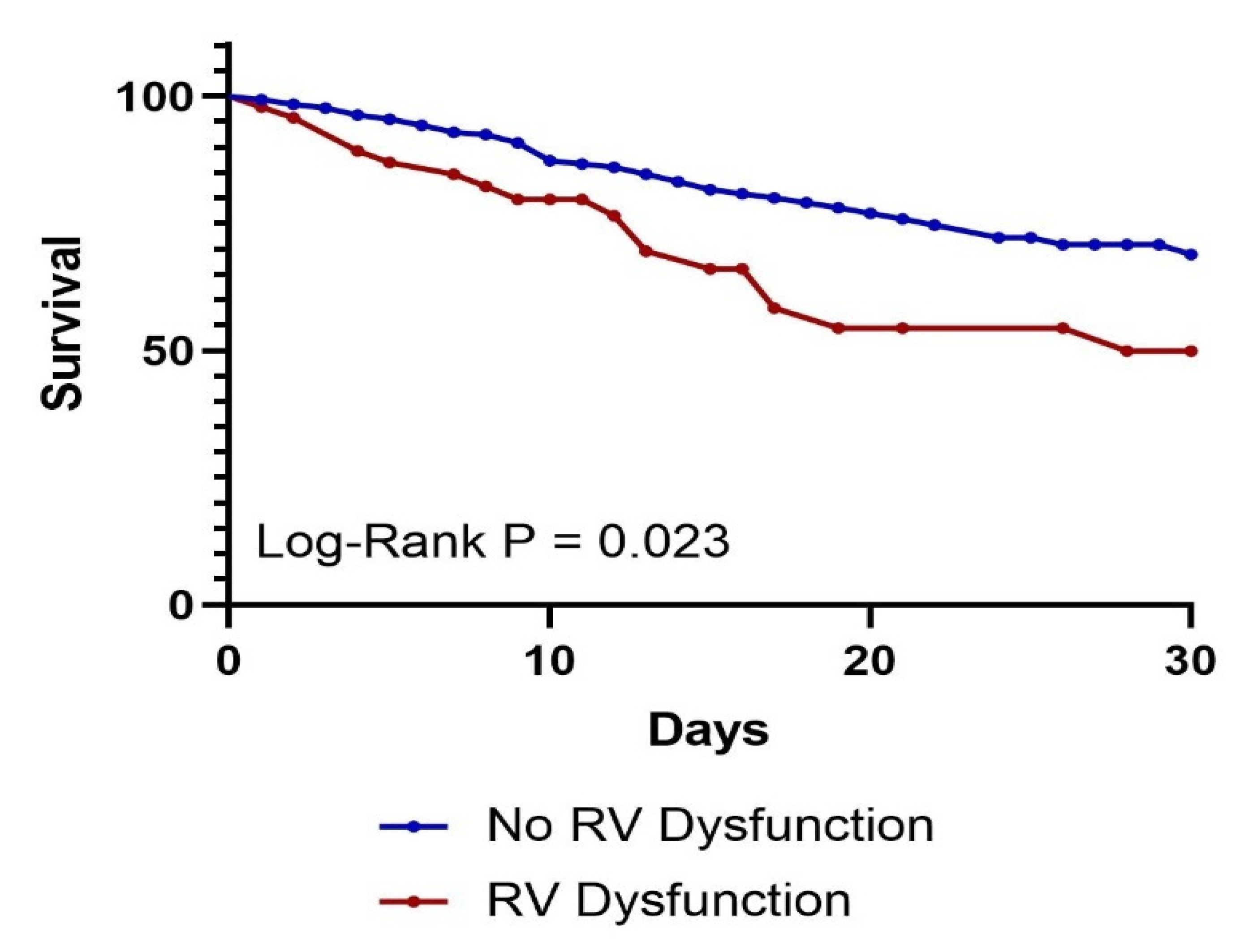
| 30-day mortality, N (%) | 68 | 18.8% | 51 | 16.2% | 17 | 36.2% | 0.001 |
| Variables | Odds Ratios | p Value |
|---|---|---|
| Age | 1.032 (1.001–1.064) | 0.041 |
| Female Sex | 0.893 (0.427–1.869) | 0.764 |
| Body Mass Index | 1.009 (0.990–1.029) | 0.356 |
| Hypertension | 1.842 (0.710–4.779) | 0.209 |
| Diabetes | 1.077 (0.490–2.369) | 0.853 |
| Hyperlipidemia | 0.895 (0.394–2.035) | 0.792 |
| HIV Infection | 5.883 (1.565–22.114) | 0.009 |
| CHF | 0.590 (0.225–1.545) | 0.283 |
| Atrial Fibrillation | 4.335 (1.827–10.286) | 0.001 |
| MAP at Admission | 0.990 (0.971–1.008) | 0.271 |
| APACHE-2 score at Admission | 0.998 (0.934–1.065) | 0.942 |
| SOFA Score at Admission | 1.125 (0.975–1.299) | 0.106 |
| LV Systolic Dysfunction | 14.395 (5.625–36.841) | <0.001 |
| Univariate Analysis | Multivariate Analysis | |||
|---|---|---|---|---|
| Variables | Hazard Ratios | p Value | Hazard Ratios | p Value |
| Age | 1.016 (1.000–1.032) | 0.045 | 1.019 (1.002–1.036) | 0.032 |
| Female sex | 1.432 (0.880–2.332) | 0.148 | - | - |
| BMI | 0.993 (0.971–1.016) | 0.559 | - | - |
| Hypertension | 1.021 (0.619–1.683) | 0.936 | - | - |
| Diabetes | 1.066 (0.656–1.733) | 0.796 | - | - |
| Hyperlipidemia | 0.631 (0.364–1.094) | 0.101 | - | - |
| Smokers | 1.131 (0.634–2.017) | 0.677 | - | - |
| Cocaine users | 0.171 (0.024–1.236) | 0.080 | - | - |
| Alcohol users | 0.931 (0.473–1.833) | 0.837 | - | - |
| Cerebrovascular accident | 1.156 (0.674–1.982) | 0.598 | - | - |
| Cirrhosis | 1.443 (0.690–3.020) | 0.330 | - | - |
| ESRD | 1.270 (0.581–2.779) | 0.549 | - | - |
| HIV | 0.533 (0.167–1.699) | 0.288 | - | - |
| COPD | 1.621 (0.951–2.761) | 0.076 | - | - |
| CHF | 1.251 (0.704–2.222) | 0.445 | - | - |
| Coronary artery disease | 0.908 (0.475–1.734) | 0.770 | - | - |
| Peripheral artery disease | 0.997 (0.362–2.741) | 0.995 | - | - |
| Atrial fibrillation | 1.549 (0.882–2.720) | 0.128 | - | - |
| MAP at admission | 0.985 (0.974–0.997) | 0.011 | 1.004 (0.992–1.017) | 0.504 |
| Shock index at admission | 1.276 (0.749–2.173) | 0.370 | - | - |
| AKI at admission | 1.883 (1.046–3.392) | 0.035 | 0.860 (0.457–1.619) | 0.641 |
| Peak lactate | 1.150 (1.114–1.188) | <0.001 | 1.161 (1.114–1.210) | <0.001 |
| APACHE-2 at admission | 1.074 (1.044–1.104) | <0.001 | 1.022 (0.981–1.064) | 0.299 |
| SOFA at admission | 1.197 (1.125–1.274) | <0.001 | 1.069 (0.957–1.194) | 0.235 |
| Pressor requirement | 4.243 (2.384–7.550) | <0.001 | 0.688 (0.144–3.285) | 0.639 |
| Ventilator requirement | 4.490 (2.283–8.832) | <0.001 | 1.778 (0.792–3.988) | 0.163 |
| RV dysfunction | 1.866 (1.076–3.233) | 0.026 | 1.576 (0.876–2.836) | 0.129 |
| LV systolic dysfunction | 0.936 (0.499–1.756) | 0.837 | - | - |
| Takotsubo cardiomyopathy | 1.397 (0.666–2.931) | 0.377 | - | - |
| Developed PE | 0.843 (0.306–2.317) | 0.740 | - | - |
| Developed ARDS | 1.684 (0.830–3.418) | 0.149 | - | - |
| Developed shock | 4.034 (2.200–7.399) | <0.001 | 1.943 (0.397–9.506) | 0.412 |
Disclaimer/Publisher’s Note: The statements, opinions and data contained in all publications are solely those of the individual author(s) and contributor(s) and not of MDPI and/or the editor(s). MDPI and/or the editor(s) disclaim responsibility for any injury to people or property resulting from any ideas, methods, instructions or products referred to in the content. |
© 2025 by the authors. Licensee MDPI, Basel, Switzerland. This article is an open access article distributed under the terms and conditions of the Creative Commons Attribution (CC BY) license (https://creativecommons.org/licenses/by/4.0/).
Share and Cite
Agarwal, R.; Yakkali, S.; Shah, P.; Vyas, R.; Kushwaha, A.; Krishnan, A.; Nair, A.S.; Hanumanthu, B.K.J.; Faillace, R.T.; Gashi, E.; et al. Predictors and Outcomes of Right Ventricular Dysfunction in Patients Admitted to the Medical Intensive Care Unit for Sepsis—A Retrospective Cohort Study. J. Clin. Med. 2025, 14, 5423. https://doi.org/10.3390/jcm14155423
Agarwal R, Yakkali S, Shah P, Vyas R, Kushwaha A, Krishnan A, Nair AS, Hanumanthu BKJ, Faillace RT, Gashi E, et al. Predictors and Outcomes of Right Ventricular Dysfunction in Patients Admitted to the Medical Intensive Care Unit for Sepsis—A Retrospective Cohort Study. Journal of Clinical Medicine. 2025; 14(15):5423. https://doi.org/10.3390/jcm14155423
Chicago/Turabian StyleAgarwal, Raksheeth, Shreyas Yakkali, Priyansh Shah, Rhea Vyas, Ankit Kushwaha, Ankita Krishnan, Anika Sasidharan Nair, Balaram Krishna Jagannayakulu Hanumanthu, Robert T. Faillace, Eleonora Gashi, and et al. 2025. "Predictors and Outcomes of Right Ventricular Dysfunction in Patients Admitted to the Medical Intensive Care Unit for Sepsis—A Retrospective Cohort Study" Journal of Clinical Medicine 14, no. 15: 5423. https://doi.org/10.3390/jcm14155423
APA StyleAgarwal, R., Yakkali, S., Shah, P., Vyas, R., Kushwaha, A., Krishnan, A., Nair, A. S., Hanumanthu, B. K. J., Faillace, R. T., Gashi, E., & Gulani, P. (2025). Predictors and Outcomes of Right Ventricular Dysfunction in Patients Admitted to the Medical Intensive Care Unit for Sepsis—A Retrospective Cohort Study. Journal of Clinical Medicine, 14(15), 5423. https://doi.org/10.3390/jcm14155423