Article

Comparison of the Effectiveness of Low-Level Laser Therapy and Therapeutic Ultrasound in Patients with Rotator Cuff Tendinopathy

by
Şeyma Diyarbakır
1,
Münevver Serdaroğlu Beyazal
2,*,
Gül Devrimsel
2,
Murat Yıldırım
2 and
Mehmet Serhat Topaloğlu
2
1
Department of Physical Medicine and Rehabilitation, Agri Training and Research Hospital, 04200 Agri, Turkey
2
Department of Physical Medicine and Rehabilitation, Faculty of Medicine, Recep Tayyip Erdogan University, 53100 Rize, Turkey
*
Author to whom correspondence should be addressed.
J. Clin. Med. 2025, 14(12), 4197; https://doi.org/10.3390/jcm14124197
Submission received: 14 May 2025 / Revised: 3 June 2025 / Accepted: 10 June 2025 / Published: 12 June 2025
(This article belongs to the Section Orthopedics)

Abstract

:
Objectives: The aims of the presented study were to investigate and compare the effectiveness of Low-Level Laser Therapy (LLLT) and therapeutic ultrasound (US) on pain, function, emotional status, and sleep disturbances in patients with rotator cuff tendinopathy (RCT). Method: A total of 84 patients with RCT were included in the study and randomly divided into the US group (n = 42) and the LLLT group (n = 42). Hot-pack, transcutaneous electrical nerve stimulation, and a home-based exercise program were also administered to patients in each group. The patients were evaluated at baseline, and at 1st, 4th, and 12th weeks after treatment by Visual Analog Scale (VAS), Shoulder Pain and Disability Index (SPADI), Constant Murley Score (CMS), Disabilities of the Arm, Shoulder, and Hand Questionnaire (DASH), Hand Grip Strength (HGS), Beck Depression Inventory (BDI), Beck Anxiety Inventory (BAI), Pittsburgh Sleep Quality Index (PSQI), and Short Form-36 (SF-36). Results: Significant improvements in VAS, SPADI, CMS, DASH, BDI, BAI, PSQI, and SF-36 scores were observed over time in both groups (p < 0.05 for all). The improvements in HGS scores were significantly greater in the US group compared to the LLLT group (p < 0.05 for all). There were no statistically significant differences between the groups in VAS, SPADI, CMS, DASH, BDI, BAI, PSQI, and SF-36 scores at each time point (p > 0.05 for all). Conclusions: Both therapeutic US and LLLT are effective and safe in the treatment of patients with RCT. However, our findings indicate no superiority of one treatment over the other in terms of pain relief or improvements in function, emotional status, sleep disturbances, or quality of life.

1. Introduction

Shoulder pain is one of the common musculoskeletal problems with a prevalence of 7–26% [1,2]. Rotator cuff tendinopathy (RCT), biceps tendon pathologies, acromioclavicular joint pathologies, calcific tendinitis, cervical pathologies, myofascial, and referred pain are among the pathologies causing shoulder pain. RCT affects the individual’s quality of life, and if left untreated, leads to restricted activity and disability in work, education, and social areas. It can develop due to a single traumatic event, overload, and degenerative processes [3]. The research suggests that the diagnoses of subacromial impingement syndrome (SIS), subacromial bursitis, rotator cuff tendonitis, and rotator cuff tears (partial- or full-thickness) arise from tendon degeneration and/or the repetitive or excessive contact of the rotator cuff tendons with other anatomic structures in the shoulder and usually result in functional loss and disability. There are many commonly employed treatments for RCT. Non-steroidal anti-inflammatory drugs (NSAIDs), steroid injections, exercise, manual therapy, physical therapy, and surgical methods are used in the treatment of RCT. Conservative treatment is 40–80% effective in reducing pain and improving function [4].
There are a limited number of studies on the effectiveness of Low-Level Laser Therapy (LLLT) and therapeutic ultrasound (US) in RCT, and conflicting results are obtained among these studies. In some studies, it was thought that LLLT could be used as an alternative to US in RCT, but in others, its superiority over home-based exercise (HBE) could not be demonstrated [5,6,7].
The aims of the presented study were to assess and compare the effectiveness of LLLT and therapeutic US combined with an HBE program on pain, function, emotional status, sleep disturbances, and quality of life in patients with RCT.

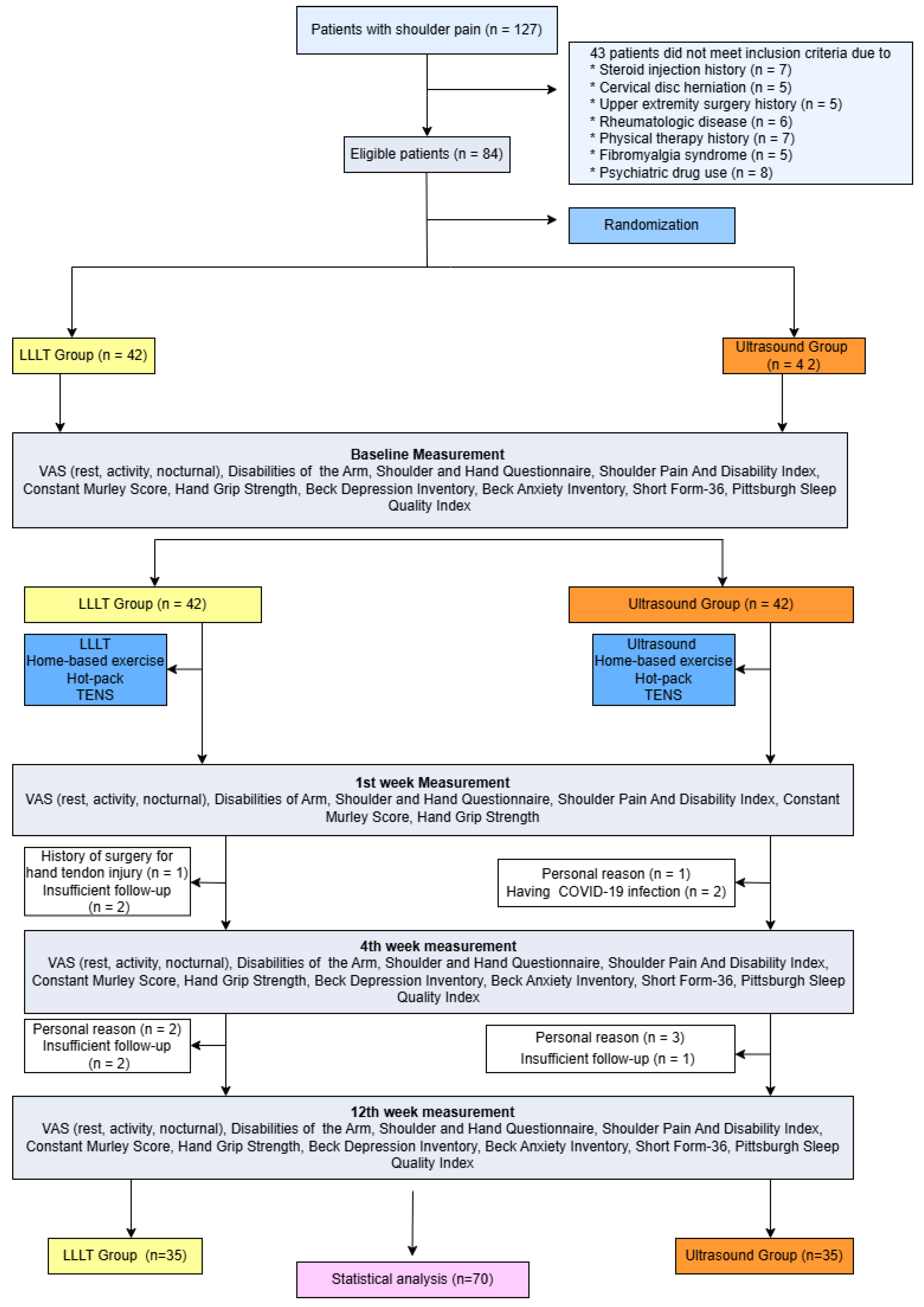
2. Patients and Methods

This prospective, observer-blinded, randomized study was conducted at Recep Tayyip Erdogan University, Faculty of Medicine, Department of Physical Medicine and Rehabilitation between April 2021 and February 2022. The study protocol was approved by the local ethics committee of our institution and performed in accordance with the principles stated in the Declaration of Helsinki. All participants provided written informed consent prior to the study. A total of 127 patients were evaluated for compliance with the inclusion criteria of the study. Of these, 84 patients diagnosed with RCT through physical examination and imaging methods were included in the study. The flow diagram of the study is shown in Figure 1. Fourteen participants did not complete the study and were not included in the analysis.
Patients were excluded from the study if they had a history of intra-articular or subacromial injection in the past three months; shoulder surgery or trauma; physical therapy in the past six months, adhesive capsulitis, hand or elbow deformity; radicular pain or cervical disk herniation; malignancy; pregnancy; or psychiatric, metabolic, or inflammatory neuromuscular diseases.

2.1. Randomization and Interventions

Randomization was performed using the sealed envelope technique, and patients were allocated into two groups: the US group (n = 42) and the LLLT group (n = 42). Both LLLT and US were applied over a total of 15 sessions, conducted five times per week. All participants also received a 20 min of hot pack, transcutaneous electrical nerve stimulation (TENS), and an HBE program. The HBE program included Codman pendulum exercises, joint range of motion (ROM) exercises, scapular stabilization exercises, and isometric strengthening exercises. The exercises were prescribed for at least three months, and participants were instructed to perform 10–15 repetitions twice daily.
A therapeutic US device (Chattanooga Medical Supply Inc., Chattanooga, TN, USA) was used for treatment. The US was applied to the painful area of the shoulder in slow circular motions, at a frequency of 1 MHz and an intensity of 1.2 W/cm2, using in pulsed mode. The therapy was applied to the painful area of the shoulder using slow circular movements at a frequency of 1 MHz and an intensity of 1.2 W/cm2, in pulsed mode. Each session lasted 8 min.
For LLLT, a gallium–aluminum–arsenide (GaAlAs) infrared diode laser device (Chattanooga Medical Supply Inc., Chattanooga, TN, USA) was used, with a wavelength of 850 nm, a power output of 100 mW in continuous wave mode, and a spot area of 0.07 cm2. Both patients and the operator wore protective eyeglasses during the procedure. LLLT was administered at a dose of 4 J/cm2 to a maximum of five painful points, each for one minute. The total duration of the LLLT session was five minutes per patient.

2.2. Outcome Measures

The clinical and sociodemographic characteristics of the patients were recorded. Patients were evaluated with the Visual Analog Scale (VAS), the Disabilities of the Arm, Shoulder, and Hand (DASH), the Shoulder Pain and Disability Index (SPADI), the Constant Murley Score (CMS), the Hand Grip Strength (HGS), the Beck Depression Inventory (BDI), the Beck Anxiety Inventory (BAI), the Pittsburgh Sleep Quality Index (PSQI), and the Short Form-36 (SF-36) at baseline and at the 1st, 4th, and 12th weeks after treatment. Emotional status, sleep disturbances, and quality of life were not evaluated at the 1st week after treatment. Rest, activity, and nocturnal pain intensity were evaluated by a 10 cm horizontal VAS [8,9].
The SPADI consists of 13 items: 5 assess shoulder pain and 8 assess shoulder disability. The total score is converted to a 100-point scale, with higher scores indicating greater impairment. The SPADI is a reliable and highly responsive tool for evaluating shoulder pain and function. Its validity and reliability in the Turkish population were demonstrated by Bumin et al. [10].
The DASH is a 30-item disability/symptom scale that evaluates a patient’s health status and includes 21 items assessing the degree of difficulty in performing various physical activities due to arm, shoulder, or hand problems. Additionally, five items were used to measure the severity of symptoms such as pain, activity-related pain, tingling, weakness, and stiffness. The remaining four items assess the impact of these issues on social activities, work, sleep, and self-image. Each item has five response options. The scores for all items are then used to calculate a scale score ranging from 0 (no disability) to 100 (most severe disability). The validity and reliability of the DASH in the Turkish population were demonstrated by Duger et al. [11].
The CMS consists of four subsections: pain, daily living activities, strength, and ROM. Pain is scored out of 15 points, daily living activities out of 20 points, strength out of 25 points, and ROM out of 40 points, yielding a total score of 100. Higher scores indicate better shoulder function. The validity and reliability of the CMS in the Turkish population were demonstrated by Celik [12].
Hand grip strength was evaluated with the patient seated in a chair, with the elbow flexed at 90 degrees and positioned close to the trunk, and the forearm and wrist in neutral position, using a hand dynamometer. Three measurements were taken, with one-minute rest intervals between them. The results were calculated as the average of the three measurements. Normative reference values have been reported as 40–47 kg for men aged 20–69 (2 kg less in the left hand), 24–30 kg for women aged 20–69 (1.5–2 kg less in the left hand) [13].
The BDI is a 21-item survey that assesses the presence and severity of depression. Higher scores reflect more severe levels of depression. The standard cutoff scores are as follows: 0–9, minimal depression; 10–18, mild depression; 19–29, moderate depression; 30–63, severe depression. The validity and reliability of the BDI in the Turkish population were demonstrated by Hisli [14].
The BAI is relatively brief, easily administered, and scored using a 21-item survey that assesses the severity of anxiety symptoms. The total score ranges from 0 to 63. Scores of 0–9 indicate normal or no anxiety; 10–18, mild to moderate anxiety; 19–29, moderate to severe anxiety; and 30–63, severe anxiety. The validity and reliability of the BAI in the Turkish population were demonstrated by Ulusoy et al. [15].
The PSQI is an 18-item questionnaire that assesses sleep quality and disturbances over a one-month period. It differentiates “poor” sleep from “good” sleep by evaluating seven components, including subjective sleep quality, sleep duration, sleep latency, sleep disturbances, habitual sleep efficiency, daytime dysfunction, and use of sleep medication. A total score of 5 or higher indicates a poor sleep quality. The validity and reliability of the PSQI in the Turkish population were demonstrated by Agargun et al. [16].
The SF-36 is a 36-item questionnaire that measures quality of life across eight domains, covering both physical and emotional aspects of health. These domains are role limitations due to physical health problems (RP), physical functioning (PF), social functioning (SF), bodily pain (BP), general mental health (MH), vitality (VT), role limitations due to emotional problems (RE), and general health perceptions (GH). The validity and reliability of the SF-36 in the Turkish population were previously demonstrated by Demiral et al. [17].

2.3. Statistical Analysis

The sample size was calculated using G*Power 3.1. A priori power analysis was conducted with a 5% significance level, 80% power, and medium effect size (dz = 0.5). Based on these assumptions, the required sample size for the study was determined to be 68 patients.
The data obtained from the study were analyzed using the SPSS 24.0 statistical software package. Continuous variables were expressed as mean ± standard deviation (SD). Compliance of the variables with normal distribution was assessed by the Kolmogorov–Smirnov test. Intra-group comparisons were performed using the Friedman and Wilcoxon signed-rank tests. Inter-group analyses were performed with Student’s t-test for normally distributed variables and the Mann–Whitney U test for non-normally distributed variables. The chi-square test was used to compare categorical data. To determine the correlation between the variables, Spearman’s rank or Pearson’s correlation analyses were performed according to the distribution of the data. A p-value < 0.05 was considered statistically significant, and an adjusted significance level of p < 0.001 was applied where appropriate. The effect size for intra-group comparisons was calculated using the Kendall’s W coefficient. For inter-group comparisons, the Mann–Whitney U test was applied, and the effect size was determined using the formula r = z/√n.

3. Results

During the follow-up period, fourteen participants did not complete the intervention protocol (Figure 1). These patients were approximately evenly distributed between the two groups (US group: n = 7; LLLT group: n = 7), and their baseline characteristics did not differ from those who completed the study. A total of 70 patients (mean age 50.8 ± 9.1 years, range 25–65 years) were included in the final analysis. The demographic and clinical characteristics of each group are presented in Table 1. There were no significant differences between groups at baseline in terms of age, gender, BMI, disease duration, pain, HGS, BAI, and PSQI scores (for all, p > 0.05). However, the mean BDI scores in the LLLT group were significantly higher compared to those in the US group (12.5 ± 8.8 vs. 7.9 ± 5.9, p = 0.010). Additionally, the mean VT, MH, and GH subdomains of SF-36 were significantly higher in the US group than in the LLLT group (p = 0.002, p = 0.006, p = 0.040, respectively).
In both groups, statistically significant increases were observed in the mean CMS, SF-36, and HGS values, and significant decreases were noted in the mean DASH, SPADI, VAS, BDI, BAI, and PSQI scores over time (for all, p < 0.05) (Table 2 and Table 3). The improvement in study parameters for each group during the follow-up period is shown in Table 4 and Table 5. There were no significant differences between the US and LLLT groups in VAS, DASH, SPADI, CMS, BDI, BAI, and PSQI scores (p > 0.05 for all). However, improvement of the HGS values was significantly greater in the US group compared to the LLLT group at the follow-up period (p < 0.05).

4. Discussion

The presented study revealed that both LLLT and therapeutic US were effective in reducing pain and improving functional status, emotional well-being, sleep quality, and quality of life in patients with RCT. However, therapeutic US demonstrated better improvement in HGS compared to LLLT.
Rotator cuff tendinopathy is one of the most common musculoskeletal disorders. RCT encompasses a range of pathologies, including bursitis, SIS, tendonitis, and partial- or full-thickness rotator cuff tears. Traditional treatment of RCT involves a variety of therapies, such as administration of analgesics and anti-inflammatory drugs, application of hot and cold packs, TENS, acupuncture, exercise, manual therapy, subacromial corticosteroid injections, nutritional supplements, as well as subacromial decompression and/or surgical repair of rotator cuff tears [18].
Therapeutic US is commonly used in the conservative treatment of RCT and is often prescribed in combination with other interventions. The physiologic effects of US are increased vascular permeability, blood flow, local metabolism, and increased fibrous tissue extensibility and muscle relaxation [19]. Several studies have reported conflicting findings regarding the efficacy of therapeutic US as a physical therapy modality [20]. Desmeules et al. reported that therapeutic US did not provide any significant benefit when compared to placebo, laser therapy, or when combined with exercise [21]. In our study, both therapeutic US and LLLT improved all clinical parameters, with greater improvement in HGS observed in the US group compared to the LLLT group during the follow-up period. Also, Yildirim et al. [22] reported that therapeutic US improved pain and functional status in patients with SIS. Similarly to our study, they also evaluated emotional status using the BDI. In addition, our study assessed HGS, anxiety, sleep disturbances, and quality of life.
The physiologic effects of the LLLT include increased collagen synthesis, cell proliferation, and blood flow, as well as promotion of tissue regeneration and reduction in inflammation [23]. Tombak et al. reported that HBE combined with LLLT for the rotator cuff calcific tendinitis was more effective than HBE alone in improving pain and disability [24]. In our study, we evaluated strength and ROM using the CMS scale. Additionally, we assessed HGS, emotional status, sleep disturbances, and quality of life. A systematic review also reported that LLLT is used as a treatment for SIS [25]. However, it has been advised that the benefits of LLLT should be further investigated in future studies. Atıcı et al. found that LLLT is not superior to placebo in the treatment of SIS [26]. In their study, LLLT was applied at a dosage of 4 joules, similar to our protocol. On the other hand, the treatment duration differed from that in our study. Additionally, our study included a longer follow-up period and further assessment, such as strength using CMS, emotional status, sleep disturbances, and quality of life.
In the present study, the efficacy of therapeutic US and LLLT was compared in patients with RCT, and no significant differences were observed between groups in terms of pain relief, functional improvement, emotional status, sleep disturbances, or quality of life. However, there was a significant baseline difference in BDI scores and some subdomains of the SF-36 between the groups. To account for these imbalances, inter-group comparisons were conducted using change scores (i.e., the difference between baseline and post-treatment values) rather than absolute values. This approach allowed for the evaluation of treatment effects while minimizing the potential bias introduced by baseline differences. Our findings were consistent with those of previous studies [5,6,7,27]. Yavuz et al. demonstrated that LLLT and therapeutic US improved pain, disability, and sleep disturbances in SIS, and suggested that LLLT could be used when US is contraindicated [5]. Unlike our study, that investigation included a smaller patient cohort. Calis et al. also reported that both LLLT and therapeutic US improved pain, function, and ROM in patients with SIS, although no significant differences were observed between the two modalities [6]. Our study differed in that it included a longer follow-up period and a more comprehensive evaluation of patient outcomes. In addition to pain, function, and ROM, we also assessed HGS, sleep disturbances, emotional status, and quality of life, and observed improvements across all parameters in both the LLLT and US groups. Sen et al. reported that both LLLT and therapeutic US, when combined with an HBE program, led to significant improvements in pain and disability in patients with SIS [7]. However, in contrast to our findings, they reported that therapeutic US had no additional benefit in the short term. In their study, LLLT combined with HBE was more effective than HBE alone in relieving activity pain and improving shoulder function in the short term. In contrast to the study by Sen et al., the absence of an HBE-only group in our study constitutes a limitation, as it precludes a clear assessment of the specific contribution of HBE compared to the active modalities (US or LLLT). In our study, therapeutic US not only improved pain and disability but also led to greater improvements in HGS compared to LLLT, and was effective across all measured parameters. Importantly, our study incorporated additional outcome measures that are often overlooked. These included emotional status, sleep disturbance, and quality of life, along with the use of the CMS to evaluate shoulder strength. The follow-up period was similar to that of Sen et al., yet our broader outcome assessment provides a more comprehensive evaluation of treatment efficacy. A study by Ranjithkumar et al. found that low-energy extra-corporeal shockwave therapy (ESWT), LLLT, and US are effective treatments for RCT, while ESWT is more effective in improving muscle thickness, shoulder function, and pain level [27]. Unlike our study, Saunders reported that laser treatment was significantly more effective in improving muscle force than US [28]. In addition, Santamato et al. also reported that high-intensity laser therapy was more effective than US therapy in reducing pain and improving joint mobility, functional status, and muscle strength in patients with SIS short-term follow-up period [29]. The discrepancies among studies may be attributed to differences in study design, sample size, treatment protocols, follow-up durations, and the range of outcome measures assessed.
This study has several limitations that should be considered when interpreting the findings. First, the absence of a placebo or control group limits the ability to determine the isolated effects of LLLT and US therapy. Second, the lack of blinding among patients, due to the nature of the interventions, may have introduced bias. Third, although the follow-up period was longer than in some prior studies, it may still be insufficient to assess long-term outcomes.

5. Conclusions

In conclusion, this study demonstrated that both therapeutic US and LLLT were effective in the management of RCT, leading to significant improvements in pain, functional status, emotional status, sleep quality, and quality of life. Notably, therapeutic US led to greater improvements in HGS compared to LLLT. These findings contribute to the growing body of evidence supporting the use of both modalities in the treatment of RCT. Therapeutic US may be preferred in cases where improving HGS is a clinical priority. However, due to limitations such as the lack of a placebo group and the single-center design, further randomized controlled trials with larger sample sizes and placebo-controlled protocols are warranted to better define the optimal treatment approach.

Author Contributions

Conceptualization, Ş.D. and M.S.B.; methodology, Ş.D. and M.S.B.; data curation, Ş.D., M.S.B., G.D., M.Y. and M.S.T.; writing—original draft, Ş.D. and M.S.B.; writing—review and editing, Ş.D. and M.S.B. All authors have read and agreed to the published version of the manuscript.

Funding

This study has been funded by Recep Tayyip Erdogan University Development Foundation (Grand number: 02025005002465).

Institutional Review Board Statement

This study was conducted in accordance with the Declaration of Helsinki and approved by the institutional review board of Recep Tayyip Erdogan University Faculty of Medicine Ethics Committee (protocol code: 2021/52 and date of approval: 1 April 2021).

Informed Consent Statement

Informed consent was obtained from all subjects involved in the study.

Data Availability Statement

The data presented in this study are available on request from the corresponding author.

Conflicts of Interest

The authors declare no conflicts of interest.

References

  1. Luime, J.J.; Koes, B.W.; Hendriksen, I.J.; Burdorf, A.; Verhagen, A.P.; Miedema, H.S.; Verhaar, J.A. Prevalence and incidence of shoulder pain in the general population; a systematic review. Scand J. Rheumatol. 2004, 33, 73–81. [Google Scholar] [CrossRef] [PubMed]
  2. Harrison, A.K.; Flatow, E.L. Subacromial Impingement Syndrome. J. Am. Acad. Orthop. Surg. 2011, 19, 701–708. [Google Scholar] [CrossRef] [PubMed]
  3. Hanchard, N.C.A.; Cummins, J.; Jeffries, C. Evidence-Based Clinical Guidelines for the Diagnosis, Assessment and Physiotherapy Management of Shoulder Impingement Syndrome; The Chartered Society of Physiotherapy: London, UK, 2004; p. 1. [Google Scholar]
  4. Burbank, K.M.; Stevenson, J.H.; Czarnecki, G.R.; Dorfman, J. Chronic Shoulder Pain: Part I. Evaluation and Diagnosis. Am. Fam. Physician 2008, 77, 453–460. [Google Scholar] [PubMed]
  5. Yavuz, F.; Duman, I.; Taskaynatan, M.A.; Tan, A.K. Low-Level Laser Therapy versus Ultrasound Therapy in the Treatment of Subacromial Impingement Syndrome: A Randomized Clinical Trial. J. Back Musculoskelet. Rehabil. 2014, 27, 315–320. [Google Scholar] [CrossRef]
  6. Calis, H.T.; Berberoglu, N.; Calis, M. Are Ultrasound, Laser and Exercise Superior to Each Other in the Treatment of Subacromial Impingement Syndrome? A Randomized Clinical Trial. Eur. J. Phys. Rehabil. Med. 2011, 47, 375–380. [Google Scholar]
  7. Sen, E.I.; Arman, S.; Tseveendorj, N.; Yılmaz, E.; Oral, A.; Capan, N. Low-Level Laser Therapy versus Ultrasound Therapy Combined with Home-Based Exercise in Patients with Subacromial Impingement Syndrome: A Randomized-Controlled Trial. Turk. J. Phys. Med. Rehabil. 2023, 69, 424–433. [Google Scholar] [CrossRef]
  8. Price, D.D.; McGrath, P.A.; Rafii, A.; Buckingham, B. The Validation of Visual Analogue Scales as Ratio Scale Measures for Chronic and Experimental Pain. Pain 1983, 17, 45–56. [Google Scholar] [CrossRef]
  9. Carlsson, A.M. Assessment of Chronic Pain. I. Aspects of the Reliability and Validity of the Visual Analogue Scale. Pain 1983, 16, 87–101. [Google Scholar] [CrossRef]
  10. Bumin, G.; Tuzun, E.H.; Tonga, E. The Shoulder Pain and Disability Index (SPADI): Cross-Cultural Adaptation, Reliability, and Validity of the Turkish Version. J. Back Musculoskelet. Rehabil. 2008, 1, 57–62. [Google Scholar] [CrossRef]
  11. Duger, T.; Yakut, E.; Oksuz, C.; Yorukan, S.; Bilgutay, B.S.; Kuru, C.S.; Leblebicioglu, G.; Kayihan, H.; Kirdi, N.; Yakut, Y.; et al. Kol Omuz ve El Sorunları Disabilities of the Arm Shoulder and Hand DASH Anketi Türkçe Uyarlamasının Güvenirliği ve Geçerliği. Turk. J. Physiother. Rehabil. -Fiz. Rehabil. 2006, 3, 99–107. [Google Scholar]
  12. Celik, D. Turkish Version of the Modified Constant-Murley Score and Standardized Test Protocol: Reliability and Validity. Acta. Orthop. Traumatol. Turc. 2016, 50, 69–75. [Google Scholar] [CrossRef] [PubMed]
  13. Hamilton, G.F.; McDonald, C.; Chenier, T.C. Measurement of Grip Strength: Validity and Reliability of the Sphygmomanometer and Jamar Grip Dynamometer. J. Orthop. Sports Phys. Ther. 1992, 16, 215–219. [Google Scholar] [CrossRef] [PubMed]
  14. Hisli, N. A Study on Validity and Reliability Test of the Beck Depression Scale. J. Psychol. 1988, 6, 118–122. [Google Scholar]
  15. Ulusoy, M.; Sahin, N.H.; Erkmen, H. Turkish Version of the Beck Anxiety Inventory: Psychometric Properties. J. Cogn. Psychother 1998, 12, 163–172. [Google Scholar]
  16. Agargun, M.Y.; Kara, H.; Anlar, O. The Validity and Reliability of the Pittsburgh Sleep Quality Index. Turk Psikiyatr. Derg. 1996, 7, 107–111. [Google Scholar]
  17. Demiral, Y.; Ergor, G.; Unal, B.; Semin, S.; Akvardar, Y.; Kıvırcık, B.; Alptekin, K. Normative Data and Discriminative Properties of Short Form 36 (SF-36) in Turkish Urban Population. BMC Public Health 2006, 6, 247. [Google Scholar] [CrossRef]
  18. Varacallo, M.A.; El Bitar, Y.; Sina, R.E.; Mair, S.D. Rotator Cuff Syndrome. In StatPearls; StatPearls Publishing: Treasure Island, FL, USA, 2025. [Google Scholar]
  19. van der Windt, D.A.W.M.; van der Heijden, G.J.M.G.; van den Berg, S.G.M.; Ter Riet, G.; de Winter, A.F.; Bouter, L.M. Ultrasound Therapy for Musculoskeletal Disorders: A Systematic Review. Pain 1999, 81, 257–271. [Google Scholar] [CrossRef]
  20. Page, M.J.; Green, S.; Mrocki, M.A.; Surace, S.J.; Deitch, J.; McBain, B.; Lyttle, N.; Buchbinder, R. Electrotherapy Modalities for Rotator Cuff Disease. Cochrane Database Syst. Rev. 2016, 10, CD012225. [Google Scholar] [CrossRef]
  21. Desmeules, F.; Boudreault, J.; Roy, J.S.; Dionne, C.; Frémont, P.; MacDermid, J.C. The Efficacy of Therapeutic Ultrasound for Rotator Cuff Tendinopathy: A Systematic Review and Meta-Analysis. Phys. Ther. Sport 2015, 16, 276–284. [Google Scholar] [CrossRef]
  22. Yildirim, M.A.; Ones, K.; Celik, E.C. Comparision of Ultrasound Therapy of Various Durations in the Treatment of Subacromial Impingement Syndrome. J. Phys. Ther. Sci. 2013, 25, 1151–1154. [Google Scholar] [CrossRef]
  23. Kingsley, J.D.; Demchak, T.; Mathis, R. Low-Level Laser Therapy as a Treatment for Chronic Pain. Front Physiol. 2014, 5, 306. [Google Scholar] [CrossRef] [PubMed] [PubMed Central]
  24. Tombak, Y.; Karaahmet, O.Z.; Tombak, A.; Gurcay, E. Effects of Low-Level Laser Therapy on Symptomatic Calcific Rotator Cuff Tendinopathy: A Prospective Randomized Controlled Study. Wien Klin. Wochenschr 2024, 5, 1–8. [Google Scholar] [CrossRef] [PubMed]
  25. Castaldo, M.; De Angelis D’Ossat, A.; Gnessi, P.; Galeoto, G. A Systematic Review on Low-Level Laser Therapy in the Management of Shoulder Impingement Syndrome. Appl. Sci. 2023, 13, 3536. [Google Scholar] [CrossRef]
  26. Atıcı Ozturk, P.; Sengül, I.; Karatepe, A.G.; Kaya, T.; Günaydın, R. Effectiveness of Low-Level Laser Therapy in Patients with Subacromial Impingement Syndrome: A Randomized, Placebo Controlled, Prospective Study. J. Tepecik Educ. Res. Hosp. 2015, 25, 78–84. [Google Scholar] [CrossRef]
  27. Ranjithkumar, N.; Paul, J.; Alagesan, J.; Viswanathan, R. Comparative Effectiveness of Extracorporeal Short-Wave Therapy, Low-Level Laser Therapy, and Ultrasound in the Treatment of Rotator Cuff Tendinopathy. Biomed. Pharmacol. J. 2025, 18, 849–866. [Google Scholar] [CrossRef]
  28. Saunders, L. Laser versus Ultrasound in the Treatment of Supraspinatus Tendinosis: Randomised Controlled Trial. Physiotherapy 2003, 89, 365–373. [Google Scholar] [CrossRef]
  29. Santamato, A.; Solfrizzi, V.; Panza, F.; Tondi, G.; Frisardi, V.; Leggin, B.G.; Ranieri, M.; Fiore, P. Short-Term Effects of High-Intensity Laser Therapy versus Ultrasound Therapy in the Treatment of People with Subacromial Impingement Syndrome: A Randomized Clinical Trial. Phys. Ther. 2009, 89, 643–652. [Google Scholar] [CrossRef]
Figure 1. Flowchart of the study. LLLT: Low-Level Laser Therapy, VAS: Visual Analog Scale.
Jcm 14 04197 g001
Table 1. Baseline demographic and clinical characteristics of participants in the US and LLLT groups.
Ultrasound Group
(n = 35)
LLLT Group
(n = 35)
p-Value
Age, years (mean ± SD)51.7 ± 8.8 49.9 ± 9.40.410
Gender, F/M (n, %)20/15(57.1/42.9)20/15 (57.1/42.9)1.000
BMI (mean ± SD)30.4 ± 5.829.1 ± 4.90.310
Disease duration, months (mean ± SD)13.6 ± 16.29.4 ± 1.20.210
Side of involvement (R/L), n (%)23/12 (65.7/34.3)24/11 (68.6/31.4)0.790
Occupation, n (%) 0.730
 Housewife18 (51.4)13 (37.2)
 Retired4 (11.4)6 (17.1)
 Active employee13 (37.2)16 (45.7)
VAS (mean ± SD)
 Activity8.3 ± 1.58.6 ± 0.80.330
 Rest5.4 ± 0.85.4 ± 0.90.680
 Night6.9 ± 1.97.6 ± 1.50.170
DASH (mean ± SD)
 Function-symptom51.8 ± 15.650.3 ± 16.90.714
 Work64.1 ± 20.955.3 ± 20.50.870
CMS (mean ± SD)
 Total60.5 ± 7.658.4 ± 4.70.170
 Objective assessment49.5 ± 5.749.1 ± 3.70.747
SPADI (mean ± SD)
 Pain80.8 ± 17.377.5 ± 10.90.342
 Disability68.6 ± 19.163.5 ± 13.10.202
 Total73.3 ± 17.768.9 ± 11.60.225
HGS, kg (mean ± SD)
 Right28.6 ± 9.629.6 ± 10.50.600
 Left28.1 ± 9.230.2 ± 9.70.300
SF-36 (mean ± SD)
 Physical functioning63.3 ± 16.9360.3 ± 21.50.500
 Role physical17.9 ± 33.5233.6 ± 39.70.070
 Role emotional56.2 ± 44.1149.5 ± 39.90.500
 Vitality64.1 ± 20.3848.6 ± 20.30.002 *
 Mental health69.3 ± 14.4158.9 ± 15.90.006 *
 Social functioning67.5 ± 25.463.6 ± 20.40.470
 Bodily pain34.8 ± 23.133.7 ± 20.90.840
 General Health55.9 ± 16.348.0 ± 16.40.040 *
BDI (mean ± SD)7.9 ± 5.912.5 ± 8.80.010 *
BAI (mean ± SD)13.1 ± 11.614.2 ± 8.50.600
PSQI (mean ± SD)7.3 ± 3.97.6 ± 3.30.700
LLLT, Low-Level Laser Therapy; US, Ultrasound; VAS, Visual Analog Scale; DASH, Disabilities of the Arm, Shoulder and Hand; CMS, Constant Murley Score; SPADI, Shoulder Pain and Disability Index; HGS, Hand Grip Strength; SF-36, Short Form-36; BDI, Beck Depression Inventory; BAI, Beck Anxiety Inventory; PSQI, Pittsburgh Sleep Quality Index; *, statistically significant.
Table 2. Intra-group comparison of the clinical characteristics over follow-up in the therapeutic ultrasound group.
Ultrasound Group
(n = 35)
BaselinePost-Treatment 1st WeekPost-Treatment 4th WeekPost-Treatment 12th Weekp-Value *Effect Size
(Kendall’s W)
DASH
 Function Symptom51.8 ± 15.634.8 ± 21.928.1 ± 18.815.1 ± 17.5<0.001 **0.665
 Work64.1 ± 20.945.9 ± 25.739.5 ± 24.319.7 ± 22.2<0.001 **0.586
SPADI
 Pain80.8 ± 17.347.5 ± 24.543.1 ± 30.225.6 ± 25.9<0.001 **0.691
 Disability68.6 ± 19.141.1 ± 27.635.0 ± 24.018.0 ± 20.6<0.001 **0.609
 Total73.3 ± 17.743.0 ± 25.938.2 ± 26.120.3 ± 22.2<0.001 **0.717
CMS
 Total60.5 ± 7.680.0 ± 13.888.9 ± 11.596.4 ± 5.2<0.001 **0.806
 Objective Assessment49.5 ± 5.758.9 ± 7.061.9 ± 5.764.5 ± 1.2<0.001 **0.733
VAS
 Activity8.3 ± 1.55.8 ± 2.44.5 ± 1.92.5 ± 2.3<0.001 **0.783
 Rest5.4 ± 0.83.0 ± 2.11.1 ± 1.70.5 ± 1.2<0.001 **0.777
 Night6.9 ± 2.03.0 ± 2.71.0 ± 1.80.9 ± 1.5<0.001 **0.788
HGS
 Right28.6 ± 9.632.2 ± 9.831.5 ± 9.933.5 ± 9.4<0.001 **0.248
 Left28.2 ± 9.232.2 ± 8.631.9 ± 9.333.1 ± 9.3<0.001 **0.306
BDI7.9 ± 6.0x5.4 ± 5.25.0 ± 6.5<0.001 **0.276
BAI13.1 ± 11.6x8.3 ± 7.38.1 ± 9.00.002 **0.181
PSQI7.3 ± 3.9x4.5 ± 3.73.1 ± 3.4<0.001 **0.325
SF-36
Physical Functioning63.3 ± 16.9x78.4 ± 15.277.6 ± 20.3<0.001 **0.290
Role Physical17.9 ± 33.5x60.7 ± 42.164.3 ± 43.0<0.001 **0.394
Role Emotional56.2 ± 44.1x84.8 ± 31.775.2 ± 39.90.001 **0.205
Vitality64.1 ± 20.4x70.7 ± 18.269.1 ± 18.00.0740.075
Mental Health69.3 ± 14.4x73.8 ± 12.872.8 ± 15.60.021 **0.110
Social Functioning67.5 ± 25.4x80.2 ± 21.384.6 ± 19.50.001 **0.217
Bodily Pain34.8 ± 23.1x58.6 ± 26.169.2 ± 26.5<0.001 **0.527
General Health55.9 ± 16.3x67.3 ± 12.770.0 ± 22.5<0.001 **0.375
DASH, Disabilities of the Arm, Shoulder and Hand; SPADI, Shoulder Pain and Disability Index; VAS, Visual Analog Scale; CMS, Constant Murley Score; HGS, Hand Grip Strength; BDI, Beck Depression Inventory; BAI, Beck Anxiety Inventory; PSQI, Pittsburgh Sleep Quality Index; SF-36, Short Form-36; *, p-values are based on the Friedman Test; x, not assessed at this time point; **, statistically significant.
Table 3. Intra-group comparison of the clinical characteristics over follow-up in the LLLT group.
LLLT Group
(n = 35)
BaselinePost-Treatment
1st Week
Post-Treatment
4th Week
Post-Treatment
12th Week
p-Value *Effect Size
(Kendall’s W)
DASH
 Function Symptom50.4 ± 16.930.4 ± 17.8 29.5 ± 22.516.4 ± 19.5<0.001 **0.530
 Work55.4 ± 20.837.5 ± 21.028.6 ± 24.016.1 ± 24.9<0.001 **0.508
SPADI
 Pain77.5 ± 10.947.5 ± 20.843.3 ± 30.025.1 ± 28.9<0.001 **0.554
 Disability63.5 ± 13.141.9 ± 19.435.8 ± 24.923.2 ± 26.6<0.001 **0.544
 Total68.9 ± 11.645.6 ± 19.038.7 ± 26.723.9 ± 27.1<0.001 **0.559
CMS
 Total58.4 ± 4.781.8 ± 10.386.7 ± 9.995.4 ± 8.8<0.001 **0.820
 Objective Assessment49.1 ± 3.761.2 ± 5.063.2 ± 3.564.3 ± 2.0<0.001 **0.790
VAS
 Activity8.6 ± 0.85.7 ± 1.84.6 ± 2.0 2.7 ± 2.4<0.001 **0.884
 Rest5.5 ± 1.02.9 ± 1.42.0 ± 1.80.9 ± 1.4<0.001 **0.785
 Night7.6 ± 1.53.5 ± 2.32.3 ± 2.11.1 ± 1.9<0.001 **0.853
HGS (kg)
 Right29.6 ± 10.530.1 ± 9.330.9 ± 8.733.1 ± 9.0<0.001 **0.243
 Left30.2 ± 9.831.1 ± 9.931.1 ± 8.732.5 ± 8.9<0.001 **0.117
BDI12.5 ± 8.8x8.8 ± 9.18.4 ± 7.9<0.001 **0.235
BAI14.2 ± 8.5x10.6 ± 8.49.4 ± 8.5<0.001 **0.319
PSQI7.6 ± 3.3x5.4 ± 4.24.9 ± 3.9<0.001 **0.245
SF-36
Physical Functioning60.3 ± 21.5x74.0 ± 19.777.4 ± 18.40.001 **0.210
Role Physical33.6 ± 39.7x64.3 ± 39.974.3 ± 36.6<0.001 **0.342
Role Emotional49.5 ± 39.9x66.7 ± 36.281.0 ± 32.60.001 **0.207
Vitality48.6 ± 20.3x63.4 ± 16.264.3 ± 23.6<0.001 **0.321
Mental Health58.9 ± 15.9x66.3 ± 14.070.7 ± 16.30.001 **0.213
Social Functioning63.6 ± 20.4x75.4 ± 21.283.5 ± 17.6<0.001 **0.337
Bodily Pain33.7 ± 21.0x63.3 ± 27.374.4 ± 25.3<0.001 **0.600
General Health48.0 ± 16.4x61.1 ± 22.965.3 ± 18.2<0.001 **0.274
Table 4. Inter-group comparison of score changes in VAS, DASH, SPADI, CMS, and HGS over time.
Ultrasound Group
(n = 35)
LLLT Group (n = 35)p-ValueEffect Size
(r)
VAS:
Activity
Baseline vs. W1−2.5 ± 2.8−2.9 ± 1.70.2800.128
Baseline vs. W4−3.8 ± 2.4−4.0 ± 1.90.9100.013
Baseline vs. W12−5.8 ± 2.6−5.9 ± 2.30.9800.002
Rest
Baseline vs. W1−2.4 ± 2.0−2.6 ± 1.40.6200.059
Baseline vs. W4−4.3 ± 1.7−3.5 ± 2.00.0500.232
Baseline vs. W12−4.9 ± 1.1−4.6 ± 1.60.4100.098
Night
Baseline vs. W1−3.9 ± 2.5−4.1 ± 2.00.6000.062
Baseline vs. W4−5.9 ± 2.3−5.3 ± 2.10.2400.139
Baseline vs. W12−6.2 ± 2.1−6.5 ± 1.80.5300.074
DASH:
Function-symptom
Baseline vs. W1−17.0 ± 16.7−20.0 ± 17.90.6600.053
Baseline vs. W4−23.7 ± 15.0−20.8 ± 20.40.3900.101
Baseline vs. W12−36.7 ± 17.9−34.0 ± 23.70.5500.070
Work
Baseline vs. W1−18.2 ± 21.6−18.2 ± 22.30.8500.022
Baseline vs. W4−24.6 ± 19.9−27.0 ± 20.30.5100.078
Baseline vs. W12−44.4 ± 25.5−39.3 ± 26.90.5200.077
SPADI:
Pain
Baseline vs. W1−33.3 ± 22.7−30.0 ± 20.70.4840.084
Baseline vs. W4−37.7 ± 25.8−34.1 ± 30.40.7420.039
Baseline vs. W12−55.2 ± 23.0−52.4 ± 29.70.9300.010
Disability
Baseline vs. W1−27.4 ± 26.3−21.6 ± 18.50.4700.086
Baseline vs. W4−33.6 ± 21.6−27.7 ± 24.40.3500.109
Baseline vs. W12−50.6 ± 21.2−40.3 ± 26.80.1000.192
Total
Baseline vs. W1−30.3 ± 24.8−23.3 ± 17.80.2600.132
Baseline vs. W4−35.1 ± 22.5−30.2 ± 26.10.4000.100
Baseline vs. W12−53.0 ± 21.1−45.0 ± 27.40.2900.125
CMS:
Total
Baseline vs. W119.5 ± 13.223.4 ± 9.60.1200.183
Baseline vs. W428.5 ± 12.328.3 ± 9.80.7100.044
Baseline vs. W1235.9 ± 7.537.0 ± 9.20.2400.138
CMS
Objective assessment
Baseline vs. W19.4 ± 6.812.1 ± 5.10.0500.229
Baseline vs. W412.5 ± 6.714.1 ± 4.20.3200.117
Baseline vs. W1246.9 ± 6.246.3 ± 9.20.5400.072
HGS
Right
Baseline vs. W13.7 ± 4.70.4 ± 5.50.010 **0.306
Baseline vs. W43.0 ± 5.11.2 ± 6.40.3700.107
Baseline vs. W124.9 ± 5.43.5 ± 7.30.4600.088
Left
Baseline vs. W14.0 ± 5.00.9 ± 3.70.007 **0.320
Baseline vs. W43.7 ± 5.10.8 ± 4.30.040 **0.240
Baseline vs. W124.9 ± 4.92.2 ± 5.00.030 **0.252
LLLT, Low-Level Laser Therapy; VAS, Visual Analog Scale; DASH, Disabilities of the Arm, Shoulder, and Hand; SPADI, Shoulder Pain and Disability Index; CMS, Constant Murley Score; HGS, Hand Grip Strength; W1, 1st week post-treatment; W4, 4th week post-treatment; W12, 12th week post-treatment; **, statistically significant.
Table 5. Inter-group comparison of score changes in SF-36, BDI, BAI, and PSQI over time.
US Group
(n = 35)
LLLT Group
(n = 35)
p ValueEffect Size
(r)
SF-36:
Physical functioning
Baseline vs. W415.1 ± 1.613.7 ± 18.20.8700.019
Baseline vs. W1214.3 ± 24.717.1 ± 25.70.7600.037
Role Physical
Baseline vs. W442.9 ± 44.030.7 ± 43.30.2300.142
Baseline vs. W1246.4 ± 45.840.7 ± 43.80.5600.068
Role emotional
Baseline vs. W428.6 ± 39.717.1 ± 54.50.4800.083
Baseline vs. W1219.0 ± 41.431.4 ± 42.70.1300.181
Vitality
Baseline vs. W46.5 ± 11.514.9 ± 21.90.0700.210
Baseline vs. W125.0 ± 22.115.7 ± 21.20.0400.238
Mental health
Baseline vs. W44.6 ± 12.27.4 ± 15.50.6400.055
Baseline vs. W123.5 ± 18.211.9 ± 16.90.1600.166
Social functioning
Baseline vs. W412.7 ± 18.411.8 ± 24.30.8800.017
Baseline vs. W1217.1 ± 32.519.9 ± 22.30.8600.021
Bodily pain
Baseline vs. W445.4 ± 26.141.6 ± 23.60.5600.070
Baseline vs. W1234.4 ± 31.540.7 ± 31.70.4800.083
General Health
Baseline vs. W411.3 ± 13.513.1 ± 21.10.8300.025
Baseline vs. W1214.1 ± 21.617.3 ± 19.50.8000.030
BDI
Baseline vs. W4−2.5 ± 5.2−3.7 ± 7.10.2900.124
Baseline vs. W12−2.9 ± 6.2−4.1 ± 6.70.6700.049
BAI
Baseline vs. W4−4.7 ± 8.9−3.6 ± 5.80.8600.021
Baseline vs. W12−4.9 ± 12.8−4.9 ± 6.50.8500.022
PSQI
Baseline vs. W4−2.8 ± 3.4−2.2 ± 3.90.4900.081
Baseline vs. W12−4.2 ± 4.5−2.6 ± 4.10.1400.173
LLLT, Low-Level Laser Therapy; SF-36, Short Form-36; BDI, Beck Depression Inventory; BAI, Beck Anxiety Inventory; PSQI, Pittsburgh Sleep Quality Index; W1, 1st week post-treatment; W4, 4th week post-treatment; W12, 12th week post-treatment.
Disclaimer/Publisher’s Note: The statements, opinions and data contained in all publications are solely those of the individual author(s) and contributor(s) and not of MDPI and/or the editor(s). MDPI and/or the editor(s) disclaim responsibility for any injury to people or property resulting from any ideas, methods, instructions or products referred to in the content.

