Case Report

Novel Compound Heterozygous Mutation of the ABCA3 Gene in a Patient with Neonatal-Onset Interstitial Lung Disease

1
Department of Health Promotion, Mother and Child Care, Internal Medicine and Medical Specialties “G. D’Alessandro”, University of Palermo, 90133 Palermo, Italy
2
Neonatal Intensive Care Unit, “Barone Ignazio Romeo” Hospital, 98066 Patti, Italy
*
Author to whom correspondence should be addressed.
J. Clin. Med. 2025, 14(11), 3704; https://doi.org/10.3390/jcm14113704
Submission received: 22 April 2025 / Revised: 16 May 2025 / Accepted: 23 May 2025 / Published: 25 May 2025
(This article belongs to the Section Respiratory Medicine)

Abstract

:
Background: Children’s interstitial and diffuse lung diseases, commonly referred to as “chILDs”, include around 200 rare conditions that disrupt normal lung function. They are classified, based on etiopathogenesis, into several subgroups, having a varied and multifaceted clinical presentation depending on the type of genetic mutation present. Methods and Results: We describe the case of a late preterm newborn presenting soon after birth with respiratory distress syndrome poorly responsive to surfactant administration, in whom a targeted gene panel analysis for pulmonary congenital diseases, performed using next-generation sequencing (NGS), revealed a novel compound heterozygous variant of the ATP-Binding-Cassette-Subfamily-A-Member-3 (ABCA3) gene. A review of the literature on the subject completes our work. Conclusions: Molecular genetic analysis has become crucial for a more targeted therapeutic treatment, along with the only current curative treatment option that is lung transplantation.

1. Introduction

Children’s interstitial and diffuse lung diseases (also known as “chILDs”) encompass about 200 rare conditions impairing lung function. They manifest with non-specific symptoms, including coughing, dyspnea, and respiratory insufficiency. Gene transmission is described as both autosomal recessive and dominant [1]. They are classified, based on etiopathogenesis, into different subgroups: diffuse developmental disorders, growth abnormalities, structural changes in the lung associated with chromosomal anomalies, specific conditions of undefined etiology, surfactant dysfunction, and other conditions related to surfactant dysfunction in the absence of a known genetic cause [2]. Diagnosis is made with clinical pictures, radiological investigations (i.e., chest X-ray and computed tomography [CT] scan), alveolar bronchoscopy, lung biopsy with histological examination, and, recently, genetic tests, including next-generation sequencing (NGS), which are increasingly used and crucial in clinical practice [3,4]. Currently, no established and effective treatments are available for these rare disorders, with the exception of lung transplantation [1,2]. The latter, however, is burdened by major comorbidities, difficulties in finding compatible donors, and high mortality rates. Research is underway to investigate potential new and promising therapeutic tools, like gene therapy [1,2]. We describe the case of a late preterm newborn presenting right after birth with respiratory distress syndrome poorly responsive to surfactant administration, in whom a targeted gene panel analysis, performed using NGS, revealed a novel compound heterozygous variant of the ATP-Binding-Cassette-Subfamily-A-Member-3 (ABCA3) gene. In addition, a bioinformatic prediction analysis on the protein function of the identified variants was performed. Finally, a review of previously reported patients carrying homozygous or compound heterozygous variants of the same gene was conducted. We focused on commonalities and differences among them, analyzing clinical manifestations, therapies administered, and outcomes. The aim of the present report is to provide a better clinical and genomic characterization of these rare diseases, contributing to enriching the current mutational spectrum of such conditions and to future research on new effective therapeutic approaches.

2. Case Report and Literature Review

Our proband is a female late preterm neonate born from vaginal delivery at 35+5 weeks of gestation due to chorioamnionitis to healthy, nonconsanguineous parents. She is the first child of the couple. The family history was unremarkable, including two healthy consanguineous sisters of the proband, daughters of the same father and a different mother.
During pregnancy, the mother reported to be a tobacco user, smoking 4–5 cigarettes/day. The proband’s birth occurred at a level I hospital in a small town in Eastern Sicily, neighboring the urban city of Palermo, the regional capital. Apgar scores were 7 and 9 at 1 and 5 min, respectively. Anthropometric measures were as follows: weight 2270 g (79th centile, SDS −0.47), length 46 cm (48th centile, SDS −0.06), head circumference 33 cm (74th centile, SDS 0.64), according to the Italian INeS Growth Charts [5]. Postnatally, due to respiratory distress and oxygen dependence, which had been present since birth, the baby was transferred through the neonatal emergency transport service to the Neonatal Intensive Care Unit (NICU) closest to the birth center. There, her respiratory function gradually worsened, so she was intubated and treated with exogenous surfactant administration. However, after an initial mild improvement, which allowed extubation, she needed non-invasive ventilatory support. Therefore, for the emerging suspicion of congenital respiratory disease, she was transferred, at around two weeks of life, to our NICU, located at the Mother and Child Department of the University Hospital of Palermo, to continue both diagnostic path and therapeutic management. During hospitalization in our unit, non-invasive respiratory assistance was provided, with a gradual increase in oxygen requirement up to 100%. Three doses of surfactant were also administered through the less invasive surfactant administration (LISA) technique, giving only partial and short benefits. The following clinical course was further marked by two episodes of severe and life-threatening blood infections caused by Serratia marcescens and Klebsiella pneumoniae (proven by blood culture), from which our patient slowly recovered after specific antibiotic therapy (amikacin and meropenem, lasting 14 and 10 days, respectively). At blood gas monitoring, a tendency to hypercapnia (with pH within the normal range, through the metabolic compensation obtained with the increase in bicarbonate levels) was observed, which slowly and gradually normalized after the first month of life. Meanwhile, laboratory tests revealed neutrophilic leukocytosis, elevated inflammatory markers, and cytolysis indexes (lactate dehydrogenase, LDH, 738 IU/L; normal values < 435), with normal hepatic and renal function tests. Neurological examination showed a generalized central type hypotonia, with normal osteotendinous and primitive reflexes. A brain ultrasound (US) revealed findings compatible with previous hypoxic-ischemic episodes (millimetric cavitated lesions of the germinal matrix of the left lateral ventricle). Abdominal US scans and ophthalmological evaluation showed no abnormalities, while echocardiography disclosed an interatrial defect, and then a mild-to-moderate pulmonary hypertension for which a diuretic treatment with furosemide, spironolactone, and hydrochlorothiazide, in addition to sildenafil, was required. Then, she underwent a chest X-ray and CT scans, which showed a lung picture with ‘ground glass’ appearance, compatible with congenital surfactant protein deficiency (Figure 1a,b).
NGS analysis of a panel of genes involved in congenital lung diseases detected the c.464 G>A and c.2921G>A variants, in compound heterozygosity, in the ABCA3 gene (for details, see the Section 2.1). Given the results of the genetic test, the diagnosis of congenital surfactant protein deficiency was made. It was not possible to test the ABCA3 protein expression in the lung, as the parents refused any further invasive investigation on their daughter, including either biopsy or the dosage of any of the surfactant proteins (SPA-SPD) in the bronchoalveolar lavage fluid (and also in the blood, also due to difficulties linked to the unavailability of a dedicated laboratory in either our center or region). Thereafter, the most recently available treatment protocol for childhood interstitial lung disease (European protocols for the diagnosis and initial treatment of interstitial lung disease in children, 2015 [6]) was started at 2 months of life. Before, it included the administration of intravenous steroids (10 mg/kg per day progressively decreased up to 2.5 mg/kg/day within one week) and then oral ones (methylprednisolone 1mg/kg/die, and every other day for the last 3 days) for an overall 14-day course, as well as oral azithromycin (10 mg/kg every other day, for 11 weeks) and, subsequently (at age of 3 months), hydroxychloroquine (10 mg/kg/day).
During the first trimester of hospitalization, the baby was mainly fed through parenteral nutrition despite effective suction, with the eventual support of high-caloric formula, to reduce energy expenditure. Later, at age 3 months, in light of frequent episodes of desaturation during feeding and the increased risk of ab ingestis pneumonia, a percutaneous endoscopic gastrostomy (PEG) was surgically placed. After about a 4-month hospitalization in the NICU, where specific physyokinesis and logopedic treatments were also provided, and having reached cardio-respiratory stability, she was allowed to stay within a rooming-in setting with her parents. There, with the support of the whole care team, including periodic psychological sessions, they progressively acquired the skills needed for home management. Her condition remained stable, the ventilatory support was weaned to high-flow oxygen therapy with an average fraction of inspired oxygen requirement of 45%, and she showed slow, mild but constant growth through PEG feeding. Finally, after the complex activation of the home medical-nursing service, she was discharged at around 5 months of age and enrolled in a follow-up program shared between the NICU closest to home (for the management of emergencies) and our outpatient service (for routine longitudinal multidisciplinary evaluations). At 9 months of age (8 months and 13 days of corrected age), our baby was admitted again to our department for the replacement and size refinement of the PEG device. On admission, she was in good clinical general condition, supported through high-flow nasal cannula (40% as a fraction of inspired oxygen requirement). The auxological parameters were as follows: weight 6350 g (2nd centile, SDS −1.98), length 69 cm (45th centile, SDS −0.13), and head circumference 44.5 cm (76th centile, SDS +0.71), according to the World Health Organization growth chart for neonatal and infant close monitoring [7]. She showed axial hypotonia, she was able to maintain the sitting position with support, and a parachute reflex was present both in the latero-lateral and antero-posterior directions. Laboratory tests prior to discharge showed a normal complete blood count, including the leukocyte differential, negative inflammatory markers, and cytolysis indices, as well as hepatic and renal function tests. Chest X-ray and heart US evidenced, as well, no significant variations from previous findings (bilateral reticular thickening of the lung parenchyma and tricuspid insufficiency and mild-moderate pulmonary hypertension, respectively), while a head US identified supratentorial triventricular dilatation, suggestive of a possible evolution of the Grade I intraventricular hemorrhage observed after birth. Auditory evoked potentials (AEPs) were performed, but they were not conclusive due to the interference of the ventilatory support. Nonetheless, transient evoked otoacoustic emissions, as well as the oriented response of the head towards acoustic stimuli as evidenced on neurological assessment, suggested a normal hearing function. Soon after, she was discharged in good general condition, with the indication to continue at home the prescribed therapy, including sildenafil and hydroxychloroquine (0.5 mg/kg 4 times in a day and 10 mg/kg/day, respectively; see above), and with a high-calorie weaning diet designed to promote more regular growth. Currently, at age 1 year, she is continuously supported through high-flow nasal cannula with a stable oxygen requirement of about 40%, and neither further clinical problems nor additional abnormalities disclosed through multiorgan US assessment are documented. The opportunity of a more invasive therapeutic approach with lung transplantation, already discussed at the time of first hospitalization, and at present more achievable, has been proposed to the parents. This will be explained to them in detail within the currently planned forthcoming multidisciplinary assessment by the medical staff of the transplant reference center of our region; they will also establish whether the patient fulfills the eligibility criteria for the procedure. At a telematic meeting, we encountered the parents for genetic counseling, providing them with recurrence risk and management opportunities for future pregnancies, including preconception and prenatal diagnosis. Actually, our team was contacted again a few weeks later, as the mother was within the first trimester of her second gestation. Therefore, we offered the couple a prenatal target NGS analysis of the known familial variants of ABCA3 on a chorionic villi sample. Such invasive genetic investigation disclosed in the embryo the presence, in homozygosity, of the only c.464G>A variant, inherited from the mother. The c.2921G>A variation was, conversely, absent. Therefore, the analysis of DNA polymorphisms (Short Tandem Repeats, STR-microsatellites) was performed, which evidenced a maternal uniparental isodisomy of chromosome 16 (see Supplementary File S1). Such an epigenetic mechanism explained the likely, although rare [8], process subtending the genetic profile of the embryo (the homozygous c.464G>A variant in the ABCA3 gene), eventually leading the couple to end the pregnancy.

2.1. Genetic Analysis

DNA was extracted from the peripheral blood of the proband and parents. A panel sequencing analysis was performed. It focused on the coding regions and exon-intron junctions (±5 bp) of genes related to the clinical suspicion, i.e., the suspected surfactant protein deficiency, including several genes, in addition to ABCA3, which are summarized in Supplementary File S2.
NGS sequencing was carried out in trio with a ClinEX pro kit (4bases) on the NovaSeq6000 platform (Illumina Inc., San Diego, CA, USA). Analytical sensitivity and specificity were >99%. Sequence analysis detected the c.464 G>A and c.2921G>A variants, in compound heterozygosity, in the ABCA3 gene. Such variants are responsible, at the protein level, for the p.Arg155Gln and p.Gly974Glu amino acid changes, respectively (rs1201230394). The c.464 G>A variant, inherited from the mother, is not present in the general population allele frequency database (gnomAD) and is described in the scientific literature as associated with surfactant protein deficiency when identified either in compound heterozygosity or homozygosity [9]. It is classified according to the guidelines of the American College of Medical Genetics and Genomics (ACMG [10]) as likely pathogenic (class 4). The c.2921G>A variant, transmitted by the father, is likewise not present in the general population polymorphism database (gnomAD), but conversely, it is not reported in the scientific literature. This latter is classified as a variant of uncertain significance (VUS, class 3), according to ACMG [10]. Next-generation sequencing analysis to search for the paternal c.2921G>A variant of the ABCA3 gene was eventually performed on both consanguineous sisters, in whom, however, it was absent.
Bioinformatic analysis was performed using the BWA Aligner/DRAGEN Germline Pipeline/DRAGEN Enrichment systems (Illumina Inc., San Diego, CA, USA). Sequences were aligned to the human reference genome GRCh37. Geneyx Analysis software latest Version 6.1 (Knowledge-Driven NGS analysis tool powered by the GeneCards Suite, Rehovot, Israel) was used to filter and prioritize variants. HPO, OMIM, and/or Gene Reviews databases were consulted to select genes associated with the clinical indication. Only variants in the selected genes, with appropriate read depth and quality parameters, were considered [11]. Variants were annotated according to the HGVS nomenclature and classified according to the ACMG standard guidelines [10,12]. For the interpretation of variants, our laboratory referred to the scientific literature, ClinVar, HGMD, LOVD databases, and gene and/or disease-specific databases; for the allelic frequency, we referred to gnomAD v2.1.1 population database and to the internal database of the current laboratory. The confirmation test was performed on a second DNA extraction. The laboratory where the analysis was performed is certified according to the reference standard UNI EN ISO 9001.2015 [13] and participates in the external quality controls GenQA (Genomics Quality Assessment).

2.2. Bioinformatic Analysis on Protein Function of Identified Variants

For bioinformatic analysis of the identified variants, tools such as SIFT [14], PolyPhen-2 [15], MutationTaster [16], and Panther [17] were used to predict their impact on protein function. The first variant (maternal allele), c.464G>A, leading to the substitution p.Arg155Gln (R155Q), and the second one (paternal allele), c.2921G>A, leading to the substitution p.Gly974Glu (G974E) (rs1201230394), have been analyzed. The analysis with the in silico server was based on the evolutionary conservation of the mutated site and on the type of variant. Both variants involve highly conserved amino acids and are, therefore, probably pathogenic, especially the first one. For the second, the response is more moderate and variable (for more in-depth results, see Supplementary Files S3 and S4).
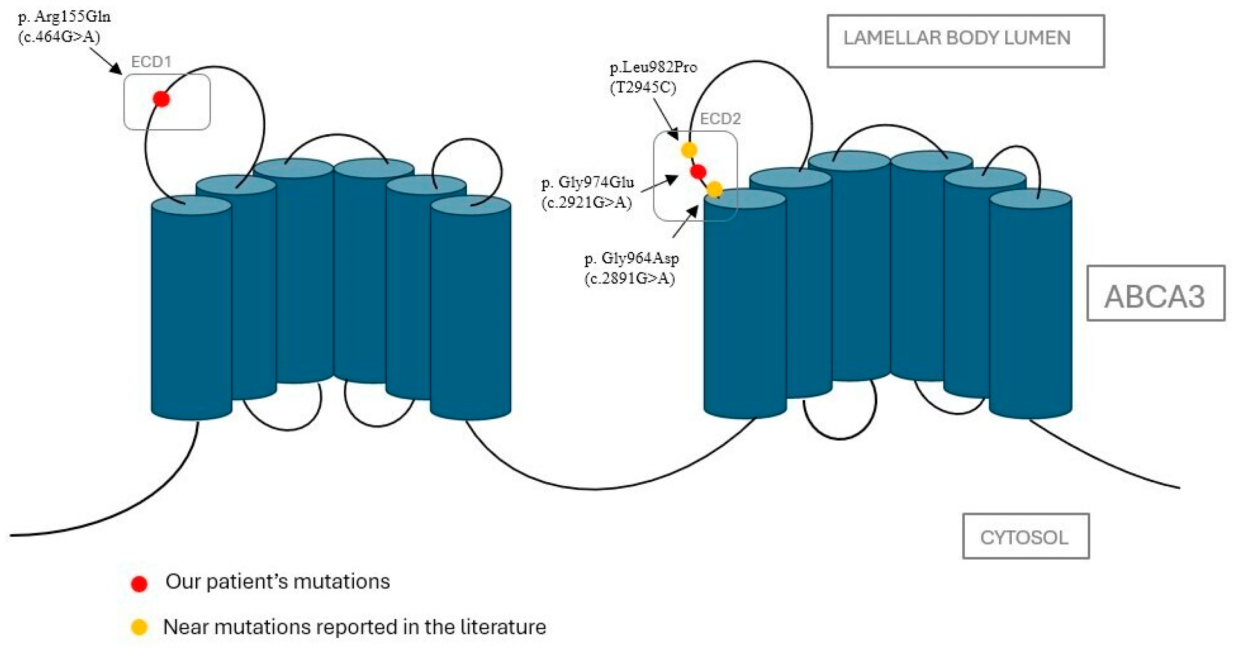
A recent study by Beers et al. [18] analyzed in detail the known variants of the ABCA3 gene. Figure 2 maps the current variants on the reconstructed 3D structure in its cellular context, showing that they are both located in the extracellular domains (ECDs), which are inside the lumen of the lamellar bodies (LBs).

3. Discussion

Different causes of interstitial lung disease may be recognized. The case herein described falls within the chILD subtype ‘surfactant dysfunction’. Actually, congenital surfactant protein deficits represent a significant cause of interstitial disease. The proteins mainly involved are surfactant protein C (SP-C), surfactant protein B (SP-B), and ABCA3. The ABCA3 protein plays a role in the transport of phosphatidylcholine and phosphatidylglycerol into the lamellar bodies, where the final processing of surfactant components prior to secretion takes place [19]. Abnormalities in this protein cause the altered transport of lipids that become trapped in the endoplasmic reticulum, triggering apoptosis in alveolar epithelial cells and the initiation of fibrogenesis [20]. Its deficiency is transmitted with autosomal recessive inheritance.
Besides presenting the clinical case of a newborn girl affected by a novel compound heterozygous variant of the ABCA3 gene, we also carried out a mini-review of the recent available literature (2004–2025) on similar pediatric cases, highlighting similarities and differences. To this aim, English reviews and case reports were included. We excluded letters and series. The following keywords (alone or in combination) were used: chILDs, newborns, interstitial disease, respiratory insufficiency, surfactant, pediatric, ABCA3, genetic, diagnosis, therapy. The electronic databases used were PubMed and Scopus. The cases described in the literature who carry both the homozygous and compound heterozygous variants are summarized in Table 1.
Our patient carried a compound heterozygous variant of ABCA3, and according to the literature reports, the prognosis seems to be better than that observed in subjects with homozygous variants. The clinical implications, however, also depend on the type of variant. The extent of the variation in the case of a deletion, or the site where it falls in the case of missense, nonsense, or frameshift ones, can modify the functionality of the protein, up to its complete loss. In the specific case of our patient’s variant, it was found that it falls into highly conserved amino acids and, therefore, may be considered probably pathogenic. The G974E variant described in our patient has never been reported in the literature, but a pathogenetic role can be assumed for it. Actually, it is located close to two already known variants: L982P and G964D. In particular, with regard to L982P, two patients carrying this variant in compound heterozygosity had an early fatal outcome, occurring during the neonatal period and at three months of age, respectively [42]. The G964D variant has been analyzed by Schindlbeck et al. [43], but its role has not been well defined. It shows discrete functionality in vitro, and Campo et al. described in 2014, indeed, the case of a girl carrying such a variant and diagnosed with pulmonary fibrosis in adulthood, leading to suggest for it a less severe pathogenic role [43,44]. Clinical pictures caused by the ABCA3 variant may also differ in severity within siblings with the same genotype [23,28]. It is likely that, in addition to genetic factors, epigenetic or environmental factors may also contribute to the severity of the respiratory picture. Moreover, a higher prevalence and mortality rate has been observed among male subjects. Actually, premature males have a higher incidence of respiratory distress syndrome (RDS) and bronchopulmonary dysplasia, as androgens are able to delay the maturation of the alveolar epithelium, and they act as positive regulators in the pathway involved in pulmonary fibrosis. In contrast, estrogens allow the promotion of the maturation of the alveolar epithelium and increase the presence of proteins in the surfactant [28]. All such aspects together might explain a mild evolution of the lung impairment observed in our baby.
Surfactant deficiency from ABCA3 protein alterations typically manifests with neonatal respiratory failure [22], as also occurred in the present patient. In the literature, however, cases with late onset (i.e., 9 months, 8 years) [31,41] or with variable manifestations (pulmonary hypertension, chronic cough) [21,41] have also been reported. Neonatal respiratory distress is a clinical condition that generally affects preterm infants due to lung immaturity, the poor development of alveolar type II cells, and, consequently, reduced surfactant production. Its incidence is greater the younger the infant’s gestational age at birth. The presence and/or persistence of symptoms in full-term or late-preterm babies can, therefore, be considered as a warning for the potential subtending occurrence of a congenital surfactant deficiency. Our patient, like several cases described in the literature [24,36], is a late preterm who had a persistent need for respiratory support. This aspect, along with the difficulty in ventilation weaning and the exclusion of other causes, led us to suspect that RDS was not linked to prematurity but rather to a congenital lung disease [30,40]. Furthermore, the absent or transient response to exogenous surfactant administration, as observed in our case and similarly reported in previous studies of the literature, represents a further suggestive element that must guide clinicians towards the suspicion of a congenital surfactant deficiency.
Chai et al. highlighted that ABCA3 protein deficiency may lead to lipid accumulation in the endoplasmic reticulum, resulting in the apoptosis of alveolar cells and the production of pro-inflammatory cytokines [19]. Furthermore, Rindler et al., in a recent study of 2017, documented and proved an anti-inflammatory function of the ABCA3 protein [45]. It has also been hypothesized that the protein defect leads to a reduction in the secretion of surfactant proteins (such as the hydrophilic collectins SP-A and SP-D), which play an immunomodulatory role by enhancing the phagocytosis of apoptotic cells and inhibiting T-cell function [19]. All these findings may explain the high susceptibility and harm experienced by such patients when facing infections, also in light of the antimicrobial role played by surfactants (in our case, indeed, two life-threatening bacterial infections were reported). To date, the only effective treatment option for these patients is lung transplantation. The operation is not free from both early and late complications, such as bleeding, infection, early rejection, bronchiolitis obliterans, and restrictive allograft syndrome. The 5-year survival rate for lung transplants remains approximately 60% for infants and 80% for children. Other therapeutic approaches are based on corticosteroids, hydroxychloroquine, or azathioprine, which can positively influence the prognosis. Not all children respond to these therapies, however, and this probably depends on the genetic defect and the type of variant. The exact mechanism of action of hydroxychloroquine is unknown, but it appears to reduce interstitial inflammation. In vitro, this drug showed efficacy, especially for the E292V, D953H, Q1045R, and A1046E variants, by increasing lipid transport [46]. Corticosteroids appear to regulate ABCA3 expression in the alveolar type II cells. Macrolides are able to inhibit the production of inflammatory cytokines and mediators involved in pulmonary fibrosis [27]. According to the above literature, survival was higher among patients receiving such triple therapy, and its benefits have also been proven in our experience. Some of the surviving children reported to date needed persistent oxygen support and showed growth impairment, in addition to neuromotor, linguistic, and cognitive delay [23,30,33,37,40,41]. In our case, the baby is dependent on high-flow oxygen therapy, while gastrostomy is, at present, able to ensure sufficient growth. No neurological, cognitive, or other developmental issues are currently noticed, except for mild motor delay. In the present case, therapy was administered according to the experimental protocol for childhood interstitial lung disease [6]. The treatment allowed a significant improvement in the patient’s condition and, finally, her discharge after around 5 months. In addition to the pharmacological approaches already analyzed, new frontiers of research are turning their gaze towards gene therapy, whereby DNA fragments or mRNA are carried by viral vectors (most frequently adenoviruses) or nanoparticles that, unlike viruses, do not trigger an immune system response [46]. Pulmonary interstitial diseases are chronic pathologies that require continuous care. It is, indeed, essential to educate parents so that they can be prepared for the home management of their children but also to provide them with medical and nursing support through the health territory services. Home management, in our experience, is provided through a complex home care service by the local health authority that is integrated with the activities of the healthcare workers of our department, who guarantee the family the necessary tools and devices to manage and protect their child.

4. Conclusions

ChILDs are a rare cause of congenital lung disease, which includes, among others, congenital surfactant deficiency disorders. They can have different age presentations but generally occur in the neonatal period with the onset of respiratory distress, completely or partially unresponsive to the administration of an exogenous surfactant. Knowing the genetic profile of these patients has a crucial role, as for other rare diseases, due to its relevant implications for diagnosis, as well as for the therapeutic approach and prognosis evaluation [47,48,49]. To date, the only curative treatment option for these patients is lung transplantation, although the use of corticosteroids, azathioprine, and hydroxychloroquine has shown a favorable effect on both prognosis and survival of affected subjects. Drugs capable of increasing the functionality or expression of the mutated proteins (i.e., ABCA3), antifibrotic drugs (already used in adult patients with pulmonary interstitial disease), and, above all, gene therapy (using viral vectors in the case of surfactant protein variants) may allow the development of targeted treatments for congenital surfactant protein deficiency. An integrated development of these pharmacological approaches may eventually improve the survival of patients and the quality of life for them and their whole families in the future.

Supplementary Materials

The following supporting information can be downloaded at: https://www.mdpi.com/article/10.3390/jcm14113704/s1, Supplementary File S1. Panel of the genes included within the current next-generation sequencing analysis. Supplementary File S2. Prenatal genetic investigations performed on chorionic villi sample during the second pregnancy of the proband’s mother. Supplementary File S3. Bioinformatic analysis of the identified variants, through SIFT, Poly-Phen-2, MutationTaster, and Panther tools. Supplementary File S4. Detail of the amino acid adjacent to each substitution and probabilities of protein function defect according to SIFT.

Author Contributions

Conceptualization, G.S. and V.N.; methodology, G.S. and V.N.; validation, M.G. and G.C.; formal analysis and data curation: G.S. and V.N.; writing—original draft preparation, G.S., V.N., V.A., C.C., M.R.D.P., D.M.M., M.P., E.P., I.A.M.S., M.S. and G.V.; writing—review and editing, G.S., V.N., D.M.M. and G.V.; supervision, M.G. and G.C. All authors have read and agreed to the published version of the manuscript.

Funding

This research received no external funding.

Institutional Review Board Statement

Not applicable.

Informed Consent Statement

Written informed consent was obtained from the child’s parents to publish this paper.

Data Availability Statement:

The original contributions presented in this study are included in the article and Supplementary Materials. Further inquiries can be directed to the corresponding author and/or to the first author.

Acknowledgments

Giuseppe and Guglielmo Puccio for having carried out the analysis of the protein function prediction test.

Conflicts of Interest

The authors declare no conflicts of interest.

References

  1. Cunningham, S.; Jaffe, A.; Young, L.R. Children’s interstitial and diffuse lung disease. Lancet Child. Adolesc. Health 2019, 3, 568–577. [Google Scholar] [CrossRef] [PubMed]
  2. Kurland, G.; Deterding, R.R.; Hagood, J.S.; Young, L.R.; Brody, A.S.; Castile, R.G.; Dell, S.; Fan, L.L.; Hamvas, A.; Hilman, B.C.; et al. American Thoracic Society Committee on Childhood Interstitial Lung Disease (chILD) and the chILD Research Network. An official American Thoracic Society clinical practice guideline: Classification, evaluation, and management of childhood interstitial lung disease in infancy. Am. J. Respir. Crit. Care Med. 2013, 188, 376–394. [Google Scholar] [CrossRef] [PubMed] [PubMed Central]
  3. Serra, G.; Memo, L.; Cavicchioli, P.; Cutrone, M.; Giuffrè, M.; La Torre, M.L.; Schierz, I.A.M.; Corsello, G. Novel mutations of the ABCA12, KRT1 and ST14 genes in three unrelated newborns showing congenital ichthyosis. Ital. J. Pediatr. 2022, 48, 145. [Google Scholar] [CrossRef] [PubMed]
  4. Serra, G.; Antona, V.; Cannata, C.; Giuffrè, M.; Piro, E.; Schierz, I.A.M.; Corsello, G. Distal Arthrogryposis type 5 in an Italian family due to an autosomal dominant gain-of-function mutation of the PIEZO2 gene. Ital. J. Pediatr. 2022, 48, 133. [Google Scholar] [CrossRef]
  5. Valutazione Antropometrica Neonatale. Riferimento Carte INeS. Available online: http://www.inescharts.com (accessed on 12 May 2025).
  6. Bush, A.; Cunningham, S.; de Blic, J.; Barbato, A.; Clement, A.; Epaud, R.; Hengst, M.; Kiper, N.; Nicholson, A.G.; Wetzke, M.; et al. chILD-EU Collaboration. European protocols for the diagnosis and initial treatment of interstitial lung disease in children. Thorax 2015, 70, 1078–1084. [Google Scholar] [CrossRef] [PubMed]
  7. World Health Organization. Child Growth Standards. 2021. Available online: https://www.who.int/tools/child-growth-standards/standards (accessed on 12 May 2025).
  8. Hamvas, A.; Nogee, L.M.; Wegner, D.J.; Depass, K.; Christodoulou, J.; Bennetts, B.; McQuade, L.R.; Gray, P.H.; Deterding, R.R.; Carroll, T.R.; et al. Inherited surfactant deficiency caused by uniparental disomy of rare mutations in the surfactant protein-B and ATP binding cassette, subfamily a, member 3 genes. J. Pediatr. 2009, 155, 854–859.e1. [Google Scholar] [CrossRef]
  9. Ciantelli, M.; Ghirri, P.; Presi, S.; Sigali, E.; Vuerich, M.; Somaschini, M.; Ferrari, M.; Boldrini, A.; Carrera, P. Fatal respiratory failure in a full-term newborn with two ABCA3 gene mutations: A case report. J. Perinatol. 2011, 31, 70–72. [Google Scholar] [CrossRef]
  10. Richards, S.; Aziz, N.; Bale, S.; Bick, D.; Das, S.; Gastier-Foster, J.; Grody, W.W.; Hegde, M.; Lyon, E.; Spector, E.; et al. Laboratory Quality Assurance Committee. Standards and guidelines for the interpretation of sequence variants: A joint consensus recommendation of the American College of Medical Genetics and Genomics and the Association for Molecular Pathology. Genet. Med. 2015, 17, 405–424. [Google Scholar] [CrossRef]
  11. Rehder, C.; Bean, L.J.H.; Bick, D.; Chao, E.; Chung, W.; Das, S.; O’Daniel, J.; Rehm, H.; Shashi, V.; Vincent, L.M.; et al. Next-generation sequencing for constitutional variants in the clinical laboratory, 2021 revision: A technical standard of the American College of Medical Genetics and Genomics (ACMG). Genet. Med. 2021, 23, 1399–1415. [Google Scholar] [CrossRef]
  12. Matthijs, G.; Souche, E.; Alders, M.; Corveleyn, A.; Eck, S.; Feenstra, I.; Race, V.; Sistermans, E.; Sturm, M.; Weiss, M.; et al. EuroGentest; European Society of Human Genetics. Guidelines for diagnostic next-generation sequencing. Eur. J. Hum. Genet. 2016, 24, 2–5. [Google Scholar] [CrossRef]
  13. ISO 9001:2015; Standard Title: Quality Management Systems. International Organization for Standardization (ISO): Geneva, Switzerland, 2015.
  14. Vaser, R.; Adusumalli, S.; Leng, S.N.; Sikic, M.; Ng, P.C. SIFT missense predictions for genomes. Nat. Protoc. 2016, 11, 1–9. [Google Scholar] [CrossRef]
  15. Adzhubei, I.; Jordan, D.M.; Sunyaev, S.R. Predicting functional effect of human missense mutations using PolyPhen-2. Curr. Protoc. Hum. Genet. 2013, 7, Unit7.20. [Google Scholar] [CrossRef]
  16. Steinhaus, R.; Proft, S.; Schuelke, M.; Cooper, D.N.; Schwarz, J.M.; Seelow, D. MutationTaster2021. Nucleic. Acids Res. 2021, 49, W446–W451. [Google Scholar] [CrossRef] [PubMed]
  17. Panther Classification System. PANTHER™ Website News. Available online: https://www.pantherdb.org (accessed on 17 September 2023).
  18. Beers, M.F.; Mulugeta, S. The biology of the ABCA3 lipid transporter in lung health and disease. Cell Tissue Res. 2017, 367, 481–493. [Google Scholar] [CrossRef] [PubMed]
  19. Chai, A.B.; Ammit, A.J.; Gelissen, I.C. Examining the role of ABC lipid transporters in pulmonary lipid homeostasis and inflammation. Respir. Res. 2017, 18, 41. [Google Scholar] [CrossRef] [PubMed]
  20. Tirelli, C.; Pesenti, C.; Miozzo, M.; Mondoni, M.; Fontana, L.; Centanni, S. The Genetic and Epigenetic Footprint in Idiopathic Pulmonary Fibrosis and Familial Pulmonary Fibrosis: A State-of-the-Art Review. Diagnostics 2022, 12, 3107. [Google Scholar] [CrossRef]
  21. Kuning, A.M.; Parker, T.A.; Nogee, L.M.; Abman, S.H.; Kinsella, J.P. ABCA3 deficiency presenting as persistent pulmonary hypertension of the newborn. J. Pediatr. 2007, 151, 322–324. [Google Scholar] [CrossRef]
  22. Anandarajan, M.; Paulraj, S.; Tubman, R. ABCA3 Deficiency: An unusual cause of respiratory distress in the newborn. Ulster Med. J. 2009, 78, 51–52. [Google Scholar]
  23. Hallik, M.; Annilo, T.; Ilmoja, M.L. Different course of lung disease in two siblings with novel ABCA3 mutations. Eur. J. Pediatr. 2014, 173, 1553–1556. [Google Scholar] [CrossRef] [PubMed]
  24. Gonçalves, J.P.; Pinheiro, L.; Costa, M.; Silva, A.; Gonçalves, A.; Pereira, A. Novel ABCA3 mutations as a cause of respiratory distress in a term newborn. Gene 2014, 534, 417–420. [Google Scholar] [CrossRef]
  25. Malý, J.; Navrátilová, M.; Hornychová, H.; Looman, A.C. Respiratory failure in a term newborn due to compound heterozygous ABCA3 mutation: The case report of another lethal variant. J. Perinatol. 2014, 34, 951–953. [Google Scholar] [CrossRef] [PubMed]
  26. Moore, G.P.; Lines, M.A.; Geraghty, M.T.; de Nanassy, J.; Kovesi, T. Novel mutation in ABCA3 resulting in fatal congenital surfactant deficiency in two siblings. Am. J. Respir. Crit. Care Med. 2014, 189, 750–752. [Google Scholar] [CrossRef] [PubMed]
  27. Jackson, T.; Wegner, D.J.; White, F.V.; Hamvas, A.; Cole, F.S.; Wambach, J.A. Respiratory failure in a term infant with cis and trans mutations in ABCA3. J. Perinatol. 2015, 35, 231–232. [Google Scholar] [CrossRef]
  28. Piersigilli, F.; Peca, D.; Campi, F.; Corsello, M.; Landolfo, F.; Boldrini, R.; Danhaive, O.; Dotta, A. New ATP-binding cassette A3 mutation causing surfactant metabolism dysfunction pulmonary type 3. Pediatr. Int. 2015, 57, 970–974. [Google Scholar] [CrossRef]
  29. Pachajoa, H.; Ruiz-Botero, F.; Meza-Escobar, L.E.; Villota-Delgado, V.A.; Ballesteros, A.; Padilla, I.; Duarte, D. Fatal respiratory disease due to a homozygous intronic ABCA3 mutation: A case report. J. Med. Case Rep. 2016, 10, 266. [Google Scholar] [CrossRef]
  30. Tan, J.K.; Murray, C.; Schultz, A. ABCA3 lung disease in an ex 27 week preterm infant responsive to systemic glucocorticosteroids. Pediatr. Pulmonol. 2016, 51, E1–E3. [Google Scholar] [CrossRef]
  31. Ota, C.; Kimura, M.; Kure, S. ABCA3 mutations led to pulmonary fibrosis and emphysema with pulmonary hypertension in an 8-year-old girl. Pediatr. Pulmonol. 2016, 51, E21–E23. [Google Scholar] [CrossRef] [PubMed]
  32. Uchida, D.A.; Wert, S.E.; Nogee, L.M.; Carroll, T.R.; Chatfield, B.A. Pulmonary nodules in a newborn with ATP-binding cassette transporter A3 (ABCA3) mutations. Pediatrics 2011, 127, e1347–e1351. [Google Scholar] [CrossRef]
  33. Akil, N.; Fischer, A.J. Surfactant deficiency syndrome in an infant with a C-terminal frame shift in ABCA3: A case report. Pediatr. Pulmonol. 2018, 53, E12–E14. [Google Scholar] [CrossRef]
  34. Mitsiakos, G.; Tsakalidis, C.; Karagianni, P.; Gialamprinou, D.; Chatziioannidis, I.; Papoulidis, I.; Tsanakas, I.; Soubasi, V. A New ABCA3 Gene Mutation c.3445G > A (p.Asp1149Asn) as a Causative Agent of Newborn Lethal Respiratory Distress Syndrome. Medicina 2019, 55, 389. [Google Scholar] [CrossRef]
  35. Oltvai, Z.N.; Smith, E.A.; Wiens, K.; Nogee, L.M.; Luquette, M.; Nelson, A.C.; Wikenheiser-Brokamp, K.A. Neonatal respiratory failure due to novel compound heterozygous mutations in the ABCA3 lipid transporter. Cold Spring Harb. Mol. Case Stud. 2020, 6, a005074. [Google Scholar] [CrossRef]
  36. Gupta, N.P.; Batra, A.; Puri, R.; Meena, V. Novel homozygous missense mutation in ABCA3 protein leading to severe respiratory distress in term infant. BMJ Case Rep. 2020, 13, e235520. [Google Scholar] [CrossRef]
  37. Shaaban, W.; Hammoud, M.; Abdulraheem, A.; Elsayed, Y.Y.; Alkazemi, N. Hydroxychloroquine, a successful treatment for lung disease in ABCA3 deficiency gene mutation: A case report. J. Med. Case Rep. 2021, 15, 54. [Google Scholar] [CrossRef]
  38. Bozkurt, H.B.; Şahin, Y. Whole exome sequencing identifies a novel variant in ABCA3 in an individual with fatal congenital surfactant protein deficiency. Turk. J. Pediatr. 2021, 63, 703–707. [Google Scholar] [CrossRef] [PubMed]
  39. Zhang, W.; Liu, Z.; Lin, Y.; Wang, R.; Xu, J.; He, Y.; Zhang, F.; Wu, L.; Chen, D. A novel synonymous ABCA3 variant identified in a Chinese family with lethal neonatal respiratory failure. BMC Med. Genom. 2021, 14, 256. [Google Scholar] [CrossRef] [PubMed]
  40. Si, X.; Steffes, L.C.; Schymick, J.C.; Hazard, F.K.; Tracy, M.C.; Cornfield, D.N. Three Infants with Pathogenic Variants in the ABCA3 Gene: Presentation, Treatment, and Clinical Course. J. Pediatr. 2021, 231, 278–283.e2. [Google Scholar] [CrossRef] [PubMed]
  41. Chen, F.; Xie, Z.; Zhang, V.W.; Chen, C.; Fan, H.; Zhang, D.; Jiang, W.; Wang, C.; Wu, P. Case Report: Report of Two Cases of Interstitial Lung Disease Caused by Novel Compound Heterozygous Variants in the ABCA3 Gene. Front. Genet. 2022, 13, 875015. [Google Scholar] [CrossRef]
  42. Shulenin, S.; Nogee, L.M.; Annilo, T.; Wert, S.E.; Whitsett, J.A.; Dean, M. ABCA3 gene mutations in newborns with fatal surfactant deficiency. N. Engl. J. Med. 2004, 350, 1296–1303. [Google Scholar] [CrossRef]
  43. Schindlbeck, U.; Wittmann, T.; Höppner, S.; Kinting, S.; Liebisch, G.; Hegermann, J.; Griese, M. ABCA3 missense mutations causing surfactant dysfunction disorders have distinct cellular phenotypes. Hum. Mutat. 2018, 39, 841–850. [Google Scholar] [CrossRef]
  44. Campo, I.; Zorzetto, M.; Mariani, F.; Kadija, Z.; Morbini, P.; Dore, R.; Kaltenborn, E.; Frixel, S.; Zarbock, R.; Liebisch, G.; et al. A large kindred of pulmonary fibrosis associated with a novel ABCA3 gene variant. Respir. Res. 2014, 15, 43. [Google Scholar] [CrossRef]
  45. Rindler, T.N.; Stockman, C.A.; Filuta, A.L.; Brown, K.M.; Snowball, J.M.; Zhou, W.; Veldhuizen, R.; Zink, E.M.; Dautel, S.E.; Clair, G.; et al. Alveolar injury and regeneration following deletion of ABCA3. JCI Insight 2017, 2, e97381. [Google Scholar] [CrossRef] [PubMed]
  46. Peers de Nieuwburgh, M.; Wambach, J.A.; Griese, M.; Danhaive, O. Towards personalized therapies for genetic disorders of surfactant dysfunction. Semin. Fetal Neonatal Med. 2023, 28, 101500. [Google Scholar] [CrossRef] [PubMed]
  47. Serra, G.; Giambrone, C.; Antona, V.; Cardella, F.; Carta, M.; Cimador, M.; Corsello, G.; Giuffrè, M.; Insinga, V.; Maggio, M.C.; et al. Congenital hypopituitarism and multiple midline defect sin a new born with non-familial Cat Eye syndrome. Ital. J. Pediatr. 2022, 48, 170. [Google Scholar] [CrossRef] [PubMed]
  48. Presti, S.; Parisi, G.F.; Papale, M.; Gitto, E.; Manti, S.; Leonardi, S. Interstitial Lung Disease in Children: “Specific Conditions of Undefined Etiology” Becoming Clearer. Children 2022, 9, 1744. [Google Scholar] [CrossRef]
  49. Drobňaková, S.; Vargová, V.; Barkai, L. The Clinical Approach to Interstitial Lung Disease in Childhood: A Narrative Review Article. Children 2024, 11, 904. [Google Scholar] [CrossRef]
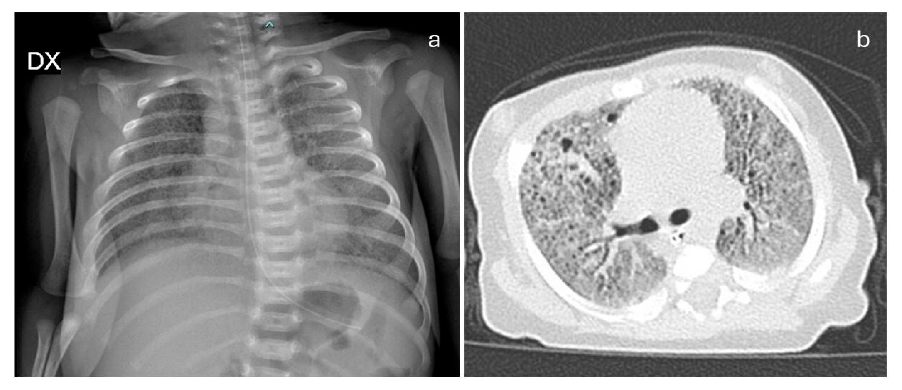
Figure 1. (a) Chest X-ray shows hypodiaphany in all lung areas, in addition to texture reinforcement with a reticulo-micronodular pattern; (b) Chest CT: in both lungs, a diffuse increase in parenchymal density is observed, with a predominantly “ground glass” appearance and associated thickening of the inter- and intralobular septa. Bilateral, multiple, subcentimeter-sized air-filled cavities are present.
Jcm 14 03704 g001
Figure 2. (Modified by Beers MF et al., 2016 [18]). A 3D reconstruction of the ABCA3 protein in its cellular context. The first variant, reported as pathogenic, is clearly shown within the ECD n.1 (left), as R155Q. The second one (G974E), never described to date, is located in the context of ECD n.2 (right), very close to two variants (G964S and L982P) already known in the literature and leading both to pathogenic effects, as described in the Section 3.
Jcm 14 03704 g002
Table 1. Comparison between our patient and those previously described in the literature affected by surfactant dysfunction due to ABCA3 variants. GA: gestational age. RDS: respiratory distress syndrome. HFNC: high-flow nasal cannula.
Authors, Year, CountryType of Mutation in the ABCA3 GeneSexGAOnsetClinical FeaturesSurfactantTreatmentOutcome
Kuning et al. [21]
2007, Colorado (USA)
p.Leu326Arg, in homozygosityMTermEarly onsetRefractory pulmonary hypertension, later parenchymal lung diseaseNo (due to clinical instability)Nitric oxide, dopamine (for pulmonary hypertension) Death due to withdrawal of intensive care
Anandarajan et al. [22]
2009, United Kingdom
p.Gly378Arg,
p.Gly1002Arg
FTermWithin few hours of birthRDSYesAntibiotics (not specified)Death at 25 days of life due to withdrawal of intensive care
Ciantelli et al. [9]
2011, Italy
p.Pro186Thr,
p.Met878Trp fs36X
MTermWithin few hours of birthRDSYes (9 doses)Dexamethasone Death at 72 days of life
Hallik et al. [23]
2013, Estonia
p.Asp507AlafsTer508, p.Asp696AsnFTermWithin few hours of birthRDSYes (1 dose)Methylprednisolone,
azithromycin, hydroxychloroquine
Discharged at age 12 months with oxygen therapy and gastrostomy. In the following months, mild neuro-motor delay.
MNot specifiedAsymptomatic for 4 years
Goncalves et al. [24]
2014, Portugal
p.Leu798Pro, p.Arg1612Pro FTermWithin few hours of birthRDSYes (4 doses)Methylprednisolone, hydroxychloroquineDeath at 101 days due to withdrawal of intensive care
Maly et al. [25]
2014, Czech Republic
p. Met1227Arg,
p. Ins1510fsTer1519
MTermWithin few hours of birthRDSYesDexamethasone and macrolidesDeath at 33 days of life due to withdrawal of intensive care
Moore et al. [26]
2014, Canada
p.Ser1116Phe, in homozygosityMTermAt three hours of lifeRDSYes (a few doses)Antibiotics, nitric oxideDeath at 48 days of life
FTermAt sixteen hours of lifeRDSYesDeath at 53 days of life
Jackson et al. [27]
2015, USA
p.Arg280Cys, p.Val1399Met, p.Gln1589TerFTermWithin few hours of birthRDSYesNitric oxideBilateral lung transplant at 9 months. At age 3 years, without oxygen support, nutritional supplementation tube, mild motor and speech delay.
Piersigilli et al. [28]
2015, Italy
p.Arg194Gly, p.Val1615GlyfsTer15FLate pretermWithin few hours of birthRDSYes (3 doses)Betamethasone, hydroxychloroquineDeath at age 5 months
MEarly onsetRDSYes (three doses)Betamethasone, hydroxychloroquineDeath at age 3 months
Pachajoa et al. [29]
2016, Colombia
IVS 25-98T in homozygosityMTermEarly onsetRDSYesAntibiotics (vancomycin, meropenem)Death at 60 days
Tan et al. [30]
2016, Australia
p.Ala307Val, in homozygosityFPretermEarly onsetRDSYes (2 doses)Hydroxychloroquine, azithromycin, methylprednisoloneOxygen therapy through nasal cannula at age 1 year
Ota et al. [31]
2016, Japan
p.Leu34Pro, p.1203_1204delFTerm8 yearsPulmonary fibrosis with emphysema, pulmonary hypertensionNot specifiedCalcium antagonist, warfarin, home oxygen therapy, beraprost sodiumImprovement of pulmonary arterial pressure. No worsening of fibrosis.
Uchida et al. [32]
2017, USA
p.(delPhe1203)4,
c.1375ins15
FTermEarly onsetRDS, perihilar and peripheral rounded masses of the right lungNot specifiedSystemic corticosteroids (not specified)Death at 113 days
Akil et al. [33]
2018, USA
p.Glu292Val,
p.Met1647fs
MTermEarly onsetRespiratory illness, cyanosis, impaired growth.Not specifiedPrednisolone, methylprednisolone, azithromycin, hydroxychloroquine.
Gastrostomy
Oxygen therapy with nasal cannula at age 1 year
Mitsiakos et al. [34]
2019, Greece
p.Asp1149Asn, in homozygosityMTermWithin few hours of birthRDSYes (10 doses)Methylprednisolone, prednisolone, azithromycin. At age 78 days hydroxychloroquine, prednisone, azithromycinDeath at 9 months
Oltvai et al. [35]
2020, USA
p.Ala1629GlyfsX15, p.Gly961Gly MTermWithin few hours of birthRDSYes (a few doses)Dexamethasone and macrolidesSuccessful bilateral lung transplantation at 15 weeks of age
Gupta et al. [36]
2020, India
p.Leu437Pro, in homozygosityFLate preterm Within few hours of birthRDSYes (2 doses)DexamethasoneDeath at 100 days
Shaaban et al. [37]
2021, Kuwait
c.875A<T in homozygosityMTermWithin few hours of birthRDSYesHydroxychloroquine, methylprednisolone, azithromycinDischarged at 160 days with hydroxychloroquine maintenance treatment
Bozkurt et al. [38]
2021, Turkey
p.Leu1226Pro, in homozygosityMTermEarly onsetRDS,
pulmonary hypertension
Yes (3 doses)Antibiotics, nitric oxide, sildenafilDeath at 24 days
Zhang et al. [39]
2021, China
c.4-7del, p.Lys291Lys Two girlsTermWithin few hours of birthRDSYesAntibiotics, nitric oxide, and methylprednisoloneDeath at 23 days
Si et al. [40]
2021, California (USA)
p.Glu292Val, p.Arg1081TrpMPre
term
Early onsetRDSYesAzithromycin, hydroxychloroquine, methylprednisolone.
Gastrostomy
fundoplicatio according to Nissen
At age 2 years without oxygen support, severe motor, language and cognitive delay, impaired growth. Steroid therapy discontinued, bilevel alternating with low-flow nasal cannula respiratory support
p.Tyr758Ter,
p.Lys915Asn
MTermWithin few hours of birthRDSYesMethylprednisolone,
hydroxychloroquine, azithromycin.
Gastrostomy fundoplicatio according to Nissen
At age 14 months, language, cognitive, and motor delay, impaired growth. Steroid discontinued at 1 year. HFNC respiratory support.
C.1285+1G>A, p.Asp200GlyMTermWithin few hours of birthRDSYesMethylprednisolone, azithromycin and hydroxychloroquine.
Gastrostomy fundoplicatio
according to Nissen
Removal of gastrostomy tube at age 18 months, impaired growth, mild language delay (at age 13 months). Steroid discontinued. Low-flow nasal cannula (LFNC).
Chen et al. [41]
2022, China
c.4035+5G>A, p.Met223Thr, c.1285+4A>CMTermAfter 24 h of birthDyspnea and coughNot specifiedAzithromycin, dexamethasone and hydroxychloroquineDeath at 94 days
p.Phe235Ser, p.Thr1346Asn
fsTer15
MTermAt 9 monthsChronic coughNot specifiedBudesonide, ipratropium bromide and acetylcysteineAt age 41 months, stable clinical condition without respiratory support
Our case (2025)p.Arg155Gln, p.Gly974GluFLate pretermEarly onsetRDS Yes (4 doses)Azithromycin, hydroxychloroquine, diuretics (furosemide, spironolactone), sildenafil. GastrostomyGood general condition with oxygen therapy through HFNC.
Impaired growth, mild motor delay.
Disclaimer/Publisher’s Note: The statements, opinions and data contained in all publications are solely those of the individual author(s) and contributor(s) and not of MDPI and/or the editor(s). MDPI and/or the editor(s) disclaim responsibility for any injury to people or property resulting from any ideas, methods, instructions or products referred to in the content.