Article

Balance or Strength? Reconsidering Muscle Metrics in Sagittal Malalignment in Adult Sagittal Deformity Patients

by
Donghua Huang
1,2,
Zhan Wang
1,2,
Mihir Dekhne
1,3,
Atahan Durbas
1,
Tejas Subramanian
1,3,
Gabrielle Dykhouse
1,3,
Robert N. Uzzo
1,3,
Luis Felipe Colón
1,
Stephane Owusu-Sarpong
1,
Han Jo Kim
1 and
Francis Lovecchio
1,*
1
Department of Orthopaedic Surgery, Hospital for Special Surgery, New York, NY 10021, USA
2
Department of Orthopedic Surgery, The Second Affiliated Hospital Zhejiang University School of Medicine, Hangzhou 310009, China
3
Weill Cornell Medicine, 1300 York Ave, New York, NY 10021, USA
*
Author to whom correspondence should be addressed.
J. Clin. Med. 2025, 14(10), 3293; https://doi.org/10.3390/jcm14103293
Submission received: 8 April 2025 / Revised: 2 May 2025 / Accepted: 6 May 2025 / Published: 9 May 2025
(This article belongs to the Special Issue Optimizing Outcomes in Scoliosis and Complex Spinal Surgery)

Abstract

:
Background/Objectives: Atrophy of the paraspinal and psoas major muscles is closely linked to sagittal malalignment in adult spinal deformity (ASD). However, most studies overlook the balance between these muscle groups. This study investigates the relationship between trunk muscle balance and sagittal alignment in ASD patients. Methods: A single-institution database was reviewed for patients with sagittal malalignment (PT > 20° and PI–LL > 10°). Standard sagittal parameters were measured based on standing X-rays. The cross-section area (CSA) of trunk posterior muscles (CSAP: erector spinae and multifidus) and anterior muscles (CSAA: psoas) at L4 were measured based on a T2-weighted MRI. Patients with prior lateral fusions were excluded. Muscle balance was evaluated by the CSA ratio of trunk posterior to anterior muscles (CSAP/A). The relationship between sagittal alignment parameters and CSAP, CSAA, as well as CSAP/A were analyzed using linear and quadratic regressions. Akaike information criteria (AIC) compared model fit. Subgroup analyses examined the relationship between sagittal alignment changes and different CSAP/A levels. Results: A total of 112 patients met inclusion and exclusion criteria. CSAP correlated linearly with SS (r2 = 0.057, p = 0.011), PT (r2 = 0.043, p = 0.028), and T4–L1PA mismatch (r2 = 0.044, p = 0.027). CSAA showed no significant linear or quadratic relationships with sagittal spinal alignment parameters. In contrast, CSAP/A was quadratically associated with LL (r2 = 0.056, p = 0.044), SS (r2 = 0.134, p < 0.001), PI (r2 = 0.096, p = 0.004), L1PA (r2 = 0.114, p = 0.001), and T4–L1PA mismatch (r2 = 0.094, p = 0.005). Quadratic models of CSAP/A consistently had higher r2 and lower AIC values compared to the linear models of CSAP for most sagittal alignment parameters, especially in SS, PI, L1PA, and T4–L1PA mismatch (AIC difference ≥4). Higher CSAP/A is correlated to larger PI (and consequently, larger LL, SS, and L1PA). Conclusions: Trunk posterior–anterior muscle balance (CSAP/A) demonstrates a stronger relationship with sagittal alignment than individual muscle metrics. Quantitative MRI-based definitions of sarcopenia may need to be adjusted for PI.

1. Introduction

Adult spinal deformity (ASD) is a complex condition characterized by abnormal spinal alignment, often leading to significant disability, pain, and decreased quality of life [1,2,3,4]. In patients with ASD, a significant proportion experience sagittal imbalance, a critical parameter reflecting the spine’s failure to maintain proper upright stance in the sagittal plane [5,6]. Sagittal malalignment is a key driver of disability and pain [7,8,9].
The factors influencing sagittal balance in ASD are multifaceted, involving skeletal [10,11], neuromuscular [12], and compensatory mechanisms [13,14]. Among these, paraspinal muscles, specifically the posterior muscles (erector spinae and multifidus) [15,16,17,18,19,20] and anterior muscles (psoas) [19,20], have been closely linked to sagittal malalignment in ASD. However, existing research has focused solely on individual muscle metrics, neglecting the balance between trunk posterior and anterior muscles, which is vital for maintaining sagittal posture [21].
The purpose of this study aims to investigate the relationship between trunk posterior–anterior muscle balance and sagittal alignment in ASD patients. We hypothesize that the balance has a stronger relationship with sagittal alignment than individual muscle metrics. Moreover, the relationship between the ratio of trunk posterior to anterior muscles and sagittal alignment may be non-linear, potentially influencing alignment at varying rates and need to be adjusted for pelvic incidence (PI).

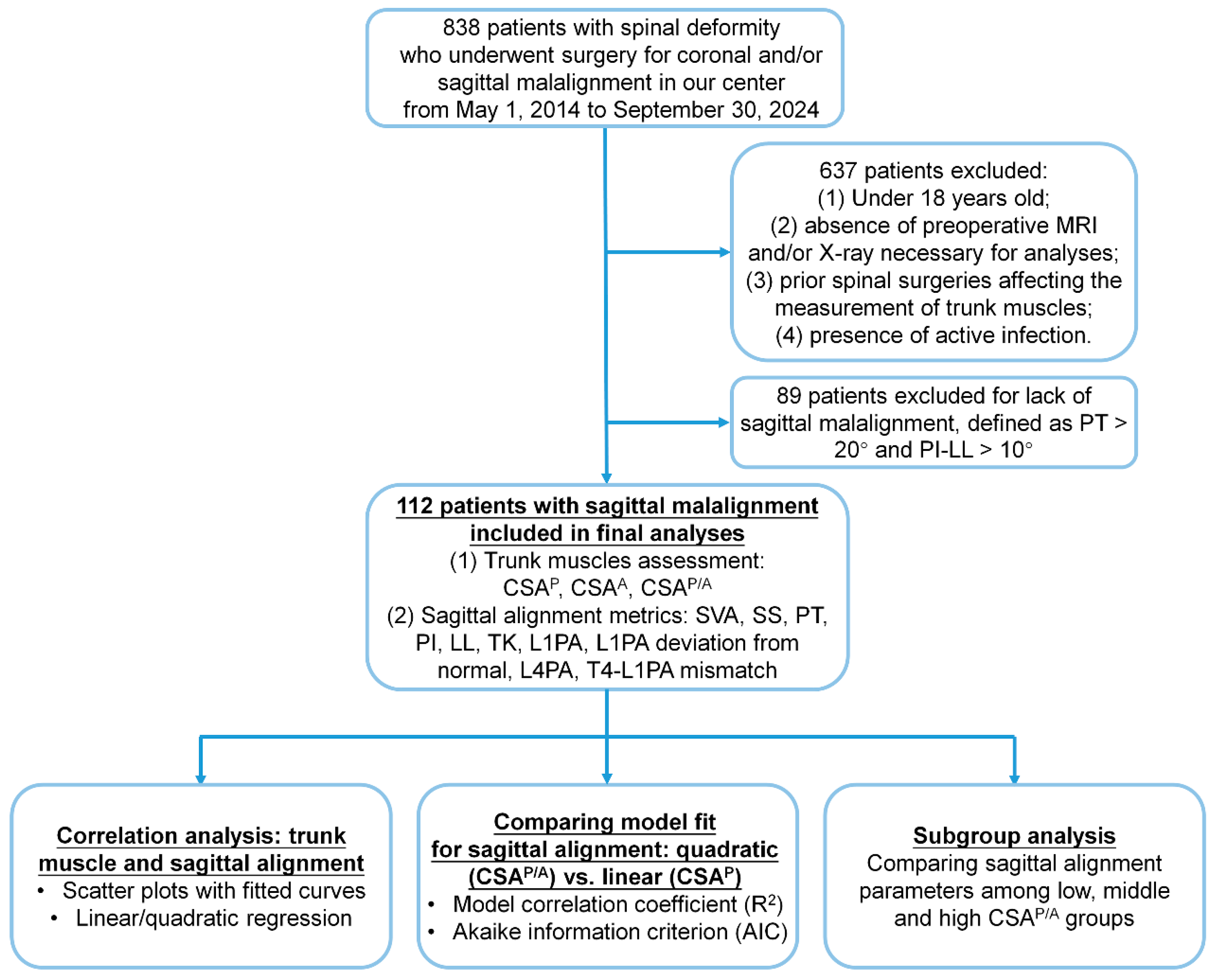
2. Materials and Methods

  • Patient Enrollment
Adult patients who underwent primary surgery for ASD from 2014 to 2023 and presented with sagittal malalignment, defined as a pelvic tilt (PT) > 20° and PI–lumbar lordosis (LL) > 10°, were included. Exclusion criteria were (1) under 18 years old; (2) lack of preoperative lumbar spine MRI or X-rays required for assessing spinal alignment or paravertebral muscles; (3) history of spinal procedures affecting the measurement of trunk muscles, such as lateral fusion with pedicle screw instrumentation; and (4) previous vertebral column infections.
  • Data Collection and Radiographic Analysis
Demographic data, including age, sex, BMI, smoking status, and medical comorbidities (diabetes, hypertension, osteoporosis, and dyslipidemia) were retrospectively obtained from electronic medical records.
  • Spinal Alignment Assessment
Spinal alignment was assessed using preoperative standing lateral and posteroanterior (PA) full-body X-rays. Standard spinopelvic parameters were measured as follows: (1) maximum coronal Cobb angle (Max Cobb); (2) thoracic kyphosis (TK; T4–T12); (3) LL (L1-S1); (4) sacral slope (SS); (5) PI; (6) PT; (7) sagittal vertical axis (SVA); (8) L1 pelvic angle (L1PA); (9) T4 pelvic angle (T4PA); (10) the L1PA offset (calculated as L1PA–normal L1PA, where normal L1PA was defined as 0.5 × PI − 21° [9]); and (11) the T4–L1PA mismatch (T4PA–L1PA).
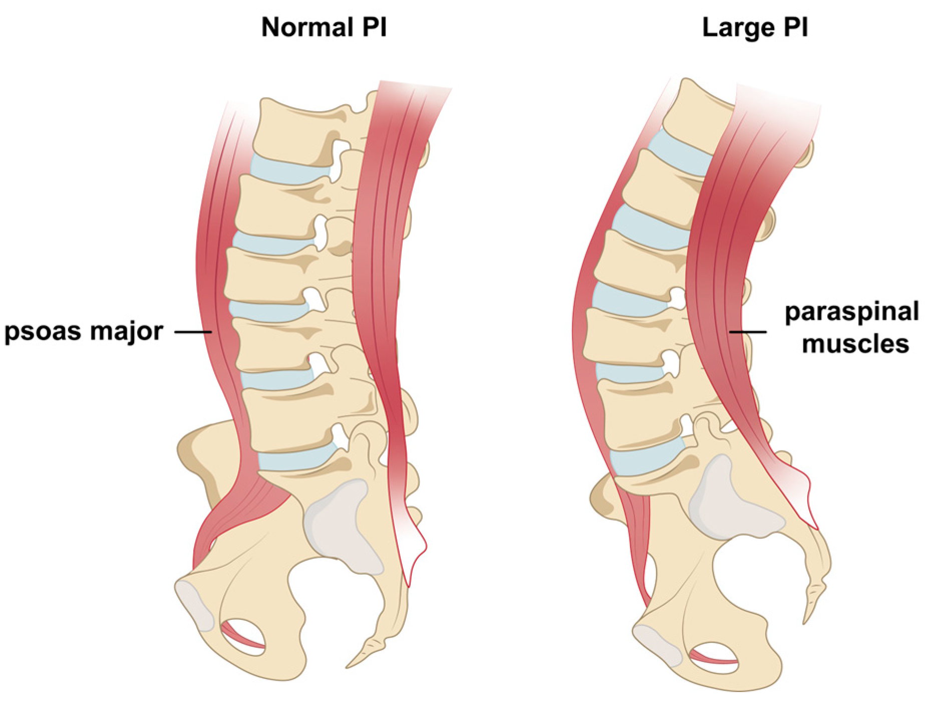
  • Trunk Muscle Assessment
To study the relationship between muscles and spinal sagittal alignment, selecting the appropriate muscle group and evaluation method is essential. Trunk muscles [17,22], particularly the paralumbar muscles at the L4 level [23,24], are more relevant and reliable than extremity muscles for analyzing degenerative ASD with sagittal imbalance. Common evaluation methods include assessing muscle strength [16,17,18], quality (fat infiltration, FI) [4,25], and quantity (cross-sectional area, CSA) [26,27]. While muscle strength directly reflects function, it is less reliable due to poor reproducibility and variability in clinical testing [28,29,30]. On the contrary, muscle quality and quantity assessments, obtained via CT or MRI, offer reliable, objective, and consistent measures [30,31]. While fat infiltration reflects muscle quality, it poses challenges in directly quantifying posterior–anterior muscle balance. In contrast, CSA serves as a practical, reliable quantitative measure of overall muscle volume, enabling precise ratio-based analyses. The bilateral CSA of trunk posterior muscles (CSAP, including the erector spinae and multifidus) and anterior muscles (CSAA, the psoas) at the L4 level were measured based on a T2-weighted MRI. The primary comparison was between the CSA ratio of posterior–anterior muscle (CSAP/A) at the L4 level and sagittal alignment.
All spinal alignment and muscle measurements were performed using Sectra Workstation software (Model IDS7, Version 24.1, Linköping, Sweden), and two independent, blinded reviewers performed these assessments for each patient.
  • Ethical Approval
This study was conducted in accordance with the Declaration of Helsinki and approved by the local institutional review board (Approval No: 2018-1599). Due to its retrospective and observational design, the requirement for informed consent was waived.
  • Statistical Analysis
The Kolmogorov–Smirnov test was used to assess normality for samples larger than 50, while the Shapiro–Wilk test was applied for samples of 50 or fewer. Continuous data were reported as mean and standard deviation (SD) or median and interquartile range (IQR), based on their distribution.
The correlations between spinal alignment parameters and trunk muscles (CSAP, CSAA, and CSAP/A) were analyzed using simple linear and quadratic regressions, with scatter plots illustrating these relationships and fitted curves generated using GraphPad Prism 9.0. The model correlation coefficient (R2) and the Akaike information criterion (AIC) were calculated as a measure of fit for each model and subsequently compared across models. AIC = −2 × (Log-Likelihood) + 2k. k in the equation is the number of free parameters in the regression model. A higher R2 and a lower AIC value indicate a better model fit [32,33]. An AIC difference of ≥4 was regarded as a notable distinction between models [32].
Additionally, patients were stratified into three subgroups based on CSAP/A values: low CSAP/A (≤mean − 1/2 SD), middle CSAP/A (mean ± 1/2 SD), and high CSAP/A (≥mean + 1/2 SD). Differences in sagittal alignment parameters across these three subgroups were assessed using one-way analysis of variance (ANOVA) or the Kruskal–Wallis test, as appropriate, depending on the distribution of the data. All statistical analyses were performed using IBM SPSS Statistics (version 22.0; IBM Corp., New York, NY, USA), with a significance level set at p-value < 0.05.

3. Results

A total of 112 patients (70.5% female) with sagittal ASD, average 66.3 ± 9.8 years of age, and BMI of 27.7 ± 6.0 kg/m2 met inclusion criteria and were included in the analysis (Figure 1). The patient demographics, radiographic measurements of spinal alignment, and CSA of trunk posterior and anterior muscles are reported in Table 1.

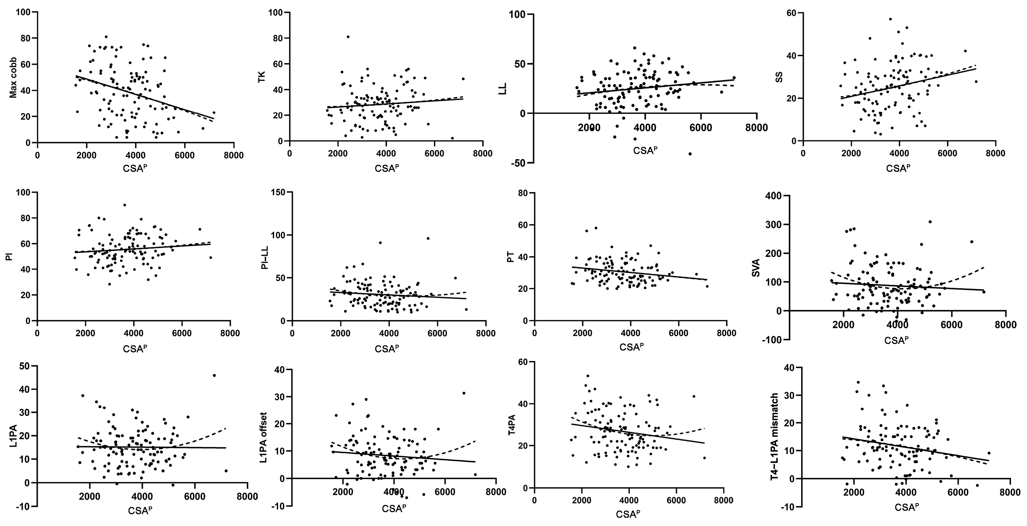
3.1. Simple Linear Regression Analysis of the Relationship Between Trunk Muscles and Sagittal Alignment

Linear regression analysis demonstrated significant associations between CSAP and SS (r2 = 0.057, p = 0.011), PT (r2 = 0.043, p = 0.028), and the T4–L1PA mismatch (r2 = 0.044, p = 0.027) (Table 2). Scatter plots and fitted results (Figure 2) indicated that CSAP exhibited linear relationships with most sagittal alignment parameters, including TK, LL, SS, PI, PI–LL, PT, T4PA, and T4–L1PA mismatch. In addition, CSAP/A (Table 2) exhibited a significant relationship with LL (r2 = 0.042, p = 0.031) and SS (r2 = 0.057, p = 0.011). However, CSAA (Table 2) showed no significant correlations with any sagittal alignment parameters. Notably, simple linear regression failed to fit the data for CSAP/A with most sagittal alignment parameters, prompting further analysis using quadratic regression.

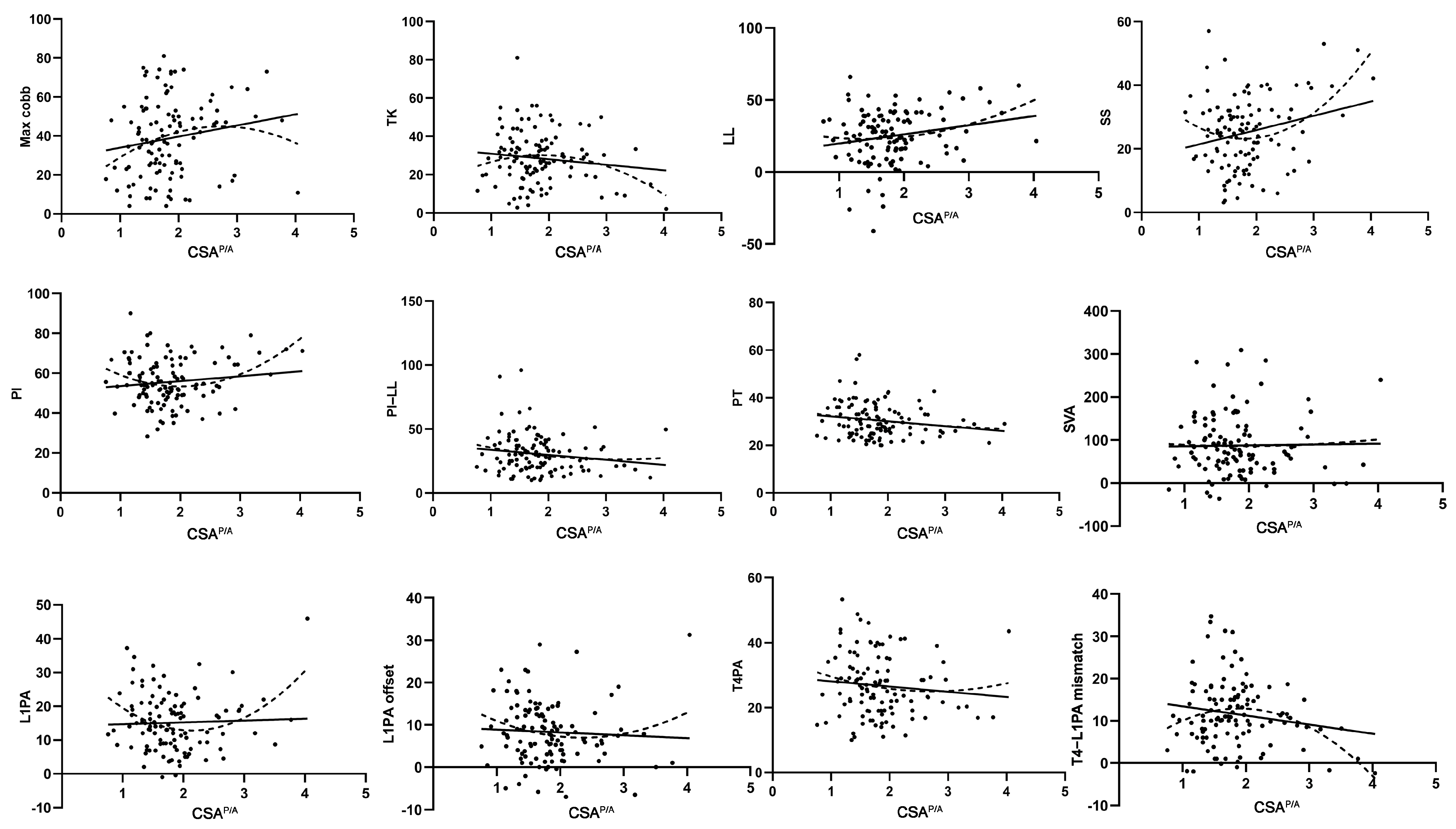
3.2. Quadratic Regression Analysis of the Relationship Between Trunk Muscles and Sagittal Alignment

Quadratic regression analysis showed a significant association between CSAP and SS (r2 = 0.058, p = 0.038) (Table 3). CSAP/A exhibited significant relationships with multiple sagittal alignment parameters, including LL (r2 = 0.056, p = 0.044), SS (r2 = 0.134, p = 0.000), PI (r2 = 0.096, p = 0.004), L1PA (r2 = 0.114, p = 0.001), and the T4–L1PA mismatch (r2 = 0.094, p = 0.005) (Table 3). Scatter plot analysis and fitted results (Figure 3) also revealed that CSAP/A exhibited quadratic relationships with most sagittal alignment parameters, particularly TK, LL, SS, PI, L1PA, L1PA offset, and T4–L1PA mismatch. Meanwhile, CSAA showed no correlations with any sagittal alignment parameter (Table 3).

3.3. Comparison of Quadratic (CSAP/A) and Linear (CSAP) Models for Sagittal Alignment

On goodness-of-fit testing, the quadratic regression model (CSAP/A) demonstrated stronger relationships than the linear regression model (CSAP) for most sagittal spinal alignment parameters, with higher r2 values and lower AIC values (Table 4), including TK, LL, PI–LL, L1PA, L1PA offset, and T4–L1PA mismatch. Notably, the differences in AIC values between the two models exceeded four for SS (7.4), PI (8.0), L1PA (11.6), and T4–L1PA mismatch (4.1).

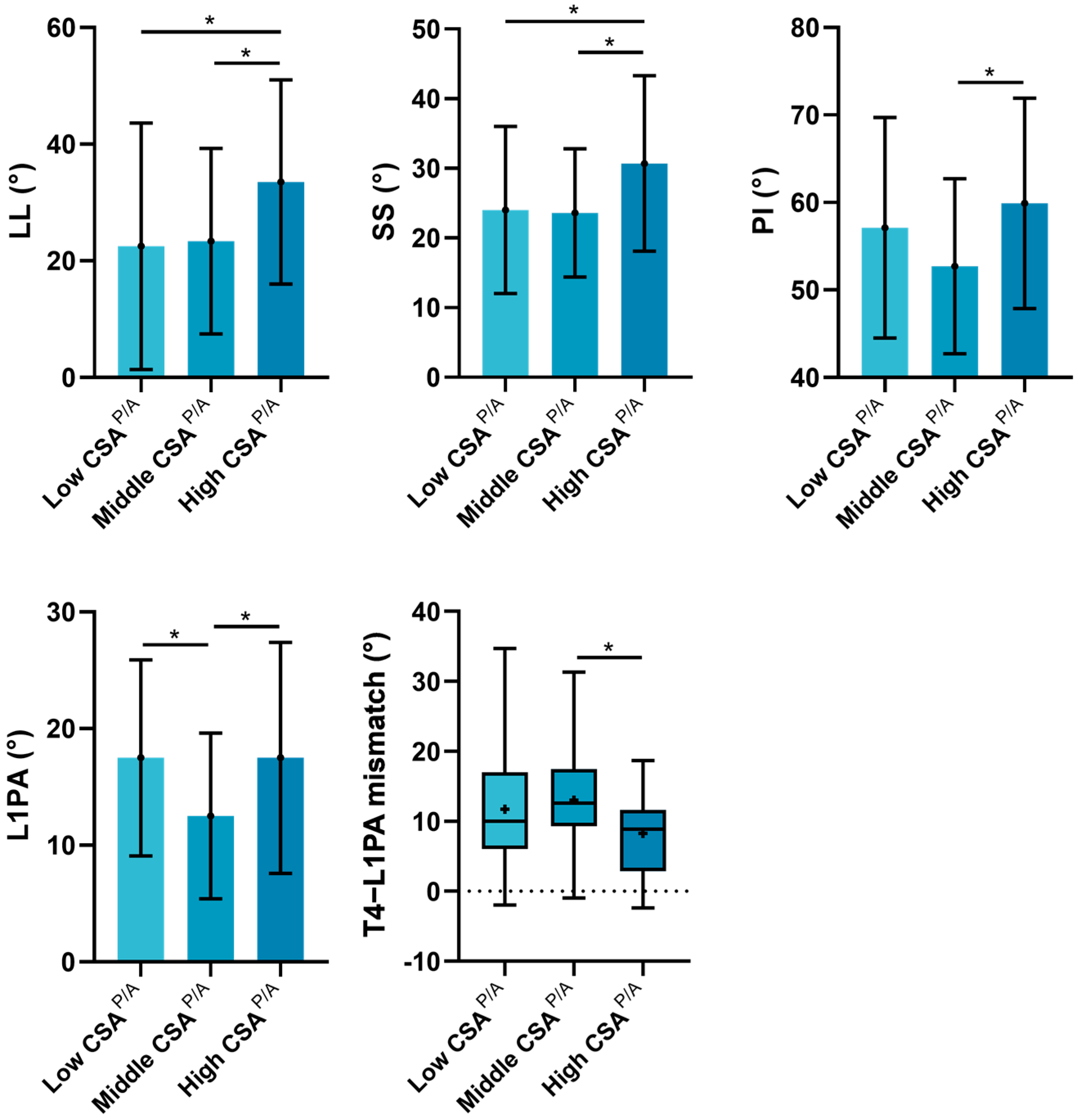
3.4. Subgroup Analyses of the Influence of CSAP/A on Sagittal Alignment

Subgroup analyses (Table 5 and Figure 4) revealed significant differences among the three groups in SS (p = 0.030), PI (p = 0.031), L1PA (p = 0.007), and T4PA–L1PA (p = 0.041), with a trend of difference in LL (p = 0.056) and PT (p = 0.056). Additionally, pairwise comparisons indicated that the high CSAP/A group exhibited significantly larger LL, SS, PI, and L1PA and smaller T4PA–L1PA compared to the middle and/or low CSAP/A groups. However, when sagittal alignment was adjusted for PI, no significant difference was observed in PI–LL and L1PA offset among the three groups.

4. Discussion

Extensive research has demonstrated that trunk muscle quality is closely related to sagittal malalignment in ASD patients [15,16,17,18,19,20]. However, most studies only focus on muscle impairment alone, neglecting the balance between trunk posterior and anterior muscles. Although trunk posterior and anterior muscles are anatomically antagonistic, their functional interaction is synergistic, working together to maintain an upright posture and counteract gravity [21]. Thus, this study aimed to explore the relationship between trunk muscle balance and sagittal malalignment in patients with ASD. We identified a linear relationship between trunk posterior muscles (CSAP) and sagittal alignment, while the ratio of posterior to anterior muscles (CSAP/A) showed a quadratic association with alignment. Notably, the quadratic model consistently outperformed the linear model, with higher r2 values and lower AIC values, emphasizing the superior predictive value of posterior–anterior muscles balance over individual muscle metrics. Furthermore, a higher ratio of posterior to anterior muscles (higher CSAP/A) in the spine is correlated to larger PI (and consequently, larger LL, SS, and L1PA).
The current literature emphasizes the role of posterior muscles in maintaining sagittal alignment and functional mobility [18,34,35,36]. We also observed that trunk posterior muscles exhibited a linear relationship with most sagittal alignment parameters, whereas anterior muscles alone showed no significant association (Table 2 and Table 3, Figure 2). Notably, after further exploration, we identified a quadratic relationship between the ratio of posterior–anterior muscles and most sagittal parameters (Table 3 and Figure 3), and this association was markedly stronger than the linear relationship observed with posterior muscles alone (Table 4). This suggests that the balance between posterior and anterior muscles may be more important than either posterior or anterior muscles alone in maintaining sagittal stability and alignment in ASD patients. Such findings align with evidence from other populations, such as patients with low back pain [37] and male students [38], identifying a similar significance of this balance in influencing lumbar lordosis across these groups. Additionally, extensor and contractor muscle balance contributes to the stability of other joints, such as the knee [39,40], elbow, and shoulder [41,42], reducing the risk of injury. Taken as a whole, our findings suggest that future study of muscle metrics must evaluate individual (e.g., anterior and posterior) muscles and the balance between the two.
Moreover, this quadratic association suggests a “U or inverted U-shaped” relationship between the ratio of posterior–anterior muscles and sagittal alignment parameters. We further explored the impact of different ranges of posterior–anterior-muscle ratio on sagittal alignment. Subgroup analysis revealed that a high posterior–anterior-muscle ratio is associated with significantly larger PI and consequently larger LL, SS, and L1PA (Table 5). A larger PI is usually accompanied by a deeper posterior concavity. It has been reported that muscles on the concave side may become shortened and stout with increased CSA, while those on the convex side may stretch, elongate, and exhibit a reduced CSA [43]. This phenomenon ultimately leads to an increased CSA ratio of the paraspinal muscles on the posterior concave side to the psoas major on the anterior convex side in cases of a large PI (Figure 5). Thus, in high-PI patients, using CSA alone may overestimate posterior muscle quantity and underestimate anterior muscle quality, a feature that could be problematic if one is studied in isolation [44,45]. To avoid these misinterpretations, MRI-based definitions of sarcopenia may need to be adjusted for PI. Furthermore, rehabilitation strategies could be personalized based on individual spinopelvic parameters. For instance, in patients with a large PI, posterior trunk muscles may exhibit compensatory hypertrophy due to structural adaptation rather than genuine functional strength. Therefore, for these high-PI patients, even when the CSA of posterior muscles appears not markedly reduced, targeted functional strengthening should remain a key focus in rehabilitation planning.
We also found that a high posterior–anterior-muscle ratio (posterior-dominant) is associated with smaller T4–L1PA mismatch (Table 5). A lower T4–L1PA mismatch has been associated with a decreased risk of mechanical failure [9]. Thus, posterior muscle dominance might be linked with better sagittal alignment compared to balanced or anterior-dominant muscle profiles. Such findings are supported by a prior study [35] that shows that trunk muscle ratios with posterior muscle predominance are associated with better physical performance and less back pain. However, a study on degenerative spondylolisthesis patients [46] presents opposing views: muscle trunk imbalance with predominance of posterior over anterior muscles was associated with functional disability. This discrepancy may originate from different outcome measures: we assessed sagittal alignment, while the opposing study used the Oswestry Disability Index (ODI) to evaluate functional disability. The ODI includes activities like lifting [47] and walking [48,49], which may bias results toward anterior muscle functions. When focusing on sagittal alignment in the present study, posterior muscle dominance means not only the inherent strength of the posterior muscles but also their dominant position in the synergistic interaction with the anterior muscles. This synergistic balance is crucial for maintaining sagittal alignment, as confirmed in a previous study [36]. Therefore, in rehabilitation exercises, focusing on strengthening trunk posterior muscles and maintaining posterior muscle dominance over anterior muscles may help reduce malalignment progression and enhance overall spinal stability in ASD patients. This may be a particularly effective strategy when employed in a “prehabilitation” manner [44].
There are limitations in the present study. First, due to the retrospective and observational design of this study, a causal relationship between muscle balance and spinal deformity cannot be inferred. This highlights the need for future prospective or interventional studies to confirm the hypotheses. In addition, although muscle characteristics are closely related to sagittal alignment, many other confounding factors (such as physical activity level, back pain, or prior spinal trauma) may also affect spinal alignment. However, these variables were not consistently available in our dataset, resulting in the failure to isolate their influence through multivariable analysis. Second, we predominantly focused on the key trunk posterior muscles (erector spinae and multifidus) and the key anterior muscle (psoas) when analyzing the relationship between posterior–anterior muscle balance and sagittal spinal malalignment. Most studies examining trunk muscle impairment in spine-related diseases have similarly focused mainly on these primary trunk posterior and/or anterior muscles [50,51,52]. However, other muscles also contribute to sagittal spinal stability, including additional posterior muscles such as the semispinalis [53] and quadratus lumborum [54] as well as anterior muscles like the rectus abdominis [55], external oblique [56], and transversus abdominis [57]. The CSAs of these muscles were not individually measured or included in our analysis due to the difficulty in accurately distinguishing and measuring them on MRI images [58] or because they were located outside the scanning range of lumbar MRI, which may limit the comprehensiveness of our findings. Future studies should expand the analysis to include additional muscle groups involved in trunk stabilization. Third, CSA does not capture muscle quality. While classifications such as the Goutallier classification have been used, there is no lumbar-specific measure of muscle quality. Future research should investigate more specific imaging modalities in the assessment of lumbar muscle quality [59]. Finally, although the paraspinal muscles at the L4 level have been shown to reliably reflect overall trunk muscle degeneration and stability [23,24], limiting measurements to this level may not fully capture muscle changes at higher deformity segments.

5. Conclusions

This study emphasizes the critical role of trunk muscle balance in sagittal alignment among ASD patients. We demonstrate that the posterior–anterior muscle balance, as captured by CSAP/A, correlates more strongly with sagittal alignment compared to individual muscle metrics. We encourage that future studies on lumbar musculature include this ratio as well as individual muscle parameters. Furthermore, the ratio between anterior and posterior muscles in the spine is correlated to PI (and consequently, LL and L1PA). Thus, MRI-based definitions of sarcopenia may need to be adjusted for PI.

Author Contributions

Conceptualization: D.H., Z.W., M.D., A.D., T.S., G.D., R.N.U., L.F.C., S.O.-S., F.L. and H.J.K.; data curation: D.H., Z.W., M.D., A.D., T.S., G.D., R.N.U., L.F.C., S.O.-S., F.L. and H.J.K.; methodology: D.H. and Z.W.; project administration: D.H. and Z.W.; roles/writing—original draft: D.H.; supervision: M.D. and F.L.; validation: D.H., Z.W., M.D., A.D., T.S., G.D., R.N.U., L.F.C., S.O.-S., F.L. and H.J.K.; writing—review and editing: M.D., Z.W., M.D., A.D., T.S., G.D., R.N.U., L.F.C., S.O.-S., H.J.K. and F.L. All authors have read and agreed to the published version of the manuscript.

Funding

This research received no external funding.

Institutional Review Board Statement

The study was conducted in accordance with the Declaration of Helsinki and approved by the Institutional Review Board (or Ethics Committee) of the Hospital for Special Surgery (HSS) (protocol code 2018-1599, approved on 17 August 2019).

Informed Consent Statement

Informed consent was waived due to the retrospective and observational nature of the research design.

Data Availability Statement

The data presented in this study are available on request from the corresponding author due to institutional policies and patient confidentiality regulations. Data sharing is restricted to protect sensitive patient information in compliance with ethical guidelines and IRB-approved protocols.

Acknowledgments

No direct funding was received for this study. However, this study used REDCap (Research Electronic Data Capture) hosted at Weill Cornell Medicine Clinical and Translational Science Center supported by the National Center for Advancing Translational Science of the National Institute of Health (NIH) under award number UL1TR002384.

Conflicts of Interest

The authors declare no conflicts of interest.

Abbreviations

The following abbreviations are used in this manuscript:
AICAkaike information criterion
ASDAdult spinal deformity
BMIBody mass index
CSACross-sectional area
CSAACross-sectional area of anterior muscles
CSAPCross-sectional area of posterior muscles
CSAP/ACross-sectional area ratio of posterior to anterior muscles
CTComputed tomography
FIFat infiltration
IQRInterquartile range
LLLumbar lordosis
L1PAL1 pelvic angle
MRIMagnetic resonance imaging
NSNo significance
PAPosteroanterior
PIPelvic incidence
PTPelvic tilt
SDStandard deviation
SSSacral slope
SVASagittal vertical axis
T4PAT4 pelvic angle
TKThoracic kyphosis

References

  1. Cerpa, M.; Lenke, L.G.; Fehlings, M.G.; Shaffrey, C.I.; Cheung, K.M.C.; Carreon, L.Y. Evolution and Advancement of Adult Spinal Deformity Research and Clinical Care: An Overview of the Scoli-RISK-1 Study. Glob. Spine J. 2019, 9 (Suppl. S1), 8s–14s. [Google Scholar] [CrossRef]
  2. Ton, A.; Hah, R.J. Postoperative complications in adult spinal deformity surgery: An overview of timing, risk factors, and management strategies. Semin. Spine Surg. 2023, 35, 101031. [Google Scholar] [CrossRef]
  3. Kim, W.J.; Shin, H.M.; Song, D.G.; Lee, J.W.; Park, K.Y.; Chang, S.H.; Bae, J.H.; Choy, W.S. Comparison of Clinical Outcomes and Complications of Primary and Revision Surgery Using a Combined Anterior and Posterior Approach in Patients with Adult Spinal Deformity and Sagittal Imbalance. Clin. Orthop. Surg. 2021, 13, 196–206. [Google Scholar] [CrossRef]
  4. Shin, J.J.; Kim, B.; Kang, J.; Choi, J.; Moon, B.J.; Ryu, D.S.; Yoon, S.H.; Chin, D.K.; Lee, J.K.; Kim, K.N.; et al. Clinical, Radiographic, and Genetic Analyses in a Population-Based Cohort of Adult Spinal Deformity in the Older Population. Neurospine 2021, 18, 608–617. [Google Scholar] [CrossRef]
  5. Lee, J.H.; Kim, K.T.; Lee, S.H.; Kang, K.C.; Oh, H.S.; Kim, Y.J.; Jung, H. Overcorrection of lumbar lordosis for adult spinal deformity with sagittal imbalance: Comparison of radiographic outcomes between overcorrection and undercorrection. Eur. Spine J. 2016, 25, 2668–2675. [Google Scholar] [CrossRef] [PubMed]
  6. Park, S.J.; Park, J.S.; Kang, D.H.; Kim, H.J.; Lim, Y.M.; Lee, C.S. Comparison of Surgical Burden, Radiographic and Clinical Outcomes According to the Severity of Baseline Sagittal Imbalance in Adult Spinal Deformity Patients. Neurospine 2024, 21, 721–731. [Google Scholar] [CrossRef] [PubMed]
  7. Pan, C.; Bourghli, A.; Larrieu, D.; Boissiere, L.; Pizones, J.; Alanay, A.; PelIise, F.; Kleinstück, F.; Obeid, I. Sagittal alignment of diverse mechanical complications following adult spinal deformity surgery. Eur. Spine J. 2024, 33, 1857–1867. [Google Scholar] [CrossRef] [PubMed]
  8. Muramoto, A.; Imagama, S.; Ito, Z.; Hirano, K.; Ishiguro, N.; Hasegawa, Y. Spinal sagittal balance substantially influences locomotive syndrome and physical performance in community-living middle-aged and elderly women. J. Orthop. Sci. 2016, 21, 216–221. [Google Scholar] [CrossRef]
  9. Hills, J.; Mundis, G.M.; Klineberg, E.O.; Smith, J.S.; Line, B.; Gum, J.L.; Protopsaltis, T.S.; Hamilton, D.K.; Soroceanu, A.; Eastlack, R.; et al. The T4-L1-Hip Axis: Sagittal Spinal Realignment Targets in Long-Construct Adult Spinal Deformity Surgery: Early Impact. J. Bone Jt. Surg. Am. Vol. 2024, 106, e48. [Google Scholar] [CrossRef]
  10. Makhni, M.C.; Shillingford, J.N.; Laratta, J.L.; Hyun, S.J.; Kim, Y.J. Restoration of Sagittal Balance in Spinal Deformity Surgery. J. Korean Neurosurg. Soc. 2018, 61, 167–179. [Google Scholar] [CrossRef]
  11. Yurube, T. Impacts of Adult Spinal Deformity Surgery on Coronal Malalignment: Commentary on “Outcomes of Surgical Treatment for Patients with Mild Scoliosis and Age-Appropriate Sagittal Alignment with Minimum 2-Year Follow-up”. Neurospine 2023, 20, 849–851. [Google Scholar] [CrossRef] [PubMed]
  12. Bae, J.; Sathe, A.; Lee, S.M.; Theologis, A.A.; Deviren, V.; Lee, S.H. Correlation of Paraspinal Muscle Mass with Decompensation of Sagittal Adult Spinal Deformity After Setting of Fatigue Post 10-Minute Walk. Neurospine 2021, 18, 495–503. [Google Scholar] [CrossRef]
  13. Lee, J.K.; Hyun, S.J.; Kim, K.J. Reciprocal Changes in the Whole-Body Following Realignment Surgery in Adult Spinal Deformity. Asian Spine J. 2022, 16, 958–967. [Google Scholar] [CrossRef] [PubMed]
  14. Cirillo Totera, J.I.; Fleiderman Valenzuela, J.G.; Garrido Arancibia, J.A.; Pantoja Contreras, S.T.; Beaulieu Lalanne, L.; Alvarez-Lemos, F.L. Sagittal balance: From theory to clinical practice. EFORT Open Rev. 2021, 6, 1193–1202. [Google Scholar] [CrossRef]
  15. Wang, G.; Li, Y.; Zhang, C.; Liu, P.; Sun, J. Erector Spinae Atrophy Correlates with Global Sagittal Imbalance and Postoperative Proximal Junctional Kyphosis Incidence in Lumbar Degenerative Kyphosis. Asian Spine J. 2024, 18, 50–57. [Google Scholar] [CrossRef]
  16. Chen, C.; Yang, S.; Tang, Y.; Yu, X.; Chen, C.; Zhang, C.; Luo, F. Correlation between strength/endurance of paraspinal muscles and sagittal parameters in patients with degenerative spinal deformity. BMC Musculoskelet. Disord. 2023, 24, 643. [Google Scholar] [CrossRef] [PubMed]
  17. Kim, W.J.; Shin, H.M.; Lee, J.S.; Song, D.G.; Lee, J.W.; Chang, S.H.; Park, K.Y.; Choy, W.S. Sarcopenia and Back Muscle Degeneration as Risk Factors for Degenerative Adult Spinal Deformity with Sagittal Imbalance and Degenerative Spinal Disease: A Comparative Study. World Neurosurg. 2021, 148, e547–e555. [Google Scholar] [CrossRef]
  18. Chen, C.; Yang, S.; Tang, Y.; Zhang, C.; Yu, X.; Li, K.; Chen, C.; Dai, W.; Rong, Z.; Luo, F. Isokinetic strength assessment of trunk muscle and its relationship with spinal-pelvic parameters in patients with degenerative spinal deformity. J. Back Musculoskelet. Rehabil. 2023, 36, 931–940. [Google Scholar] [CrossRef]
  19. Kobayashi, T.; Chiba, H.; Jimbo, S.; Senoo, I.; Shimizu, M.; Atsuta, Y.; Ito, H.; Sugisawa, H.; Sugawara, T.; Habaguchi, T. Clinical, physical, and radiographic analyses of lumbar degenerative kyphosis and spondylolisthesis among community-based cohort. Eur. Spine J. 2016, 25, 2384–2389. [Google Scholar] [CrossRef]
  20. Ryu, D.S.; Shinn, J.K.; Kim, B.W.; Moon, B.J.; Ha, Y.; Lee, J.K.; Kim, K.N.; Chin, D.K.; Yoon, S.H. Prospective Observational Cohort Study of Health-related Quality of Life: Marked Adult Sagittal Deformity (ASD) in Comparison with Mild to Moderate ASD. Spine 2019, 44, 1723–1730. [Google Scholar] [CrossRef]
  21. Cholewicki, J.; Panjabi, M.M.; Khachatryan, A. Stabilizing function of trunk flexor-extensor muscles around a neutral spine posture. Spine 1997, 22, 2207–2212. [Google Scholar] [CrossRef]
  22. Tanishima, S.; Hagino, H.; Matsumoto, H.; Tanimura, C.; Nagashima, H. Relationship among Osteoporosis, Sarcopenia, Locomotive Syndrome, and Spinal Kyphosis in Older Individuals Living in a Local Mountain Area. Asian Spine J. 2023, 17, 1074–1081. [Google Scholar] [CrossRef]
  23. Tomov, M.; Alvi, M.A.; Elminawy, M.; Currier, B.; Yaszemski, M.; Nassr, A.; Huddleston, P.; Sebastian, A.; Bydon, M.; Freedman, B. An Objective and Reliable Method for Identifying Sarcopenia in Lumbar Spine Surgery Patients: Using Morphometric Measurements on Computed Tomography Imaging. Asian Spine J. 2020, 14, 814–820. [Google Scholar] [CrossRef] [PubMed]
  24. Jun, H.S.; Kim, J.H.; Ahn, J.H.; Chang, I.B.; Song, J.H.; Kim, T.H.; Park, M.S.; Chan Kim, Y.; Kim, S.W.; Oh, J.K.; et al. The Effect of Lumbar Spinal Muscle on Spinal Sagittal Alignment: Evaluating Muscle Quantity and Quality. Neurosurgery 2016, 79, 847–855. [Google Scholar] [CrossRef] [PubMed]
  25. Lee, D.; Kuroki, T.; Nagai, T.; Kawano, K.; Higa, K.; Kurogi, S.; Hamanaka, H.; Chosa, E. Sarcopenia, Ectopic Fat Infiltration into the Lumbar Paravertebral Muscles, and Lumbo-Pelvic Deformity in Older Adults Undergoing Lumbar Surgery. Spine 2022, 47, E46–E57. [Google Scholar] [CrossRef] [PubMed]
  26. Banno, T.; Yamato, Y.; Hasegawa, T.; Kobayashi, S.; Togawa, D.; Oe, S.; Mihara, Y.; Kurosu, K.; Yamamoto, N.; Matsuyama, Y. Assessment of the Cross-Sectional Areas of the Psoas Major and Multifidus Muscles in Patients with Adult Spinal Deformity: A Case-Control Study. Clin. Spine Surg. 2017, 30, E968–E973. [Google Scholar] [CrossRef]
  27. Xia, W.; Fu, H.; Zhu, Z.; Liu, C.; Wang, K.; Xu, S.; Liu, H. Association between back muscle degeneration and spinal-pelvic parameters in patients with degenerative spinal kyphosis. BMC Musculoskelet. Disord. 2019, 20, 454. [Google Scholar] [CrossRef]
  28. Paalanne, N.P.; Korpelainen, R.; Taimela, S.P.; Remes, J.; Salakka, M.; Karppinen, J.I. Reproducibility and reference values of inclinometric balance and isometric trunk muscle strength measurements in Finnish young adults. J. Strength Cond. Res. 2009, 23, 1618–1626. [Google Scholar] [CrossRef]
  29. Hyytiäinen, K.; Salminen, J.J.; Suvitie, T.; Wickström, G.; Pentti, J. Reproducibility of nine tests to measure spinal mobility and trunk muscle strength. Scand. J. Rehabil. Med. 1991, 23, 3–10. [Google Scholar] [CrossRef]
  30. Ropponen, A.; Videman, T.; Battié, M.C. The reliability of paraspinal muscles composition measurements using routine spine MRI and their association with back function. Man. Ther. 2008, 13, 349–356. [Google Scholar] [CrossRef]
  31. Kaplan, S.J.; Zhao, K.L.; Koren, M.; Bentov, I.; Reed, M.J.; Pham, T.N. Thresholds and Mortality Associations of Paraspinous Muscle Sarcopenia in Older Trauma Patients. JAMA Surg. 2020, 155, 662–664. [Google Scholar] [CrossRef]
  32. Vadhera, A.S.; Sachdev, R.; Andrade, N.S.; Ren, M.; Zhang, B.; Kebaish, K.M.; Cohen, D.B.; Skolasky, R.L.; Neuman, B.J. Predicting major complications and discharge disposition after adult spinal deformity surgery. Spine J. 2024, 24, 325–329. [Google Scholar] [CrossRef]
  33. Snipes, M.; Taylor, D.C. Model selection and Akaike Information Criteria: An example from wine ratings and prices. Wine Econ. Policy 2014, 3, 3–9. [Google Scholar] [CrossRef]
  34. Liang, C.; Sun, J.; Cui, X.; Jiang, Z.; Zhang, W.; Li, T. Spinal sagittal imbalance in patients with lumbar disc herniation: Its spinopelvic characteristics, strength changes of the spinal musculature and natural history after lumbar discectomy. BMC Musculoskelet. Disord. 2016, 17, 305. [Google Scholar] [CrossRef] [PubMed]
  35. Takenaka, H.; Ikemoto, T.; Suzuki, J.; Inoue, M.; Arai, Y.C.; Ushida, T.; Deie, M.; Kamiya, M. Association between Trunk Muscle Strength, Lumbar Spine Bone Mineral Density, Lumbar Scoliosis Angle, and Skeletal Muscle Volume and Locomotive Syndrome in Elderly Individuals: A Dual-Energy X-ray Absorptiometry Study. Spine Surg. Relat. Res. 2020, 4, 164–170. [Google Scholar] [CrossRef] [PubMed]
  36. Takahashi, S.; Hoshino, M.; Ohyama, S.; Hori, Y.; Yabu, A.; Kobayashi, A.; Tsujio, T.; Kotake, S.; Nakamura, H. Relationship of back muscle and knee extensors with the compensatory mechanism of sagittal alignment in a community-dwelling elderly population. Sci. Rep. 2021, 11, 2179. [Google Scholar] [CrossRef]
  37. Kim, H.J.; Chung, S.; Kim, S.; Shin, H.; Lee, J.; Kim, S.; Song, M.Y. Influences of trunk muscles on lumbar lordosis and sacral angle. Eur. Spine J. 2006, 15, 409–414. [Google Scholar] [CrossRef]
  38. Marijančić, V.; Peharec, S.; Starčević-Klasan, G.; Grubić Kezele, T. Gender Differences in the Relationship between Physical Activity, Postural Characteristics and Non-Specific Low Back Pain in Young Adults. J. Funct. Morphol. Kinesiol. 2024, 9, 189. [Google Scholar] [CrossRef] [PubMed]
  39. Lee, J.W.Y.; Mok, K.M.; Chan, H.C.K.; Yung, P.S.H.; Chan, K.M. Eccentric hamstring strength deficit and poor hamstring-to-quadriceps ratio are risk factors for hamstring strain injury in football: A prospective study of 146 professional players. J. Sci. Med. Sport 2018, 21, 789–793. [Google Scholar] [CrossRef]
  40. Heinert, B.L.; Collins, T.; Tehan, C.; Ragan, R.; Kernozek, T.W. Effect of Hamstring-to-quadriceps Ratio on Knee Forces in Females During Landing. Int. J. Sports Med. 2021, 42, 264–269. [Google Scholar] [CrossRef]
  41. Lin, Y.C.; Thompson, A.; Kung, J.T.; Chieh, L.W.; Chou, S.W.; Lin, J.C. Functional isokinetic strength ratios in baseball players with injured elbows. J. Sport Rehabil. 2010, 19, 21–29. [Google Scholar] [CrossRef]
  42. Kochanowicz, A.; Niespodziński, B.; Mieszkowski, J.; Kochanowicz, K.; Sawczyn, S. The effect of gymnastic training on muscle strength and co-activation during isometric elbow and glenohumeral flexion/extension. J. Sports Med. Phys. Fit. 2018, 58, 966–973. [Google Scholar] [CrossRef] [PubMed]
  43. Kim, H.; Lee, C.K.; Yeom, J.S.; Lee, J.H.; Cho, J.H.; Shin, S.I.; Lee, H.J.; Chang, B.S. Asymmetry of the cross-sectional area of paravertebral and psoas muscle in patients with degenerative scoliosis. Eur. Spine J. 2013, 22, 1332–1338. [Google Scholar] [CrossRef]
  44. Hirase, T.; Lovecchio, F.C.; Allen, M.R.J.; Achebe, C.C.; Mazzucco, M.; Uzzo, R.N.; Kazarian, G.S.; Asada, T.; Nakarai, H.; Subramanian, T.; et al. Preoperative Physical Therapy is Associated with Decreased Length of Stay and Improved Postoperative Mobility in Patients with Sarcopenia Undergoing Adult Spinal Deformity Surgery. Spine 2025, 50, 172–178. [Google Scholar] [CrossRef] [PubMed]
  45. Lidar, S.; Salame, K.; Chua, M.; Khashan, M.; Ofir, D.; Grundstein, A.; Hochberg, U.; Lidar, Z.; Regev, G.J. Sarcopenia Is an Independent Risk Factor for Subsequent Osteoporotic Vertebral Fractures Following Percutaneous Cement Augmentation in Elderly Patients. J. Clin. Med. 2022, 11, 5778. [Google Scholar] [CrossRef]
  46. Nava-Bringas, T.I.; Ramírez-Mora, I.; Coronado-Zarco, R.; Macías-Hernández, S.I.; Cruz-Medina, E.; Arellano-Hernández, A.; Hern Ndez-López, M.; León-Hernández, S.R. Association of strength, muscle balance, and atrophy with pain and function in patients with degenerative spondylolisthesis. J. Back Musculoskelet. Rehabil. 2014, 27, 371–376. [Google Scholar] [CrossRef]
  47. Deering, R.E.; Senefeld, J.W.; Pashibin, T.; Neumann, D.A.; Hunter, S.K. Muscle function and fatigability of trunk flexors in males and females. Biol. Sex Differ. 2017, 8, 12. [Google Scholar] [CrossRef] [PubMed]
  48. Sullivan, M.S. Back support mechanisms during manual lifting. Phys. Ther. 1989, 69, 38–45. [Google Scholar] [CrossRef]
  49. Sturdy, J.T.; Rizeq, H.N.; Silder, A.; Sessoms, P.H.; Silverman, A.K. Concentric and eccentric hip musculotendon work depends on backpack loads and walking slopes. J. Biomech. 2024, 163, 111942. [Google Scholar] [CrossRef]
  50. Muellner, M.; Haffer, H.; Chiapparelli, E.; Dodo, Y.; Shue, J.; Tan, E.T.; Zhu, J.; Pumberger, M.; Sama, A.A.; Cammisa, F.P.; et al. Fat infiltration of the posterior paraspinal muscles is inversely associated with the fat infiltration of the psoas muscle: A potential compensatory mechanism in the lumbar spine. BMC Musculoskelet. Disord. 2023, 24, 846. [Google Scholar] [CrossRef]
  51. Choi, M.K.; Kim, S.B.; Park, C.K.; Malla, H.P.; Kim, S.M. Cross-Sectional Area of the Lumbar Spine Trunk Muscle and Posterior Lumbar Interbody Fusion Rate: A Retrospective Study. Clin. Spine Surg. 2017, 30, E798–E803. [Google Scholar] [CrossRef] [PubMed]
  52. Zhu, W.P.; Huang, Y.; Hu, P.; Lin, W. Lumbar Extensor and Flexor Muscle Structural Changes in Young Female Nurses with Chronic Bilateral Non-Specific Low Back Pain: A Case-Control Study. Discov. Med. 2023, 35, 444–450. [Google Scholar] [CrossRef]
  53. Kim, P.; Murata, H.; Kurokawa, R.; Takaishi, Y.; Asakuno, K.; Kawamoto, T. Myoarchitectonic spinolaminoplasty: Efficacy in reconstituting the cervical musculature and preserving biomechanical function. J. Neurosurg. Spine 2007, 7, 293–304. [Google Scholar] [CrossRef]
  54. Hides, J.A.; Lambrecht, G.; Richardson, C.A.; Stanton, W.R.; Armbrecht, G.; Pruett, C.; Damann, V.; Felsenberg, D.; Belavý, D.L. The effects of rehabilitation on the muscles of the trunk following prolonged bed rest. Eur. Spine J. 2011, 20, 808–818. [Google Scholar] [CrossRef] [PubMed]
  55. Zmyślna, A.; Żurawski, A.; Rosiński, T.; Pogorzelska, J.; Śliwiński, Z.; Śliwiński, G.; Kiebzak, W. The Relationship Between the Shape of the Spine and the Width of Linea Alba in Children Aged 6-9 Years. Case-Control Study. Front. Pediatr. 2022, 10, 839171. [Google Scholar] [CrossRef] [PubMed]
  56. Banno, T.; Yamato, Y.; Nojima, O.; Hasegawa, T.; Yoshida, G.; Arima, H.; Oe, S.; Ushirozako, H.; Yamada, T.; Ide, K.; et al. Comparison of the postoperative changes in trunk and lower extremity muscle activities between patients with adult spinal deformity and age-matched controls using surface electromyography. Spine Deform. 2022, 10, 141–149. [Google Scholar] [CrossRef]
  57. Swanenburg, J.; Easthope, C.A.; Meinke, A.; Langenfeld, A.; Green, D.A.; Schweinhardt, P. Lunar and mars gravity induce similar changes in spinal motor control as microgravity. Front. Physiol. 2023, 14, 1196929. [Google Scholar] [CrossRef]
  58. Bao, H.; Moal, B.; Vira, S.; Bronsard, N.; Amabile, C.; Errico, T.; Schwab, F.; Skalli, W.; Dubousset, J.; Lafage, V. Spino-femoral muscles affect sagittal alignment and compensatory recruitment: A new look into soft tissues in adult spinal deformity. Eur. Spine J. 2020, 29, 2998–3005. [Google Scholar] [CrossRef]
  59. Tan, E.T.; Serrano, K.C.; Bhatti, P.; Pishgar, F.; Vanderbeek, A.M.; Milani, C.J.; Sneag, D.B. Quantitative MRI Differentiates Electromyography Severity Grades of Denervated Muscle in Neuropathy of the Brachial Plexus. J. Magn. Reson. Imaging JMRI 2022, 56, 1104–1115. [Google Scholar] [CrossRef]
Figure 1. Flowchart of patient inclusion, exclusion, and analysis process.
Jcm 14 03293 g001
Figure 2. Scatter plot of the relationship between CSAP and sagittal alignment parameters with linear and quadratic fit curves.
Jcm 14 03293 g002
Figure 3. Scatter plot of the relationship between CSAP/A and sagittal alignment parameters with linear and quadratic fit curves.
Jcm 14 03293 g003
Figure 4. Comparison of sagittal alignment parameters (LL, SS, PI, L1PA, and T4–L1PA mismatch) among low, middle, and high CSAP/A subgroups. *, p < 0.05 between the two groups.
Jcm 14 03293 g004
Figure 5. Illustration of asymmetric muscular morphological changes associated with large pelvic incidence (PI). A large PI typically results in increased lumbar lordosis (LL), which in turn leads to elongation and thinning of the psoas major on the convex side, reducing its cross-sectional area (CSA). Simultaneously, the paraspinal muscles on the posterior concave side become shortened and thickened, increasing their CSA. This asymmetric adaptation ultimately results in an elevated CSA ratio of the posterior concave paraspinal muscles to the anterior convex psoas major (CSAP/A).
Jcm 14 03293 g005
Table 1. Measurements of trunk posterior and anterior muscles, and spinal alignment for the 112 adult patients with sagittal spinal deformity.
Variable *Mean (SD)
Age (year)66.3 ± 9.8
Sex (M/F) (no.)33:79
BMI (kg/m2) &27.7 ± 6.0
Smoker (current or former), n(%)28(25.0%)
Diabetes, n(%)5(4.5%)
Tumor, n(%)13(11.6%)
Hypertension, n(%)45(40.1%)
Osteoporosis, n(%)20(17.9%)
Dyslipidemia, n(%)32(28.6%)
Coronary artery disease, n(%)11(9.8%)
Peripheral neuropathy, n(%)1(0.9%)
Autoimmune, n(%)4(3.6%)
Anxiety, n(%)7(6.3%)
Spinal alignment parameters
   Max Cobb (°)38.7 ± 19.9
   TK (T4–T12) (°)25.6 ± 13.9
   LL (L1–S1) (°)25.1 ± 18.5
   SS (°)25.1 ± 11.1
   PI (°)55.6 ± 11.6
   PI–LL (°)30.5 ± 15.1
   PT (°)30.5 ± 7.2
   SVA (mm)87.7 ± 69.8
   L1PA (°)15.1 ± 8.5
   L1PA offset (°)8.3 ± 7.3
   T4PA (°)26.8 ± 9.6
   T4–L1PA mismatch (°)11.6 ± 7.7
Trunk muscle assessment #
   CSAP3707.9 ± 1074.5
   CSAA2148.7 ± 718.4
   CSAP/A1.83 ± 0.59
Abbreviations: * Values are given as the mean and SD unless otherwise noted; SD, standard deviation; M, male; F, female; BMI, body mass index; &, the data of four patients are missing; TK, thoracic kyphosis; LL, lumbar lordosis; SS, sacral slope; PI, pelvic incidence; PT, pelvic tilt; SVA, sagittal vertical axis; L1PA, L1 pelvic angle; L1PA offset, calculated as L1PA-(0.5 × PI − 21°); T4PA, T4 pelvic angle; T4–L1PA mismatch, calculated as T4PA–L1PA; #, trunk muscles were evaluated at L4 level; CSA, the cross-sectional area; CSAP, CSA of posterior muscles (erector spinae and multifidus); CSAA, CSA of anterior muscles (psoas); and CSAP/A, the CSA ratio of trunk posterior to anterior muscles.
Table 2. Simple linear regression of the trunk muscles and radiological parameters of spinal alignment.
AssessmentMax CobbTKLLSSPIPI–LLPTSVAL1PAL1PA OffsetT4PAT4–L1PA Mismatch
CSAPr2 = 0.100r2 = 0.009r2 = 0.021r2 = 0.057r2 = 0.011r2 = 0.009r2 = 0.043r2 = 0.005r2 = 0.000r2 = 0.010r2 = 0.032r2 = 0.044
p = 0.001p = 0.329p = 0.124p = 0.011p = 0.266p = 0.308p = 0.028p = 0.464p = 0.901p = 0.301p = 0.057p = 0.027
CSAAr2 = 0.210r2 = 0.024r2 = 0.000r2 = 0.004r2 = 0.002r2 = 0.003r2 = 0.001r2 = 0.011r2 = 0.000r2 = 0.001r2 = 0.007r2 = 0.011
p = 0.000p = 0.101p = 0.877p = 0.502p = 0.646p = 0.590p = 0.713p = 0.268p = 0.981p = 0.735p = 0.383p = 0.270
CSAP/Ar2 = 0.029r2 = 0.015r2 = 0.042r2 = 0.057r2 = 0.016r2 = 0.023r2 = 0.029r2 = 0.000r2 = 0.001r2 = 0.003r2 = 0.010r2 = 0.028
p = 0.073p = 0.196p = 0.031p = 0.011p = 0.187p = 0.108p = 0.074p = 0.859p = 0.687p = 0.563p = 0.292p = 0.080
Abbreviations: TK, thoracic kyphosis; LL, lumbar lordosis; SS, sacral slope; PI, pelvic incidence; PT, pelvic tilt; SVA, sagittal vertical axis; L1PA offset, calculated as L1PA-(0.5 × PI − 21°); T4PA, T4 pelvic angle; T4–L1PA mismatch, calculated as T4PA–L1PA; CSA, the cross-sectional area; CSAP, CSA of posterior muscles (erector spinae and multifidus); CSAA, CSA of anterior muscles (psoas); CSAP/A, the CSA ratio of trunk posterior to anterior muscles. The correlations with significance (p < 0.05) are highlighted with bold; The darker the red background, the stronger the correlation (the higher r² value).
Table 3. Quadratic regression of the trunk muscles and radiological parameters of spinal alignment.
AssessmentMax CobbTKLLSSPIPI–LLPTSVAL1PAL1PA OffsetT4PAT4–L1PA Mismatch
CSAPr2 = 0.100r2 = 0.009r2 = 0.024r2 = 0.058r2 = 0.012r2 = 0.016r2 = 0.043r2 = 0.042r2 = 0.029r2 = 0.042r2 = 0.048r2 = 0.045
p = 0.003p = 0.608p = 0.261p = 0.038p = 0.525p = 0.404p = 0.089p = 0.096p = 0.202p = 0.096p = 0.068p = 0.083
CSAAr2 = 0.212r2 = 0.024r2 = 0.000r2 = 0.005r2 = 0.003r2 = 0.005r2 = 0.001r2 = 0.013r2 = 0.009r2 = 0.007r2 = 0.008r2 = 0.016
p = 0.000p = 0.259p = 0.974p = 0.762p = 0.841p = 0.768p = 0.931p = 0.481p = 0.623p = 0.664p = 0.658p = 0.423
CSAP/Ar2 = 0.050r2 = 0.048r2 = 0.056r2 = 0.134r2 = 0.096r2 = 0.028r2 = 0.029r2 = 0.001r2 = 0.114r2 = 0.031r2 = 0.018r2 = 0.094
p = 0.060p = 0.070p = 0.044p = 0.000p = 0.004p = 0.209p = 0.196p = 0.945p = 0.001p = 0.184p = 0.376p = 0.005
Abbreviations: TK, thoracic kyphosis; LL, lumbar lordosis; SS, sacral slope; PI, pelvic incidence; PT, pelvic tilt; SVA, sagittal vertical axis; L1PA offset, calculated as L1PA-(0.5 × PI − 21°); T4PA, T4 pelvic angle; T4–L1PA mismatch, calculated as T4PA–L1PA; CSA, the cross-sectional area; CSAP, CSA of posterior muscles (erector spinae and multifidus); CSAA, CSA of anterior muscles (psoas); CSAP/A, the CSA ratio of trunk posterior to anterior muscles. The correlations with significance (p < 0.05) are highlighted with bold; The darker the red background, the stronger the correlation (the higher r² value).
Table 4. Comparison of AIC values between the linear model (CSAP) and quadratic model (CSAP/A) in relation to spinal alignment parameters.
Regression ModelMuscle ParameterMax CobbTKLLSSPIPI–LLPTSVAL1PAL1PA OffsetT4PAT4–L1PA Mismatch
LinearCSAP867.4 798.0 859.5 742.3 756.2 816.5 646.3 1159.3 687.2 651.6 711.0 662.4
QuadraticCSAP/A875.4 795.5 857.5 734.8 748.1 816.3 649.9 1161.7 675.6 651.2 714.7 658.4
Decreased AIC value from linear to quadratic model−8.0 2.5 2.0 7.4 8.0 0.2 −3.6 −2.4 11.6 0.4 −3.7 4.1
Abbreviations: AIC, Akaike information criterion; TK, thoracic kyphosis; LL, lumbar lordosis; SS, sacral slope; PI, pelvic incidence; PT, pelvic tilt; SVA, sagittal vertical axis; L1PA offset, calculated as L1PA-(0.5 × PI − 21°); T4PA, L4 pelvic angle; T4–L1PA mismatch, calculated as T4PA–L1PA; CSAP, CSA of posterior muscles (erector spinae and multifidus); CSAP/A, the CSA ratio of trunk posterior to anterior muscles.
Table 5. Comparisons of spinal alignment parameters in subdividing three CSAP/A groups.
ParametersCSAP/Ap-Value
Low Group
(n = 38)
Middle Group
(n = 52)
High Group
(n = 22)
TotalL Versus ML Versus HM Versus H
Max Cobb (°)36.3 (17.2, 49.3)36.5 (25.3, 52.5)47.5 (34.2, 55.0)0.207 $NSNSNS
TK (°) #30.8 ± 15.927.8 ± 12.726.6 ± 13.20.459 *NSNSNS
LL (°)22.5 ± 21.123.4 ± 15.933.5 ± 17.50.056 *NS0.0260.031
SS (°)24.0 ± 12.023.6 ± 9.230.7 ± 12.60.030 *NS0.0230.012
PI (°)57.1 ± 12.652.7 ± 10.059.9 ± 12.00.031 *NSNS0.015
PI–LL (°)30.7 (21.5, 41.9)29.4 (19.9, 37.8)21.4 (17.6, 34.3)0.226 $NSNSNS
PT (°) #31.0 (26.1, 38.6)27.7 (24.9, 34.0)29.0 (25.2, 32.9)0.056 $NSNSNS
SVA (mm) #85.5 (48.9, 136.7)70.4 (36.7, 112.6)66.2 (34.1, 137.1)0.790 $NSNSNS
L1PA (°)17.5 ± 8.412.5 ± 7.117.5 ± 9.90.007 *0.005NS0.018
L1PA offset (°) #9.6 (3.9, 16.0)6.5 (3.0, 11.8)6.4 (3.2, 10.5)0.195 $NSNSNS
T4PA (°) #28.9 (22.0, 36.3)24.1 (18.9, 31.3)23.3 (18.6, 30.6)0.147 $NSNSNS
T4–L1PA mismatch (°) #10.0 (6.0, 17.0)12.6 (9.3, 17.5)8.9 (2.9, 11.7)0.041 $NSNS0.040
Abbreviations: Mean and standard deviation is shown except for #, where median and interquartile range (Q1–Q3) are given; CSA, the cross-sectional area; CSAP/A, the CSA ratio of trunk posterior to anterior muscles; TK, thoracic kyphosis; LL, lumbar lordosis; SS, sacral slope; PI, pelvic incidence; PT, pelvic tilt; SVA, sagittal vertical axis; L1PA offset, calculated as L1PA-(0.5 × PI − 21°); T4PA, L4 pelvic angle; T4–L1PA mismatch, calculated as T4PA–L1PA; L, low group; M, middle group; H, high group; *, a one-way analysis of variance (ANOVA); $, Kruskal–Wallis test; and NS, no significance. Significances (p < 0.05) are highlighted with bold.
Disclaimer/Publisher’s Note: The statements, opinions and data contained in all publications are solely those of the individual author(s) and contributor(s) and not of MDPI and/or the editor(s). MDPI and/or the editor(s) disclaim responsibility for any injury to people or property resulting from any ideas, methods, instructions or products referred to in the content.

