Next Article in Journal
Current Concepts of Cervical Spine Alignment, Sagittal Deformity, and Cervical Spine Surgery
Previous Article in Journal
Myocardial Metabolism in Heart Failure with Preserved Ejection Fraction
 
 
jcm-logo
Article Menu

Article Menu

Article

Validation of SeptiCyte RAPID to Discriminate Sepsis from Non-Infectious Systemic Inflammation

by
Robert Balk
1,*,
Annette M. Esper
2,
Greg S. Martin
2,
Russell R. Miller III
3,
Bert K. Lopansri
4,5,
John P. Burke
4,5,
Mitchell Levy
6,
Steven Opal
6,
Richard E. Rothman
7,
Franco R. D’Alessio
7,
Venkataramana K. Sidhaye
7,
Neil R. Aggarwal
8,
Jared A. Greenberg
1,
Mark Yoder
1,
Gourang Patel
1,
Emily Gilbert
9,
Jorge P. Parada
9,
Majid Afshar
10,
Jordan A. Kempker
2,
Tom van der Poll
11,
Marcus J. Schultz
11,
Brendon P. Scicluna
12,13,
Peter M. C. Klein Klouwenberg
14,
Janice Liebler
15,16,
Emily Blodget
15,16,
Santhi Kumar
15,16,
Krupa Navalkar
17,
Thomas D. Yager
17,*,
Dayle Sampson
17,
James T. Kirk
17,
Silvia Cermelli
17,
Roy F. Davis
17 and
Richard B. Brandon
17,*
add Show full author list remove Hide full author list
1
Rush Medical College and Rush University Medical Center, Chicago, IL 60612, USA
2
Grady Memorial Hospital and Emory University School of Medicine, Atlanta, GA 30322, USA
3
FirstHealth of the Carolinas, Pinehurst, NC 28374, USA
4
Intermountain Medical Center, Murray, UT 84107, USA
5
School of Medicine, University of Utah, Salt Lake City, UT 84132, USA
6
Warren Alpert Medical School, Brown University, Providence, RI 02912, USA
7
School of Medicine, Johns Hopkins University, Baltimore, MD 21205, USA
8
Anschutz Medical Campus, University of Colorado, Denver, CO 80045, USA
9
Loyola University Medical Center, Maywood, IL 60153, USA
10
School of Medicine and Public Health, University of Wisconsin, Madison, WI 53705, USA
11
Amsterdam UMC, University of Amsterdam, 1105 AZ Amsterdam, The Netherlands
12
Centre for Molecular Medicine and Biobanking, University of Malta, Msida MSD 2080, Malta
13
Department of Applied Biomedical Science, Faculty of Health Sciences, Mater Dei Hospital, University of Malta, Msida MSD 2080, Malta
14
Fundashon Mariadal, Kralendijk, Bonaire, Netherlands Antilles
15
Keck Hospital of University of Southern California (USC), Los Angeles, CA 90033, USA
16
Los Angeles General Medical Center, Los Angeles, CA 90033, USA
17
Immunexpress Inc., Seattle, DC 98109, USA
*
Authors to whom correspondence should be addressed.
J. Clin. Med. 2024, 13(5), 1194; https://doi.org/10.3390/jcm13051194
Submission received: 1 January 2024 / Revised: 8 February 2024 / Accepted: 18 February 2024 / Published: 20 February 2024
(This article belongs to the Section Intensive Care)

Abstract

:
(1) Background: SeptiCyte RAPID is a molecular test for discriminating sepsis from non-infectious systemic inflammation, and for estimating sepsis probabilities. The objective of this study was the clinical validation of SeptiCyte RAPID, based on testing retrospectively banked and prospectively collected patient samples. (2) Methods: The cartridge-based SeptiCyte RAPID test accepts a PAXgene blood RNA sample and provides sample-to-answer processing in ~1 h. The test output (SeptiScore, range 0–15) falls into four interpretation bands, with higher scores indicating higher probabilities of sepsis. Retrospective (N = 356) and prospective (N = 63) samples were tested from adult patients in ICU who either had the systemic inflammatory response syndrome (SIRS), or were suspected of having/diagnosed with sepsis. Patients were clinically evaluated by a panel of three expert physicians blinded to the SeptiCyte test results. Results were interpreted under either the Sepsis-2 or Sepsis-3 framework. (3) Results: Under the Sepsis-2 framework, SeptiCyte RAPID performance for the combined retrospective and prospective cohorts had Areas Under the ROC Curve (AUCs) ranging from 0.82 to 0.85, a negative predictive value of 0.91 (sensitivity 0.94) for SeptiScore Band 1 (score range 0.1–5.0; lowest risk of sepsis), and a positive predictive value of 0.81 (specificity 0.90) for SeptiScore Band 4 (score range 7.4–15; highest risk of sepsis). Performance estimates for the prospective cohort ranged from AUC 0.86–0.95. For physician-adjudicated sepsis cases that were blood culture (+) or blood, urine culture (+)(+), 43/48 (90%) of SeptiCyte scores fell in Bands 3 or 4. In multivariable analysis with up to 14 additional clinical variables, SeptiScore was the most important variable for sepsis diagnosis. A comparable performance was obtained for the majority of patients reanalyzed under the Sepsis-3 definition, although a subgroup of 16 patients was identified that was called septic under Sepsis-2 but not under Sepsis-3. (4) Conclusions: This study validates SeptiCyte RAPID for estimating sepsis probability, under both the Sepsis-2 and Sepsis-3 frameworks, for hospitalized patients on their first day of ICU admission.

1. Introduction

Sepsis is an important and expensive global health problem with high morbidity and mortality. According to the World Health Organization, more than 11 million people die from sepsis worldwide annually—more than the deaths caused by all cancers combined [1] and comprising almost 20% of all deaths annually [2]. Sepsis is the leading cause of death in U.S. hospitals and is ranked as the most expensive disease state to manage for admitted patients [3], with total annual costs of treatment and rehabilitation estimated at $62 billion [4]. Before the COVID-19 pandemic, at least 1.7 million adults in the United States developed sepsis annually [5]. During the pandemic, this problem was exacerbated as a large fraction of COVID-19 deaths appeared to be due to viral and/or bacterial sepsis [6,7].
The early identification of sepsis and implementation of treatment bundles have been shown to improve outcomes for sepsis patients [8]. However, the early identification of sepsis is difficult for many reasons. In the early stages of the disease, patients present with inflammatory clinical signs which may be early indicators of a septic response, but which are also common to many other non-infectious conditions [9]. Proof of infection by the conventional ‘gold standard’ criterion of culture positivity lacks timeliness [10] and cultures are negative in a significant fraction of retrospectively diagnosed sepsis cases [11,12]. The presence of infection could perhaps be inferred from the clinician’s act of therapeutic antibiotic administration, but this is also problematic, as antibiotics are found in retrospect to often be overprescribed [13,14,15]. Also, in the early stages of sepsis, organ dysfunction (the sine qua non for sepsis under the Sepsis-3 definition) may not yet be highly evident or easily detected. In short, the detection of sepsis in its early stages where treatment could be most effective (i.e., before organ damage is extensive) remains a challenge. This point of concern has been raised by a number of authors in connection with the Sepsis-3 definition [16,17,18,19,20,21].
We previously reported on the development and validation of a diagnostic test (SeptiCyte LAB) to differentiate patients with sepsis from those exhibiting a non-infectious systemic inflammatory response syndrome (SIRS). The test provided a probability of sepsis, based on measurement of four host immune response biomarkers—PLA2G7, PLAC8, CEACAM4 and LAMP1 [22]. Here, we describe SeptiCyte RAPID, a simplified and improved cartridge-based version of the earlier SeptiCyte LAB test, which achieves the simultaneous amplification and detection of two of the original four RNA transcripts (PLA2G7 and PLAC8) in human blood samples. The specific aim of the present work is to provide clinical validation data demonstrating the robust performance of SeptiCyte RAPID for the estimation of sepsis probabilities, under both the Sepsis-2 and Sepsis-3 frameworks, in a combined cohort of 419 hospitalized patients on their first day of ICU admission. SeptiCyte RAPID addresses a clinical need for more rapid and accurate differentiation of sepsis from SIRS within a clinically actionable (~1 h) time frame. Some of these results have been presented earlier in the form of an abstract [23].

2. Materials and Methods

2.1. Study Cohorts

The clinical validation of SeptiCyte RAPID for the discrimination of sepsis from SIRS according to the Sepsis-2 definition used PAXgene blood RNA samples from retrospective (N = 356) and prospective (N = 63) patient cohorts. A flow diagram describing the origin of all samples used in the study is provided in the Supplementary Information, Section S1.
The retrospective cohort was drawn from the observational MARS, VENUS, and VENUS Supplement trials (NCT01905033 and NCT02127502; clinicaltrials.gov (accessed on 17 February 2024)) which have been previously described [22]. The retrospective cohort comprised 80% of the 447 patients used for the 510(k) clearance of SeptiCyte LAB, for which duplicate banked PAXgene blood RNA samples remained available. The recruitment dates were January 2011–December 2013 (MARS), May 2014–April 2015 (VENUS) and March–August 2016 (VENUS Supplement).
The prospective cohort consisted of 63 critically ill adult subjects enrolled in an observational trial (NEPTUNE, NCT05469048; clinicaltrials.gov (accessed on 17 February 2024)) between the dates 26 May 2020 and 25 April 2021 at Emory University/Grady Memorial Hospital (Atlanta, GA, USA), Rush University Medical Center (Chicago, IL, USA) and University of Southern California (USC) Medical Center (with two separate sites, Keck Hospital of USC, and Los Angeles General Medical Center, in Los Angeles, CA, USA). By design, the NEPTUNE inclusion/exclusion criteria matched the criteria used earlier in the MARS, VENUS, and VENUS Supplement studies. Subjects were considered for inclusion in NEPTUNE if they were adults (≥18 years old), exhibited two or more SIRS criteria, and received an ICU admission order. Subjects were operationally defined as “suspected of sepsis” if microbiological tests were ordered within 24 h of the ICU admission order. Subjects were excluded if therapeutic intravenous antibiotic treatment was initiated >24 h before ICU admission order, as this would be expected to decrease blood culture positivity [24,25], thus potentially confounding the retrospective physician diagnosis (RPD) process. Extensive early antibiotic treatment could also potentially affect the host response to infection, introducing bias into the SeptiCyte RAPID test scores. As much as possible, enrolments were consecutive; however, during the COVID-19 pandemic, screening did not happen every day, and initial consenting sometimes resulted in later refusal to participate. PAXgene blood samples were collected within 24 h of ICU admission order and run fresh on Idylla instruments installed at the sites. Clinical data were collected on case report forms as described previously [22].

2.2. Ethics Approval and Consent to Participate

Ethics approval for the MARS trial was given by the Medical Ethics Committee of the Amsterdam Medical Center (approval # 10-056C). Ethics approvals for the VENUS trial were given by the relevant Institutional Review Boards as follows: Intermountain Medical Center/Latter Day Saints Hospital (approval # 1024931); Johns Hopkins Hospital (approval # IRB00087839); Rush University Medical Center (approval # 15111104-IRB01); Loyola University Medical Center (approval # 208291); Northwell Healthcare (approval #16-02-42-03). Ethics approvals for the NEPTUNE trial were given by the relevant Institutional Review Boards as follows: Emory University (approval # IRB00115400); Grady Memorial Hospital (approval # 00-115400); Rush University Medical Center (approval # 19101603-IRB01); University of Southern California Medical Center (approval # HS-19-0884-CR001). All subjects, or their legally authorized representatives, gave informed consent for participation in this study. All methods used in this study were carried out in accordance with the relevant guidelines and regulations.

2.3. The SeptiCyte RAPID Test

SeptiCyte RAPID is run on the Idylla hardware platform, using version 1.2 of the test specific software (Biocartis NV, Mechelen, Belgium). The test is performed by pipetting 0.9 mL of PAXgene-stabilized blood (corresponding to 0.24 mL of drawn blood) into a custom cartridge, which performs all assay steps including sample extraction/purification and RT-qPCR for the detection and relative quantification of the PLAC8 and PLA2G7 mRNA targets. Test results are calculated and presented automatically through a software-generated report, which includes a quantitative score (SeptiScore, range 0–15), calculated as the difference between the RT-qPCR Cq values for PLA2G7 and PLAC8 and proportional to sepsis probability. The test has a hands-on time of ~2 min and a turnaround time of ~1 h.

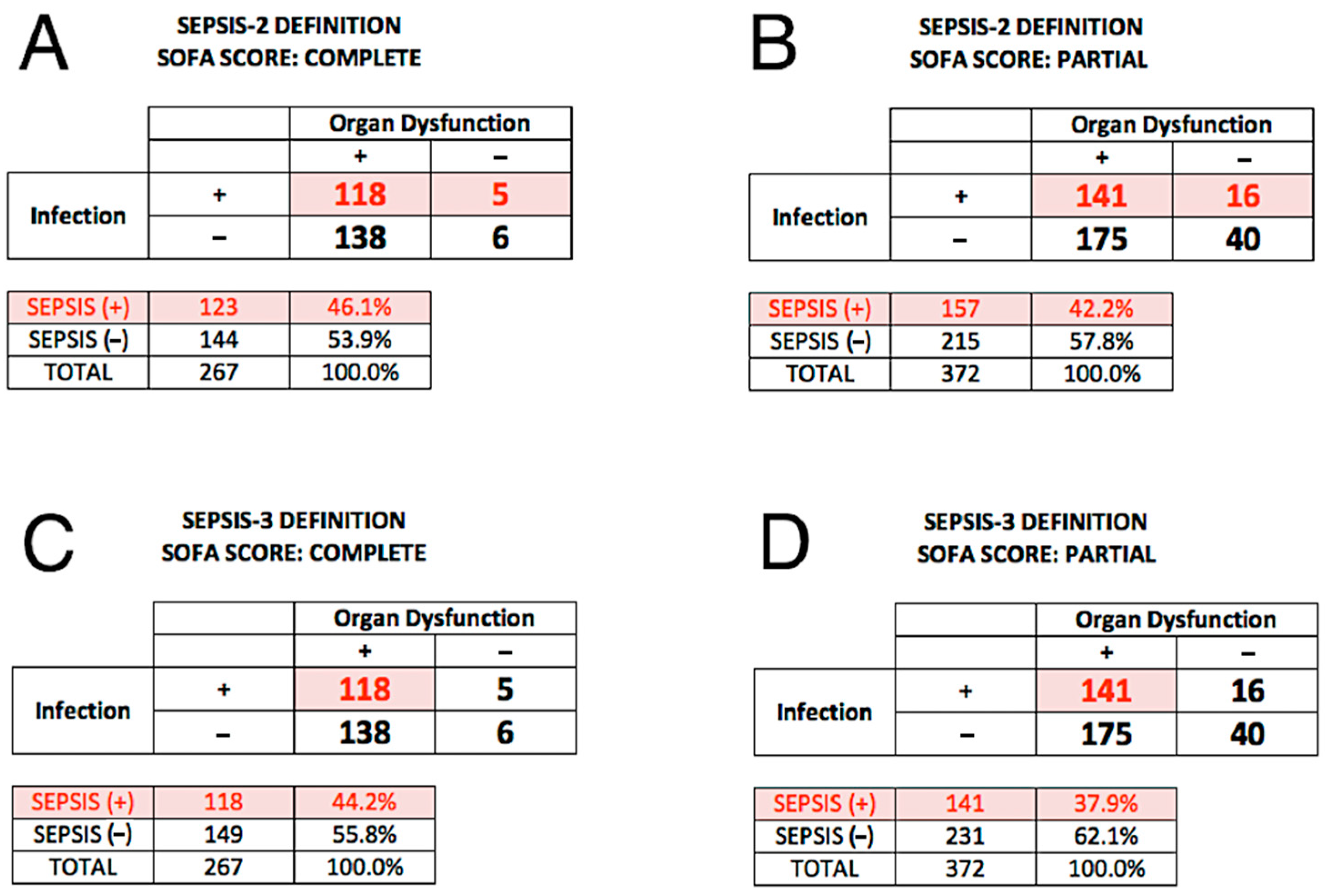
2.4. Reference Diagnosis (Comparator) under Sepsis-2

The clinical performance of SeptiCyte RAPID was evaluated by comparison to a ‘gold standard’ of clinical adjudication. The adjudication process, termed retrospective physician diagnosis (RPD), consisted of clinical evaluations by an external three-member panel of experienced physicians not involved in the care of the patients. For each patient, a chart review was conducted independently by each panel member, leading to a three-way classification (sepsis, SIRS or indeterminate) in accordance with the Sepsis-2 definition under which sepsis is defined as ≥2 SIRS criteria + infection [26]. The vote tally for each patient was either unanimous, consensus (2 of 3), or indeterminate. There were 41/419 (9.8%) indeterminates, which were then reanalyzed by forcing into either the sepsis or SIRS categories by a second RPD. The Sepsis-2 definition was used in this initial analysis because the majority of patients were recruited before the Sepsis-3 definition became available. Additional details are provided in the Supplementary Information, Section S2.

2.5. Reference Diagnosis (Comparator) under Sepsis-3

An additional analysis was conducted under the Sepsis-3 definition. Figure 1 in the publication of Singer et al. (2016) [27] describes how we operationalized the Sepsis-3 definition. Singer et al. proposed that patients with organ dysfunction (SOFA scores ≥ 2) and with definite or suspected infection could be considered to have sepsis. Therefore, the key to transitioning from a Sepsis-2 to a Sepsis-3 framework lies in determining whether patients with organ dysfunction (SOFA scores ≥ 2) are infection-positive or infection-negative. Under the Sepsis-2 framework, when the RPD panelists called a patient septic, this implied that the patient exhibited ≥2 SIRS criteria and had a definite or probable infection. Therefore, a call of sepsis by the RPD panelists implied that they classified the patient as infection-positive.
As measures of organ dysfunction, we considered both the complete SOFA score [28] that had been collected for 289/419 (69.0%) of patients and also, following Grissom et al. [29] and Lambden et al. [30], a partial SOFA score (pSOFA) in which 4 or more of the 7 SOFA components were present. The pSOFA could be computed for 372/419 (88.8%) of patients.

2.6. Statistical Calculations

Sepsis probability as a function of SeptiScore was calculated using a “sliding window” approach [31]. A custom R script was written to conduct a sliding window calculation of the sepsis probability (P) across the 0–15 SeptiScore range. The sliding window was 4 SeptiScore units wide and was shifted in 1-unit increments from the lower to the upper limit of the SeptiScore range. In each 4-unit wide window, the numbers of sepsis calls (Nsepsis) and SIRS calls (NSIRS) were tabulated according to each RPD method, and P = (Nsepsis)/(Nsepsis + NSIRS) was computed for that window. The process was repeated stepwise over the entire 0–15 SeptiScore range.
Sepsis probability as a function of SeptiScore was parsed into four bands, with higher SeptiScores representing higher probabilities of sepsis. Band boundaries were pre-defined on the basis of an independent set of 195 clinical samples from the MARS consortium. The samples for setting the band boundaries were independent of the 419 samples in the validation dataset and were not used in the performance evaluation. The band boundaries were set to give 90% sensitivity for binarization at the Band 1/2 boundary (SeptiScore 4.95) and 80% specificity for binarization at the Band 3/4 boundary (SeptiScore 7.45), using site clinical adjudications as ground truth values. The intermediate zone between the Band 1/2 and Band 3/4 boundaries was divided in half to define the Band 2/3 boundary (SeptiScore 6.15).
Receiver Operating Characteristic (ROC) curve analysis was performed in accordance with Clinical and Laboratory Standards Institute EP24-A2 using the pROC package [32], with comparisons between ROC curves performed by DeLong’s method [33]. Other clinical performance measures (clinical sensitivity, clinical specificity, sepsis and SIRS probabilities, and likelihood ratios) were also calculated. Additional statistical methods for data analyses, including methods for imputing missing data and for combining the SeptiScore with other clinical variables, are described in the Supplementary Information, Sections S3–S5.

3. Results

3.1. Description of the SeptiCyte RAPID Test

SeptiCyte RAPID is a fully automated sample-to-result test, with all reagents integrated in a single-use cartridge. The test provides an actionable result in ~1 h, in the form of a SeptiScore ranging from 0 to 15 and falling into one of four “SeptiScore Bands” of increasing sepsis probability. SeptiCyte RAPID provides a significant technical advance over the earlier SeptiCyte LAB assay, which was based upon the conventional 96- well Applied Biosystems 7500 Fast Dx format with a turnaround time of ~8 h [22]. The correlation of SeptiScore values between SeptiCyte LAB and SeptiCyte RAPID, based upon the retrospective sample set (N = 356) run on both platforms, was high with a Pearson’s sample correlation coefficient r = 0.88 (Supplementary Information, Section S6).
SeptiCyte RAPID quantitatively measures the relative expression levels of two host immune response genes PLAC8 (Placenta Associated 8) and PLA2G7 (Phospholipase A2 Group 7). This expression signature was discovered using a purely bioinformatic approach, which compared SIRS patients that had been retrospectively diagnosed as having either infection or no infection. With respect to biological roles in the host immune response to infection, PLAC8 is reportedly an interferon inducible gene expressed in a variety of tissues rich in immune cells (e.g., whole blood, spleen, lymph node, colon), and up-regulated in sepsis across a broad range of different peripheral blood cell types including plasmacytoid dendritic and natural killer cells. PLA2G7 encodes the protein platelet-activating factor (PAF) acetylhydrolase, a secreted enzyme primarily produced by macrophages that catalyzes the degradation of PAF and hydrolyses the oxidized short chain phospholipids of low-density lipoproteins (LDL), thereby releasing pro-inflammatory mediators (lysophospholipids and oxidized fatty acids). Further discussion and literature references are provided in the Supplementary Information, Section S7.

3.2. Demographic and Clinical Characteristics of the Study Cohorts

Clinical validation of SeptiCyte RAPID was based on testing samples from retrospective (n = 356) and prospective (n = 63) patient cohorts. Demographic characteristics of the study cohorts are presented in Table 1. The majority of patients were recruited over the dates January 2011–December 2013 (MARS), May 2014–April 2015 (VENUS) and March–August 2016 (VENUS Supplement), which largely predate the Sepsis-3 definition [27]. Sepsis, SIRS or indeterminate diagnoses were assigned by RPD under the Sepsis-2 definition, although reanalysis under Sepsis-3 was also performed and is described below. When compared to patients with SIRS, patients adjudicated as septic tended to be older (p = 0.016). There were no significant differences by sex or race/ethnicity. Although the patients were evaluated in intensive care, they originated from a variety of hospital locations. Patients coming from hospital wards exhibited a higher proportion of sepsis relative to patients coming from the ED (p = 0.003), the post-anesthesia unit/post-operating room (p < 0.00001), or the ICU/cardiac care unit (p = 0.02).
Clinical characteristics of the study cohorts are presented in Table 2 and Table 3. Of particular note are positive culture results of high statistical significance. Positive results for blood+, urine+ and blood+/urine+ cultures were very highly associated with RPD diagnoses of sepsis as opposed to SIRS, with p-values of <0.00001, <0.00001, and <0.001, respectively. Also, positive results for blood+, urine+, and blood+/urine+ cultures were associated with elevated SeptiScores, especially SeptiScores in Band 4 (see Methods for Band definitions). Of the 48 patients that were called septic by RPD with positive blood+ or blood+/urine+ culture results, 34/48 (71%) of SeptiScores fell in Band 4 and 9/48 (19%) fell in Band 3. Of the 42 patients with positive blood+ culture results, only one had a SeptiScore in Band 1; this patient was diagnosed as SIRS by RPD, with the blood culture result being considered a probable contaminant by the attending physician and the RPD panelists.
A taxonomic analysis of the culture-confirmed pathogens for the sepsis cases is presented in the Supplementary Information, Sections S8 and S9. In our study cohorts, SeptiScore does not appear to be significantly affected by the type of pathogen (bacterial, viral, fungal) underlying a sepsis event. In particular, we observed no significant difference (p = 0.264) between the SeptiScore distributions for septic patients with Gram-positive infections (n = 55) versus Gram-negative infections (n = 36).

3.3. SeptiCyte RAPID Performance under Sepsis-2

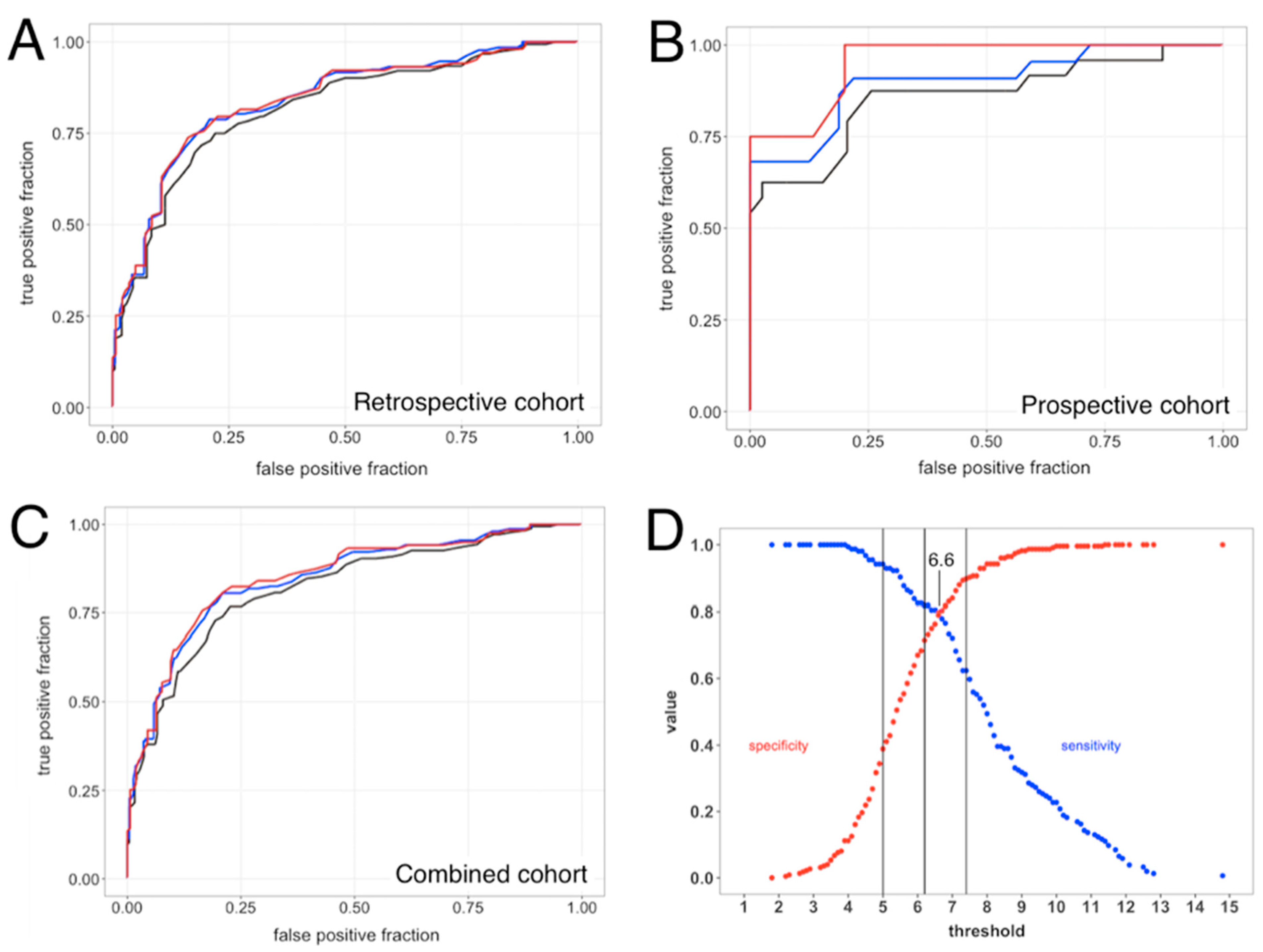
3.3.1. ROC Curve Analyses

Receiver Operating Characteristic (ROC) curve analyses for sepsis vs. SIRS were performed for the retrospective cohort (Figure 1A), the prospective cohort (Figure 1B), and the combined retrospective + prospective cohorts (Figure 1C). Performance estimates for the retrospective cohort ranged from AUC 0.82 to 0.85 (depending upon RPD method) and were statistically indistinguishable, both from each other and from the AUC values previously obtained for SeptiCyte LAB [22]. Performance estimates for the prospective cohort ranged from AUC 0.86 to 0.95 (depending upon RPD method) and again were statistically indistinguishable from each other.
DeLong’s test [33] showed no significant difference between the ROC curves from Figure 1A (retrospective cohort) vs. 1B (prospective cohort), when either forced or consensus RPD was used as comparator. When unanimous RPD was used as comparator, DeLong’s test gave p = 0.013. For the retrospective + prospective dataset, using consensus RPD as a comparator and assuming a binary distinction between sepsis and SIRS, the relationship between cutpoint, sensitivity, and specificity is shown in Figure 1D.

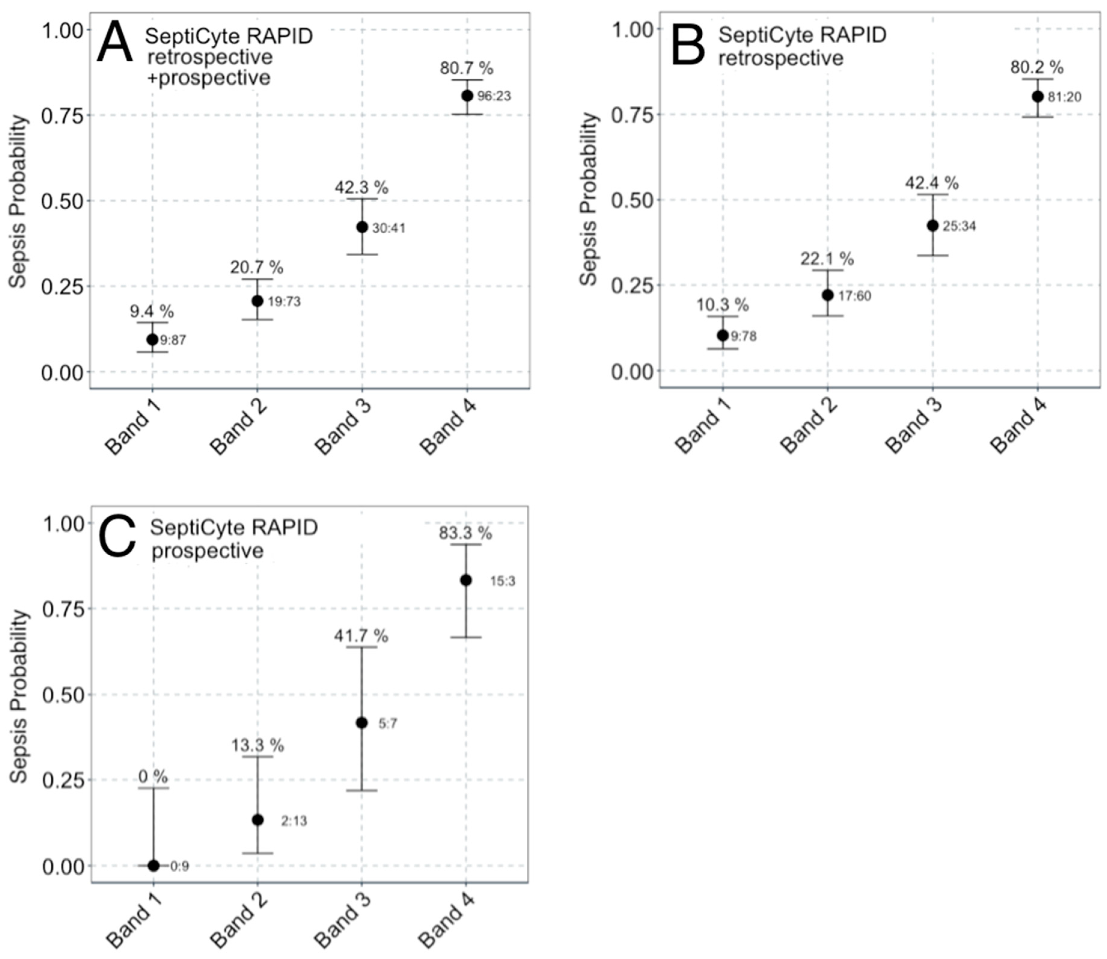
3.3.2. Sepsis Probability Distributions

The distribution of SeptiScores for septic patients falls above that for patients with SIRS (Figure 2A). A sliding window analysis (Figure 2B) was used to calculate the probability of sepsis as a function of SeptiScore (Figure 2C). Patients with higher SeptiScores had higher probabilities of sepsis. As described in the Methods Section, the 0–15 SeptiScore range has been divided into four probability bands (Figure 2D) defined by the pre-set band boundaries of 4.95, 6.15, and 7.45. Figure 3 and Table 4 present SeptiScores for the complete (retrospective + prospective) cohort, and for the retrospective and prospective cohorts separately, after parsing into the four bands. Using consensus RPD as a comparator, Band 1 had a negative predictive value of 0.91 (sensitivity 0.94), and Band 4 had a positive predictive value of 0.81 (specificity 0.90). Chi-squared analyses (Table 4) indicated that, after sorting patients into the four SeptiScore bands, no significant influences were found with respect to the baseline demographic criteria or sex, age, race, or ethnicity (Table 3).

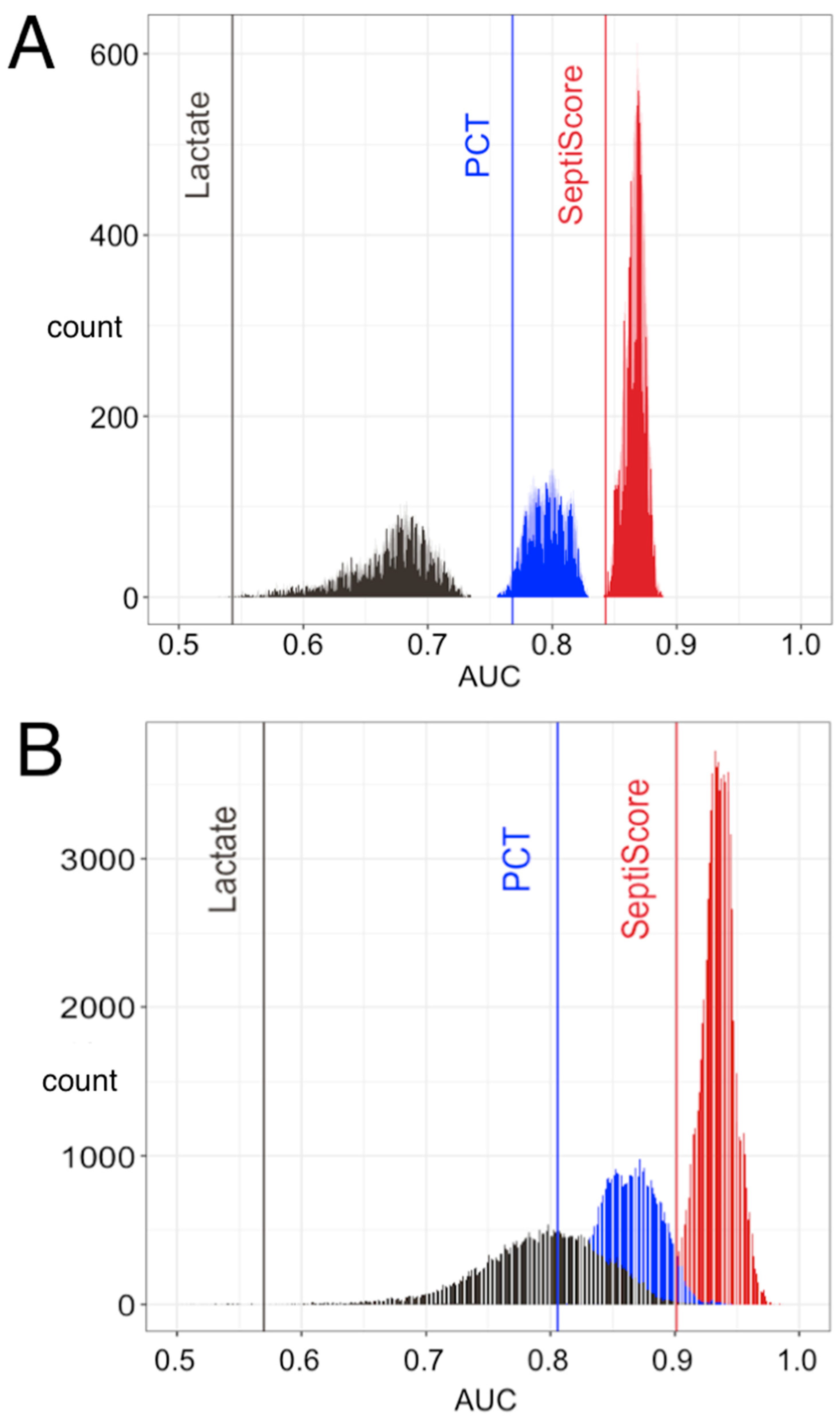
3.3.3. Multivariable Analysis

Under the Sepsis-2 framework, we asked if SeptiCyte RAPID could provide diagnostic clinical utility for discriminating between sepsis and SIRS patients, beyond that provided by other clinical variables and laboratory assessments available on the first day of ICU admission. Fourteen variables in addition to SeptiCyte RAPID were examined (see Supplementary Information, Sections S4 and S5). We evaluated all 32,767 possible combinations of the fifteen variables, and performance (sepsis vs. SIRS under the Sepsis-2 framework) was assessed by AUC against consensus RPD. Procalcitonin was included in this analysis, as well as lactate, which is commonly used for determining which patients should receive sepsis treatment bundles [34,35]. Figure 4A shows the results of the multivariable analysis for the combined retrospective and prospective cohorts, while Figure 4B shows the comparable results for just the prospective cohort. The AUC distributions for the prospective cohort appear somewhat high compared to those for the full patient cohort, possibly because of overfitting (the prospective cohort has only 54 patients with SIRS or sepsis by consensus RPD, as compared to 154 in the full patient cohort). The potential for inflation of performance measures, including AUC, as a consequence of adding new markers to a predefined test, has been noted by others [36]. With respect to individual variables, SeptiScore was found to rank highest by AUC. When combinations of clinical variables were considered, those containing SeptiScore were found to have higher AUCs than all combinations lacking SeptiScore.

3.4. SeptiCyte RAPID Performance under Sepsis-3

In 2016, Singer et al. [27] proposed that the Sepsis-2 definition be replaced with Sepsis-3, which identifies sepsis as “life-threatening organ dysfunction caused by a dysregulated host response to infection”. We compared the performance of SeptiCyte RAPID for discriminating sepsis vs. non-sepsis under the Sepsis-2 vs. Sepsis-3 definitions. Figure 5 displays 2 × 2 tables for classifying patients in terms of (+/− organ dysfunction) × (+/− infection). Panels A, B employ the Sepsis-2 definition, while Panels C, D employ the Sepsis-3 definition. In each panel, the numbers in red indicate septic patients, according to whether organ dysfunction was indicated by complete SOFA score ≥ 2 (Panels A, C) or by partial SOFA score ≥ 2 (Panels B, D). A striking feature of these tables is that they reveal that nearly all the patients in the study cohort display some amount of organ dysfunction, indicated by SOFA ≥ 2 (256/267 = 95.9% of patients) or pSOFA ≥ 2 (316/372 = 84.9% of patients). This however might be expected, since all patients in the cohort were admitted to ICU.
Figure 6 builds upon Figure 5, using ROC analysis to evaluate SeptiCyte RAPID performance for each patient grouping. Performance under the Sepsis-2 vs. Sepsis-3 definitions is compared when organ dysfunction is indicated either by complete SOFA score ≥ 2 (panel A) or by partial SOFA score ≥ 2 (panel B). The ROC curves were found to be statistically equivalent across all these comparisons, with AUC values ranging from 0.807 to 0.820. Additional analyses, concerning the relative independence of SeptiScore on the extent of organ dysfunction, are presented in the Supplementary Material, Section S10.

4. Discussion

We have presented data validating the use of SeptiCyte RAPID for discriminating sepsis from SIRS and for estimating the probability of sepsis in critically ill adult patients, within a clinically actionable time frame. Analysis was performed under both the Sepsis-2 and Sepsis-3 frameworks. Patients in this study came from the ED, post-anesthesia unit, post-operating rooms and wards, and were tested in the ICU. For the full (retrospective + prospective) cohort, the diagnostic performance of SeptiCyte RAPID under the Sepsis-2 definition was equivalent or superior to that previously reported for SeptiCyte LAB [22].
In a multivariable analysis under the Sepsis-2 framework, we examined all possible combinations of SeptiScore and up to 14 additional clinical or laboratory variables, including lactate and PCT. We found that SeptiScore alone had a greater performance than any combination of variables without SeptiScore, for differentiating sepsis vs. SIRS (Figure 4). However, the analysis also indicated it should be possible to moderately boost the performance of SeptiCyte RAPID by combining the SeptiScore with other clinical parameters.
We reanalyzed our data under the Sepsis-3 framework, using the SOFA or pSOFA score as a quantitative measure of organ dysfunction [28,29,30], and interpreting an RPD call of sepsis as indicating probable or definite infection. The results (Figure 6) indicated that SeptiScore has a high diagnostic performance in terms of detecting sepsis (AUC 0.807–0.820) under either the Sepsis-2 or Sepsis-3 framework. Because SeptiCyte RAPID performance appears independent of organ dysfunction, we hypothesize that the SeptiCyte RAPID expression signature may be responding to some process or condition in the septic trajectory occurring earlier than organ damage, likely related to the presence of an infection. A similar hypothesis has been stated by Lukaszewski et al. [16] in connection with an independently discovered sepsis signature.
Our reanalysis under Sepsis-3 also identified a subset of 16 patients called septic by the RPD panelists under the Sepsis-2 definition, but without appreciable organ dysfunction (Figure 5B). Under the Sepsis-3 framework, these patients would not be called septic, and therefore represent a discordance between the two sepsis definitions. We are continuing to study the clinical characteristics of these patients, to understand why they were called sepsis under Sepsis-2 but not Sepsis-3. We note that Engoren et al. [37] has identified a similar discordance: in a large retrospective analysis of over 29,000 hospitalized patients with suspected infection, 44% were called septic according to Sepsis-2, and 41% called septic according to Sepsis-3, but only 23% satisfied both sets of criteria, implying poor agreement (kappa 0.213) between Sepsis-2 and Sepsis-3 in their cohort. These investigators hypothesized that Sepsis-2 and Sepsis-3 represent different phenotypes that only partially overlap.
Our study has several limitations. Our comparator method (RPD) is imperfect. In the complete (n = 419) validation dataset, three expert clinicians failed to reach either a unanimous or consensus diagnosis for 41/419 (9.8%) of patients. The use of an imperfect comparator sets an upper limit on the reportable diagnostic performance of a new test [38]. Our cohort consisted of adult patients within 24 h of ICU admission, so generalization to other patient cohorts has not been established. We have not included pediatric patients in the present study. However, we previously reported the strong diagnostic performance of SeptiCyte LAB in children [39], and the high correlation between SeptiCyte LAB and SeptiCyte RAPID suggests that an equivalently strong performance in children will be found for SeptiCyte RAPID. To avoid potential confounding effects, the study excluded subjects who received therapeutic antibiotic treatment > 24 h before ICU admission, which could comprise a significant fraction of patients transferred from the floor to the ICU. Most of the study was based on a re-analysis of banked samples (356/419 = 85%), while only 63/419 (15%) were prospectively collected. SeptiScores falling in Bands 2 or 3 do not provide definitive conclusions (i.e., very high or low probabilities) regarding the absence or presence of sepsis. The interpretation of SeptiScores in this range would be enhanced by combining with other clinical variables to adjust the post-test sepsis probabilities (see Figure 4).
During the discovery and initial validation of the PLAC8 and PLA2G7 biomarkers [22], patients with a broad range of co-morbidities were examined, including septic patients with confirmed bacterial, viral, fungal infections and malignancies, and non-septic patients with non-infectious systemic inflammation of varying etiologies. The present validation cohort also included the use of a broad range of prescribed medications such as immunosuppressants, anti-neoplastic drugs, antithrombotics, corticosteroids, and statins. To our knowledge, SeptiCyte RAPID results are unaffected by these factors, as reported in a previous preliminary analysis [40]. However, there may be other specific conditions and treatments we have not yet examined that could affect SeptiCyte RAPID’s performance.
Although the dynamic range of SeptiCyte RAPID is broad and extends well below and above the 4000–11,000 WBC/uL normal reference range, we have not yet completed an evaluation of the assay on severely neutropenic patients, although in a previous preliminary analysis we reported that SeptiCyte performance was maintained in patients treated with anti-neoplastic drugs [40]. It is possible that SeptiCyte RAPID scores might be skewed by selective leukopenias due to disease or medications, for example T cell depletion in HIV/AIDS. However, it is known from single cell sequencing studies that both PLAC8 and PLA2G7 are expressed across a range of different white cell types [41], which would mitigate the effect of a selective leukopenia.
SeptiCyte RAPID was not designed to predict mortality, and this falls outside the intended use of the test. Also, the clinical studies described herein collected only a single blood sample within 24 h of ICU admission, and SeptiCyte RAPID testing was not repeated serially during the hospital stay. However, in parallel with the present work, we have conducted additional studies on other patient cohorts, in which longitudinal testing was performed. The results of these studies, including a determination of whether continued detection of high SeptiCyte RAPID scores had prognostic ability for mortality or other adverse events, will be described in future publications.
The recently updated Surviving Sepsis Campaign (SSC) guidelines (2021) divide patients into three groups (low, intermediate, and high) based on sepsis probability, and recommend appropriate evaluation and treatment of these groups [42]. SeptiCyte RAPID aligns well with these guidelines and could have a role in supporting their implementation. The SSC ‘low sepsis probability’ group has, under previous guidelines, been treated early resulting in poor antibiotic stewardship. The new guidelines recommend deferring antibiotics, and monitoring and evaluating for other etiologies that may underlie the presenting symptoms. A SeptiScore < 5 (Band 1) with a sensitivity of 0.94 in this group of patients could potentially support the deferral of antibiotics, at least until clinical microbiology results become available, thereby facilitating antibiotic and diagnostic stewardship. For patients falling in the SSC ‘intermediate sepsis probability (without shock)’ group, the SSC recommendation is a rapid assessment of infectious versus non-infectious causes of the illness. In this probability range, the information typically obtained from other clinical variables could be augmented by information from SeptiCyte RAPID to shift the post-test probability of sepsis to either a lower or a higher value (Figure 4). Per the SSC guidelines, those patients in the ‘high sepsis probability’ group, differentiated by the presence of shock, should appropriately be treated within 1 h of recognition. SeptiScores in Band 4 (>7.5) in this patient group, with specificity 0.9 or greater, would provide evidence for a high probability of sepsis and the continuation of antibiotics.
Although SeptiCyte RAPID provides a fast turnaround time (~1 h) between pipetting a blood sample into the SeptiCyte RAPID cartridge and generating a test report, by the time patient blood is drawn and delivered, e.g., to a STAT lab, the total time between blood draw and presentation of the test result to an attending clinician is likely to be 1.5–2 h. Although this is longer than requirements to implement a 1 h sepsis bundle, SeptiCyte RAPID would provide timely information with respect to implementing a 3 h sepsis bundle, which has been shown to have a low level of compliance in US hospitals [43,44]. Compliance with a 3 h sepsis bundle in patients with severe sepsis and septic shock has been shown to improve survival and reduce overall costs [44].

5. Conclusions

This study validates SeptiCyte RAPID for the discrimination of sepsis from non-infectious systemic inflammation, and for the estimation of the probability of sepsis on the first day of ICU admission, under either the Sepsis-2 or Sepsis-3 frameworks. With a turnaround time of ~1 h, SeptiCyte RAPID provides actionable test results within a clinically relevant timeframe. SeptiCyte RAPID may thus provide clinical utility through helping to guide patient management decisions on the implementation and timing of sepsis bundles.

Supplementary Materials

The following supporting information can be downloaded at: https://www.mdpi.com/article/10.3390/jcm13051194/s1, Balk et al. (2024) Supplementary Information. References [45,46,47,48,49,50,51,52,53,54,55,56,57,58,59,60,61,62,63,64,65,66,67] are cited in the supplementary materials.

Author Contributions

Conceptualization: R.R.M.III, J.P.B., K.N., T.D.Y., J.T.K., S.C., R.F.D. and R.B.B.; Data curation: J.P.B., V.K.S., J.A.K., B.P.S., S.K., K.N., D.S., J.T.K., S.C. and R.B.B.; Formal analysis: M.L., S.O., R.E.R., M.J.S., K.N., T.D.Y., D.S., S.C., R.F.D. and R.B.B.; Funding acquisition: M.J.S.; Investigation: R.B., A.M.E., G.S.M., B.K.L., F.R.D., V.K.S., N.R.A., J.A.G., M.Y., G.P., E.G., J.P.P., M.A., J.A.K., T.v.d.P., M.J.S., P.M.C.K.K., J.L., E.B., S.K., J.T.K. and S.C.; Methodology: M.L., S.O., R.E.R., K.N., T.D.Y., D.S., J.T.K. and R.B.B.; Project administration: A.M.E., F.R.D., J.L., S.K., T.D.Y., S.C. and R.B.B.; Resources: R.B., A.M.E., F.R.D., V.K.S., T.v.d.P., B.P.S., J.L., S.K., D.S. and S.C.; Software: K.N. and D.S.; Supervision: R.B., A.M.E., B.K.L., F.R.D., E.G., J.P.P., M.A., T.v.d.P., S.K., S.C. and R.B.B.; Validation: R.B., M.L., S.O., R.E.R., N.R.A., G.P., B.P.S., E.B., K.N., S.C. and R.B.B.; Visualization: R.B., K.N., T.D.Y. and D.S.; Writing—original draft: K.N., T.D.Y., S.C. and R.B.B.; Writing—review & editing: R.B., G.S.M., R.R.M.III, J.P.B., M.L., R.E.R., V.K.S., N.R.A., J.A.G., M.Y., E.G., J.P.P., M.A., J.A.K., T.v.d.P., M.J.S., B.P.S., P.M.C.K.K., E.B., S.K., K.N., T.D.Y., D.S., J.T.K., S.C., R.F.D. and R.B.B. All authors have read and agreed to the published version of the manuscript.

Funding

This work was funded by Immunexpress, Inc.

Institutional Review Board Statement

Ethics approval for the MARS trial was given by the medical ethics committee of AMC, Amsterdam (approval # 10-056C, 16 June 2010). Ethics approvals for the VENUS trial were given by the relevant Institutional Review Boards as follows: Intermountain Medical Center/Latter Day Saints Hospital (approval # 1024931, 21 January 2014); Johns Hopkins Hospital (approval # IRB00087839, 28 January 2016); Rush University Medical Center (approval # 15111104-IRB01, 11 March 2016); Loyola University Medical Center (approval # 208291, 10 March 2016); Northwell Healthcare (approval #16-02-42-03, 1 April 2016). Ethics approvals for the NEPTUNE trial were given by the relevant Institutional Review Boards as follows: Emory University (approval # IRB00115400, 3 December 2019); Grady Memorial Hospital (approval # 00-115400, 14 January 2020); Rush University Medical Center (approval # 19101603-IRB01, 16 January 2020); University of Southern California Medical Center (approval # HS-19-0884-CR001, 10 February 2020). All methods used in this study were carried out in accordance with the relevant guidelines and regulations.

Informed Consent Statement

All subjects, or their legally authorized representatives, gave informed consent for participation in this study.

Data Availability Statement

The datasets used and/or analyzed during the current study are available from the corresponding author upon reasonable request.

Acknowledgments

The authors thank the clinical study coordinators of the NEPTUNE study: Joyce D. Brown (Rush University), Liliacna Jara (University of Southern California), Leona Wells and Maya C. Whaley (Grady Memorial Hospital). The authors also thank the laboratory technical staff at each institution, at Reach Bio (Seattle, WA, USA), and at Biocartis (Mechelen, Belgium).

Conflicts of Interest

K.N., T.D.Y., D.S., J.T.K., S.C., R.F.D. and R.B.B. declare they are present or past employees or shareholders of Immunexpress, Inc. R.R.M. III discloses that, over the time period of relevance to this work he was paid (as a consultant) an honorarium by Immunexpress for conducting a critical review of a “Clinical Evaluation Report” of SeptiCyte technology, submitted by Immunexpress to the Therapeutic Goods Administration (TGA) of Australia. N.R.A. declares that he has received grants from the NIH and Department of Defense for non-related work, and that payments from these grants are made to his institution, the University of Colorado. No other competing interests are declared.

References

  1. Vincent, J.L. Highlighting the huge global burden of sepsis. Anaesth. Crit. Care Pain Med. 2020, 39, 171–172. [Google Scholar] [CrossRef]
  2. Rudd, K.E.; Johnson, S.C.; Agesa, K.M.; Shackelford, K.A.; Tsoi, D.; Kievlan, D.R.; Colombara, D.V.; Ikuta, K.S.; Kissoon, N.; Finfer, S.; et al. Global, regional, and national sepsis incidence and mortality, 1990–2017: Analysis for the Global Burden of Disease Study. Lancet 2020, 395, 200–211. [Google Scholar] [CrossRef] [PubMed]
  3. Torio, C.M.; Moore, B.J. National Inpatient Hospital Costs: The Most Expensive Conditions by Payer, 2013. In Healthcare Cost and Utilization Project (HCUP) Statistical Briefs [Internet]; Statistical Brief #204; Agency for Healthcare Research and Quality (US): Rockville, MD, USA, 2016. Available online: https://www.ncbi.nlm.nih.gov/books/NBK368492/ (accessed on 17 February 2024).
  4. Paoli, C.J.; Reynolds, M.A.; Sinha, M.; Gitlin, M.; Crouser, E. Epidemiology and Costs of Sepsis in the United States—An Analysis Based on Timing of Diagnosis and Severity Level. Crit. Care Med. 2018, 46, 1889–1897. [Google Scholar] [CrossRef] [PubMed]
  5. Rhee, C.; Dantes, R.; Epstein, L.; Murphy, D.J.; Seymour, C.W.; Iwashyna, T.J.; Kadri, S.S.; Angus, D.C.; Danner, R.L.; Fiore, A.E.; et al. CDC Prevention Epicenter Program. Incidence and Trends of Sepsis in US Hospitals Using Clinical vs Claims Data, 2009–2014. JAMA 2017, 318, 1241–1249. [Google Scholar] [CrossRef] [PubMed]
  6. Elezkurtaj, S.; Greuel, S.; Ihlow, J.; Michaelis, E.G.; Bischoff, P.; Kunze, C.A.; Sinn, B.V.; Gerhold, M.; Hauptmann, K.; Ingold-Heppner, B.; et al. Causes of death and comorbidities in hospitalized patients with COVID-19. Sci. Rep. 2021, 11, 4263. [Google Scholar] [CrossRef] [PubMed]
  7. Shappell, C.N.; Klompas, M.; Chan, C.; Chen, T.; Kanjilal, S.; McKenna, C.; Rhee, C.; CDC Prevention Epicenters Program. Use of Electronic Clinical Data to Track Incidence and Mortality for SARS-CoV-2-Associated Sepsis. JAMA Netw. Open 2023, 6, e2335728. [Google Scholar] [CrossRef] [PubMed]
  8. Levy, M.M.; Evans, L.E.; Rhodes, A. The Surviving Sepsis Campaign Bundle: 2018 update. Intensive Care Med. 2018, 44, 925–928. [Google Scholar] [CrossRef] [PubMed]
  9. Liu, V.X.; Bhimarao, M.; Greene, J.D.; Manickam, R.N.; Martinez, A.; Schuler, A.; Barreda, F.; Escobar, G.J. The Presentation, Pace, and Profile of Infection and Sepsis Patients Hospitalized Through the Emergency Department: An Exploratory Analysis. Crit. Care Explor. 2021, 3, e0344. [Google Scholar] [CrossRef]
  10. Lambregts, M.M.C.; Bernards, A.T.; Beek MT van der Visser, L.G.; de Boer, M.G. Time to positivity of blood cultures supports early re-evaluation of empiric broad-spectrum antimicrobial therapy. PLoS ONE 2019, 14, e0208819. [Google Scholar] [CrossRef]
  11. Panday, R.S.N.; Lammers, E.M.J.; Alam, N.; Nanayakkara, P.W.B. An overview of positive cultures and clinical outcomes in septic patients: A sub-analysis of the Prehospital Antibiotics Against Sepsis (PHANTASi) trial. Crit. Care 2019, 23, 182. [Google Scholar] [CrossRef]
  12. Panday, R.S.N.; Wang, S.; Ven PM van de Hekker, T.A.M.; Alam, N.; Nanayakkara, P.W.B. Evaluation of blood culture epidemiology and efficiency in a large European teaching hospital. PLoS ONE 2019, 14, e0214052. [Google Scholar] [CrossRef]
  13. Minderhoud, T.C.; Spruyt, C.; Huisman, S.; Oskam, E.; Schuit, S.C.E.; Levin, M.D. Microbiological outcomes and antibiotic overuse in Emergency Department patients with suspected sepsis. Neth. J. Med. 2017, 75, 196–203. [Google Scholar] [PubMed]
  14. Shappell, C.N.; Klompas, M.; Ochoa, A.; Rhee, C.; CDC Prevention Epicenters Program. Likelihood of Bacterial Infection in Patients Treated with Broad-Spectrum IV Antibiotics in the Emergency Department. Crit. Care Med. 2021, 49, e1144–e1150. [Google Scholar] [CrossRef]
  15. Bae, E.Y.; Smith, T.T.; Monogue, M.L. A case-control study evaluating the unnecessary use of intravenous broad-spectrum antibiotics in presumed sepsis and septic-shock patients in the emergency department. Antimicrob. Steward. Healthc. Epidemiol. 2022, 2, e193. [Google Scholar] [CrossRef] [PubMed]
  16. Lukaszewski, R.A.; Jones, H.E.; Gersuk, V.H.; Russell, P.; Simpson, A.; Brealey, D.; Walker, J.; Thomas, M.; Whitehouse, T.; Ostermann, M.; et al. Presymptomatic diagnosis of postoperative infection and sepsis using gene expression signatures. Intensive Care Med. 2022, 48, 1133–1143. [Google Scholar] [CrossRef] [PubMed]
  17. Zhang, Z.; Smischney, N.J.; Zhang, H.; Van Poucke, S.; Tsirigotis, P.; Rello, J.; Honore, P.M.; Kuan, W.S.; Ray, J.J.; Zhou, J.; et al. AME evidence series 001-The Society for Translational Medicine: Clinical practice guidelines for diagnosis and early identification of sepsis in the hospital. J. Thorac. Dis. 2016, 8, 2654–2665. [Google Scholar] [CrossRef] [PubMed]
  18. Tusgul, S.; Carron, P.N.; Yersin, B.; Calandra, T.; Dami, F. Low sensitivity of qSOFA, SIRS criteria and sepsis definition to identify infected patients at risk of complication in the prehospital setting and at the emergency department triage. Scand. J. Trauma Resusc. Emerg. Med. 2017, 25, 108. [Google Scholar] [CrossRef] [PubMed]
  19. Dorsett, M.; Kroll, M.; Smith, C.S.; Asaro, P.; Liang, S.Y.; Moy, H.P. qSOFA has poor sensitivity for Prehospital identification of severe sepsis and septic shock. Prehosp. Emerg. Car. 2017, 21, 489–497. [Google Scholar] [CrossRef] [PubMed]
  20. Sartelli, M.; Kluger, Y.; Ansaloni, L.; Hardcastle, T.C.; Rello, J.; Watkins, R.R.; Bassetti, M.; Giamarellou, E.; Coccolini, F.; Abu-Zidan, F.M.; et al. Raising concerns about the Sepsis-3 definitions. World J. Emerg. Surg. 2018, 13, 6. [Google Scholar] [CrossRef] [PubMed]
  21. Kim, H.I.; Park, S. Sepsis: Early Recognition and Optimized Treatment. Tuberc. Respir. Dis. 2019, 82, 6–14. [Google Scholar] [CrossRef]
  22. Miller, R.R., 3rd; Lopansri, B.K.; Burke, J.P.; Levy, M.; Opal, S.; Rothman, R.E.; D’Alessio, F.R.; Sidhaye, V.K.; Aggarwal, N.R.; Balk, R.; et al. Validation of a Host Response Assay, SeptiCyte LAB, for Discriminating Sepsis from Systemic Inflammatory Response Syndrome in the ICU. Am. J. Respir. Crit. Care Med. 2018, 198, 903–913. [Google Scholar] [CrossRef] [PubMed]
  23. Kirk, J.; Yager, T.; Cermelli, S.; Sampson, D.; Brandon, R.; Sillekens, P.; Keuleers, I.; Vanhoey, T. Clinical performance of a rapid sepsis test on a near-patient molecular testing platform. In Proceedings of the ISICEM 2020, Brussels, Belgium, 19–22 March 2020. Abstract # P481. [Google Scholar]
  24. Rand, K.H.; Beal, S.G.; Rivera, K.; Allen, B.; Payton, T.; Lipori, G.P. Hourly Effect of Pretreatment with IV Antibiotics on Blood Culture Positivity Rate in Emergency Department Patients. Open Forum Infect. Dis. 2019, 6, ofz179. [Google Scholar] [CrossRef] [PubMed]
  25. Scheer, C.S.; Fuchs, C.; Gründling, M.; Vollmer, M.; Bast, J.; Bohnert, J.A.; Zimmermann, K.; Hahnenkamp, K.; Rehberg, S.; Kuhn, S.O. Impact of antibiotic administration on blood culture positivity at the beginning of sepsis: A prospective clinical cohort study. Clin. Microbiol. Infect. 2019, 25, 326–331. [Google Scholar] [CrossRef] [PubMed]
  26. Verdonk, F.; Blet, A.; Mebazaa, A. The new sepsis definition: Limitations and contribution to research and diagnosis of sepsis. Curr. Opin. Anesthesiol. 2017, 30, 200–204. [Google Scholar] [CrossRef]
  27. Singer, M.; Deutschman, C.S.; Seymour, C.W.; Shankar-Hari, M.; Annane, D.; Bauer, M.; Bellomo, R.; Bernard, G.R.; Chiche, J.D.; Coopersmith, C.M.; et al. The Third International Consensus Definitions for Sepsis and Septic Shock (Sepsis-3). JAMA 2016, 315, 801–810. [Google Scholar] [CrossRef] [PubMed]
  28. Seymour, C.W.; Liu, V.X.; Iwashyna, T.J.; Brunkhorst, F.M.; Rea, T.D.; Scherag, A.; Rubenfeld, G.; Kahn, J.M.; Shankar-Hari, M.; Singer, M.; et al. Assessment of Clinical Criteria for Sepsis: For the Third International Consensus Definitions for Sepsis and Septic Shock (Sepsis-3). JAMA 2016, 315, 762–774, Erratum in JAMA 2016, 315, 2237. [Google Scholar] [CrossRef] [PubMed]
  29. Grissom, C.K.; Brown, S.M.; Kuttler, K.G.; Boltax, J.P.; Jones, J.; Jephson, A.R.; Orme, J.F., Jr. A modified sequential organ failure assessment score for critical care triage. Disaster Med. Public Health Prep. 2010, 4, 277–284. [Google Scholar] [CrossRef]
  30. Lambden, S.; Laterre, P.F.; Levy, M.M.; Francois, B. The SOFA score-development, utility and challenges of accurate assessment in clinical trials. Crit. Care 2019, 23, 374–382. [Google Scholar] [CrossRef]
  31. Arasu, A.; Manku, G.S. Approximate Counts and Quantiles over Sliding Windows. In Proceedings of the PODS 2004: Twenty-third ACM SIGMOD-SIGACT-SIGART Symposium on Principles of Database Systems, Paris, France, 14–16 June 2004; pp. 286–296. [Google Scholar] [CrossRef]
  32. Robin, X.; Turck, N.; Hainard, A.; Tiberti, N.; Lisacek, F.; Sanchez, J.C.; Müller, M. pROC: An open-source package for R and S+ to analyze and compare ROC curves. BMC Bioinform. 2011, 12, 77–84. [Google Scholar] [CrossRef]
  33. DeLong, E.R.; DeLong, D.M.; Clarke-Pearson, D.L. Comparing the areas under two or more correlated receiver operating characteristic curves: A nonparametric approach. Biometrics 1988, 44, 837–845. [Google Scholar] [CrossRef]
  34. Faust, J.S.; Weingart, S.D. The Past, Present, and Future of the Centers for Medicare and Medicaid Services Quality Measure SEP-1: The Early Management Bundle for Severe Sepsis/Septic Shock. Emerg. Med. Clin. N. Am. 2017, 35, 219–231. [Google Scholar] [CrossRef]
  35. Rhee, C.; Chiotos, K.; Cosgrove, S.E.; Heil, E.L.; Kadri, S.S.; Kalil, A.C.; Gilbert, D.N.; Masur, H.; Septimus, E.J.; Sweeney, D.A.; et al. Infectious Diseases Society of America Position Paper: Recommended Revisions to the National Severe Sepsis and Septic Shock Early Management Bundle (SEP-1) Sepsis Quality Measure. Clin. Infect. Dis. 2020, 72, ciaa059. [Google Scholar] [CrossRef]
  36. Pepe, M.S.; Janes, H.; Li, C.I. Net Risk Reclassification P Values: Valid or Misleading? J. Natl. Cancer Inst. 2014, 106, dju041. [Google Scholar] [CrossRef] [PubMed]
  37. Engoren, M.; Seelhammer, T.; Freundlich, R.E.; Maile, M.D.; Sigakis, M.J.G.; Schwann, T.A. A Comparison of Sepsis-2 (Systemic Inflammatory Response Syndrome Based) to Sepsis-3 (Sequential Organ Failure Assessment Based) Definitions-A Multicenter Retrospective Study. Crit. Care Med. 2020, 48, 1258–1264. [Google Scholar] [CrossRef]
  38. Valenstein, P.N. Evaluating diagnostic tests with imperfect standards. Am. J. Clin. Pathol. 1990, 93, 252–258. [Google Scholar] [CrossRef] [PubMed]
  39. Zimmerman, J.J.; Sullivan, E.; Yager, T.D.; Cheng, C.; Permut, L.; Cermelli, S.; McHugh, L.; Sampson, D.; Seldon, T.; Brandon, R.B.; et al. Diagnostic Accuracy of a Host Gene Expression Signature That Discriminates Clinical Severe Sepsis Syndrome and Infection-Negative Systemic Inflammation Among Critically Ill Children. Crit. Care Med. 2017, 45, e418–e425. [Google Scholar] [CrossRef] [PubMed]
  40. Davis, R.; Navalkar, K.; van der Poll, T.; Schultz, M.; Zimmerman, J.; Cremer, O.; Bonton, M. SeptiCyte RAPID in Sepsis Cases with Malignancy or Treated with Antineoplastics/Immunosuppressants. Crit. Care Med. 2021, 49, 643. [Google Scholar] [CrossRef]
  41. Reyes, M.; Filbin, M.R.; Bhattacharyya, R.P.; Billman, K.; Eisenhaure, T.; Hung, D.T.; Levy, B.D.; Baron, R.M.; Blainey, P.C.; Goldberg, M.B.; et al. An immune-cell signature of bacterial sepsis. Nat. Med. 2020, 26, 333–340. [Google Scholar] [CrossRef] [PubMed]
  42. Evans, L.; Rhodes, A.; Alhazzani, W.; Antonelli, M.; Coopersmith, C.M.; French, C.; Machado, F.R.; Mcintyre, L.; Ostermann, M.; Prescott, H.C.; et al. Surviving Sepsis Campaign: International Guidelines for Management of Sepsis and Septic Shock 2021. Crit. Care Med. 2021, 49, e1063–e1143. [Google Scholar] [CrossRef] [PubMed]
  43. Deis, A.S.; Whiles, B.B.; Brown, A.R.; Satterwhite, C.L.; Simpson, S.Q. Three-Hour Bundle Compliance and Outcomes in Patients with Undiagnosed Severe Sepsis. Chest 2018, 153, 39–45. [Google Scholar] [CrossRef]
  44. Leisman, D.E.; Doerfler, M.E.; Ward, M.F.; Masick, K.D.; Wie, B.J.; Gribben, J.L.; Hamilton, E.; Klein, Z.; Bianculli, A.R.; Akerman, M.B.; et al. Survival Benefit and Cost Savings from Compliance with a Simplified 3-Hour Sepsis Bundle in a Series of Prospective, Multisite, Observational Cohorts. Crit. Care Med. 2017, 45, 395–406. [Google Scholar] [CrossRef]
  45. Balk, R.A. Systemic inflammatory response syndrome (SIRS): Where did it come from and is it still relevant today? Virulence 2014, 5, 20–26. [Google Scholar] [CrossRef]
  46. Bone, R.C.; Balk, R.A.; Cerra, F.B.; Dellinger, R.P.; Fein, A.M.; Knaus, W.A.; Schein, R.M.H.; Sibbald, W.J. Definitions for sepsis and organ failure and guidelines for the use of innovative therapies in sepsis. Chest 1992, 101, 1644–1655. [Google Scholar] [CrossRef] [PubMed]
  47. Gomes, R.N.; Bozza, F.A.; Amâncio, R.T.; Japiassú, A.M.; Vianna, R.C.S.; Larangeira, A.P.; Gouvêa, J.M.; Bastos, M.S.; Zimmerman, G.A.; Stafforini, D.M.; et al. Exogenous platelet- activating factor acetylhydrolase reduces mortality in mice with systemic inflammatory response syndrome and sepsis. Shock 2006, 26, 41–49. [Google Scholar] [CrossRef] [PubMed]
  48. Gul, F.; Arslantas, M.K.; Cinel, I.; Kumar, A. Changing Definitions of Sepsis. Turk. J. Anesthesia Reanim. 2017, 45, 129–138. [Google Scholar] [CrossRef] [PubMed]
  49. Honaker, J.; King, G.; Blackwell, M. AmeliaII: A Program for Missing Data. J. Stat. Softw. 2011, 45, 1–47. [Google Scholar] [CrossRef]
  50. Huang, Z.; Jiang, H.; Cui, X.; Liang, G.; Chen, Y.; Wang, T.; Sun, Z.; Qi, L. Elevated serum levels of lipoprotein-associated phospholipase A2 predict mortality rates in patients with sepsis. Mol. Med. Rep. 2017, 17, 1791–1798. [Google Scholar] [CrossRef] [PubMed]
  51. Huang, F.; Wang, K.; Shen, J. Lipoprotein-associated phospholipase A2: The story continues. Med. Res. Rev. 2019, 40, 79–134. [Google Scholar] [CrossRef] [PubMed]
  52. Johnson, R.M.; Kerr, M.S.; Slaven, J.E. Plac8-Dependent and Inducible NO Synthase-Dependent Mechanisms Clear Chlamydia muridarum Infections from the Genital Tract. J. Immunol. 2012, 188, 1896–1904. [Google Scholar] [CrossRef]
  53. Johnson, R.M.; Kerr, M.S.; Slaven, J.E. Perforin Is Detrimental to Controllinγ C. muridarum Replication In Vitro, but Not In Vivo. PLOS ONE 2013, 8, e63340. [Google Scholar] [CrossRef]
  54. Karlowsky, J.A.; Jones, M.E.; Draghi, D.C.; Thornsberry, C.; Sahm, D.F.; Volturo, G.A. Prevalence and antimicrobial susceptibilities of bacteria isolated from blood cultures of hospitalized patients in the United States in 2002. Ann. Clin. Microbiol. Antimicrob. 2004, 3, 7. [Google Scholar] [CrossRef]
  55. Klouwenberg, P.M.C.K.; Cremer, O.L.; van Vught, L.A.; Ong, D.S.Y.; Frencken, J.F.; Schultz, M.J.; Bonten, M.J.; van der Poll, T. Likelihood of infection in patients with presumed sepsis at the time of intensive care unit admission: A cohort study. Crit. Care 2015, 19, 319. [Google Scholar] [CrossRef] [PubMed]
  56. Klein Klouwenberg, P.M.; Ong, D.S.; Bonten, M.J.; Cremer, O.L. Classification of sepsis, severe sepsis and septic shock: The impact of minor variations in data capture and definition of SIRS criteria. Intensive Care Med. 2012, 38, 811–819. [Google Scholar] [CrossRef] [PubMed]
  57. Klouwenberg, P.M.C.K.; Ong, D.S.Y.; Bos, L.D.J.; de Beer, F.M.; van Hooijdonk, R.T.M.; Huson, M.A.; Straat, M.; van Vught, L.A.; Wieske, L.; Horn, J.; et al. Interobserver Agreement of Centers for Disease Control and Prevention Criteria for Classifying Infections in Critically Ill Patients. Crit. Care Med. 2013, 41, 2373–2378. [Google Scholar] [CrossRef]
  58. Ledford, J.G.; Kovarova, M.; Koller, B.H. Impaired Host Defense in Mice Lacking ONZIN. J. Immunol. 2007, 178, 5132–5143. [Google Scholar] [CrossRef] [PubMed]
  59. Levy, M.M.; Fink, M.P.; Marshall, J.C.; Abraham, E.; Angus, D.; Cook, D.; Cohen, J.; Opal, S.M.; Vincent, J.-L.; Ramsay, G. 2001 SCCM/ESICM/ACCP/ATS/SIS International Sepsis Definitions Conference. Crit. Care Med. 2003, 31, 1250–1256. [Google Scholar] [CrossRef]
  60. Levy, M.M.; Dellinger, R.P.; Townsend, S.R.; Linde-Zwirble, W.T.; Marshall, J.C.; Bion, J.; Schorr, C.; Artigas, A.; Ramsay, G.; Beale, R.; et al. The Surviving Sepsis Campaign: Results of an international guideline-based performance improvement program targeting severe sepsis. Intensive Care Med. 2010, 38, 367–374. [Google Scholar] [CrossRef]
  61. Lopansri, B.K.; Iii, R.R.M.; Burke, J.P.; Levy, M.; Opal, S.; Rothman, R.E.; D’alessio, F.R.; Sidhaye, V.K.; Balk, R.; Greenberg, J.A.; et al. Physician agreement on the diagnosis of sepsis in the intensive care unit: Estimation of concordance and analysis of underlying factors in a multicenter cohort. J. Intensive Care. 2019, 7, 13. [Google Scholar] [CrossRef]
  62. McHugh, L.; Seldon, T.A.; Brandon, R.A.; Kirk, J.T.; Rapisarda, A.; Sutherland, A.J.; Presneill, J.J.; Venter, D.J.; Lipman, J.; Thomas, M.R.; et al. A Molecular Host Response Assay to Discriminate between Sepsis and Infection-Negative Systemic Inflammation in Critically Ill Patients: Discovery and Validation in Independent Cohorts. PLOS Med. 2015, 12, e1001916. [Google Scholar] [CrossRef]
  63. Pankla, R.; Buddhisa, S.; Berry, M.; Blankenship, D.M.; Bancroft, G.J.; Banchereau, J.; Lertmemongkolchai, G.; Chaussabel, D. Genomic transcriptional profiling identifies a candidate blood biomarker signature for the diagnosis of septicemic melioidosis. Genome Biol. 2009, 10, R127. [Google Scholar] [CrossRef]
  64. Velásquez, S.Y.; Coulibaly, A.; Sticht, C.; Schulte, J.; Hahn, B.; Sturm, T.; Schefzik, R.; Thiel, M.; Lindner, H.A. Key Signature Genes of Early Terminal Granulocytic Differentiation Distinguish Sepsis from Systemic Inflammatory Response Syndrome on Intensive Care Unit Admission. Front. Immunol. 2022, 13, 864835. [Google Scholar] [CrossRef] [PubMed]
  65. Vincent, J.L.; Opal, S.M.; Marshall, J.C.; Tracey, K.J. Sepsis definitions: Time for change. Lancet 2013, 381, 774–775. [Google Scholar] [CrossRef]
  66. Wieland, S.; Thimme, R.; Purcell, R.H.; Chisari, F.V. Genomic analysis of the host response to hepatitis B virus infection. Proc. Natl. Acad. Sci. USA 2004, 101, 6669–6674. [Google Scholar] [CrossRef] [PubMed]
  67. Yang, J.; Xu, J.; Chen, X.; Zhang, Y.; Jiang, X.; Guo, X.; Zhao, G. Decrease of plasma platelet-activating factor acetylhydrolase activity in lipopolysaccharide induced Mongolian gerbil sepsis model. PLoS ONE 2010, 5, e9190. [Google Scholar] [CrossRef] [PubMed]
Figure 1. Diagnostic performance of SeptiCyte RAPID. ROC curve analyses for SeptiCyte RAPID were performed using three different RPD methods (consensus, forced and unanimous), for either the retrospective, prospective, or full cohorts. (A) Retrospective cohort ROC curves for forced (black), consensus (blue) and unanimous (red) RPD, with AUC = 0.82, 0.85 and 0.85, respectively. (B) Prospective cohort ROC curves for forced (black), consensus (blue), and unanimous (red) RPD, with AUC = 0.86, 0.90, and 0.95, respectively. (C) Combined cohort ROC curves for forced (black), consensus (blue), and unanimous (red) RPD, with AUC = 0.82, 0.85, and 0.85, respectively. (D) Sensitivity (blue) and specificity (red) as a function of threshold SeptiScore. Comparator = consensus RPD. Band boundaries at 5.0, 6.2, 7.4 are shown as vertical black lines.
Jcm 13 01194 g001
Figure 2. Probability of sepsis as a function of SeptiScore. (A) Cumulative distributions of the SeptiScore for SIRS patients (black) and sepsis patients (red), as evaluated by consensus RPD. (B) Basis of sliding window calculation of P(sepsis) across the 0–15 SeptiScore range. A sliding window was defined to be 3 score-units wide initially (from 0 to 3), and 2-score-units wide thereafter. It was shifted in 1-unit increments. The number of sepsis and SIRS in each placement of the window was counted, and P(sepsis) calculated as Nsepsis/(Nsepsis + NSIRS). (C) Probability of sepsis as a function of SeptiScore, from the sliding window analysis. Key: black curve, forced RPD; blue curve, consensus RPD; red curve, unanimous RPD. (D) Division of SeptiScore range into four sepsis probability bands.
Jcm 13 01194 g002
Figure 3. Comparison of sepsis probabilities per band for (A) SeptiCyte RAPID combined retrospective + prospective cohorts (N = 378), (B) SeptiCyte RAPID retrospective cohort (N = 324), and (C) SeptiCyte RAPID prospective cohort (N = 54). Assignment of sepsis or SIRS was by consensus RPD. Subjects called “indeterminate” by consensus RPD are not included. The ratio to the right of each whisker (x:y) indicates the number of patients called as sepsis (x) or SIRS (y) in the associated SeptiScore band. The number above each whisker describes the percentage of sepsis calls in each band. The error bars represent the lower and upper bounds of the 80% confidence interval for the sample proportion, calculated using the exact binomial formula.
Jcm 13 01194 g003
Figure 4. Comparison of lactate, procalcitonin, and SeptiScore, without or with additional clinical variables, for discrimination of sepsis vs. SIRS. (A) Data from the combined retrospective and prospective cohorts. (B) Data from just the prospective cohort. Area under curve (AUC) distributions are shown for all 32,767 possible logistic combinations of the following variables: age, race (African-American or not), sex, MAP max, T min, T max, HR min, HR max, WBC min, WBC max, glucose max, lactate, procalcitonin, SeptiScore, num.SIRS. (Abbreviations: MAP, mean arterial pressure; T, core body temperature; HR, heart rate; WBC, white blood cell count; num.SIRS, number of SIRS criteria met.) Performance was assessed against consensus RPD. Key: grey vertical line, lactate alone; grey distribution, lactate combined with other clinical variables except SeptiScore or PCT; blue vertical line, PCT alone; blue distribution, PCT combined with other clinical variables except SeptiScore; red vertical line, SeptiScore alone; red distribution, SeptiScore combined with other clinical variables.
Jcm 13 01194 g004
Figure 5. Classification of patients in terms of 2 × 2 tables (+/− organ dysfunction) × (+/− infection), according to the Sepsis-2 vs. Sepsis-3 definitions. The numbers in red indicate the septic patients according to the respective definitions. (A) Sepsis-2 definition, with complete SOFA score ≥ 2 indicating organ dysfunction. (B) Sepsis-2 definition, with partial SOFA score ≥ 2 indicating organ dysfunction. (C) Sepsis-3 definition, with complete SOFA score ≥ 2 indicating organ dysfunction. (D) Sepsis-3 definition, with partial SOFA score ≥ 2 indicating organ dysfunction.
Jcm 13 01194 g005
Figure 6. SeptiCyte RAPID performance by ROC analysis. (A) ROC curves for discriminating sepsis vs. non-sepsis under the Sepsis-2 vs. Sepsis-3 definitions when organ dysfunction is indicated by complete SOFA score ≥ 2. Blue curve: Sepsis-2 definition, AUC 0.812. Red curve: Sepsis-3 definition, AUC 0.809. (B) ROC curves for discriminating sepsis vs. non-sepsis under the Sepsis-2 vs. Sepsis-3 definitions when organ dysfunction is indicated by partial SOFA score ≥2. Blue curve: Sepsis-2 definition, AUC 0.820. Red curve: Sepsis-3 definition, AUC 0.807.
Jcm 13 01194 g006
Table 1. Demographic characteristics of study population. There were 41 (9.8%) indeterminates by consensus RPD, which are not shown in this table or included in the p-value calculations. Student’s t-test was used to calculate p-values for the indicated comparisons.
ParameterSIRS (N = 224)
(Consensus)
Sepsis (N = 154)
(Consensus)
p-Value
N (%)N (%)
AgeMedian (Interquartile range, IQR))57 (42–69)62 (49–71)p = 0.016
SexFemale98 (58%)71 (42%)female vs. male
p = 0.65
Male126 (60%)83 (40%)
Race/EthnicityAsian10 (53%)9 (47%)white vs. non-white 1
p = 0.75
white vs. black
p = 0.39
Black (American or European of African descent)65 (64%)37 (36%)
Hispanic11 (52%)10 (48%)
Other1 (50%)1 (50%)
Unknown2 (50%)2 (50%)
White135 (59%)95 (41%)
Study SiteEurope (1 site)76 (60%)50 (40%)Europe vs. USA
p = 0.76
United States (9 sites)148 (59%)104 (41%)
Source of AdmissionEmergency department138 (59%)95 (41%)ED vs. ward
p = 0.0026
(post-anesthesia or
post-op) vs. ward
p < 0.00001
ICU or CCF
vs. ward
p = 0.024
Post Anesthesia Unit or Post-Operating Room34 (83%)7 (17%)
ICU7 (58%)5 (42%)
ICU, other hospital14 (54%)12 (46%)
Coronary care facility (CCF)4 (80%)1 (20%)
Wards13 (33%)26 (67%)
Other13 (62%)8 (38%)
1 Hispanic grouped with non-white for this comparison.
Table 2. Clinical characteristics of the study population. Consensus RPD was used as the comparator. There were 41/419 (9.8%) indeterminates by consensus RPD, which are not shown in this table or included in the p-value calculations. For categorical variables, the chi-squared test was used to calculate p-values for the difference in prevalence (%) of observations of each type across the SIRS and sepsis categories. For continuous variables, Welch’s two sample t-test was used to calculate p-values across the SIRS and sepsis categories.
CategorySIRS (n = 224; 59.3%)Sepsis (n = 154; 40.7%)p-Value
N (%) or Median (IQR)N (%) or Median (IQR)
Culture results
Blood (+/− Other)3 (1.34%)38 (24.7%)<0.0001
Urine (+/− Other)2 (0.89%)21 (13.6%)<0.0001
Blood + Urine (+/− Other)(double positive)0 (0%)10 (6.49%)<0.0001
Sputum (+/− Other)13 (5.8%)19 (12.3%)0.03
Other culture *19 (8.5%)20 (13.0%)0.2
Viral positives/coinfections (by PCR)9 (4.0%)16 (10.4%)0.02
No culture data recorded **178 (79.5%)30 (19.5%)<0.0001
Presumed initial site of infection
Pulmonary (N = 58)8 (3.6%)50 (32.5%)<0.0001
Abdominal (N = 28)1 (0.45%)27 (17.5%)<0.0001
Blood (N = 16)0 (0%)16 (10.4%)<0.0001
Urinary Tract (N = 25)2 (0.89%)23 (14.9%)<0.0001
Central Nervous System (N = 9)4 (1.8%)5 (3.25%)0.4
Other (N = 16)2 (0.89%)14 (9.1%)0.0001
Multiple (N = 5)0 (0%)5 (3.25%)0.007
Not Identified at Initial Evaluation (N = 226)207 (92.4%)19 (12.3%)<0.0001
Clinical parameters (Median, IQR)
Minimum Temperature (°C) 36 (35.5–36.4)36.1 (35.2–36.7)0.9
Maximum Temperature (°C)37.2 (36.9–37.8)37.8 (37.2–38.6)<0.001
SOFA6.0 (4.0–8.0)7.0 (5.0–9.0)0.004
Procalcitonin (ng/mL)0.28 (0.05–1.33) 5.19 (0.85–25.72)<0.0001
WBC max (cells/uL × 10−3)12.65 (8.3–17.20)14.9 (9.94–21.15)0.0002
Lactate (mmol/L)2.05 (1.40–3.27)2.40 (1.55–4.05)0.5
Interventions (Median, IQR)Interventions (Median, IQR)
Invasive mechanical ventilation 86 (38.4%)52 (33.8%)0.4
Antibiotic administration 134 (59.8%)149 (96.8%)<0.0001
Vasopressor Use 59 (26.3%)68 (44.2%)0.003
Outcomes (Median, IQR)Outcomes (Median, IQR)
Hospital length of stay (days)4 (3–7)8 (4–15)<0.001
ICU length of stay (days)1.8 (1.2–3.3)3.2 (1.7–7.0)<0.001
Death19 (8.5%)22 (14.3%)0.08
SeptiCyte RAPID Score ***(Median, IQR)5.3 (4.6–6.3)7.9 (6.8–9.6)<0.0001
* Other culture includes the following sample types: bronchial combicath, bronchoalveolar lavage (BAL), cerebrospinal fluid (CSF), pus, nares, stool, throat swab, drain fluid, body fluid, interstitial fluid, skin swab. This category also includes one Cryptococcal antigen positive result. ** This includes 7 positive blood cultures deemed to be false positives. *** Clinicians did not have access to SeptiCyte RAPID Scores during their clinical assessments of patients.
Table 3. Characteristics of all patients stratified by RPD, demographic and clinical parameters, and SeptiScore band. A chi-squared test or ANOVA was used for categorical variables or continuous variables, respectively, to estimate p-values across or within the SeptiScore bands as appropriate. In the chi-squared analyses, p(Bi, i = 1…4) means the probability of the observed vs. expected distributions of patients across sub-categories (e.g., male, female; or Black, White, Other) within band Bi.
CategorySubcategorySeptiScore 0–4.9
(Band B1)
(N = 104; 24.8%)
SeptiScore 5–6.1
(Band B2)
(N = 103; 24.6%)
SeptiScore 6.2–7.3
(Band B3)
(N = 82; 19.6%)
SeptiScore 7.4–15
(Band B4)
(N = 130; 31.0%)
Significance
RPD Process
RPD: Consensus
(sepsis + SIRS = 378)
Indeterminate (N = 41; 9.8%)8 (7.7%)11 (10.7%)11 (13.4%)11 (8.5%)p(B1,B2,B3,B4) =
1.6 × 10⁻⁹, 4.7 × 10⁻⁴,
0.53, <1.0 × 10⁻¹⁵
Sepsis (N = 154; 36.8%)9 (8.6%)19 (18.4%)30 (36.6%)96 (73.8%)
SIRS (N = 224; 53.5%)87 (83.6%)73 (70.9%)41 (50.0%)23 (17.7%)
RPD: Forced
(sepsis + SIRS = 419)
Sepsis (N = 176; 42.0%)13 (12.5%)23 (22.3%)37 (45.1%)103 (79.2%)p(B1,B2,B3,B4) =
1.1 × 10⁻⁹, 5.2 × 10⁻⁵,
0.57, <1.0 × 10⁻¹⁵
SIRS (N = 243; 58.0%)91 (87.5%)80 (77.7%)45 (54.9%)27 (20.8%)
RPD: Unanimous
(sepsis + SIRS = 276)
7 (6.7%)12 (11.6%)23 (28.0%)77 (59.2%)p(B1,B2,B3,B4) =
6.3 × 10⁻⁷, 6.0 × 10⁻⁴,
0.97, 3.2 × 10⁻¹⁵
SIRS (N = 157; 37.5%)60 (57.7%)51 (49.5%)30 (36.6%)16 (12.3%)
N/A (N = 143; 34.1%)37 (35.6%)40 (38.8%)29 (35.4%)37 (28.5%)
Demographics
AgeMedian, (Q1–Q3)56.5 (42.8–68.2)59 (44.5–69)58 (41.2–74)62 (48.2–70)p = 0.6
Sexfemale (N = 187; 44.6%)46 (44.2%)41 (39.8%)37 (45.1%)63 (48.5%)p(B1,B2,B3,B4) =
0.93, 0.33, 0.92, 0.38
male (N = 232; 55.4%)58 (55.8%)62 (60.2%)45 (54.9%)67 (51.5%)
RaceBlack (N = 115; 27.4%)25 (24.0%)33 (32.0%)21 (25.6%)36 (27.7%)p(B1,B2,B3,B4) =
0.70, 0.22, 0.20, 0.92
White (N = 254; 60.6%)65 (62.5%)63 (61.2%)46 (56.1%)80 (61.5%)
Other (N = 50; 11.9%)14 (13.5%)7 (6.8%)15 (18.3%)14 (10.8%)
Culture Results *
Blood (+/− secondary) Number (%) positive (out of 42)1 (2.4%)7 (16.6%)8 (19.5%)26 (63.4%)p < 0.0001
Urine (+/− secondary)Number (%) positive (out of 24)4 (16.6%)4 (16.6%)3 (12.5%)13 (54.2%)p = 0.1
Blood + Urine
(+/− tertiary)
Number (%) double positive (out of 10)0 (0.0%)1 (10%)1 (10%)8 (80%)p = 0.008
Sputum (+/− secondary)Number (%) positive (out of 35)8 (22.9%)1 (2.9%)9 (25.7%)17 (48.5%) p = 0.02
Other culture/results **Number (%) positive (out of 42)11 (26%)10 (24%)8 (19%)13 (31%)p = 0.99
Viral acute (by PCR)Number (%) positive (out of 24)1 (4%)3 (12%)8 (32%)12 (52%)p = 0.01
No positive culture or
PCR data recorded ***
Number (%) without positive culture
or PCR data recorded (out of 242)
79 (32.8%)77 (32%)45 (18.7%)41 (16.5%)p < 0.0001
Presumed Initial Site
of Infection *
PulmonaryNumber (%) out of 746 (8%)11 (15%)17 (23%)40 (54%)p < 0.0001
AbdominalNumber (%) out of 342 (5.9%)7 (20.6%)6 (17.6%)19 (55.9%)p = 0.01
BloodNumber (%) out of 171 (5.9%)2 (11.8%)3 (17.6%)11 (64.7%)p = 0.02
Urinary TractNumber (%) out of 263 (11.5%)5 (19.2%)2 (7.7%)16 (61.5%)p = 0.008
Central Nervous SystemNumber (%) out of 112 (18.2%)2 (18.2%)3 (27.3%)4 (36.4%)p = 0.8
OtherNumber (%) out of 161 (6.3%)1 (6.3%)6 (37.5%)8 (50%)p = 0.03
MultipleNumber (%) out of 50 (0.0%)2 (40.0%)0 (0.0%)3 (60.0%)p = 0.3
Not Identified at Initial Evaluation Number (%) out of 24189 (36.9%)75 (31.1%)45 (18.7%)32 (13.3%)p < 0.0001
Clinical Parameters
Temperature (Min)Median, (Q1–Q3)36.0 (35.2–36.4)36.2 (35.6–36.6)36.2 (35.5–36.7)36.0 (35.4–36.7)p = 0.2
Temperature (Max)Median, (Q1–Q3)37.2 (37.0–37.8)37.3 (36.8–37.9)37.3 (36.9–38.2)37.8 (37.2–38.4)p < 0.001
LactateMedian, (Q1–Q3)1.9 (1.3–3.3)2.1 (1.6–3.3)2.2 (1.5–3.3)2.6 (1.6–4.1)p = 0.7
WBC.MaxMedian, (Q1–Q3)13.5 (8.5–17.2)12.2 (8.4–17.4)13.9 (10.2–17.9)14.6 (9.1–21.1)p = 0.02
ProcalcitoninMedian, (Q1, Q3)0.3 (0.1–0.9)0.3 (0.1–1.1)0.6 (0.2–2.6)5.6 (1.7–25.8)p < 0.0001
SeptiScoreMedian, (Q1–Q3)4.3 (3.8–4.7)5.6 (5.3–5.8)6.8 (6.5–7.0)8.7 (8.0–10.1)p < 0.0001
* If small sample size (<5) in one or more categories, implying an increased uncertainty in the quantitative value of p, then calculation of p was performed by a Monte Carlo simulation-based method [applet and R code available at https://www.statskingdom.com/310GoodnessChi.html (accessed on 17 February 2024)]. ** “Other culture/results” includes the following sample types: bronchial combicath, bronchoalveolar lavage (BAL), cerebrospinal fluid (CSF), pus, nares, stool, throat swab, drain fluid, body fluid, interstitial fluid, skin. *** This includes 7 blood culture false positives.
Table 4. Interpretation of SeptiCyte RAPID scores. This table provides the performance metrics for SeptiScores falling within each band. The consensus RPD method was used as comparator in all analyses.
SeptiScore
Band
CohortAverage ProbabilitySepsis
Likelihood
Ratio
Percentage
of Cohort
SIRSSepsis
Band 4
(7.4–15)
High Risk of Sepsis
Retrospective20 (20%)81 (80%)5.8931%
Prospective3 (17%)15 (83%)7.2733%
Combined23 (19%)96 (81%)6.0732%
Band 3
(6.2–7.3)
Retrospective34 (58%)25 (42%)1.0718%
Prospective7 (58%)5 (42%)1.0422%
Combined41 (58%)30 (42%)1.0619%
Band 2
(5.0–6.1)
Retrospective60 (78%)17 (22%)0.4124%
Prospective13 (87%)2 (13%)0.2228%
Combined73 (79%)19 (21%)0.3824%
Band 1
(0–4.9)
Low Risk of Sepsis
Retrospective78 (90%)9 (10%)0.1727%
Prospective9 (100%)0 (0%)0.0017%
Combined87 (91%)9 (9%)0.1525%
Disclaimer/Publisher’s Note: The statements, opinions and data contained in all publications are solely those of the individual author(s) and contributor(s) and not of MDPI and/or the editor(s). MDPI and/or the editor(s) disclaim responsibility for any injury to people or property resulting from any ideas, methods, instructions or products referred to in the content.

