Characteristic Differences between Normotensive and Hypertensive Pseudoexfoliative Glaucoma
Abstract
1. Introduction
2. Methods
2.1. Patients
2.2. PXG
2.3. Hypertensive and Normotensive PXG
2.4. Posterior Pole Profile
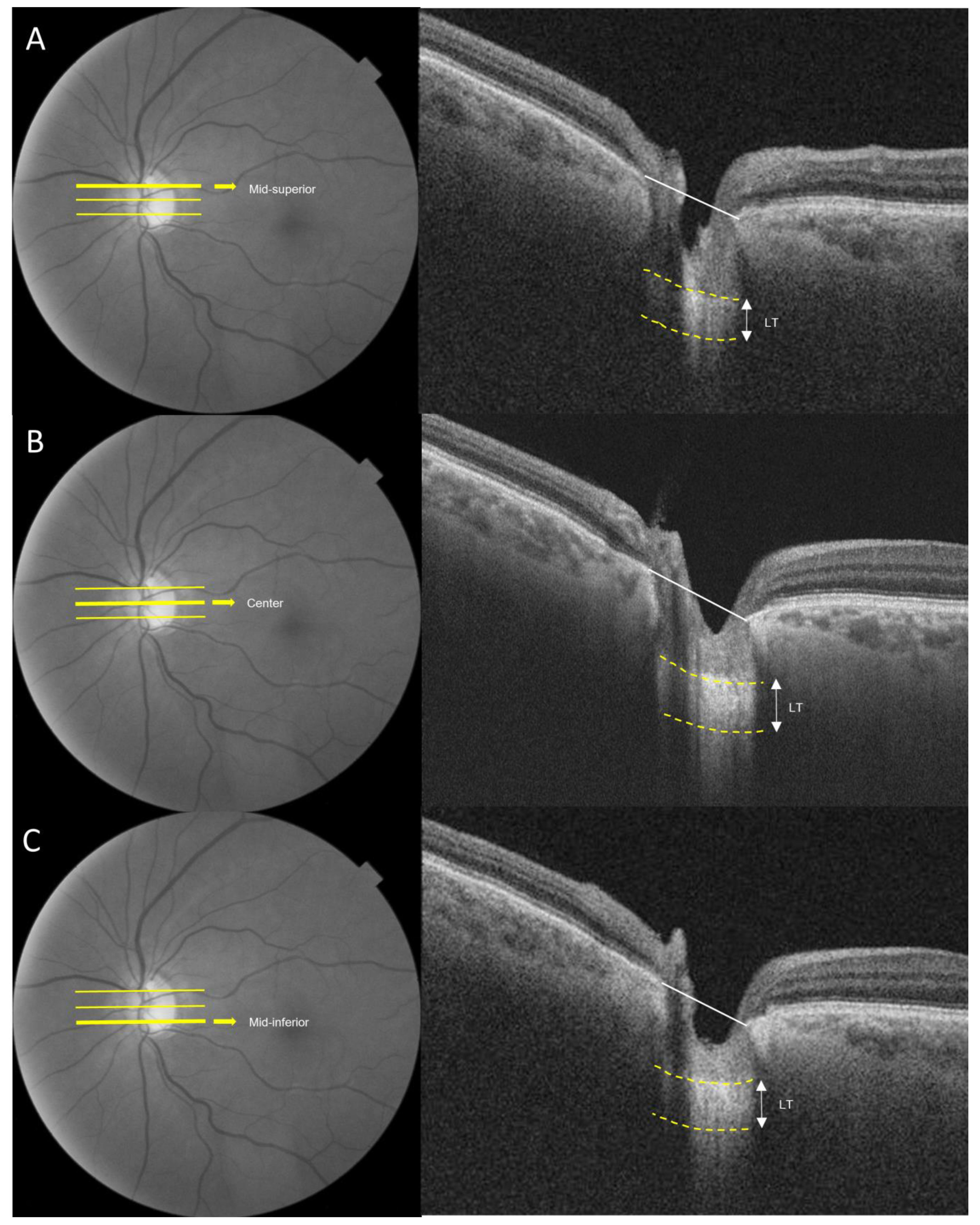
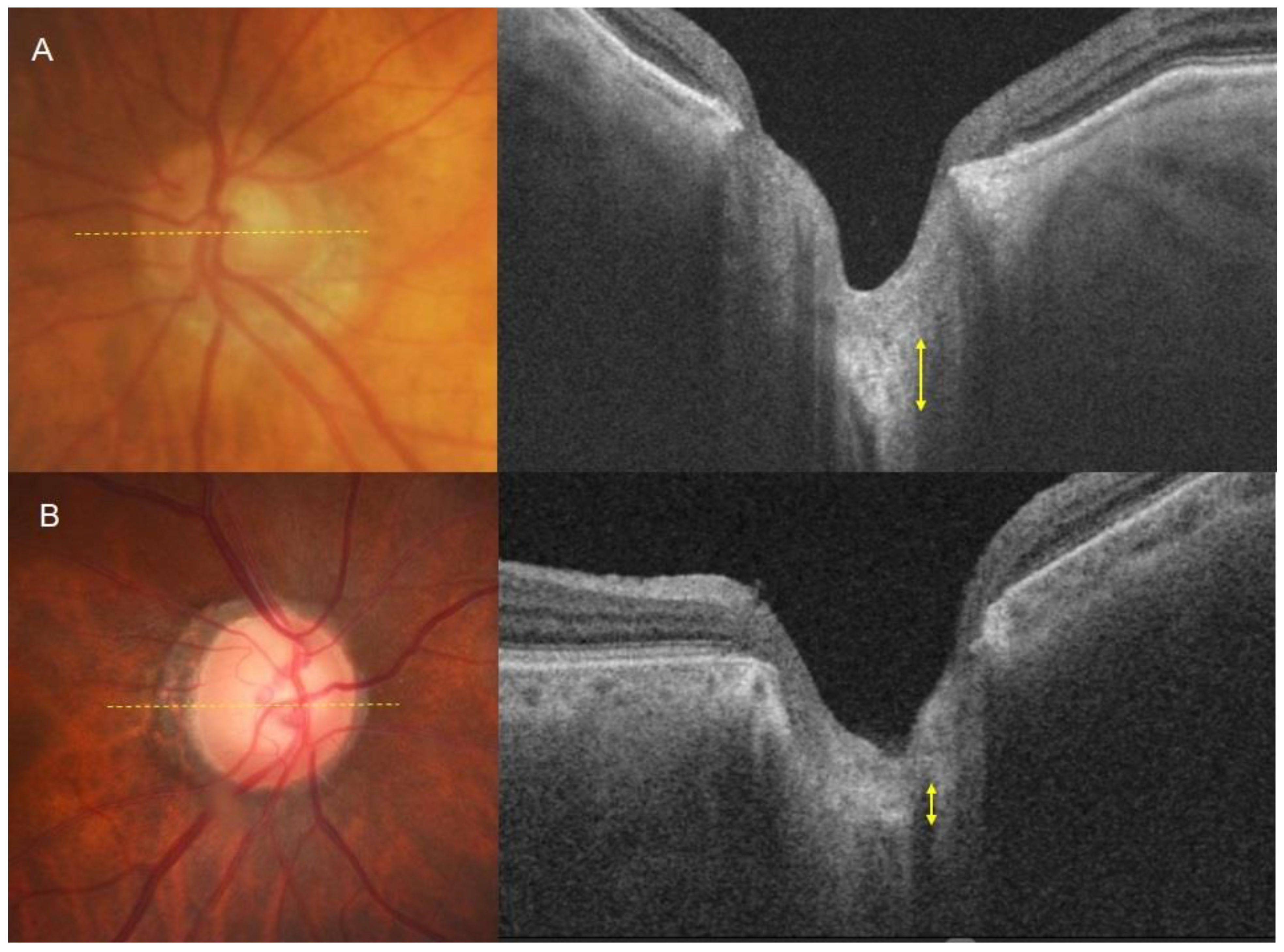
2.5. Laminar Cribrosa Assessment by SS-OCT
2.6. Statistical Analysis
3. Results
4. Discussion
Author Contributions
Funding
Institutional Review Board Statement
Informed Consent Statement
Data Availability Statement
Conflicts of Interest
References
- Naumann, G.O.; Schlötzer-Schrehardt, U.; Küchle, M. Pseudoexfoliation syndrome for the comprehensive ophthalmologist. Intraocular and systemic manifestations. Ophthalmology 1998, 105, 951–968. [Google Scholar] [CrossRef]
- Prince, A.M.; Ritch, R. Clinical signs of the pseudoexfoliation syndrome. Ophthalmology 1986, 93, 803–807. [Google Scholar] [CrossRef]
- Konstas, A.G.; Mantziris, D.A.; Stewart, W.C. Diurnal intraocular pressure in untreated exfoliation and primary open-angle glaucoma. Arch. Ophthalmol. 1997, 115, 182–185. [Google Scholar] [CrossRef]
- Ritch, R.; Schlötzer-Schrehardt, U. Exfoliation syndrome. Surv. Ophthalmol. 2001, 45, 265–315. [Google Scholar] [CrossRef] [PubMed]
- Hyman, L.; Heijl, A.; Leske, M.C.; Bengtsson, B.; Yang, Z. Natural history of intraocular pressure in the early manifest glaucoma trial: A 6-year follow-up. Arch. Ophthalmol. 2010, 128, 601–607. [Google Scholar] [CrossRef]
- Heijl, A.; Bengtsson, B.; Hyman, L.; Leske, M.C. Natural history of open-angle glaucoma. Ophthalmology 2009, 116, 2271–2276. [Google Scholar] [CrossRef]
- Grødum, K.; Heijl, A.; Bengtsson, B. Risk of glaucoma in ocular hypertension with and without pseudoexfoliation. Ophthalmology 2005, 112, 386–390. [Google Scholar] [CrossRef]
- Jünemann, A.G. Diagnosis and therapy of pseudoexfoliation glaucoma. Ophthalmol. Z. Dtsch. Ophthalmol. Ges. 2012, 109, 962–975. [Google Scholar]
- Yarangümeli, A.; Davutluoglu, B.; Köz, O.G.; Elhan, A.H.; Yaylaci, M.; Kural, G. Glaucomatous damage in normotensive fellow eyes of patients with unilateral hypertensive pseudoexfoliation glaucoma: Normotensive pseudoexfoliation glaucoma? Clin. Exp. Ophthalmol. 2006, 34, 15–19. [Google Scholar] [CrossRef] [PubMed]
- Puska, P.; Vesti, E.; Tomita, G.; Ishida, K.; Raitta, C. Optic disc changes in normotensive persons with unilateral exfoliation syndrome: A 3-year follow-up study. Graefe’s Arch. Clin. Exp. Ophthalmol. Albrecht Von Graefes Arch. Klin. Exp. Ophthalmol. 1999, 237, 457–462. [Google Scholar] [CrossRef]
- Koz, O.G.; Turkcu, M.F.; Yarangumeli, A.; Koz, C.; Kural, G. Normotensive glaucoma and risk factors in normotensive eyes with pseudoexfoliation syndrome. J. Glaucoma 2009, 18, 684–688. [Google Scholar] [CrossRef]
- Rao, A. Normotensive pseudoexfoliation glaucoma: A new phenotype? Semin. Ophthalmol. 2012, 27, 48–51. [Google Scholar] [CrossRef]
- Shih, C.Y.; Graff Zivin, J.S.; Trokel, S.L.; Tsai, J.C. Clinical significance of central corneal thickness in the management of glaucoma. Arch Ophthalmol 2004, 122, 1270–1275. [Google Scholar] [CrossRef]
- Shin, D.Y.; Jeon, S.J.; Park, H.Y.L.; Park, C.K. Posterior scleral deformation and autonomic dysfunction in normal tension glaucoma. Sci. Rep. 2020, 10, 8203. [Google Scholar] [CrossRef]
- Tay, E.; Seah, S.K.; Chan, S.P.; Lim, A.T.; Chew, S.J.; Foster, P.J.; Aung, T. Optic disk ovality as an index of tilt and its relationship to myopia and perimetry. Am. J. Ophthalmol. 2005, 139, 247–252. [Google Scholar] [CrossRef]
- How, A.C.; Tan, G.S.; Chan, Y.H.; Wong, T.T.; Seah, S.K.; Foster, P.J.; Aung, T. Population prevalence of tilted and torted optic discs among an adult Chinese population in Singapore: The Tanjong Pagar Study. Arch. Ophthalmol. 2009, 127, 894–899. [Google Scholar] [CrossRef]
- Park, H.Y.; Lee, K.; Park, C.K. Optic disc torsion direction predicts the location of glaucomatous damage in normal-tension glaucoma patients with myopia. Ophthalmology 2012, 119, 1844–1851. [Google Scholar] [CrossRef]
- Lee, E.J.; Kim, T.W.; Weinreb, R.N.; Park, K.H.; Kim, S.H.; Kim, D.M. β-Zone parapapillary atrophy and the rate of retinal nerve fiber layer thinning in glaucoma. Investig. Ophthalmol. Vis. Sci. 2011, 52, 4422–4427. [Google Scholar] [CrossRef]
- Omodaka, K.; Horii, T.; Takahashi, S.; Kikawa, T.; Matsumoto, A.; Shiga, Y.; Maruyama, K.; Yuasa, T.; Akiba, M.; Nakazawa, T. 3D evaluation of the lamina cribrosa with swept-source optical coherence tomography in normal tension glaucoma. PLoS ONE 2015, 10, e0122347. [Google Scholar] [CrossRef]
- Omodaka, K.; Takahashi, S.; Matsumoto, A.; Maekawa, S.; Kikawa, T.; Himori, N.; Takahashi, H.; Maruyama, K.; Kunikata, H.; Akiba, M.; et al. Clinical Factors Associated with Lamina Cribrosa Thickness in Patients with Glaucoma, as Measured with Swept Source Optical Coherence Tomography. PLoS ONE 2016, 11, e0153707. [Google Scholar] [CrossRef][Green Version]
- Shin, J.W.; Jo, Y.H.; Song, M.K.; Won, H.J.; Kook, M.S. Nocturnal blood pressure dip and parapapillary choroidal microvasculature dropout in normal-tension glaucoma. Sci. Rep. 2021, 11, 206. [Google Scholar] [CrossRef] [PubMed]
- Linnér, E.; Schwartz, B.; Araujo, D. Optic disc pallor and visual field defect in exfoliative and non-exfoliative, untreated ocular hypertension. Int. Ophthalmol. 1989, 13, 21–24. [Google Scholar] [CrossRef] [PubMed]
- Xu, H.; Zhai, R.; Zong, Y.; Kong, X.; Jiang, C.; Sun, X.; He, Y.; Li, X. Comparison of retinal microvascular changes in eyes with high-tension glaucoma or normal-tension glaucoma: A quantitative optic coherence tomography angiographic study. Graefe’s Arch. Clin. Exp. Ophthalmol. Albrecht Von Graefes Arch. Klin. Exp. Ophthalmol. 2018, 256, 1179–1186. [Google Scholar] [CrossRef]
- Park, H.Y.; Jeon, S.H.; Park, C.K. Enhanced depth imaging detects lamina cribrosa thickness differences in normal tension glaucoma and primary open-angle glaucoma. Ophthalmology 2012, 119, 10–20. [Google Scholar] [CrossRef] [PubMed]
- Park, H.Y.; Lee, K.I.; Lee, K.; Shin, H.Y.; Park, C.K. Torsion of the optic nerve head is a prominent feature of normal-tension glaucoma. Investig. Ophthalmol. Vis. Sci. 2014, 56, 156–163. [Google Scholar] [CrossRef]
- Sung, M.S.; Kang, Y.S.; Heo, H.; Park, S.W. Optic Disc Rotation as a Clue for Predicting Visual Field Progression in Myopic Normal-Tension Glaucoma. Ophthalmology 2016, 123, 1484–1493. [Google Scholar] [CrossRef]
- Quigley, H.; Anderson, D.R. The dynamics and location of axonal transport blockade by acute intraocular pressure elevation in primate optic nerve. Investig. Ophthalmol. 1976, 15, 606–616. [Google Scholar]
- Quigley, H.A.; Addicks, E.M.; Green, W.R.; Maumenee, A.E. Optic nerve damage in human glaucoma. II. The site of injury and susceptibility to damage. Arch. Ophthalmol. 1981, 99, 635–649. [Google Scholar] [CrossRef]
- Quigley, H.A.; Anderson, D.R. Distribution of axonal transport blockade by acute intraocular pressure elevation in the primate optic nerve head. Investig. Ophthalmol. Vis. Sci. 1977, 16, 640–644. [Google Scholar]
- Thorleifsson, G.; Magnusson, K.P.; Sulem, P.; Walters, G.B.; Gudbjartsson, D.F.; Stefansson, H.; Jonsson, T.; Jonasdottir, A.; Jonasdottir, A.; Stefansdottir, G.; et al. Common sequence variants in the LOXL1 gene confer susceptibility to exfoliation glaucoma. Science 2007, 317, 1397–1400. [Google Scholar] [CrossRef]
- Liu, X.; Zhao, Y.; Gao, J.; Pawlyk, B.; Starcher, B.; Spencer, J.A.; Yanagisawa, H.; Zuo, J.; Li, T. Elastic fiber homeostasis requires lysyl oxidase-like 1 protein. Nat. Genet. 2004, 36, 178–182. [Google Scholar] [CrossRef]
- Streeten, B.W.; Bookman, L.; Ritch, R.; Prince, A.M.; Dark, A.J. Pseudoexfoliative fibrillopathy in the conjunctiva. A relation to elastic fibers and elastosis. Ophthalmology 1987, 94, 1439–1449. [Google Scholar] [CrossRef] [PubMed]
- Rehnberg, M.; Ammitzböll, T.; Tengroth, B. Collagen distribution in the lamina cribrosa and the trabecular meshwork of the human eye. Br. J. Ophthalmol. 1987, 71, 886–892. [Google Scholar] [CrossRef]
- Hernandez, M.R. Ultrastructural immunocytochemical analysis of elastin in the human lamina cribrosa. Changes in elastic fibers in primary open-angle glaucoma. Investig. Ophthalmol. Vis. Sci. 1992, 33, 2891–2903. [Google Scholar]
- Netland, P.A.; Ye, H.; Streeten, B.W.; Hernandez, M.R. Elastosis of the lamina cribrosa in pseudoexfoliation syndrome with glaucoma. Ophthalmology 1995, 102, 878–886. [Google Scholar] [CrossRef]
- Pena, J.D.; Netland, P.A.; Vidal, I.; Dorr, D.A.; Rasky, A.; Hernandez, M.R. Elastosis of the lamina cribrosa in glaucomatous optic neuropathy. Exp. Eye Res. 1998, 67, 517–524. [Google Scholar] [CrossRef]
- Schlötzer-Schrehardt, U.; Hammer, C.M.; Krysta, A.W.; Hofmann-Rummelt, C.; Pasutto, F.; Sasaki, T.; Kruse, F.E.; Zenkel, M. LOXL1 deficiency in the lamina cribrosa as candidate susceptibility factor for a pseudoexfoliation-specific risk of glaucoma. Ophthalmology 2012, 119, 1832–1843. [Google Scholar] [CrossRef]
- Kim, S.; Sung, K.R.; Lee, J.R.; Lee, K.S. Evaluation of lamina cribrosa in pseudoexfoliation syndrome using spectral-domain optical coherence tomography enhanced depth imaging. Ophthalmology 2013, 120, 1798–1803. [Google Scholar] [CrossRef] [PubMed]
- Herndon, L.W.; Weizer, J.S.; Stinnett, S.S. Central corneal thickness as a risk factor for advanced glaucoma damage. Arch. Ophthalmol. 2004, 122, 17–21. [Google Scholar] [CrossRef] [PubMed]
- Medeiros, F.A.; Sample, P.A.; Weinreb, R.N. Corneal thickness measurements and visual function abnormalities in ocular hypertensive patients. Am. J. Ophthalmol. 2003, 135, 131–137. [Google Scholar] [CrossRef] [PubMed]
- Medeiros, F.A.; Sample, P.A.; Zangwill, L.M.; Bowd, C.; Aihara, M.; Weinreb, R.N. Corneal thickness as a risk factor for visual field loss in patients with preperimetric glaucomatous optic neuropathy. Am. J. Ophthalmol. 2003, 136, 805–813. [Google Scholar] [CrossRef] [PubMed]
- Shah, S.; Chatterjee, A.; Mathai, M.; Kelly, S.P.; Kwartz, J.; Henson, D.; McLeod, D. Relationship between corneal thickness and measured intraocular pressure in a general ophthalmology clinic. Ophthalmology 1999, 106, 2154–2160. [Google Scholar] [CrossRef] [PubMed]
- Aghaian, E.; Choe, J.E.; Lin, S.; Stamper, R.L. Central corneal thickness of Caucasians, Chinese, Hispanics, Filipinos, African Americans, and Japanese in a glaucoma clinic. Ophthalmology 2004, 111, 2211–2219. [Google Scholar] [CrossRef] [PubMed]
- Lee, E.S.; Kim, C.Y.; Ha, S.J.; Seong, G.J.; Hong, Y.J. Central corneal thickness of Korean patients with glaucoma. Ophthalmology 2007, 114, 927–930. [Google Scholar] [CrossRef] [PubMed]
- Brandt, J.D.; Gordon, M.O.; Gao, F.; Beiser, J.A.; Miller, J.P.; Kass, M.A. Adjusting intraocular pressure for central corneal thickness does not improve prediction models for primary open-angle glaucoma. Ophthalmology 2012, 119, 437–442. [Google Scholar] [CrossRef] [PubMed]
- Medeiros, F.A.; Weinreb, R.N. Is corneal thickness an independent risk factor for glaucoma? Ophthalmology 2012, 119, 435–436. [Google Scholar] [CrossRef] [PubMed]
- Congdon, N.G.; Broman, A.T.; Bandeen-Roche, K.; Grover, D.; Quigley, H.A. Central corneal thickness and corneal hysteresis associated with glaucoma damage. Am. J. Ophthalmol. 2006, 141, 868–875. [Google Scholar] [CrossRef]
- Chuang, L.-H.; Koh, Y.-Y.; Chen, H.S.L.; Lin, Y.-H.; Lai, C.-C. Thinner Central Corneal Thickness is Associated with a Decreased Parapapillary Vessel Density in Normal Tension Glaucoma. J. Ophthalmol. 2022, 2022, 1937431. [Google Scholar] [CrossRef]
- Ariga, M.; Nivean, M.; Utkarsha, P. Pseudoexfoliation Syndrome. J. Curr. Glaucoma Pract. 2013, 7, 118–120. [Google Scholar] [CrossRef]
- Jammal, H.; Abu Ameera, M.; Al Qudah, N.; Aldalaykeh, M.; Abukahel, A.; Al Amer, A.; Al Bdour, M. Characteristics of Patients with Pseudoexfoliation Syndrome at a Tertiary Eye Care Center in Jordan: A Retrospective Chart Review. Ophthalmol. Ther. 2021, 10, 51–61. [Google Scholar] [CrossRef]
- Musch, D.C.; Shimizu, T.; Niziol, L.M.; Gillespie, B.W.; Cashwell, L.F.; Lichter, P.R. Clinical characteristics of newly diagnosed primary, pigmentary and pseudoexfoliative open-angle glaucoma in the Collaborative Initial Glaucoma Treatment Study. Br. J. Ophthalmol. 2012, 96, 1180–1184. [Google Scholar] [CrossRef] [PubMed]
- Kang, J.H.; Loomis, S.; Wiggs, J.L.; Stein, J.D.; Pasquale, L.R. Demographic and geographic features of exfoliation glaucoma in 2 United States-based prospective cohorts. Ophthalmology 2012, 119, 27–35. [Google Scholar] [CrossRef] [PubMed]
- Hylton, C.; Congdon, N.; Friedman, D.; Kempen, J.; Quigley, H.; Bass, E.; Jampel, H. Cataract after glaucoma filtration surgery. Am. J. Ophthalmol. 2003, 135, 231–232. [Google Scholar] [CrossRef]


| Variable | Hypertensive PXG (86 Eyes) | Normotensive PXG (80 Eyes) | p-Value |
|---|---|---|---|
| Age, year | 73.78 ± 8.41 | 73.58 ± 10.08 | 0.890 |
| Sex, M/F | 53/33 | 28/53 | <0.001 |
| CCT, μm | 544.71 ± 61.84 | 523.30 ± 32.15 | 0.021 |
| Axial length, mm | 23.70 ± 1.04 | 23.85 ± 1.24 | 0.451 |
| Hypertension, n (%) | 35 (40.7%) | 28 (34.6%) | 0.414 |
| Diabetes, n (%) | 14(13.3%) | 8 (9.9%) | 0.200 |
| Pseudophakia, n | 37 (44.0%) | 23 (28.4%) | 0.037 |
| Medication eyedrop | 1.63 ± 0.75 | 1.93 ± 1.02 | 0.032 |
| Glaucoma surgery history | 52 (60.5%) | 4 (4.9%) | <0.001 |
| Mean IOP, mmHg | 17.95 ± 6.60 | 14.30 ± 2.95 | <0.001 |
| Corrected mean IOP, mmHg | 18.22 ± 6.95 | 15.17 ± 3.37 | 0.001 |
| Peak IOP, mmHg | 30.68 ± 8.25 | 17.78 ± 2.82 | <0.001 |
| Corrected peak IOP, mmHg | 30.84 ± 8.33 | 18.53 ± 3.27 | <0.001 |
| RNFL thickness, μm | 59.08 ± 11.75 | 66.74 ± 9.84 | <0.001 |
| VF MD, dB | −17.59 ± 10.91 | −8.69 ± 8.32 | <0.001 |
| Follow-up period, year | 5.69 ± 3.81 | 5.26 ± 3.13 | 0.470 |
| Tilt ratio | 1.12 ± 0.94 | 1.14 ± 0.12 | 0.361 |
| Torsion ratio | 90.73 ± 6.96 | 89.44 ± 8.63 | 0.611 |
| Disc fovea angle | −6.68 ± 5.36 | −6.20 ± 4.06 | 0.563 |
| PPA/Disc area > 0.4 | 18 (20.5%) | 20 (25.0%) | 0.533 |
| Variable | Hypertensive PXG (32 Eyes) | Normotensive PXG (32 Eyes) | p Value |
|---|---|---|---|
| Age, year | 74.78 ± 8.91 | 75.22 ± 7.99 | 0.837 |
| Axial length, mm | 23.47± 0.81 | 23.68 ± 1.02 | 0.434 |
| RNFL thickness, μm | 68.13 ± 11.45 | 69.00 ± 10.76 | 0.754 |
| VF MD, dB | −9.02 ± 8.78 | −8.92 ± 8.71 | 0.962 |
| VF PSD, dB | 5.68 ± 4.14 | 6.01 ± 3.48 | 0.737 |
| Lamina Cribrosa Thickness | |||
| Mid-superior, μm | 176.16 ± 22.74 | 151.66 ± 20.88 | <0.001 |
| Center, μm | 188.50 ± 29.21 | 176.63 ± 24.70 | 0.084 |
| Mid-inferior, μm | 171.19 ± 31.70 | 151.25 ± 22.31 | 0.005 |
| Mean, μm | 178.61 ± 15.74 | 159.84 ± 15.60 | <0.001 |
Disclaimer/Publisher’s Note: The statements, opinions and data contained in all publications are solely those of the individual author(s) and contributor(s) and not of MDPI and/or the editor(s). MDPI and/or the editor(s) disclaim responsibility for any injury to people or property resulting from any ideas, methods, instructions or products referred to in the content. |
© 2024 by the authors. Licensee MDPI, Basel, Switzerland. This article is an open access article distributed under the terms and conditions of the Creative Commons Attribution (CC BY) license (https://creativecommons.org/licenses/by/4.0/).
Share and Cite
Shin, D.Y.; Park, C.K.; Lee, N.Y. Characteristic Differences between Normotensive and Hypertensive Pseudoexfoliative Glaucoma. J. Clin. Med. 2024, 13, 1078. https://doi.org/10.3390/jcm13041078
Shin DY, Park CK, Lee NY. Characteristic Differences between Normotensive and Hypertensive Pseudoexfoliative Glaucoma. Journal of Clinical Medicine. 2024; 13(4):1078. https://doi.org/10.3390/jcm13041078
Chicago/Turabian StyleShin, Da Young, Chan Kee Park, and Na Young Lee. 2024. "Characteristic Differences between Normotensive and Hypertensive Pseudoexfoliative Glaucoma" Journal of Clinical Medicine 13, no. 4: 1078. https://doi.org/10.3390/jcm13041078
APA StyleShin, D. Y., Park, C. K., & Lee, N. Y. (2024). Characteristic Differences between Normotensive and Hypertensive Pseudoexfoliative Glaucoma. Journal of Clinical Medicine, 13(4), 1078. https://doi.org/10.3390/jcm13041078


