Article

Potential Causal Association Between Atrial Fibrillation/Flutter and Primary Open-Angle Glaucoma: A Two-Sample Mendelian Randomisation Study

Veterans Medical Research Institute, Veterans Health Service Medical Center, Seoul 05368, Republic of Korea
*
Author to whom correspondence should be addressed.
These authors contributed equally to this work.
J. Clin. Med. 2024, 13(24), 7670; https://doi.org/10.3390/jcm13247670
Submission received: 8 October 2024 / Revised: 12 December 2024 / Accepted: 14 December 2024 / Published: 16 December 2024
(This article belongs to the Special Issue New Insights into Glaucoma)

Abstract

:
Background: A few studies have reported controversial relationships between atrial fibrillation/flutter (AF/L) and primary open-angle glaucoma (POAG). This study aimed to investigate the potential causal relationship between AF/L and POAG. Methods: Single-nucleotide polymorphisms associated with exposure to AF/L were selected as instrumental variables with significance (p < 5.0 × 10 8 ) from a genome-wide association study (GWAS) by FinnGen. The GWAS summary of POAG from the UK Biobank was used as the outcome dataset. A two-sample Mendelian randomisation (MR) study was performed to assess the causal effects of AF/L on POAG. In addition, potential confounders, including hypertension, autoimmune hyperthyroidism, sleep apnoea, and alcohol use disorder, were assessed using multivariable MR analysis. Results: There was a significant causal association of AF/L with POAG (odds ratio [OR] = 1.26, 95% confidence interval [CI] = 1.07–1.48, p = 0.005 using inverse-variance weighting [IVW]). Multivariable MR analysis confirmed a causal association of AF/L with POAG (OR = 1.24, 95% CI = 1.02–1.51, p = 0.034 using IVW), but hypertension, hyperthyroidism, sleep apnoea and alcohol use disorder did not show significant causal associations with POAG (all p > 0.05). Conclusions: This established causal relationship between AF/L and POAG supports the need for further investigation into the role of AF/L as a possible risk factor for POAG. Further research is required to confirm these findings.

1. Introduction

Primary open-angle glaucoma (POAG), a major cause of permanent vision loss, is a progressive optic neuropathy characterised by the degeneration of retinal ganglion cells and their axons [1]. Its pathogenesis remains unclear, despite the established roles of multiple factors in its pathophysiology. Elevated intraocular pressure (IOP) is a major risk factor for POAG [2], although the precise mechanisms underlying glaucomatous optic neuropathy and the associated visual field loss remain unclear [1]. However, reports indicate that lowering IOP with long-acting drug delivery reduces the risk of glaucoma progression, highlighting that IOP is a critical factor in glaucoma [3]. Studies identifying the causal risk factors for POAG may enable the early detection and prevention of glaucoma; consequently, they form the foundation for research in ophthalmology. Several risk factors, such as ageing, hypoxia, neuroprotection, and environmental factors, have been suggested as contributing to the pathogenesis of POAG [1,4,5,6,7,8,9]. Minor injuries from repeated reperfusion, such as fluctuating IOP or disrupted autoregulation, may eventually cause oxidative stress and glaucomatous damage [10]. Cardiovascular disorders, including vasospasm, hypertension, and hypotension, are also potential risk factors for glaucoma [11].
Atrial fibrillation (AF) and atrial flutter (AFL) are disorders of significant importance to public health, especially when considering their associations with ischaemic stroke and heart failure [12,13]. AF is the most prevalent type of cardiac arrhythmia, caused by aberrant electrical activity within the atria of the heart, leading to fibrillation. AFL is similar to AF, but the rapid upper-chamber cardiac arrhythmias encountered in clinical practice are often more regular [14]. Additionally, many patients with AFL develop AF over time [15]. Recent advances in the understanding of the unique electrophysiological processes underlying AF and AFL have led to anatomy-based treatment approaches. Owing to their mechanistic similarities, AF and AFL (AF/L) are often classified and studied together. AF prevalence ranges from 2% in the general population to 10–12% in those aged ≥80 years. AF/L presents as an erratic heartbeat that may cause instability in ocular perfusion pressure. AF/L plays a significant role in heart failure, stroke, cardiovascular morbidity, and sudden death. However, the associations between AF/L, as a major cardiovascular disease, and ophthalmological diseases have not been intensively examined. A recent analysis from the Ural Eye and Medical Study showed that AF/L was not associated with major ocular diseases, including glaucoma (p = 0.90) [16]. However, Flammer et al. suggested that ischaemia and reperfusion damage caused by unstable ocular perfusion may play a role in the vascular pathophysiology of POAG [17]. An observational study also revealed that patients with normal-tension glaucoma had a higher AF incidence than controls (p = 0.022) [18]. Another recent longitudinal study evaluating the association of AF and glaucoma using Korean National Health Insurance data determined that AF increased the risk of developing glaucoma (hazard ratio = 1.31; 95% confidence interval [CI] = 1.15–1.48) [19]. Evidently, research on the relationship between AF and glaucoma is not enough.
Mendelian randomisation (MR) is a genetic epidemiological technique that uses genetic variants associated with potential risk factors (exposures) as instrumental variables (IVs) to assess their causal effects on disease outcomes [20,21]. Several studies have used MR to investigate glaucoma and its potential risk factors [22,23,24]. However, no MR studies have assessed the association between AF/L and POAG. Therefore, this study aimed to analyse the association between AF/L and POAG using univariate MR analysis and to conduct multivariate MR analysis to account for potential confounding factors, such as hypertension, hyperthyroidism, sleep apnoea, and alcohol use disorder. To achieve this goal, a two-sample MR technique was used to examine the causal effects of AF/L on POAG, utilising summary statistics from FinnGen [25] for exposures and from the UK Biobank (UKB) for outcomes [26].

2. Materials and Methods

2.1. Study Design

The study protocol was approved by the Institutional Review Board of the Veterans Health Service Medical Center (approval number. 2023-12-030), and owing to the retrospective study design, the requirement for informed consent was waived. This study was conducted in accordance with the principles of the Declaration of Helsinki.

2.2. Data Sources

The analytical study design used to investigate the causal relationship between AF/L and POAG is presented in Figure 1. The following summary datasets were used: exposure data from the FinnGen genome-wide association study (GWAS) for AF/L, which included 237,690 participants (45,766 cases vs. 191,924 controls), and outcome data from the UKB POAG GWAS, which included 456,351 participants (654 cases vs. 455,697 controls). To assess the causal relationship between AF/L and glaucoma, additional FinnGen data from the following participant types were incorporated in the multivariable MR analysis to assess potential confounders: 377,209 participants with hypertension (111,581 cases vs. 265,626 controls), 281,683 participants with autoimmune hyperthyroidism (1828 cases vs. 279,855 controls), 375,657 participants with sleep apnoea (38,998 cases vs. 336,659 controls) and 377,277 participants with alcohol use disorder (15,715 cases vs. 361,562 controls). Detailed sources of the summary data are presented in Table 1.

2.3. Selection of the Genetic IVs

The study used single-nucleotide polymorphisms (SNPs) associated with exposure at the GWAS significance threshold (p < 5.0 × 10 8 ) as IVs. To maintain the independence of each IV, SNPs were selected after pruning for linkage disequilibrium (R2 < 0.001, clumping distance = 10,000 kb). Data from the 1000 Genomes Phase III European database were used as a reference to calculate linkage disequilibrium during the clumping process. The F-statistic was calculated using the formula
F = R2(n − 2)/(1 − R2)
where n represents the sample size, and R2 is the proportion of variance in the exposure explained by the genetic variants [29]. An F-statistic >10 suggests no indication of weak instrument bias [30]. For the multivariable MR analysis, the strength of the IVs was evaluated using conditional F-statistics, with values of >10 indicating sufficient instrument strength for the analysis [31].

2.4. MR Analysis

In this study, a two-sample MR approach with SNPs was used as IVs to investigate the causal effect of AF/L on POAG. For valid causal inference, IVs were required to meet the following three assumptions: (1) SNPs should be strongly associated with AF/L; (2) SNPs should not be associated with confounders of the AF/L–POAG relationship; and (3) SNPs should influence POAG exclusively through AF/L, with no evidence of directional horizontal pleiotropy. Our primary analytical method was inverse-variance weighting (IVW) with a multiplicative random effects model [30,32,33]. The IVW approach is most efficient when all genetic variations satisfy the three IV assumptions [34]. Recognising the potential bias that may arise from invalid IVs if the assumptions were not fully met, sensitivity analyses were conducted using the weighted median method [35] and MR-Egger regression [36,37]. The weighted median method can provide a consistent estimate even if more than 50% of the IVs are invalid [35]. MR-Egger regression, which is less sensitive to pleiotropy, estimates the average horizontal pleiotropy through its intercept, providing an unbiased estimate of the causal effect [36]. Further, MR-Egger analysis with simulation extrapolation (SIMEX) was employed as it allows bias correction in scenarios where the ‘no measurement error’ assumption is violated, such as when the I2 value is <90% [37]. To assess heterogeneity and potential pleiotropy, we used Cochran’s Q statistics from the IVW method and Rücker’s Q statistics from the MR-Egger analysis [32,38]. We also employed the MR pleiotropy residual sum and outlier (MR-PRESSO) [39] test and conducted a ‘leave-one-SNP-out’ analysis to identify the influence of potentially pleiotropic SNPs on our estimates. Considering that hypertension, autoimmune hyperthyroidism, sleep apnoea, and alcohol use disorder are common risk factors for POAG, multivariable MR IVW [20] and MR-Egger [40] approaches were used to adjust for these factors and isolate the effects of AF/L on POAG. The QA statistic, a modification of Cochran’s Q, was used to assess the heterogeneity and potential pleiotropy among IVs [31]. When the conditional F-statistic or QA statistics indicated the presence of weak instruments or potential pleiotropy, the Q-minimisation approach (Q-het) was applied to estimate robust causal associations, supplementing the MVMR-IVW approach. The standard errors were calculated using the bootstrap method [31]. For exposures with overlapping samples, covariances for the effect of each SNP on each exposure were required to calculate the conditional F-statistic and QA statistics [31]. One approach to obtain these covariances is to use a phenotypic correlation matrix [31], which can be derived from the intercept of the bivariate LD score regression [41,42,43]. Causal effects are expressed as odds ratios (ORs) with 95% CIs. Statistical significance was established at a two-tailed p-value of <0.05. All MR analyses were performed using the TwoSampleMR, MVMR, Mendelian randomisation, and SIMEX packages in R version 3.6.3 (R Core Team, Vienna, Austria).

3. Results

3.1. Genetic IVs in Univariable MR

In the MR study, 85 SNPs that were significant at the GWAS level and were independent of each other were selected to serve as IVs. The F-statistic values for all the selected SNPs exceeded 10, with a mean value of 114.53, confirming that they were strong instruments. Detailed information on the IVs is provided in Supplementary Table S1. According to the results, the assumption of no measurement error was satisfied, with an I2 value of 97.93% (Table 2). Heterogeneity among the IVs was evaluated using Cochran’s Q test from the IVW method and Rücker’s Q’ test from the MR-Egger analysis and found no significant heterogeneity (p = 0.338 for Cochran’s Q and p = 0.312 for Rücker’s Q’; Table 2). Horizontal pleiotropy was assessed using the MR-PRESSO global test and MR-Egger regression, both with and without SIMEX adjustments; the MR-PRESSO global test indicated no significant pleiotropic effects (p = 0.338), and the MR-Egger intercepts showed no evidence of horizontal pleiotropy (intercept = 0.004, p = 0.797 without SIMEX; Intercept = 0.004, p = 0.848 with SIMEX), as shown in Table 2. Given that the F-statistic indicated no evidence of weak instrument bias, the I2 statistic exceeded 90, and Cochran’s Q test was not significant, the IVW method was selected for primary analysis because of its superior statistical power when core MR assumptions were satisfied [44].

3.2. Univariable MR for the Causal Effects of AF/L on POAG

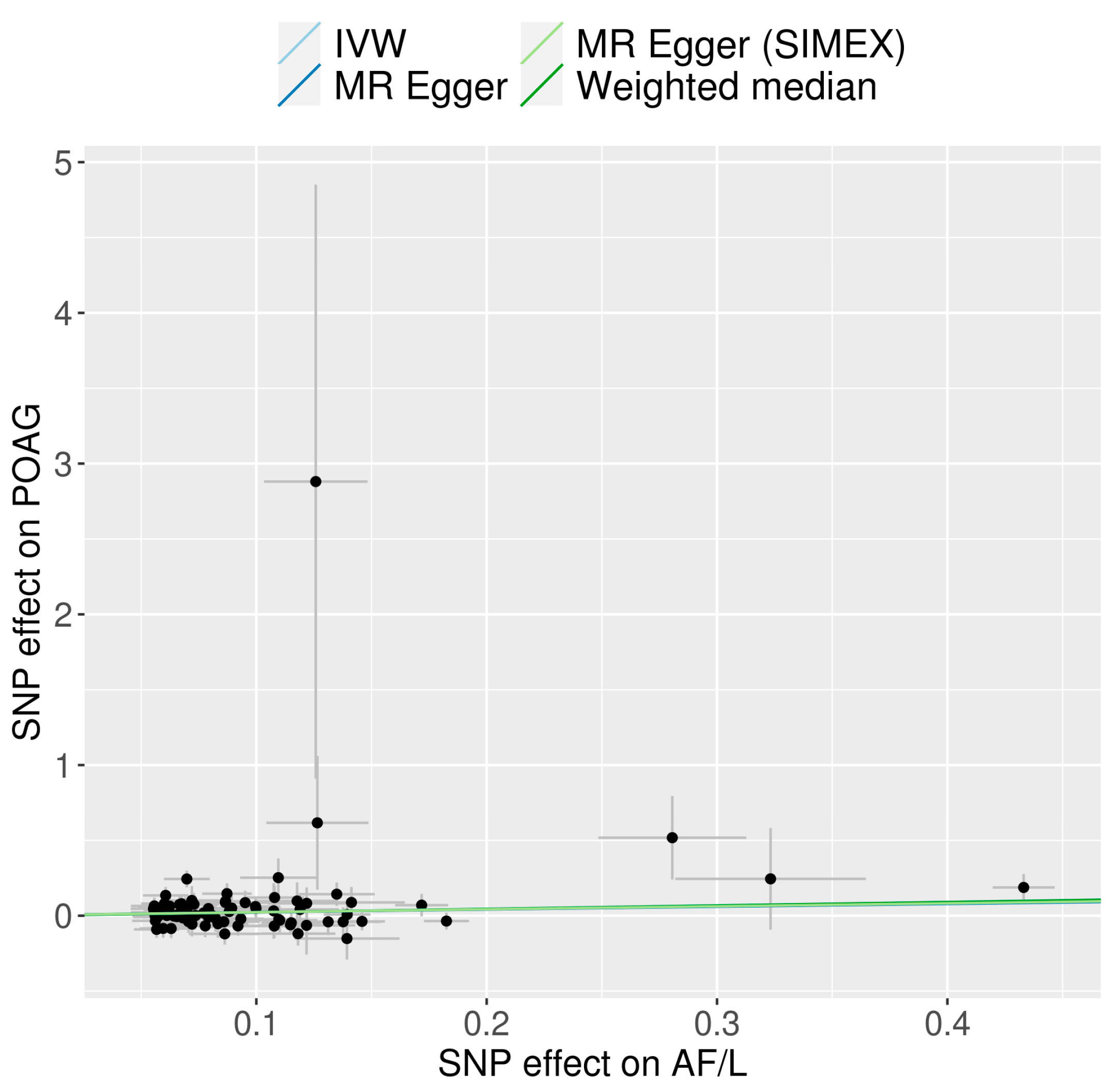
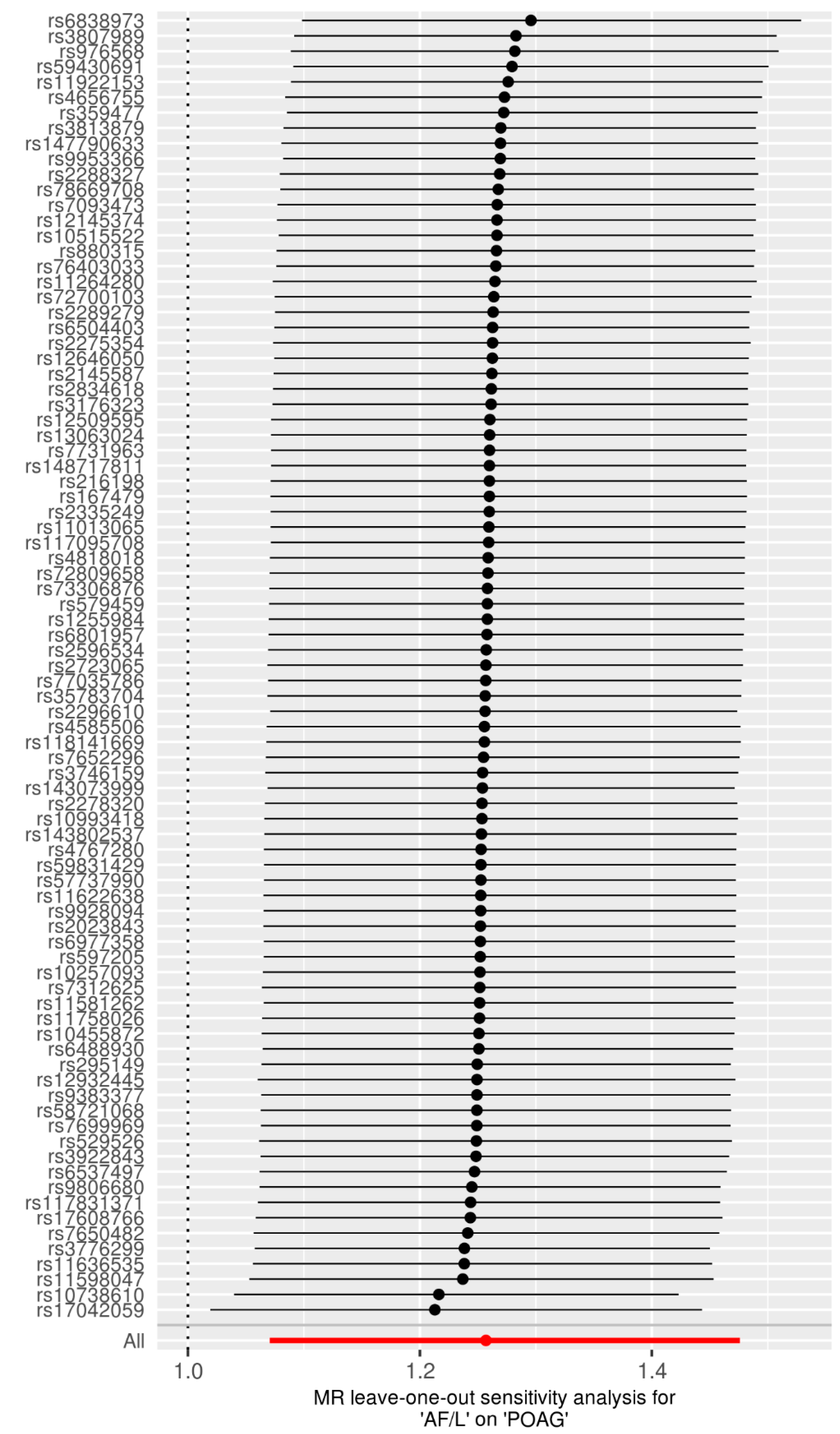
We explored the potential causal relationship between AF/L and POAG by employing 85 SNPs as IVs in a univariable MR analysis. The results, displayed in Figure 2, indicate that AF/L increases the risk of POAG according to the IVW method (OR =1.26, 95% CI = 1.07–1.48, p = 0.005). The weighted median approach revealed a similar trend, though it did not achieve statistical significance (OR = 1.25, 95% CI = 0.98–1.60, p = 0.068). No significant causal relationship was observed with the MR-Egger method, with or without SIMEX adjustment (OR = 1.20, 95% CI = 0.84–1.74, p = 0.323 without SIMEX; OR = 1.22, 95% CI = 0.82–1.81, p = 0.329 with SIMEX). Figure 3 displays a scatter plot of the effects of SNPs on AF/L and their corresponding effects on POAG. The leave-one-out analysis corroborated the primary IVW MR findings, as depicted in Figure 4.

3.3. Multivariable MR for the Causal Effects of AF/L on POAG

In the multivariable MR analysis (controlled for hypertension, autoimmune hyperthyroidism, sleep apnoea, and alcohol use disorder), the association between AF/L and POAG remained significant (IVW OR = 1.24, 95% CI = 1.02–1.51, p = 0.034), as presented in Table 3 (Model 1). Hypertension, autoimmune hyperthyroidism, sleep apnoea, and alcohol use disorder were not significantly associated with POAG in Model 1. The causal effects estimated from the multivariate MR-Egger results were consistent with those of the IVW analysis (Table 3). The QA statistic for Model 1 was 211.99, with a p-value of 0.192, indicating no substantial heterogeneity among the IVs. The conditional F-statistics for autoimmune hyperthyroidism, sleep apnoea, and alcohol use disorder were below the conventional threshold of 10 (F = 4.08, 3.19, and 3.21, respectively), indicating potentially weak instruments for these exposures. In contrast, the conditional F-statistics for AF/L and hypertension were above 10 (F = 22.48 and 14.16, respectively), suggesting sufficient instrument strength for these variables. Given the potential bias introduced by weak instruments, we performed an additional multivariate MR analysis (Model 2) that included only AF/L and hypertension, which had conditional F-statistics above the threshold of 10. In Model 2, the association between AF/L and POAG remained significant (IVW OR = 1.24, 95% CI = 1.02–1.52, p = 0.032), consistent with the findings from Model 1. Hypertension continued to show no significant association with POAG in Model 2 (IVW OR = 0.90, 95% CI = 0.69–1.18, p = 0.449). The QA statistic for Model 2 was 212.86, with a p-value of 0.133, indicating no evidence of heterogeneity. To address potentially weak instruments, we also performed an IVW analysis with Q-minimisation (Q-het) for Model 1. The results of this analysis are presented in Supplementary Table S2 and are consistent with the standard IVW results for AF/L (OR = 1.38, 95% CI = 1.02–2.05).

4. Discussion

The findings suggest a potential causal relationship between AF/L and POAG. Moreover, after controlling for the risk factors for AF/L and POAG, including hypertension, autoimmune hyperthyroidism, sleep apnoea, and alcohol use disorders, AF/L demonstrated a causal association with POAG. However, studies on the relationship between AF/L and glaucoma are limited. Potential risk factors for cardiovascular disorders include systemic hypertension, hypotension, vasospasm, elevated blood viscosity, and diabetes, particularly in the absence of elevated IOP [45]. Considering that vascular dysfunction is one of the known mechanisms of glaucoma, AF/L may be considered a cause of glaucoma. According to a longitudinal study on the association between AF and risk of glaucoma [19], AF leads to an increased risk of glaucoma (hazard ratio = 1.31, 95% CI = 1.15–1.48) with variable adjustment. In the sequence of AF onset, conditions are amenable to the formation of an intracardiac thrombus or embolus [46]. According to the literature, AF/L remains one of the main contributors to stroke, unexpected death, and cardiovascular morbidity [13,47]. AF is independently linked to a 1.5-fold higher risk of all-cause mortality in men and a 2-fold higher risk in women [48]. In clinical ophthalmological practice, microembolic complications may remain unreported [49,50]. Reperfusion injury is caused by repeated microemboli occlusion of the central retinal artery, branched posterior ciliary artery, or ophthalmic artery at any level followed by reperfusion, resulting in unstable ocular perfusion of the retina or choroid [18]. Recurrent transient ischaemic episodes can cause retinal ganglion cell death and perfusion disruption, and subsequently glaucoma. Considering that migraine and orthostatic hypotension are risk factors for POAG [51], the presence of AF/L is potentially associated with POAG.
AF/L is characterised by a highly irregular heart rate, which may contribute to unstable ocular perfusion. Given that high IOP is the primary risk factor for POAG, researchers may be curious about the effects of AF/L on IOP as a mechanism of POAG. To address these concerns, the effect of AF/L on IOP through MR analysis indicated that there was no effect on IOP changes (Supplementary Tables S3–S5). These results support the vascular theory of POAG aetiology, and AF/L may cause POAG because of unstable perfusion pressure rather than IOP. Several studies have suggested a potential association between glaucoma and AF [17]. One study identified correlations between cardiac arrhythmias, particularly AF, with visual field abnormalities and a decline in visual acuity in older patients with glaucoma. AF was also significantly more common in patients with glaucoma than in control participants [52]. Another hospital-based study of Polish patients found that AF, independent of other established cardiovascular risk factors, was associated with an increased risk of normal-tension glaucoma [18]. Normal-tension glaucoma may be influenced by factors other than IOP; however, it is included in the POAG spectrum. Recently, a study using Korean National Health Insurance Service data determined via cross-sectional analysis showed that the AF group had higher incidence rates of diabetes, hypertension, glaucoma, and chronic nephropathy [19]. In addition, a longitudinal analysis showed that patients with AF had a significantly higher cumulative incidence of glaucoma at 11 years (4.37% in the control group vs. 6.42% in the AF group) [19]. In addition, a reduced retinal capillary plexus precedes retinal ganglion cell loss in ocular hypertension [53], and macular microvascular damage is highly associated with visual field defects in POAG [54]. Considering these studies and our results simultaneously, it can be concluded that AF/L and vascular dysregulation can independently cause POAG. Although the primary focus of this study was POAG, angle-closure glaucoma should be considered when interpreting the results. Nevertheless, because this study analysed a large dataset for the risk of glaucoma compared with the severity of AF, its results are expected to be meaningful if interpreted in comparison with the study results.
Hypertension has been suggested as a risk factor for glaucoma, with a higher incidence in patients with POAG (OR = 1.55, p < 0.001) [55]. In addition, a Cox regression study found that untreated hypertension led to a greater risk of POAG [56]. AF/L and systemic hypertension frequently coexist with AF because arterial hypertension increases the risk of developing new-onset AF, and because both conditions share common risk factors and underlying pathophysiological mechanisms that contribute to their incidence [57]. A recent MR study indicated that hyperthyroidism is associated with AF, reporting that a genetically elevated FT3:FT4 ratio and hyperthyroidism, rather than FT4 levels within the reference range, are associated with an increased risk of developing AF [58]. Additionally, one study reported that individuals with thyroid eye disease in the All-of-Us Research Program were significantly more likely to be diagnosed with glaucoma [59]. Sleep apnoea is associated with POAG [60], and obstructive sleep apnoea is considered a complex and dynamic substrate for AF [61,62]. Moreover, alcohol consumption is a common trigger for AF/L, especially habitual alcohol intake [63]. A meta-analysis suggested a harmful association between alcohol use and OAG, with IOP elevation [64]. These variables are relevant confounders in studies of the association between AF/L and glaucoma. However, incorporating additional covariates, such as hypertension, autoimmune hyperthyroidism, sleep apnoea, and alcohol use disorder, into our multivariable MR analysis did not alter the significant association between AF/L and POAG. This suggests that the observed relationship between AF/L and POAG was not confounded by these factors, further supporting the causality of the association.
The primary strength of our study lies in the use of a relatively large cohort dataset, which provided evidence suggesting a causal effect of AF/L on POAG. While the study provides robust evidence for a causal relationship between these conditions, it is essential to consider limitations such as the generalisability of the findings to non-European populations, as the dataset was predominantly derived from European cohorts (Finland and the UK). Further research is required to determine whether these results can be generalised to other ethnic groups. Additionally, potential residual confounding factors cannot be ruled out despite multivariable adjustments. This study lacked access to individual-level data, which limited our ability to account for various confounding factors because our analysis relied on summary statistics derived from two-sample MR methods. We were also unable to assess the potential interactions between AF/L and other risk factors, such as hypertension, diabetes, or sleep apnoea, owing to the aggregated nature of the GWAS summary statistics used in our study. Future studies with access to individual-level data are warranted to explore these interactions and their impact on causal pathways. However, the methods employed to validate the MR hypotheses did not provide absolute confirmation. Violations of underlying MR assumptions may lead to erroneous conclusions, necessitating a cautious interpretation of the findings. The conditional F-statistics for the confounders included in the multivariable MR analysis were below the conventional threshold of 10 (F = 4.08 for autoimmune hyperthyroidism; F = 3.19 for sleep apnoea; and F = 3.21 for alcohol use disorder), indicating the presence of potentially weak instruments. To address this issue, we performed an IVW analysis with Q-minimisation (Q-het), which is robust against weak instruments and heterogeneity, to validate our findings. Even with the application of robust methods, the non-significant results for certain confounders require cautious interpretation as they may reflect limited statistical power due to weak instruments. Finally, because the prevalence rates of AF/L and POAG were not high (2% and 3%, respectively), GWAS of rare diseases using biobank data may face a severe imbalance in the number of cases and controls [65], which may produce false-positive GWAS hits [66]. When these hits are selected as IVs, they can bias the MR estimates. However, the GWAS summary statistics used in this study employed methods designed to address case-control imbalances, such as SAIGE and the firth test [65,67]. These approaches have been shown to efficiently control false-positive associations and enhance the reliability of selected IVs. Future studies could benefit from the inclusion of larger and more diverse cohorts, as well as the investigation of the specific pathways through which AF/L influences POAG risk.

5. Conclusions

The findings contribute to the understanding of the relationship between AF/L and POAG and suggest that AF/L may increase the risk of POAG. This has potential clinical implications as it highlights the importance of monitoring glaucoma risk in patients with AF/L. In addition, the value of this study lies in the recommendation that, in cases where AF/L is diagnosed in a clinical setting, ophthalmological screening should be advised to monitor the development of glaucoma. Conversely, when glaucoma is diagnosed, AF/L should be included in the medical history questionnaire for further evaluation. Further research should explore the biological mechanisms underlying this association and determine whether the effective management of AF/L could reduce the incidence of POAG.

Supplementary Materials

The following supporting information can be downloaded at https://www.mdpi.com/article/10.3390/jcm13247670/s1. Table S1: List of SNPs used as IVs in univariate MR analysis; Table S2: Multivariate MR-IVW analysis with minimised Q statistics; Table S3: Summary of intraocular pressure data; Table S4: Univariate MR results of the causal effects of AF/L on intraocular pressure; Table S5: Multivariable MR results of the causal effects of AF/L on intraocular pressure.

Author Contributions

J.H.S. and Y.L. designed the study and collected the data, Y.L. performed the statistical analysis, and J.H.S. drafted the manuscript. All authors have reviewed and edited the previous versions of the manuscript. All authors have read and agreed to the published version of the manuscript.

Funding

This study was supported by a National Research Foundation of Korea (NRF) grant funded by the Korean Government (Ministry of Science and ICT) (no. 2022R1C1C1002929) and a Veterans Health Service Medical Centre research grant (no. VHSMC24002). The funders had no role in the study design, collection, analyses, interpretation of data, writing of the manuscript, or decision to publish the results.

Institutional Review Board Statement

This study was conducted in accordance with the tenets of the Declaration of Helsinki and approved by the Institutional Review Board of the Veterans Health Service Medical Centre (no. 2023-12-030, approved on 11 January 2024).

Informed Consent Statement

Informed consent was not required because anonymised and deidentified data were used in the analyses. The requirement for patient consent was waived because of the retrospective nature of the study.

Data Availability Statement

The datasets used and/or analysed in the current study are available from FinnGen (https://finngen.gitbook.io/documentation/data-download, accessed on 4 November 2023) and the GWAS catalogue (https://www.ebi.ac.uk/gwas/summary-statistics, accessed on 4 November 2023).

Acknowledgments

We would like to thank FinnGen (https://www.finngen.fi/en) and the GWAS catalogue (https://www.ebi.ac.uk/gwas).

Conflicts of Interest

The authors declare no conflicts of interest. The funders had no role in the study design; collection, analyses, or interpretation of data; writing of the manuscript; or decision to publish the results.

References

  1. Weinreb, R.N.; Aung, T.; Medeiros, F.A. The pathophysiology and treatment of glaucoma: A review. JAMA 2014, 311, 1901–1911. [Google Scholar] [CrossRef] [PubMed]
  2. Li, T.; Lindsley, K.; Rouse, B.; Hong, H.; Shi, Q.; Friedman, D.S.; Wormald, R.; Dickersin, K. Comparative Effectiveness of First-Line Medications for Primary Open-Angle Glaucoma: A Systematic Review and Network Meta-analysis. Ophthalmology 2016, 123, 129–140. [Google Scholar] [CrossRef] [PubMed]
  3. Luo, L.J.; Nguyen, D.D.; Lai, J.Y. Benzoic acid derivative-modified chitosan-g-poly(N-isopropylacrylamide): Methoxylation effects and pharmacological treatments of Glaucoma-related neurodegeneration. J. Control Release 2020, 317, 246–258. [Google Scholar] [CrossRef] [PubMed]
  4. Jonas, J.B.; Aung, T.; Bourne, R.R.; Bron, A.M.; Ritch, R.; Panda-Jonas, S. Glaucoma. Lancet 2017, 390, 2183–2193. [Google Scholar] [CrossRef] [PubMed]
  5. Shin, H.T.; Yoon, B.W.; Seo, J.H. Analysis of risk allele frequencies of single nucleotide polymorphisms related to open-angle glaucoma in different ethnic groups. BMC Med. Genom. 2021, 14, 80. [Google Scholar] [CrossRef]
  6. Seo, J.H.; Kim, T.W.; Weinreb, R.N. Lamina cribrosa depth in healthy eyes. Investig. Ophthalmol. Vis. Sci. 2014, 55, 1241–1251. [Google Scholar] [CrossRef]
  7. Yanagi, M.; Kawasaki, R.; Wang, J.J.; Wong, T.Y.; Crowston, J.; Kiuchi, Y. Vascular risk factors in glaucoma: A review. Clin. Exp. Ophthalmol. 2011, 39, 252–258. [Google Scholar] [CrossRef]
  8. Jonas, J.B. Role of cerebrospinal fluid pressure in the pathogenesis of glaucoma. Acta Ophthalmol. 2011, 89, 505–514. [Google Scholar] [CrossRef]
  9. Seo, J.H.; Kim, T.W.; Weinreb, R.N.; Kim, Y.A.; Kim, M. Relationship of intraocular pressure and frequency of spontaneous retinal venous pulsation in primary open-angle glaucoma. Ophthalmology 2012, 119, 2254–2260. [Google Scholar] [CrossRef]
  10. Flammer, J.; Mozaffarieh, M. What is the present pathogenetic concept of glaucomatous optic neuropathy? Surv. Ophthalmol. 2007, 52 (Suppl. S2), S162–S173. [Google Scholar] [CrossRef]
  11. Wierzbowska, J.; Wierzbowski, R.; Stankiewicz, A.; Siesky, B.; Harris, A. Cardiac autonomic dysfunction in patients with normal tension glaucoma: 24-h heart rate and blood pressure variability analysis. Br. J. Ophthalmol. 2012, 96, 624–628. [Google Scholar] [CrossRef] [PubMed]
  12. Li, X.; Liu, Z.; Jiang, X.; Xia, R.; Li, Y.; Pan, X.; Yao, Y.; Fan, X. Global, regional, and national burdens of atrial fibrillation/flutter from 1990 to 2019: An age-period-cohort analysis using the Global Burden of Disease 2019 study. J. Glob. Health 2023, 13, 04154. [Google Scholar] [CrossRef] [PubMed]
  13. Elsheikh, S.; Hill, A.; Irving, G.; Lip, G.Y.H.; Abdul-Rahim, A.H. Atrial fibrillation and stroke: State-of-the-art and future directions. Curr. Probl. Cardiol. 2024, 49, 102181. [Google Scholar] [CrossRef] [PubMed]
  14. Mareedu, R.K.; Abdalrahman, I.B.; Dharmashankar, K.C.; Granada, J.F.; Chyou, P.H.; Sharma, P.P.; Smith, P.N.; Hayes, J.J.; Greenlee, R.T.; Vidaillet, H. Atrial flutter versus atrial fibrillation in a general population: Differences in comorbidities associated with their respective onset. Clin. Med. Res. 2010, 8, 1–6. [Google Scholar] [CrossRef]
  15. Waldo, A.L.; Feld, G.K. Inter-relationships of atrial fibrillation and atrial flutter mechanisms and clinical implications. J. Am. Coll. Cardiol. 2008, 51, 779–786. [Google Scholar] [CrossRef]
  16. Bikbov, M.M.; Kazakbaeva, G.M.; Rakhimova, E.M.; Panda-Jonas, S.; Fakhretdinova, A.A.; Tuliakova, A.M.; Rusakova, I.A.; Jonas, J.B. Atrial fibrillation and flutter and ocular diseases. The Ural eye and medical study and the Ural very old study. Acta Ophthalmol. 2024, 102, e1057–e1065. [Google Scholar] [CrossRef]
  17. Flammer, J.; Orgul, S.; Costa, V.P.; Orzalesi, N.; Krieglstein, G.K.; Serra, L.M.; Renard, J.P.; Stefansson, E. The impact of ocular blood flow in glaucoma. Prog. Retin. Eye Res. 2002, 21, 359–393. [Google Scholar] [CrossRef]
  18. Zaleska-Zmijewska, A.; Janiszewski, M.; Wawrzyniak, Z.M.; Kuch, M.; Szaflik, J.; Szaflik, J.P. Is atrial fibrillation a risk factor for normal-tension glaucoma? Medicine 2017, 96, e8347. [Google Scholar] [CrossRef]
  19. Cho, H.K.; Han, J.C.; Choi, J.A.; Chae, J.E.; Kim, R.B. Association between atrial fibrillation and the risk of glaucoma development: A 12-year Nationwide cohort study. Eye 2023, 37, 2033–2041. [Google Scholar] [CrossRef]
  20. Burgess, S.; Thompson, S.G. Multivariable Mendelian randomization: The use of pleiotropic genetic variants to estimate causal effects. Am. J. Epidemiol. 2015, 181, 251–260. [Google Scholar] [CrossRef]
  21. Burgess, S.; Thompson, S.G. Interpreting findings from Mendelian randomization using the MR-Egger method. Eur. J. Epidemiol. 2017, 32, 377–389. [Google Scholar] [CrossRef]
  22. Seo, J.H.; Lee, Y. Possible Causal Association between Type 2 Diabetes and Glycaemic Traits in Primary Open-Angle Glaucoma: A Two-Sample Mendelian Randomisation Study. Biomedicines 2024, 12, 866. [Google Scholar] [CrossRef]
  23. Wang, K.; Yang, F.; Liu, X.; Lin, X.; Yin, H.; Tang, Q.; Jiang, L.; Yao, K. Appraising the Effects of Metabolic Traits on the Risk of Glaucoma: A Mendelian Randomization Study. Metabolites 2023, 13, 109. [Google Scholar] [CrossRef]
  24. Chong, R.S.; Li, H.; Cheong, A.J.Y.; Fan, Q.; Koh, V.; Raghavan, L.; Nongpiur, M.E.; Cheng, C.Y. Mendelian Randomization Implicates Bidirectional Association between Myopia and Primary Open-Angle Glaucoma or Intraocular Pressure. Ophthalmology 2023, 130, 394–403. [Google Scholar] [CrossRef]
  25. Sakaue, S.; Kanai, M.; Tanigawa, Y.; Karjalainen, J.; Kurki, M.; Koshiba, S.; Narita, A.; Konuma, T.; Yamamoto, K.; Akiyama, M.; et al. A cross-population atlas of genetic associations for 220 human phenotypes. Nat. Genet. 2021, 53, 1415–1424. [Google Scholar] [CrossRef]
  26. Choquet, H.; Paylakhi, S.; Kneeland, S.C.; Thai, K.K.; Hoffmann, T.J.; Yin, J.; Kvale, M.N.; Banda, Y.; Tolman, N.G.; Williams, P.A.; et al. A multiethnic genome-wide association study of primary open-angle glaucoma identifies novel risk loci. Nat. Commun. 2018, 9, 2278. [Google Scholar] [CrossRef]
  27. Kurki, M.I.; Karjalainen, J.; Palta, P.; Sipila, T.P.; Kristiansson, K.; Donner, K.M.; Reeve, M.P.; Laivuori, H.; Aavikko, M.; Kaunisto, M.A.; et al. FinnGen provides genetic insights from a well-phenotyped isolated population. Nature 2023, 613, 508–518. [Google Scholar] [CrossRef]
  28. Jiang, L.; Zheng, Z.; Fang, H.; Yang, J. A generalized linear mixed model association tool for biobank-scale data. Nat. Genet. 2021, 53, 1616–1621. [Google Scholar] [CrossRef]
  29. Burgess, S.; Thompson, S.G.; CRP CHD Genetics Collaboration. Avoiding bias from weak instruments in Mendelian randomization studies. Int. J. Epidemiol. 2011, 40, 755–764. [Google Scholar] [CrossRef]
  30. Burgess, S.; Butterworth, A.; Thompson, S.G. Mendelian randomization analysis with multiple genetic variants using summarized data. Genet. Epidemiol. 2013, 37, 658–665. [Google Scholar] [CrossRef]
  31. Sanderson, E.; Spiller, W.; Bowden, J. Testing and correcting for weak and pleiotropic instruments in two-sample multivariable Mendelian randomization. Stat. Med. 2021, 40, 5434–5452. [Google Scholar] [CrossRef]
  32. Bowden, J.; Del Greco, M.F.; Minelli, C.; Davey Smith, G.; Sheehan, N.; Thompson, J. A framework for the investigation of pleiotropy in two-sample summary data Mendelian randomization. Stat. Med. 2017, 36, 1783–1802. [Google Scholar] [CrossRef]
  33. Lee, Y.; Kim, Y.A.; Seo, J.H. Causal Association of Obesity and Dyslipidemia with Type 2 Diabetes: A Two-Sample Mendelian Randomization Study. Genes 2022, 13, 2407. [Google Scholar] [CrossRef]
  34. Burgess, S.; Davey Smith, G.; Davies, N.M.; Dudbridge, F.; Gill, D.; Glymour, M.M.; Hartwig, F.P.; Holmes, M.V.; Minelli, C.; Relton, C.L.; et al. Guidelines for performing Mendelian randomization investigations. Wellcome Open Res. 2019, 4, 186. [Google Scholar] [CrossRef]
  35. Bowden, J.; Davey Smith, G.; Haycock, P.C.; Burgess, S. Consistent Estimation in Mendelian Randomization with Some Invalid Instruments Using a Weighted Median Estimator. Genet. Epidemiol. 2016, 40, 304–314. [Google Scholar] [CrossRef]
  36. Bowden, J.; Davey Smith, G.; Burgess, S. Mendelian randomization with invalid instruments: Effect estimation and bias detection through Egger regression. Int. J. Epidemiol. 2015, 44, 512–525. [Google Scholar] [CrossRef]
  37. Bowden, J.; Del Greco, M.F.; Minelli, C.; Davey Smith, G.; Sheehan, N.A.; Thompson, J.R. Assessing the suitability of summary data for two-sample Mendelian randomization analyses using MR-Egger regression: The role of the I2 statistic. Int. J. Epidemiol. 2016, 45, 1961–1974. [Google Scholar] [CrossRef]
  38. Greco, M.F.; Minelli, C.; Sheehan, N.A.; Thompson, J.R. Detecting pleiotropy in Mendelian randomisation studies with summary data and a continuous outcome. Stat. Med. 2015, 34, 2926–2940. [Google Scholar] [CrossRef]
  39. Verbanck, M.; Chen, C.Y.; Neale, B.; Do, R. Publisher Correction: Detection of widespread horizontal pleiotropy in causal relationships inferred from Mendelian randomization between complex traits and diseases. Nat. Genet. 2018, 50, 1196. [Google Scholar] [CrossRef]
  40. Rees, J.M.B.; Wood, A.M.; Burgess, S. Extending the MR-Egger method for multivariable Mendelian randomization to correct for both measured and unmeasured pleiotropy. Stat. Med. 2017, 36, 4705–4718. [Google Scholar] [CrossRef]
  41. Bulik-Sullivan, B.K.; Loh, P.R.; Finucane, H.K.; Ripke, S.; Yang, J.; Patterson, N.; Daly, M.J.; Price, A.L.; Neale, B.M.; Schizophrenia Working Group of the Psychiatric Genomics Consortium. Score regression distinguishes confounding from polygenicity in genome-wide association studies. Nat. Genet. 2015, 47, 291–295. [Google Scholar] [CrossRef] [PubMed]
  42. Bulik-Sullivan, B.; Finucane, H.K.; Anttila, V.; Gusev, A.; Day, F.R.; Loh, P.R.; ReproGen, C.; Psychiatric Genomics Consortium; Duncan, L.; Genetic Consortium for Anorexia Nervosa of the Wellcome Trust Case Control Consortium; et al. An atlas of genetic correlations across human diseases and traits. Nat. Genet. 2015, 47, 1236–1241. [Google Scholar] [CrossRef]
  43. Zheng, J.; Richardson, T.G.; Millard, L.A.C.; Hemani, G.; Elsworth, B.L.; Raistrick, C.A.; Vilhjalmsson, B.; Neale, B.M.; Haycock, P.C.; Smith, G.D.; et al. PhenoSpD: An integrated toolkit for phenotypic correlation estimation and multiple testing correction using GWAS summary statistics. Gigascience 2018, 7, giy090. [Google Scholar] [CrossRef]
  44. Jin, H.; Lee, S.; Won, S. Causal Evaluation of Laboratory Markers in Type 2 Diabetes on Cancer and Vascular Diseases Using Various Mendelian Randomization Tools. Front. Genet. 2020, 11, 597420. [Google Scholar] [CrossRef]
  45. Broadway, D.C.; Drance, S.M. Glaucoma and vasospasm. Br. J. Ophthalmol. 1998, 82, 862–870. [Google Scholar] [CrossRef]
  46. Migdady, I.; Russman, A.; Buletko, A.B. Atrial Fibrillation and Ischemic Stroke: A Clinical Review. Semin. Neurol. 2021, 41, 348–364. [Google Scholar] [CrossRef]
  47. Naser, N.; Kulic, M.; Dilic, M.; Dzubur, A.; Durak, A.; Pepic, E.; Smajic, E.; Kusljugic, Z. The Cumulative Incidence of Stroke, Myocardial infarction, Heart Failure and Sudden Cardiac Death in Patients with Atrial Fibrillation. Med. Arch. 2017, 71, 316–319. [Google Scholar] [CrossRef]
  48. Lip, G.Y.H.; Proietti, M.; Potpara, T.; Mansour, M.; Savelieva, I.; Tse, H.F.; Goette, A.; Camm, A.J.; Blomstrom-Lundqvist, C.; Gupta, D.; et al. Atrial fibrillation and stroke prevention: 25 years of research at EP Europace journal. Europace 2023, 25, euad226. [Google Scholar] [CrossRef]
  49. Hayreh, S.S.; Podhajsky, P.A.; Zimmerman, M.B. Retinal artery occlusion: Associated systemic and ophthalmic abnormalities. Ophthalmology 2009, 116, 1928–1936. [Google Scholar] [CrossRef]
  50. Yen, J.C.; Lin, H.L.; Hsu, C.A.; Li, Y.C.; Hsu, M.H. Atrial Fibrillation and Coronary Artery Disease as Risk Factors of Retinal Artery Occlusion: A Nationwide Population-Based Study. BioMed Res. Int. 2015, 2015, 374616. [Google Scholar] [CrossRef]
  51. Flammer, J.; Konieczka, K.; Flammer, A.J. The primary vascular dysregulation syndrome: Implications for eye diseases. EPMA J. 2013, 4, 14. [Google Scholar] [CrossRef] [PubMed]
  52. Perasalo, R.; Perasalo, J.; Raitta, C. Electrocardiographic changes in institutionalized geriatric glaucoma patients. Graefes Arch. Clin. Exp. Ophthalmol. 1992, 230, 213–217. [Google Scholar] [CrossRef] [PubMed]
  53. Pitale, P.M.; Shen, G.; Sigireddi, R.R.; Polo-Prieto, M.; Park, Y.H.; Gibson, S.E.; Westenskow, P.D.; Channa, R.; Frankfort, B.J. Selective vulnerability of the intermediate retinal capillary plexus precedes retinal ganglion cell loss in ocular hypertension. Front. Cell Neurosci. 2022, 16, 1073786. [Google Scholar] [CrossRef] [PubMed]
  54. Tao, A.; Liang, Y.; Chen, J.; Hu, H.; Huang, Q.; Zheng, J.; Ye, C.; Lu, F. Structure-function correlation of localized visual field defects and macular microvascular damage in primary open-angle glaucoma. Microvasc. Res. 2020, 130, 104005. [Google Scholar] [CrossRef]
  55. Wu, J.; Wang, H.; Yu, S. Hypertension as a Risk Factor for Primary Open-angle Glaucoma: A Meta-analysis. J. Coll. Physicians Surg. Pak. 2023, 33, 915–918. [Google Scholar] [CrossRef]
  56. Lee, J.S.; Kim, Y.J.; Kim, S.; Bae, H.W.; Kim, S.S.; Lee, S.W.; Kim, C.Y. Increased Risks of Open-Angle Glaucoma in Untreated Hypertension. Am. J. Ophthalmol. 2023, 252, 111–120. [Google Scholar] [CrossRef]
  57. Kallistratos, M.S.; Poulimenos, L.E.; Manolis, A.J. Atrial fibrillation and arterial hypertension. Pharmacol. Res. 2018, 128, 322–326. [Google Scholar] [CrossRef]
  58. Ellervik, C.; Roselli, C.; Christophersen, I.E.; Alonso, A.; Pietzner, M.; Sitlani, C.M.; Trompet, S.; Arking, D.E.; Geelhoed, B.; Guo, X.; et al. Assessment of the Relationship Between Genetic Determinants of Thyroid Function and Atrial Fibrillation: A Mendelian Randomization Study. JAMA Cardiol. 2019, 4, 144–152. [Google Scholar] [CrossRef]
  59. Delavar, A.; Saseendrakumar, B.R.; Lee, T.C. Associations Between Thyroid Eye Disease and Glaucoma Among Those Enrolled in the National Institutes of Health All of Us Research Program: Corrigendum. Ophthalmic Plast. Reconstr. Surg. 2023, 39, 405. [Google Scholar] [CrossRef]
  60. Leggewie, B.; Gouveris, H.; Bahr, K. A Narrative Review of the Association between Obstructive Sleep Apnea and Glaucoma in Adults. Int. J. Mol. Sci. 2022, 23, 10080. [Google Scholar] [CrossRef]
  61. Linz, D.; Nattel, S.; Kalman, J.M.; Sanders, P. Sleep Apnea and Atrial Fibrillation. Card. Electrophysiol. Clin. 2021, 13, 87–94. [Google Scholar] [CrossRef] [PubMed]
  62. Chung, M.K.; Eckhardt, L.L.; Chen, L.Y.; Ahmed, H.M.; Gopinathannair, R.; Joglar, J.A.; Noseworthy, P.A.; Pack, Q.R.; Sanders, P.; Trulock, K.M.; et al. Lifestyle and Risk Factor Modification for Reduction of Atrial Fibrillation: A Scientific Statement From the American Heart Association. Circulation 2020, 141, e750–e772. [Google Scholar] [CrossRef] [PubMed]
  63. Voskoboinik, A.; Marcus, G.M. The Impact of Alcohol Intake on Atrial Fibrillation. Curr. Cardiol. Rep. 2020, 22, 111. [Google Scholar] [CrossRef] [PubMed]
  64. Stuart, K.V.; Madjedi, K.; Luben, R.N.; Chua, S.Y.L.; Warwick, A.N.; Chia, M.; Pasquale, L.R.; Wiggs, J.L.; Kang, J.H.; Hysi, P.G.; et al. Alcohol, Intraocular Pressure, and Open-Angle Glaucoma: A Systematic Review and Meta-analysis. Ophthalmology 2022, 129, 637–652. [Google Scholar] [CrossRef]
  65. Zhou, W.; Nielsen, J.B.; Fritsche, L.G.; Dey, R.; Gabrielsen, M.E.; Wolford, B.N.; LeFaive, J.; VandeHaar, P.; Gagliano, S.A.; Gifford, A.; et al. Efficiently controlling for case-control imbalance and sample relatedness in large-scale genetic association studies. Nat. Genet. 2018, 50, 1335–1341. [Google Scholar] [CrossRef]
  66. Zhao, S.S.; Burgess, S. Use of Mendelian randomization to assess the causal status of modifiable exposures for rheumatic diseases. Best Pract. Res. Clin. Rheumatol. 2024, 38, 101967. [Google Scholar] [CrossRef]
  67. Wang, X. Firth logistic regression for rare variant association tests. Front. Genet. 2014, 5, 187. [Google Scholar] [CrossRef]
Figure 1. Diagram of two-sample Mendelian randomisation analysis. Solid lines indicate an association, while dashed lines indicate none. Abbreviation: SNP, single-nucleotide polymorphism.
Jcm 13 07670 g001
Figure 2. Forest plot of causal association of AF/L with POAG. The plot presents causal effect estimates from various MR methods, including IVW, MR-Egger, and MR-Egger with SIMEX correction. Each method is represented with its corresponding causal estimate and 95% CI. The x-axis indicates the OR for POAG associated with the presence of AF/L. Odds ratios greater than 1 suggest that AF/L increases the risk of POAG. Abbreviations: AF/L, atrial fibrillation and flutter; CI, confidence interval; OR, odds ratio; SNP, single-nucleotide polymorphism; IVW, inverse-variance weighting; MR, Mendelian randomisation; POAG, primary open-angle glaucoma; SIMEX, simulation extrapolation.
Jcm 13 07670 g002
Figure 3. Scatterplot of MR tests showing the effect of AF/L on POAG. Each dot corresponds to an SNP, with the x-axis representing the association between the SNP and the exposure and the y-axis representing the association between the SNP and the outcome. The regression lines are colour-coded: light blue for IVW, dark blue for MR-Egger, light green for MR-Egger (SIMEX), and dark green for the Weighted Median method. The slope of each line indicates the causal effect estimate obtained through the respective method. Abbreviations: MR, Mendelian randomisation; AF/L, atrial fibrillation and flutter; IVW, inverse-variance weighting; SIMEX, simulation extrapolation; SNP, single-nucleotide polymorphism; POAG, primary open-angle glaucoma.
Jcm 13 07670 g003
Figure 4. Leave-one-out plot in univariable MR analysis for the effect of AF/L on POAG. Each black dot represents the OR estimate of the causal effect after excluding a specific SNP, with the horizontal line around each dot indicating the corresponding 95% CI. The red dot and horizontal line represent the overall causal estimate (OR and CI) calculated using all SNPs. The x-axis represents ORs, where values greater than 1 suggest that AF/L increases the risk of POAG. Abbreviations: MR, Mendelian randomisation; AF/L, atrial fibrillation and flutter; POAG, primary open-angle glaucoma; OR, odds ratio; CI, confidence interval.
Jcm 13 07670 g004
Table 1. Summary statistics of data sources.
TraitsData SourceNo. of ParticipantsPopulationNo. of VariantsReference
Atrial fibrillation and flutterFinnGen237,690 (45,766 cases + 191,924 controls)European (Finland)20,164,886[27]
HypertensionFinnGen377,209 (111,581 cases + 265,626 controls)European (Finland)20,170,234
Autoimmune hyperthyroidismFinnGen281,683 (1828 cases + 279,855 controls)European (Finland)20,167,370
Sleep apnoeaFinnGen375,657 (38,998 cases + 336,659 controls)European (Finland)20,170,208
Alcohol use disorder, ICD-basedFinnGen377,277 (15,715 cases + 361,562 controls)European (Finland)20,170,236
Primary open-angle glaucomaUKB456,351 (654 cases + 455,697 controls)European (UK)11,831,932[28]
Abbreviations: ICD, International Classification of Diseases; UK, United Kingdom; UKB, UK Biobank.
Table 2. Heterogeneity and horizontal pleiotropy of instrumental variables.
Exposure HeterogeneityHorizontal Pleiotropy
MR-EggerMR-Egger (SIMEX)
NFI2 (%)p-Value *p-Value #p-Value †Intercept,
β (SE)
p-ValueIntercept,
β (SE)
p-Value
Atrial fibrillation and flutter85114.5397.930.3380.3120.3380.004 (0.017)0.7970.004 (0.018)0.848
Abbreviations: β, beta coefficient; F, mean F-statistic; MR, Mendelian randomisation; N, number of instruments; PRESSO, pleiotropy residual sum and outlier; SE, standard error; SIMEX, simulation extrapolation. * Cochran’s Q test from the inverse-variance weighting. # Rücker’s Q’ test from MR-Egger. † MR-PRESSO global test.
Table 3. Multivariable MR analysis.
IVWMR-Egger
ExposuresConditional FOR (95% CI)p-ValueOR (95% CI)p-Value
Model 1
Atrial fibrillation and flutter22.481.24 (1.02, 1.51)0.0341.24 (1.02, 1.51)0.034
Hypertension14.161.00 (0.74, 1.34)0.9841.00 (0.74, 1.34)0.983
Autoimmune hyperthyroidism4.081.03 (0.91, 1.17)0.6171.04 (0.90, 1.20)0.607
Sleep apnoea3.190.60 (0.33, 1.09)0.0920.60 (0.33, 1.09)0.094
Alcohol use disorder, ICD-based3.211.09 (0.80, 1.48)0.5961.09 (0.80, 1.48)0.595
Model 2
Atrial fibrillation and flutter20.751.24 (1.02, 1.52)0.0321.28 (1.04, 1.57)0.019
Hypertension25.840.90 (0.69, 1.18)0.4491.12 (0.69, 1.83)0.637
Abbreviations: MR, Mendelian randomisation; F, F-statistic; IVW, inverse-variance weighting; OR, odds ratio; CI, confidence interval; ICD, International Classification of Diseases.
Disclaimer/Publisher’s Note: The statements, opinions and data contained in all publications are solely those of the individual author(s) and contributor(s) and not of MDPI and/or the editor(s). MDPI and/or the editor(s) disclaim responsibility for any injury to people or property resulting from any ideas, methods, instructions or products referred to in the content.