Article

Wide-Field Optical Coherence Tomography in Patients with Diabetic Macular Edema

by
Krzysztof Kiciński
1 and
Maciej Gawęcki
1,2,*
1
Department of Ophthalmology, Pomeranian Hospitals, 84-120 Wejherowo, Poland
2
Dobry Wzrok Ophthalmological Clinic, 80-822 Gdansk, Poland
*
Author to whom correspondence should be addressed.
J. Clin. Med. 2024, 13(14), 4242; https://doi.org/10.3390/jcm13144242
Submission received: 30 June 2024 / Revised: 16 July 2024 / Accepted: 19 July 2024 / Published: 20 July 2024
(This article belongs to the Special Issue Causes and Advanced Treatments of Macular Edema)

Abstract

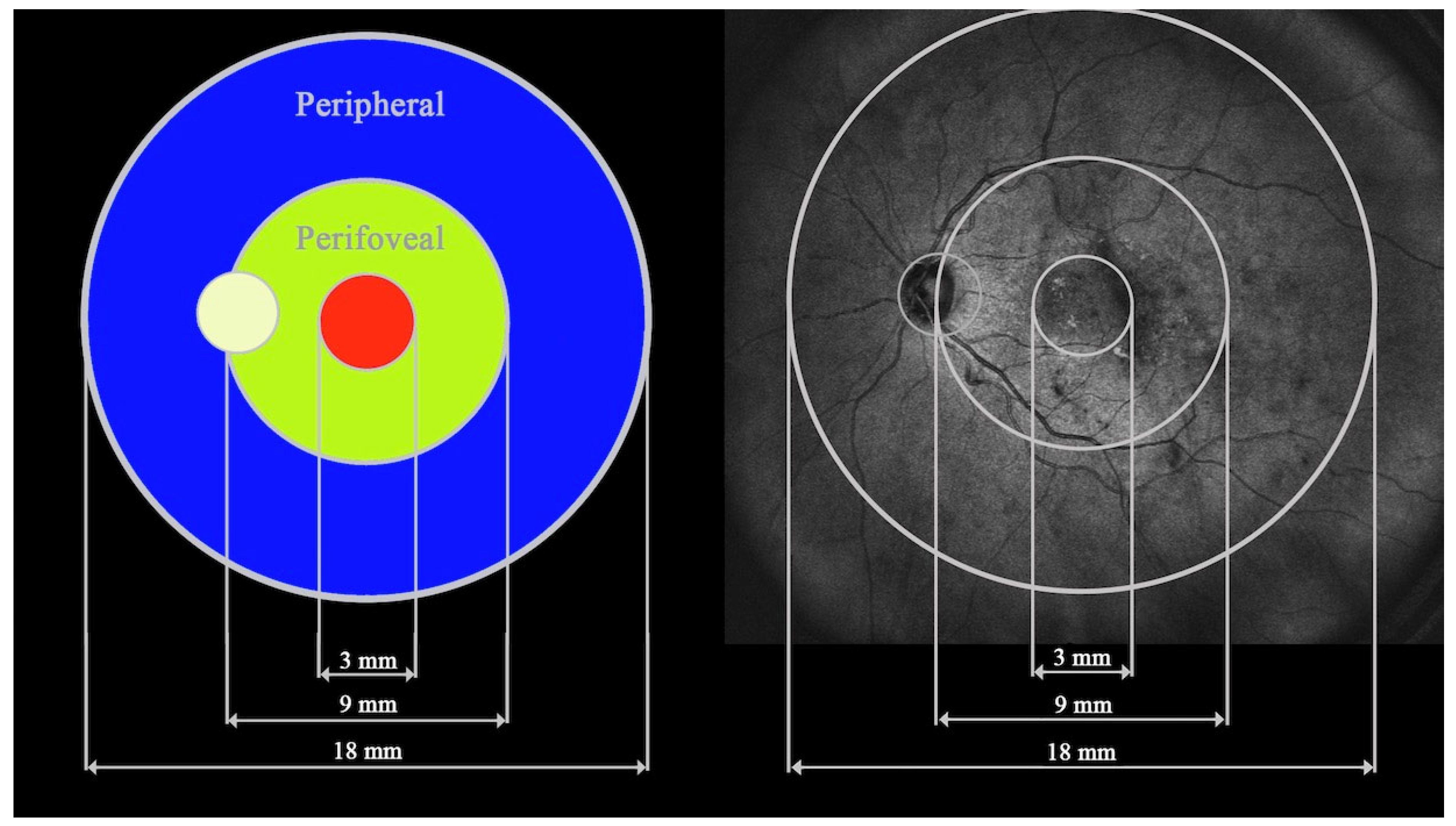
Background: The goal of the study was to analyze variations in central, perifoveal, and peripheral retinal thickness (RT) and choroidal thickness (CT) in patients with diabetic macular edema (DME) measured with ultra-wide-field optical coherence tomography (UWF-OCT). Additionally, correlations between RT and CT in the central, perifoveal, and peripheral sectors and the presence of selected systemic factors were evaluated. Methods: A total of 74 consecutive adult diabetic patients with DME and 75 healthy controls were included. Study participants were divided into three groups: DME patients without panretinal photocoagulation (PRP; 84 eyes), DME patients after PRP (56 eyes), and healthy controls (125 eyes). RT and CT were analyzed in three zones: a central circle of 3 mm diameter (central), a ring contained between a centered 9 mm circle and the central 3 mm circle (perifoveal), and a second, more peripheral ring between centered 18 mm and 9 mm circles (peripheral). Additionally, DME subgroups were analyzed according to the correlation of RT and CT with age, axial length, best corrected visual acuity (BCVA), diabetes duration, insulin therapy duration, body mass index (BMI), glycosylated hemoglobin (HbA1c) values, intravitreal injection (IVI) count, and the advancement of retinopathy assessed by the simplified diabetic retinopathy severity scale (DRSS). Results: The increase in RT in the far peripheral sectors in DME patients was not significant. The increases in central and perifoveal RT and lower values of CT in PRP-naive DME patients were strongly associated with poorer BCVA. Patients with DME after PRP presented with BCVA improvements significantly related to the number of IVIs. The amount of DME and RT in peripheral sectors were both independent of systemic factors such as BMI, duration of diabetes, duration of insulin intake, retinopathy severity, and HbA1c levels. Conclusions: Peripheral retinal sectors in DME patients are less affected in terms of increase in their thickness compared to central ones. Functional and morphological associations of DME with UWF-OCT testing refer to central and perifoveal sectors.
Keywords: ultra-wide-field optical coherence tomography; choroidal thickness; retinal thickness; diabetic macular edema; diabetes ultra-wide-field optical coherence tomography; choroidal thickness; retinal thickness; diabetic macular edema; diabetes