
Dual-Energy Computed Tomography in Urological Diseases: A Narrative Review

 , , ,
, , ,  , , , , , and
Abstract
1. Introduction
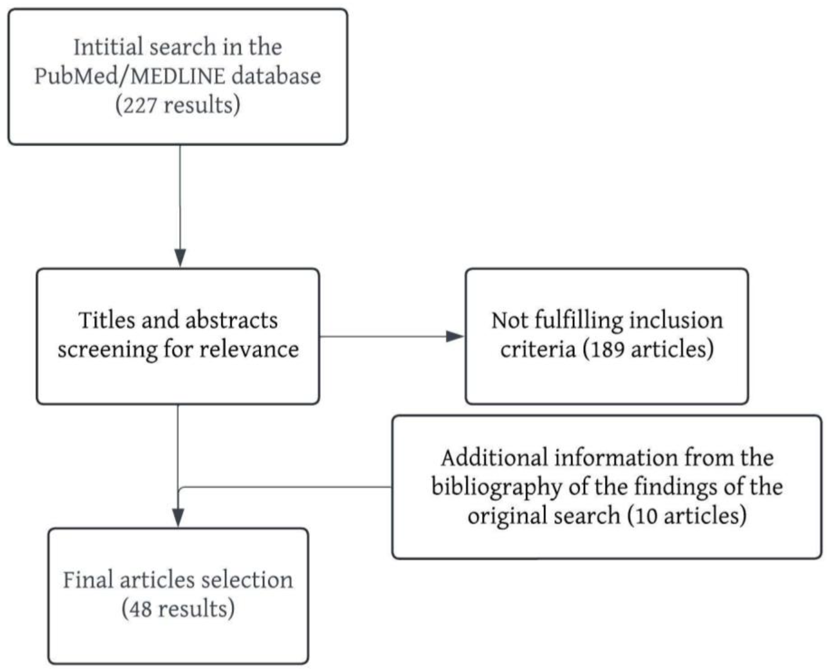
2. Materials and Methods
3. Results and Discussion
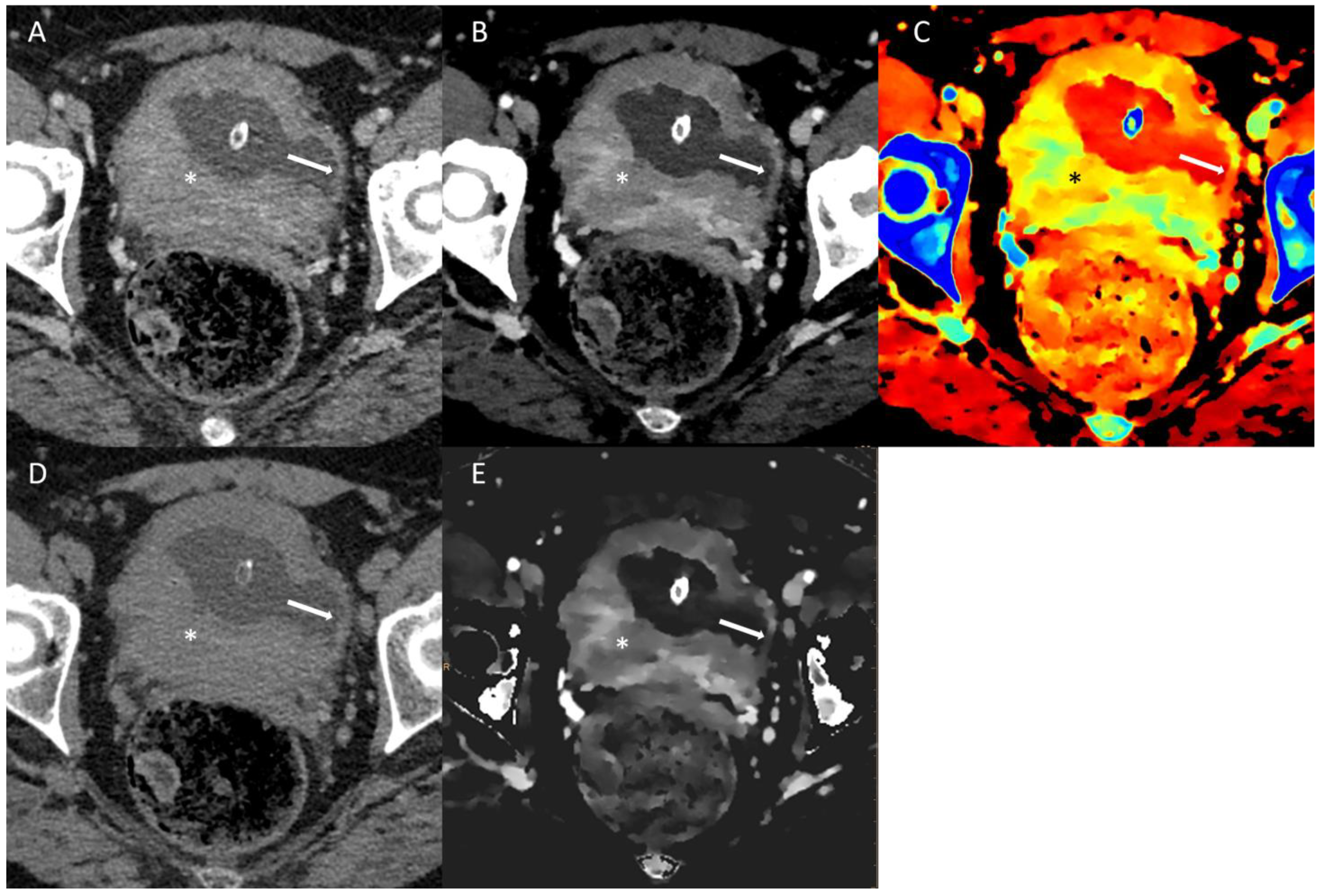
3.1. Renal Cell Carcinoma
3.2. Polycystic Kidney Disease
3.3. Urothelial Cancer
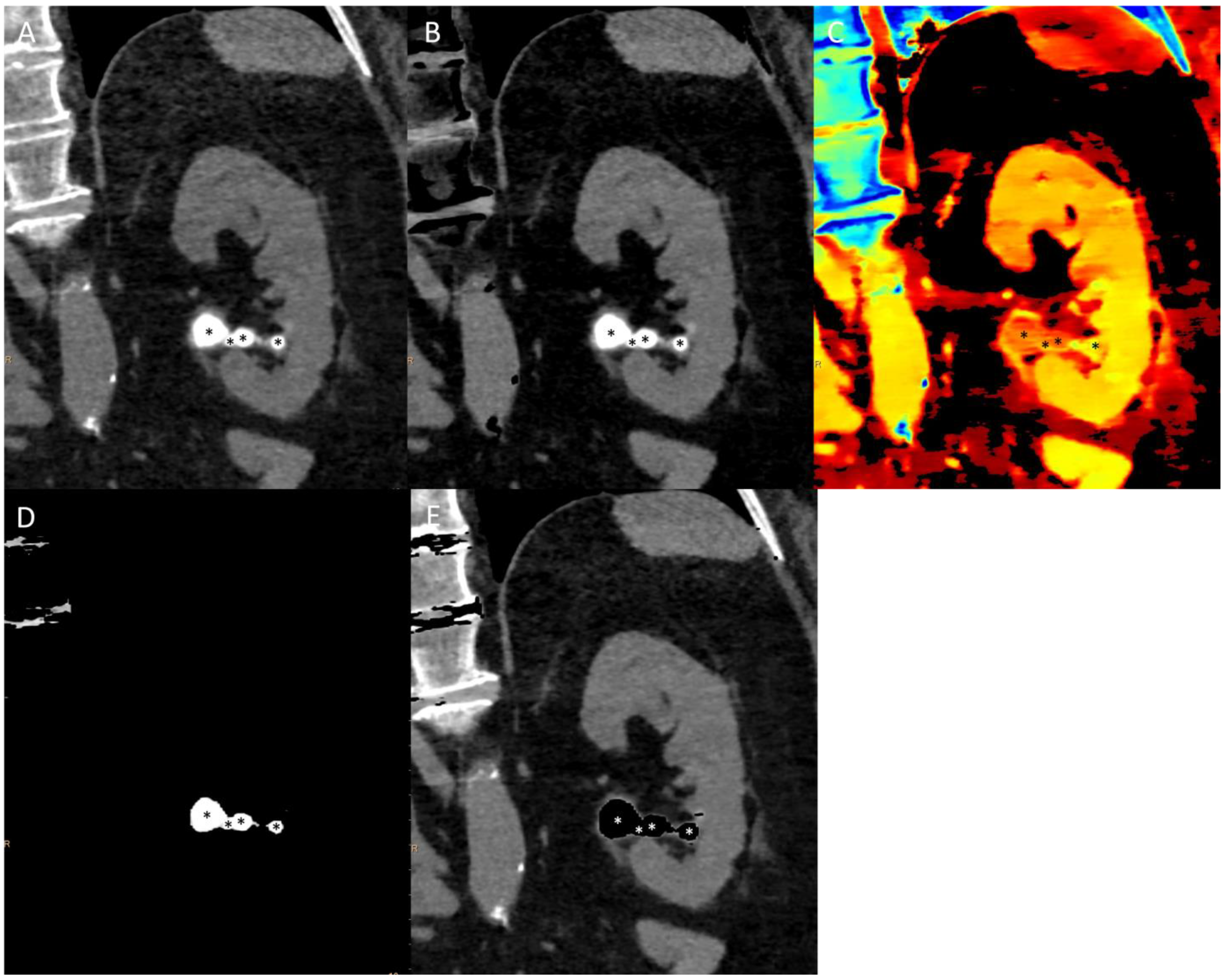
3.4. Urolithiasis
3.5. Lymph Nodes
3.6. Limitations
4. Conclusions

| Author | Journal | Year | Pathology | Technique | Study Design | |
|---|---|---|---|---|---|---|
| Salameh JP [16] | AJR | 2019 | Renal Cell Carcinoma | qIDI | Systematic Review and Meta-Analysis | \ |
| Wang D. [17] | Acta Radiol | 2020 | Renal Cell Carcinoma | qIDI | Retrospective | in vivo |
| Manoharan D. [18] | AJR | 2018 | Renal Cell Carcinoma | qIDI | Prospective | in vivo |
| Manoharan D. [19] | AJR | 2020 | Renal Cell Carcinoma | qIDI | Prospective | in vivo |
| Dai C. [20] | Abdom Radiol (NY) | 2018 | Renal Cell Carcinoma | qIDI | Retrospective | in vivo |
| Obmann M.M. [21] | Abdom Radiol (NY) | 2020 | Renal Cell Carcinoma | qIDI | Retrospective | in vivo |
| Udare A. [22] | Eur Radiol | 2020 | Renal Cell Carcinoma | qIDI | Prospective | in vivo |
| Zhang B. [23] | BMC Cancer | 2021 | Renal Cell Carcinoma | IDI | Retrospective | in vivo |
| Mileto A. [24] | AJR | 2017 | Renal Cell Carcinoma | Z-eff; Z-maps | Retrospective | in vivo |
| Bucolo G.M. [25] | JCM | 2023 | Renal Cell Carcinoma | VNC | Retrospective | in vivo |
| Han D. [26] | Clin Radiol | 2021 | Renal Cell Carcinoma | VMI; Radiomics | Retrospective | in vivo |
| Ding Y. [27] | Acta Radiol | 2022 | Renal Cell Carcinoma | VMI; IDI; Radiomics | Retrospective | in vivo |
| Park S.Y. [30] | Eur J Radiol | 2014 | Renal Cell Carcinoma | VMI; VNC | Retrospective | in vivo |
| Arndt N. [34] | Eur Radiol | 2012 | Polycystic Kidney Disease | VMI; VNC; qIDI; IDI | Prospective | in vivo |
| Graser A. [35] | Invest Radiol | 2010 | Polycystic Kidney Disease | VNC; IDI | Prospective | in vivo |
| Hansen C. [39] | AJR | 2014 | Urothelial Carcinoma | VNC; qIDI | Retrospective | in vivo |
| Zopfs, D. [42] | Eur J Radiol | 2019 | Urothelial Carcinoma | VMI | Prospective | in vivo |
| Chen A. [48] | Medicine (Baltimore) | 2016 | Urothelial Carcinoma | VMI; Z-eff | Retrospective | in vivo |
| Mansouri M. [53] | Curr Probl Diagn Radiol | 2015 | Urolithiasis | VMI; qIDI | Review | \ |
| Hidas G. [54] | Radiology | 2010 | Urolithiasis | VMI | Prospective | in vivo |
| Leng S. [55] | AJR | 2015 | Urolithiasis | VMI | Prospective | in vivo |
| Manglaviti G. [56] | AJR | 2011 | Urolithiasis | VMI | Retrospective | in vivo |
| Primak A.N. [57] | Acad Radiol | 2007 | Urolithiasis | VMI | Prospective | ex vivo |
| Stolzmann P. [58] | Abdom Imaging | 2010 | Urolithiasis | VMI | Prospective | in vivo |
| Wisenbaugh E.S. [59] | Urology | 2014 | Urolithiasis | VMI | Prospective | ex vivo |
| Ferrero A. [60] | Acad Radiol | 2016 | Urolithiasis | VMI | Prospective | ex vivo |
| Moon J.W. [61] | Br J Radiol | 2012 | Urolithiasis | VNC | Retrospective | in vivo |
| Takahashi N. [62] | AJR | 2008 | Urolithiasis | VMI; VNC; IDI | Prospective | ex vivo |
| Takahashi N. [63] | Radiology | 2010 | Urolithiasis | VMI; VNC | Retrospective | in vivo |
| Mangold S. [64] | Radiology | 2012 | Urolithiasis | VNC | Retrospective | in vivo |
| Zorzetto G. [6] | Eur Radiol Exp | 2022 | Lymph nodes | VMI; Z-eff | Retrospective | in vivo |
| Hu X. [65] | J Comput Assist Tomogr | 2021 | Lymph nodes | VMI; VNC; IDI | Retrospective | in vivo |
| Nagano H. [66] | AJR | 2022 | Lymph nodes | IDI | Retrospective | in vivo |
| Liu J. [67] | Abdom Radiol (NY) | 2023 | Lymph nodes | IDI; qIDI; Z-eff; λ | Retrospective | in vivo |
| Jin D. [68] | Front Oncol | 2022 | Lymph nodes | VMI; IDI | Retrospective | in vivo |
| Yoon J. [69] | PLoS One | 2021 | Lymph nodes | Z-eff; IDI; λ | Retrospective | in vivo |
| An C. [70] | Eur J Nucl Med Mol Imaging | 2022 | Lymph nodes | VMI | Prospective | in vivo |
| Lennartz S. [71] | Clin Nucl Med | 2021 | Lymph nodes | IDI | Retrospective | in vivo |
| Rizzo S. [72] | Eur Radiol | 2018 | Lymph nodes | VMI; IDI | Retrospective | in vivo |
| Yang Z. [73] | AJR | 2019 | Lymph nodes | Z-eff | Prospective | in vivo |
| Fan S. [74] | Eur J Radiol | 2017 | Lymph nodes | VMI; qIDI | Retrospective | in vivo |
| Liu H. [76] | Eur J Radiol | 2015 | Lymph nodes | VMI; qIDI | Retrospective | in vivo |
| Chen W.-B. [77] | Int J Colorectal Dis | 2022 | Lymph nodes | VMI; qIDI; Z-eff; λ | Retrospective | in vivo |
| Tawfik A.M. [78] | Eur Radiol | 2014 | Lymph nodes | IDI | Retrospective | in vivo |
| Pan Z. [79] | PLoS One | 2013 | Lymph nodes | VMI; qIDI | Prospective | in vivo |
| Lennartz S. [80] | Br J Radiol | 2020 | Lymph nodes | VMI | Retrospective | in vivo |
Author Contributions
Funding
Institutional Review Board Statement
Informed Consent Statement
Data Availability Statement
Conflicts of Interest
Abbreviations
| ADPKD | Autosomal Dominant Polycystic Kidney Disease |
| APKD | Acquired Polycystic Kidney Disease |
| AUC | Area Under the Curve |
| BPH | Benign Prostate Hyperplasia |
| ccRCC | Clear-cell Renal Cell Carcinoma |
| chRCC | Chromophobe Renal Cell Carcinoma |
| CT | Computed Tomography |
| DECT | Dual-Energy Computed Tomography |
| GSI | Gemstone Spectral Imaging |
| IDI | Iodine Density Index |
| λ | Slope of the Attenuation Curve |
| LNs | Lymph Nodes |
| MRI | Magnetic Resonance Imaging |
| PKD | Polycystic Kidney Disorders |
| pRCC | Papillary Renal Cell Carcinoma |
| qIDI | Quantitative Iodine Density Imaging Analysis |
| RCC | Renal Cell Carcinoma |
| ROC | Receiver Operating Characteristic |
| VMI | Virtual Monoenergetic Imaging |
| VNC | Virtual Non-Contrast |
| Z-eff | Effective Atomic Number |
| Z-Maps | Maps of Prevalent Atomic Number |
References
- Moch, H.; Amin, M.B.; Berney, D.M.; Compérat, E.M.; Gill, A.J.; Hartmann, A.; Menon, S.; Raspollini, M.R.; Rubin, M.A.; Srigley, J.R.; et al. The 2022 World Health Organization Classification of Tumours of the Urinary System and Male Genital Organs-Part A: Renal, Penile, and Testicular Tumours. Eur. Urol. 2022, 82, 458–468. [Google Scholar] [CrossRef] [PubMed]
- Netto, G.J.; Amin, M.B.; Berney, D.M.; Compérat, E.M.; Gill, A.J.; Hartmann, A.; Menon, S.; Raspollini, M.R.; Rubin, M.A.; Srigley, J.R.; et al. The 2022 World Health Organization Classification of Tumors of the Urinary System and Male Genital Organs-Part B: Prostate and Urinary Tract Tumors. Eur. Urol. 2022, 82, 469–482. [Google Scholar] [CrossRef] [PubMed]
- Goo, H.W.; Goo, J.M. Dual-Energy CT: New Horizon in Medical Imaging. Korean J. Radiol. 2017, 18, 555–569. [Google Scholar] [CrossRef] [PubMed]
- Bicci, E.; Mastrorosato, M.; Danti, G.; Lattavo, L.; Bertelli, E.; Cozzi, D.; Pradella, S.; Agostini, S.; Miele, V. Dual-Energy CT Applications in UrinaryTractCancers: An Update. Tumori 2023, 109, 148–156. [Google Scholar] [CrossRef] [PubMed]
- D’Angelo, T.; Cicero, G.; Mazziotti, S.; Ascenti, G.; Albrecht, M.H.; Martin, S.S.; Othman, A.E.; Vogl, T.J.; Wichmann, J.L. Dual Energy ComputedTomography Virtual Monoenergetic Imaging: Technique and Clinical Applications. Br. J. Radiol. 2019, 92, 20180546. [Google Scholar] [CrossRef] [PubMed]
- Zorzetto, G.; Coppola, A.; Molinelli, V.; Angeretti, M.G.; Casarin, J.; Fontana, F.; Piacentino, F.; Carcano, G.; Ghezzi, F.; Venturini, M. Spectral CT in Peritoneal Carcinomatosis from Ovarian Cancer: A Tool for Differential Diagnosis of Small Nodules? Eur. Radiol. Exp. 2022, 6, 45. [Google Scholar] [CrossRef] [PubMed]
- Curti, M.; Fontana, F.; Piacentino, F.; Ossola, C.; Coppola, A.; Carcano, G.; Venturini, M. Dual-Layer Spectral CT Fusion Imaging for LungBiopsies: More Accurate Targets, Diagnostic Samplings, and Biomarker Information? Eur. Radiol. Exp. 2022, 6, 34. [Google Scholar] [CrossRef] [PubMed]
- Ferlay, J.; Colombet, M.; Soerjomataram, I.; Dyba, T.; Randi, G.; Bettio, M.; Gavin, A.; Visser, O.; Bray, F. Cancer Incidence and Mortality Patterns in Europe: Estimates for 40 Countries and 25 Major Cancers in 2018. Eur. J. Cancer 2018, 103, 356–387. [Google Scholar] [CrossRef] [PubMed]
- Bukavina, L.; Bensalah, K.; Bray, F.; Carlo, M.; Challacombe, B.; Karam, J.A.; Kassouf, W.; Mitchell, T.; Montironi, R.; O’Brien, T.; et al. Epidemiology of Renal Cell Carcinoma: 2022 Update. Eur. Urol. 2022, 82, 529–542. [Google Scholar] [CrossRef] [PubMed]
- Capitanio, U.; Bensalah, K.; Bex, A.; Boorjian, S.A.; Bray, F.; Coleman, J.; Gore, J.L.; Sun, M.; Wood, C.; Russo, P. Epidemiology of Renal Cell Carcinoma. Eur. Urol. 2019, 75, 74–84. [Google Scholar] [CrossRef] [PubMed]
- Lane, B.R.; Kattan, M.W. Predicting Outcomes in Renal Cell Carcinoma. Curr. Opin. Urol. 2005, 15, 289–297. [Google Scholar] [CrossRef] [PubMed]
- Prasad, S.R.; Humphrey, P.A.; Catena, J.R.; Narra, V.R.; Srigley, J.R.; Cortez, A.D.; Dalrymple, N.C.; Chintapalli, K.N. Common and Uncommon Histologic Subtypes of Renal Cell Carcinoma: Imaging Spectrum with Pathologic Correlation. Radiographics 2006, 26, 1795–1806; discussion 1806–1810. [Google Scholar] [CrossRef] [PubMed]
- Low, G.; Huang, G.; Fu, W.; Moloo, Z.; Girgis, S. Review of Renal Cell Carcinoma and Its Common Subtypes in Radiology. World J. Radiol. 2016, 8, 484–500. [Google Scholar] [CrossRef] [PubMed]
- Sheir, K.Z.; El-Azab, M.; Mosbah, A.; El-Baz, M.; Shaaban, A.A. Differentiation of Renal Cell Carcinoma Subtypes by Multislice Computerized Tomography. J. Urol. 2005, 174, 451–455; discussion 455. [Google Scholar] [CrossRef] [PubMed]
- Kondo, T.; Nakazawa, H.; Sakai, F.; Kuwata, T.; Onitsuka, S.; Hashimoto, Y.; Toma, H. Spoke-Wheel-like Enhancement as an Important Imaging Finding of Chromophobe Cell Renal Carcinoma: A Retrospective Analysis on Computed Tomography and Magnetic Resonance Imaging Studies. Int. J. Urol. 2004, 11, 817–824. [Google Scholar] [CrossRef] [PubMed]
- Salameh, J.-P.; McInnes, M.D.F.; McGrath, T.A.; Salameh, G.; Schieda, N. Diagnostic Accuracy of Dual-Energy CT for Evaluation of Renal Masses: Systematic Review and Meta-Analysis. AJR Am. J. Roentgenol. 2019, 212, W100–W105. [Google Scholar] [CrossRef] [PubMed]
- Wang, D.; Huang, X.; Bai, L.; Zhang, X.; Wei, J.; Zhou, J. Differential Diagnosis of Chromophobe Renal Cell Carcinoma and Papillary Renal Cell Carcinoma with Dual-Energy Spectral Computed Tomography. Acta Radiol. 2020, 61, 1562–1569. [Google Scholar] [CrossRef] [PubMed]
- Manoharan, D.; Sharma, S.; Das, C.J.; Kumar, R.; Singh, G.; Kumar, P. Single-Acquisition Triple-Bolus Dual-Energy CT Protocol for Comprehensive Evaluation of Renal Masses: A Single-Center Randomized Noninferiority Trial. AJR Am. J. Roentgenol. 2018, 211, W22–W32. [Google Scholar] [CrossRef] [PubMed]
- Manoharan, D.; Netaji, A.; Das, C.J.; Sharma, S. Iodine Parameters in Triple-Bolus Dual-Energy CT Correlate with Perfusion CT Biomarkers of Angiogenesis in Renal Cell Carcinoma. AJR Am. J. Roentgenol. 2020, 214, 808–816. [Google Scholar] [CrossRef] [PubMed]
- Dai, C.; Cao, Y.; Jia, Y.; Ding, Y.; Sheng, R.; Zeng, M.; Zhou, J. Differentiation of Renal Cell Carcinoma Subtypes with Different Iodine Quantification Methods Using Single-Phase Contrast-Enhanced Dual-Energy CT: Areal vs. Volumetric Analyses. Abdom. Radiol. 2018, 43, 672–678. [Google Scholar] [CrossRef] [PubMed]
- Obmann, M.M.; Cosentino, A.; Cyriac, J.; Hofmann, V.; Stieltjes, B.; Boll, D.T.; Yeh, B.M.; Benz, M.R. Quantitative Enhancement Thresholds and Machine Learning Algorithms for the Evaluation of Renal Lesions Using Single-Phase Split-Filter Dual-Energy CT. Abdom. Radiol. 2020, 45, 1922–1928. [Google Scholar] [CrossRef] [PubMed]
- Udare, A.; Walker, D.; Krishna, S.; Chatelain, R.; McInnes, M.D.; Flood, T.A.; Schieda, N. Characterization of Clear Cell Renal Cell Carcinoma and Other Renal Tumors: Evaluation of Dual-Energy CT Using Material-Specific Iodine and Fat Imaging. Eur. Radiol. 2020, 30, 2091–2102. [Google Scholar] [CrossRef] [PubMed]
- Zhang, B.; Wu, Q.; Qiu, X.; Ding, X.; Wang, J.; Li, J.; Sun, P.; Hu, X. Effect of Spectral CT on Tumor Microvascular Angiogenesis in Renal Cell Carcinoma. BMC Cancer 2021, 21, 874. [Google Scholar] [CrossRef] [PubMed]
- Mileto, A.; Allen, B.C.; Pietryga, J.A.; Farjat, A.E.; Zarzour, J.G.; Bellini, D.; Ebner, L.; Morgan, D.E. Characterization of Incidental Renal Mass With Dual-Energy CT: Diagnostic Accuracy of Effective Atomic Number Maps for Discriminating Nonenhancing Cysts From Enhancing Masses. AJR Am. J. Roentgenol. 2017, 209, W221–W230. [Google Scholar] [CrossRef] [PubMed]
- Bucolo, G.M.; Ascenti, V.; Barbera, S.; Fontana, F.; Aricò, F.M.; Piacentino, F.; Coppola, A.; Cicero, G.; Marino, M.A.; Booz, C.; et al. Virtual Non-Contrast Spectral CT in Renal Masses: Is It Time to Discard Conventional Unenhanced Phase? J. Clin. Med. 2023, 12, 4718. [Google Scholar] [CrossRef] [PubMed]
- Han, D.; Yu, Y.; He, T.; Yu, N.; Dang, S.; Wu, H.; Ren, J.; Duan, X. Effect of Radiomics from Different Virtual Monochromatic Images in Dual-Energy Spectral CT on the WHO/ISUP Classification of Clear Cell Renal Cell Carcinoma. Clin. Radiol. 2021, 76, 627.e23–627.e29. [Google Scholar] [CrossRef] [PubMed]
- Ding, Y.; Meyer, M.; Lyu, P.; Rigiroli, F.; Ramirez-Giraldo, J.C.; Lafata, K.; Yang, S.; Marin, D. Can Radiomic Analysis of a Single-Phase Dual-Energy CT Improve the Diagnostic Accuracy of Differentiating Enhancing from Non-Enhancing Small Renal Lesions? Acta Radiol. 2022, 63, 828–838. [Google Scholar] [CrossRef] [PubMed]
- EAU Guidelines. Edn. Presented at the EAU Annual Congress Paris 2024. ISBN 978-94-92671-23-3. Available online: https://uroweb.org/guidelines/renal-cell-carcinoma/chapter/citation-information (accessed on 15 January 2024).
- De Cobelli, F.; Papa, M.; Panzeri, M.; Colombo, M.; Steidler, S.; Ambrosi, A.; Cao, R.; Gusmini, S.; Marra, P.; Capitanio, U.; et al. Percutaneous Microwave Ablation Versus Cryoablation in the Treatment of T1a Renal Tumors. Cardiovasc. Intervent. Radiol. 2020, 43, 76–83. [Google Scholar] [CrossRef] [PubMed]
- Park, S.Y.; Kim, C.K.; Park, B.K. Dual-Energy CT in Assessing Therapeutic Response to Radiofrequency Ablation of Renal Cell Carcinomas. Eur. J. Radiol. 2014, 83, e73–e79. [Google Scholar] [CrossRef] [PubMed]
- Suwabe, T.; Shukoor, S.; Chamberlain, A.M.; Killian, J.M.; King, B.F.; Edwards, M.; Senum, S.R.; Madsen, C.D.; Chebib, F.T.; Hogan, M.C.; et al. Epidemiology of Autosomal Dominant Polycystic Kidney Disease in Olmsted County. CJASN 2020, 15, 69–79. [Google Scholar] [CrossRef] [PubMed]
- Hateboer, N.; v Dijk, M.A.; Bogdanova, N.; Coto, E.; Saggar-Malik, A.K.; San Millan, J.L.; Torra, R.; Breuning, M.; Ravine, D. Comparison of Phenotypes of Polycystic Kidney Disease Types 1 and 2. European PKD1-PKD2 Study Group. Lancet 1999, 353, 103–107. [Google Scholar] [CrossRef] [PubMed]
- Hajj, P.; Ferlicot, S.; Massoud, W.; Awad, A.; Hammoudi, Y.; Charpentier, B.; Durrbach, A.; Droupy, S.; Benoît, G. Prevalence of Renal Cell Carcinoma in Patients with Autosomal Dominant Polycystic Kidney Disease and Chronic Renal Failure. Urology 2009, 74, 631–634. [Google Scholar] [CrossRef]
- Arndt, N.; Staehler, M.; Siegert, S.; Reiser, M.F.; Graser, A. Dual Energy CT in Patients with Polycystic Kidney Disease. Eur. Radiol. 2012, 22, 2125–2129. [Google Scholar] [CrossRef] [PubMed]
- Graser, A.; Becker, C.R.; Staehler, M.; Clevert, D.A.; Macari, M.; Arndt, N.; Nikolaou, K.; Sommer, W.; Stief, C.; Reiser, M.F.; et al. Single-Phase Dual-Energy CT Allows for Characterization of Renal Masses as Benign or Malignant. Investig. Radiol. 2010, 45, 399–405. [Google Scholar] [CrossRef] [PubMed]
- IARC, Cancer Today. Estimated Number of New Cases in 2020, Worldwide, Both Sexes, All Ages. 2021. Available online: https://gco.iarc.fr/today/online-analysis-table (accessed on 15 January 2024).
- Ferlay, J.; Shin, H.-R.; Bray, F.; Forman, D.; Mathers, C.; Parkin, D.M. Estimates of Worldwide Burden of Cancer in 2008: GLOBOCAN 2008. Int. J. Cancer 2010, 127, 2893–2917. [Google Scholar] [CrossRef]
- Blick, C.G.T.; Nazir, S.A.; Mallett, S.; Turney, B.W.; Onwu, N.N.; Roberts, I.S.D.; Crew, J.P.; Cowan, N.C. Evaluation of Diagnostic Strategies for Bladder Cancer Using Computed Tomography (CT) Urography, Flexible Cystoscopy and Voided Urine Cytology: Results for 778 Patients from a Hospital Haematuria Clinic. BJU Int. 2012, 110, 84–94. [Google Scholar] [CrossRef] [PubMed]
- Hansen, C.; Becker, C.D.; Montet, X.; Botsikas, D. Diagnosis of Urothelial Tumors with a Dedicated Dual-Source Dual-Energy MDCT Protocol: Preliminary Results. AJR Am. J. Roentgenol. 2014, 202, W357–W364. [Google Scholar] [CrossRef] [PubMed]
- Park, S.B.; Kim, J.K.; Lee, H.J.; Choi, H.J.; Cho, K.-S. Hematuria: Portal Venous Phase Multi Detector Row CT of the Bladder—A Prospective Study. Radiology 2007, 245, 798–805. [Google Scholar] [CrossRef] [PubMed]
- Metser, U.; Goldstein, M.A.; Chawla, T.P.; Fleshner, N.E.; Jacks, L.M.; O’Malley, M.E. Detection of Urothelial Tumors: Comparison of Urothelial Phase with Excretory Phase CT Urography—A Prospective Study. Radiology 2012, 264, 110–118. [Google Scholar] [CrossRef] [PubMed]
- Zopfs, D.; Laukamp, K.R.; Pinto Dos Santos, D.; Sokolowski, M.; Große Hokamp, N.; Maintz, D.; Borggrefe, J.; Persigehl, T.; Lennartz, S. Low-keV Virtual Monoenergetic Imaging Reconstructions of Excretory Phase Spectral Dual-Energy CT in Patients with Urothelial Carcinoma: A Feasibility Study. Eur. J. Radiol. 2019, 116, 135–143. [Google Scholar] [CrossRef] [PubMed]
- Bushman, W. Etiology, Epidemiology, and Natural History of Benign Prostatic Hyperplasia. Urol. Clin. N. Am. 2009, 36, 403–415. [Google Scholar] [CrossRef]
- Gosling, J.A.; Kung, L.S.; Dixon, J.S.; Horan, P.; Whitbeck, C.; Levin, R.M. Correlation between the Structure and Function of the Rabbit Urinary Bladder Following Partial Outlet Obstruction. J. Urol. 2000, 163, 1349–1356. [Google Scholar] [CrossRef]
- Shinagare, A.B.; Sadow, C.A.; Sahni, V.A.; Silverman, S.G. Urinary Bladder: Normal Appearance and Mimics of Malignancy at CT Urography. Cancer Imaging 2011, 11, 100–108. [Google Scholar] [CrossRef] [PubMed]
- Raman, S.P.; Fishman, E.K. Bladder Malignancies on CT: The Underrated Role of CT in Diagnosis. AJR Am. J. Roentgenol. 2014, 203, 347–354. [Google Scholar] [CrossRef]
- MacVicar, A.D. Bladder Cancer Staging. BJU Int. 2000, 86 (Suppl. S1), 111–122. [Google Scholar] [CrossRef] [PubMed]
- Chen, A.; Liu, A.; Liu, J.; Tian, S.; Wang, H.; Liu, Y. Application of Dual-Energy Spectral CT Imaging in Differential Diagnosis of Bladder Cancer and Benign Prostate Hyperplasia. Medicine 2016, 95, e5705. [Google Scholar] [CrossRef] [PubMed]
- Rutt, B.K.; Cunningham, I.A.; Fenster, A. Selective Iodine Imaging Using Lanthanum K Fluorescence. Med. Phys. 1983, 10, 801–808. [Google Scholar] [CrossRef]
- Stamatelou, K.K.; Francis, M.E.; Jones, C.A.; Nyberg, L.M.; Curhan, G.C. Time Trends in Reported Prevalence of Kidney Stones in the United States: 1976–199411.See Editorial by Goldfarb, p. 1951. Kidney Int. 2003, 63, 1817–1823. [Google Scholar] [CrossRef]
- Kambadakone, A.R.; Eisner, B.H.; Catalano, O.A.; Sahani, D.V. New and Evolving Concepts in the Imaging and Management of Urolithiasis: Urologists’ Perspective. Radiographics 2010, 30, 603–623. [Google Scholar] [CrossRef] [PubMed]
- Motley, G.; Dalrymple, N.; Keesling, C.; Fischer, J.; Harmon, W. Hounsfield Unit Density in the Determination of Urinary Stone Composition. Urology 2001, 58, 170–173. [Google Scholar] [CrossRef]
- Mansouri, M.; Aran, S.; Singh, A.; Kambadakone, A.R.; Sahani, D.V.; Lev, M.H.; Abujudeh, H.H. Dual-Energy Computed Tomography Characterization of Urinary Calculi: Basic Principles, Applications and Concerns. Curr. Probl. Diagn. Radiol 2015, 44, 496–500. [Google Scholar] [CrossRef] [PubMed]
- Hidas, G.; Eliahou, R.; Duvdevani, M.; Coulon, P.; Lemaitre, L.; Gofrit, O.N.; Pode, D.; Sosna, J. Determination of Renal Stone Composition with Dual-Energy CT: In Vivo Analysis and Comparison with x-Ray Diffraction. Radiology 2010, 257, 394–401. [Google Scholar] [CrossRef] [PubMed]
- Leng, S.; Shiung, M.; Ai, S.; Qu, M.; Vrtiska, T.J.; Grant, K.L.; Krauss, B.; Schmidt, B.; Lieske, J.C.; McCollough, C.H. Feasibility of Discriminating Uric Acid from Non-Uric Acid Renal Stones Using Consecutive Spatially Registered Low- and High-Energy Scans Obtained on a Conventional CT Scanner. AJR Am. J. Roentgenol. 2015, 204, 92–97. [Google Scholar] [CrossRef] [PubMed]
- Manglaviti, G.; Tresoldi, S.; Guerrer, C.S.; Di Leo, G.; Montanari, E.; Sardanelli, F.; Cornalba, G. In Vivo Evaluation of the Chemical Composition of Urinary Stones Using Dual-Energy CT. AJR Am. J. Roentgenol. 2011, 197, W76–W83. [Google Scholar] [CrossRef] [PubMed]
- Primak, A.N.; Fletcher, J.G.; Vrtiska, T.J.; Dzyubak, O.P.; Lieske, J.C.; Jackson, M.E.; Williams, J.C.; McCollough, C.H. Noninvasive Differentiation of Uric Acid versus Non-Uric Acid Kidney Stones Using Dual-Energy CT. Acad. Radiol. 2007, 14, 1441–1447. [Google Scholar] [CrossRef] [PubMed]
- Stolzmann, P.; Kozomara, M.; Chuck, N.; Müntener, M.; Leschka, S.; Scheffel, H.; Alkadhi, H. In Vivo Identification of Uric Acid Stones with Dual-Energy CT: Diagnostic Performance Evaluation in Patients. Abdom. Imaging 2010, 35, 629–635. [Google Scholar] [CrossRef] [PubMed]
- Wisenbaugh, E.S.; Paden, R.G.; Silva, A.C.; Humphreys, M.R. Dual-Energy vs Conventional Computed Tomography in Determining Stone Composition. Urology 2014, 83, 1243–1247. [Google Scholar] [CrossRef] [PubMed]
- Ferrero, A.; Montoya, J.C.; Vaughan, L.E.; Huang, A.E.; McKeag, I.O.; Enders, F.T.; Williams, J.C.; McCollough, C.H. Quantitative Prediction of Stone Fragility From Routine Dual Energy CT: Ex Vivo Proof of Feasibility. Acad. Radiol. 2016, 23, 1545–1552. [Google Scholar] [CrossRef] [PubMed]
- Moon, J.W.; Park, B.K.; Kim, C.K.; Park, S.Y. Evaluation of Virtual Unenhanced CT Obtained from Dual-Energy CT Urography for Detecting Urinary Stones. Br. J. Radiol. 2012, 85, e176–e181. [Google Scholar] [CrossRef] [PubMed]
- Takahashi, N.; Hartman, R.P.; Vrtiska, T.J.; Kawashima, A.; Primak, A.N.; Dzyubak, O.P.; Mandrekar, J.N.; Fletcher, J.G.; McCollough, C.H. Dual-Energy CT Iodine-Subtraction Virtual Unenhanced Technique to Detect Urinary Stones in an Iodine-Filled Collecting System: A Phantom Study. AJR Am. J. Roentgenol. 2008, 190, 1169–1173. [Google Scholar] [CrossRef] [PubMed]
- Takahashi, N.; Vrtiska, T.J.; Kawashima, A.; Hartman, R.P.; Primak, A.N.; Fletcher, J.G.; McCollough, C.H. Detectability of Urinary Stones on Virtual Nonenhanced Images Generated at Pyelographic-Phase Dual-Energy CT. Radiology 2010, 256, 184–190. [Google Scholar] [CrossRef] [PubMed]
- Mangold, S.; Thomas, C.; Fenchel, M.; Vuust, M.; Krauss, B.; Ketelsen, D.; Tsiflikas, I.; Claussen, C.D.; Heuschmid, M. Virtual Nonenhanced Dual-Energy CT Urography with Tin-Filter Technology: Determinants of Detection of Urinary Calculi in the Renal Collecting System. Radiology 2012, 264, 119–125. [Google Scholar] [CrossRef] [PubMed]
- Hu, X.; Gu, Q.; Zhang, K.; Deng, D.; Li, L.; Li, P.; Shen, H. Dual-Energy Computed Tomography for the Diagnosis of Mediastinal Lymph Node Metastasis in Lung Cancer Patients: A Preliminary Study. J. Comput. Assist. Tomogr. 2021, 45, 490–494. [Google Scholar] [CrossRef] [PubMed]
- Nagano, H.; Takumi, K.; Nakajo, M.; Fukukura, Y.; Kumagae, Y.; Jinguji, M.; Tani, A.; Yoshiura, T. Dual-Energy CT-Derived Electron Density for Diagnosing Metastatic Mediastinal Lymph Nodes in Non-Small Cell Lung Cancer: Comparison With Conventional CT and FDG PET/CT Findings. AJR Am. J. Roentgenol. 2022, 218, 66–74. [Google Scholar] [CrossRef] [PubMed]
- Liu, J.; Pan, H.; Lin, Q.; Chen, X.; Huang, Z.; Huang, X.; Tang, L. Added Value of Spectral Parameters in Diagnosing Metastatic Lymph Nodes of pT1-2 Rectal Cancer. Abdom. Radiol. 2023, 48, 1260–1267. [Google Scholar] [CrossRef] [PubMed]
- Jin, D.; Ni, X.; Zhang, X.; Yin, H.; Zhang, H.; Xu, L.; Wang, R.; Fan, G. Multiphase Dual-Energy Spectral CT-Based Deep Learning Method for the Noninvasive Prediction of Head and Neck Lymph Nodes Metastasis in Patients With Papillary Thyroid Cancer. Front. Oncol. 2022, 12, 869895. [Google Scholar] [CrossRef] [PubMed]
- Yoon, J.; Choi, Y.; Jang, J.; Shin, N.-Y.; Ahn, K.-J.; Kim, B.-S. Preoperative Assessment of Cervical Lymph Node Metastases in Patients with Papillary Thyroid Carcinoma: Incremental Diagnostic Value of Dual-Energy CT Combined with Ultrasound. PLoS ONE 2021, 16, e0261233. [Google Scholar] [CrossRef] [PubMed]
- An, C.; Li, D.; Li, S.; Li, W.; Tong, T.; Liu, L.; Jiang, D.; Jiang, L.; Ruan, G.; Hai, N.; et al. Deep Learning Radiomics of Dual-Energy Computed Tomography for Predicting Lymph Node Metastases of Pancreatic Ductal Adenocarcinoma. Eur. J. Nucl. Med. Mol. Imaging 2022, 49, 1187–1199. [Google Scholar] [CrossRef] [PubMed]
- Lennartz, S.; Täger, P.; Zopfs, D.; Iuga, A.-I.; Reimer, R.P.; Zäske, C.; Große Hokamp, N.; Maintz, D.; Heidenreich, A.; Drzezga, A.; et al. Lymph Node Assessment in Prostate Cancer: Evaluation of Iodine Quantification With Spectral Detector CT in Correlation to PSMA PET/CT. Clin. Nucl. Med. 2021, 46, 303–309. [Google Scholar] [CrossRef] [PubMed]
- Rizzo, S.; Radice, D.; Femia, M.; De Marco, P.; Origgi, D.; Preda, L.; Barberis, M.; Vigorito, R.; Mauri, G.; Mauro, A.; et al. Metastatic and Non-Metastatic Lymph Nodes: Quantification and Different Distribution of Iodine Uptake Assessed by Dual-Energy CT. Eur. Radiol. 2018, 28, 760–769. [Google Scholar] [CrossRef] [PubMed]
- Yang, Z.; Zhang, X.; Fang, M.; Li, G.; Duan, X.; Mao, J.; Shen, J. Preoperative Diagnosis of Regional Lymph Node Metastasis of Colorectal Cancer With Quantitative Parameters From Dual-Energy CT. AJR Am. J. Roentgenol. 2019, 213, W17–W25. [Google Scholar] [CrossRef] [PubMed]
- Fan, S.; Li, X.; Zheng, L.; Hu, D.; Ren, X.; Ye, Z. Correlations between the Iodine Concentrations from Dual Energy Computed Tomography and Molecular Markers Ki-67 and HIF-1α in Rectal Cancer: A Preliminary Study. Eur. J. Radiol. 2017, 96, 109–114. [Google Scholar] [CrossRef] [PubMed]
- Jeong, H.-S.; Jones, D.; Liao, S.; Wattson, D.A.; Cui, C.H.; Duda, D.G.; Willett, C.G.; Jain, R.K.; Padera, T.P. Investigation of the Lack of Angiogenesis in the Formation of Lymph Node Metastases. J. Natl. Cancer Inst. 2015, 107, djv155. [Google Scholar] [CrossRef] [PubMed]
- Liu, H.; Yan, F.; Pan, Z.; Lin, X.; Luo, X.; Shi, C.; Chen, X.; Wang, B.; Zhang, H. Evaluation of Dual Energy Spectral CT in Differentiating Metastatic from Non-Metastatic Lymph Nodes in Rectal Cancer: Initial Experience. Eur. J. Radiol. 2015, 84, 228–234. [Google Scholar] [CrossRef] [PubMed]
- Chen, W.-B.; Shi, Q.-Q.; Li, Z.-M.; Li, Z.-Y.; Kang, L.-Q. Diagnostic Value of Spiral CT Energy Spectrum Imaging in Lymph Node Metastasis of Colorectal Cancer. Int. J. Color. Dis. 2022, 37, 2021–2029. [Google Scholar] [CrossRef] [PubMed]
- Tawfik, A.M.; Razek, A.A.; Kerl, J.M.; Nour-Eldin, N.E.; Bauer, R.; Vogl, T.J. Comparison of Dual-Energy CT-Derived Iodine Content and Iodine Overlay of Normal, Inflammatory and Metastatic Squamous Cell Carcinoma Cervical Lymph Nodes. Eur. Radiol. 2014, 24, 574–580. [Google Scholar] [CrossRef] [PubMed]
- Pan, Z.; Pang, L.; Ding, B.; Yan, C.; Zhang, H.; Du, L.; Wang, B.; Song, Q.; Chen, K.; Yan, F. Gastric Cancer Staging with Dual Energy Spectral CT Imaging. PLoS ONE 2013, 8, e53651. [Google Scholar] [CrossRef]
- Lennartz, S.; Große Hokamp, N.; Zäske, C.; Zopfs, D.; Bratke, G.; Glauner, A.; Maintz, D.; Persigehl, T.; Chang, D.-H.; Hickethier, T. Virtual Monoenergetic Images Preserve Diagnostic Assessability in Contrast Media Reduced Abdominal Spectral Detector CT. Br. J. Radiol. 2020, 93, 20200340. [Google Scholar] [CrossRef] [PubMed]

Disclaimer/Publisher’s Note: The statements, opinions and data contained in all publications are solely those of the individual author(s) and contributor(s) and not of MDPI and/or the editor(s). MDPI and/or the editor(s) disclaim responsibility for any injury to people or property resulting from any ideas, methods, instructions or products referred to in the content. |
© 2024 by the authors. Licensee MDPI, Basel, Switzerland. This article is an open access article distributed under the terms and conditions of the Creative Commons Attribution (CC BY) license (https://creativecommons.org/licenses/by/4.0/).
Share and Cite
Coppola, A.; Tessitore, L.; Fontana, F.; Piacentino, F.; Recaldini, C.; Minenna, M.; Capogrosso, P.; Minici, R.; Laganà, D.; Ierardi, A.M.; et al. Dual-Energy Computed Tomography in Urological Diseases: A Narrative Review. J. Clin. Med. 2024, 13, 4069. https://doi.org/10.3390/jcm13144069
Coppola A, Tessitore L, Fontana F, Piacentino F, Recaldini C, Minenna M, Capogrosso P, Minici R, Laganà D, Ierardi AM, et al. Dual-Energy Computed Tomography in Urological Diseases: A Narrative Review. Journal of Clinical Medicine. 2024; 13(14):4069. https://doi.org/10.3390/jcm13144069
Chicago/Turabian StyleCoppola, Andrea, Luigi Tessitore, Federico Fontana, Filippo Piacentino, Chiara Recaldini, Manuela Minenna, Paolo Capogrosso, Roberto Minici, Domenico Laganà, Anna Maria Ierardi, and et al. 2024. "Dual-Energy Computed Tomography in Urological Diseases: A Narrative Review" Journal of Clinical Medicine 13, no. 14: 4069. https://doi.org/10.3390/jcm13144069
APA StyleCoppola, A., Tessitore, L., Fontana, F., Piacentino, F., Recaldini, C., Minenna, M., Capogrosso, P., Minici, R., Laganà, D., Ierardi, A. M., Carrafiello, G., D’Angelo, F., Carcano, G., Cacioppa, L. M., Dehò, F., & Venturini, M. (2024). Dual-Energy Computed Tomography in Urological Diseases: A Narrative Review. Journal of Clinical Medicine, 13(14), 4069. https://doi.org/10.3390/jcm13144069