
Hip Axis Length and Femoral Neck-Shaft Angle as Risk Factors for Proximal Femur Fractures in Octogenarians to Centenarians


Abstract
1. Introduction
2. Materials and Methods
2.1. Data Acquisition
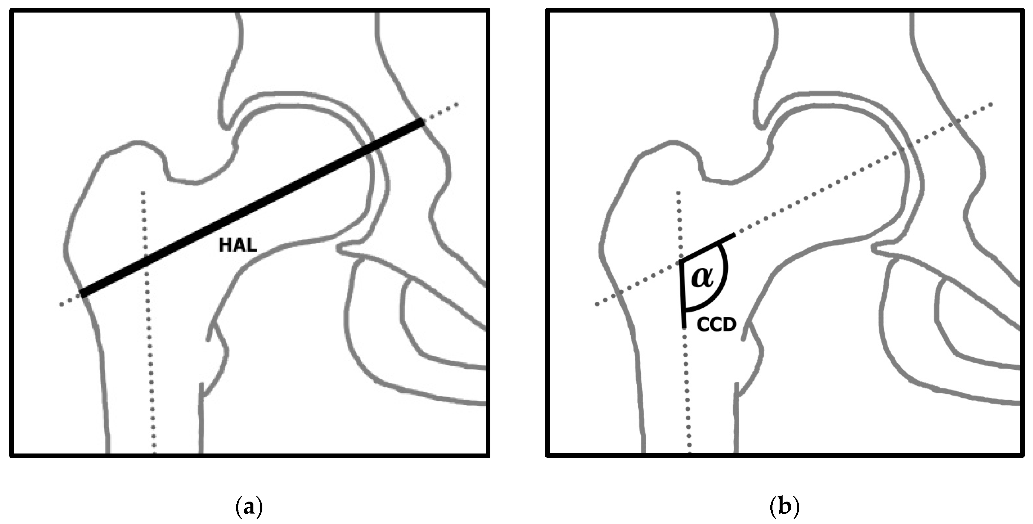
2.2. Radiological Measurements
2.3. Statistical Analysis
3. Results
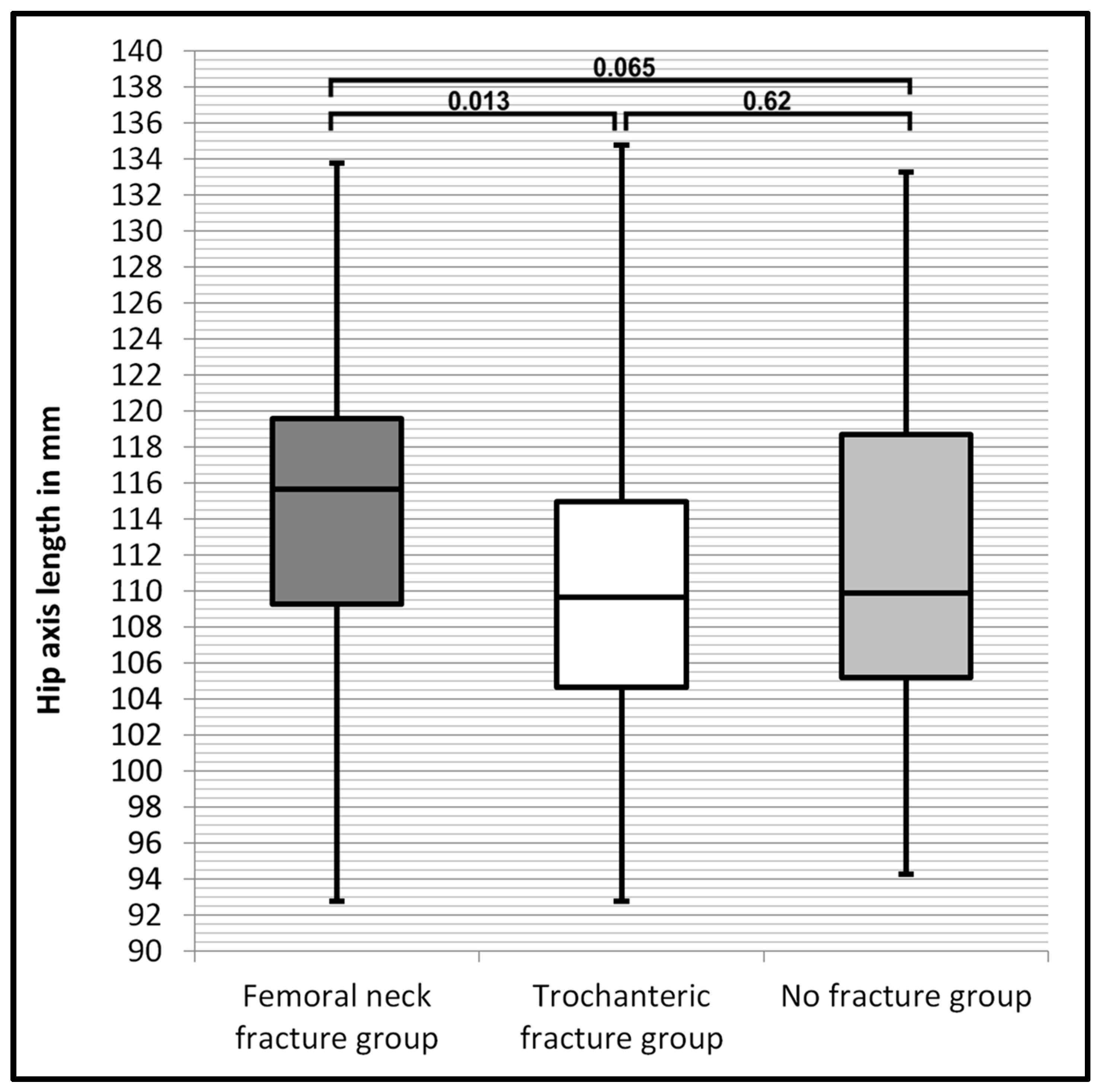
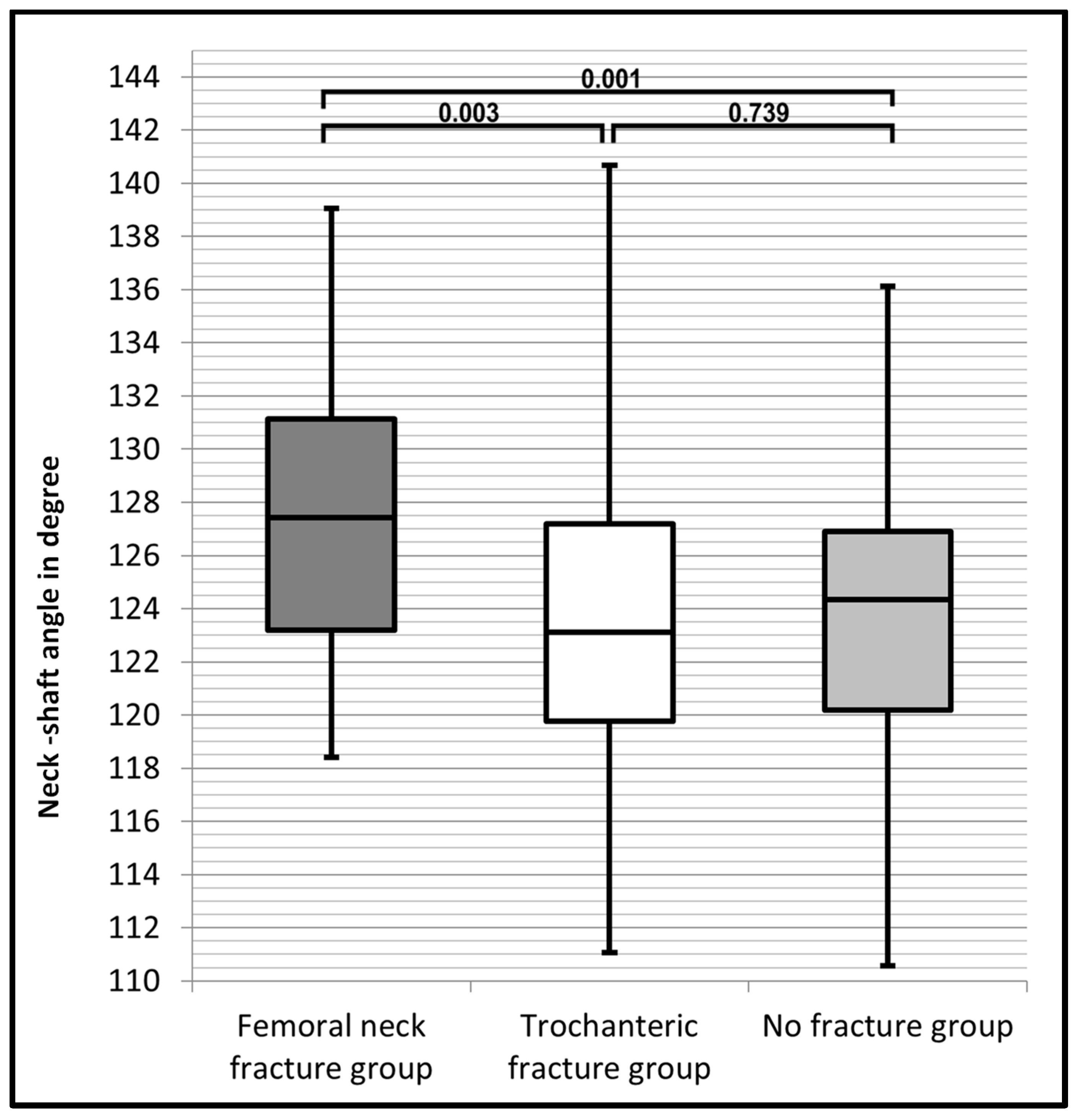
3.1. Collected Data and Group Comparison
3.2. Inter- and Intra-Rater Reliability
4. Discussion
5. Conclusions
Author Contributions
Funding
Institutional Review Board Statement
Informed Consent Statement
Data Availability Statement
Conflicts of Interest
References
- Lima, A.; Miranda, S.C.; Vasconcelos, H.F.O. Radiographic anatomy of the proximal femur: Femoral neck fracture vs. transtrochanteric fracture. Rev. Bras. Ortop. 2017, 52, 651–657. [Google Scholar] [CrossRef]
- Thalmann, B.H.; Latz, D.; Schiffner, E.; Jungbluth, P.; Windolf, J.; Grassmann, J. CCD angle & hip fractures—Predictor of fracture symmetry? J. Orthop. 2021, 24, 1–4. [Google Scholar]
- Alnemer, M.S.; Kotliar, K.E.; Neuhaus, V.; Pape, H.C.; Ciritsis, B.D. Cost-effectiveness analysis of surgical proximal femur fracture prevention in elderly: A Markov cohort simulation model. Cost Eff. Resour. Alloc. 2023, 21, 77. [Google Scholar] [CrossRef] [PubMed]
- Pugely, A.J.; Martin, C.T.; Gao, Y.; Klocke, N.F.; Callaghan, J.J.; Marsh, J.L. A risk calculator for short-term morbidity and mortality after hip fracture surgery. J. Orthop. Trauma 2014, 28, 63–69. [Google Scholar] [CrossRef]
- Tosteson, A.N.; Gabriel, S.E.; Grove, M.R.; Moncur, M.M.; Kneeland, T.S.; Melton, L.J., 3rd. Impact of hip and vertebral fractures on quality-adjusted life years. Osteoporos. Int. 2001, 12, 1042–1049. [Google Scholar] [CrossRef]
- Blain, H.; Miot, S.; Bernard, P.L. How Can We Prevent Falls? In Orthogeriatrics: The Management of Older Patients with Fragility Fractures, 2nd ed.; Falaschi, P., Marsh, D., Eds.; Springer: Cham, Switzerland, 2021; pp. 273–290. [Google Scholar]
- Montero-Odasso, M.M.; Kamkar, N.; Pieruccini-Faria, F.; Osman, A.; Sarquis-Adamson, Y.; Close, J.; Hogan, D.B.; Hunter, S.W.; Kenny, R.A.; Lipsitz, L.A.; et al. Evaluation of Clinical Practice Guidelines on Fall Prevention and Management for Older Adults: A Systematic Review. JAMA Netw. Open 2021, 4, e2138911. [Google Scholar] [CrossRef] [PubMed]
- Kendrick, D.; Kumar, A.; Carpenter, H.; Zijlstra, G.A.; Skelton, D.A.; Cook, J.R.; Stevens, Z.; Belcher, C.M.; Haworth, D.; Gawler, S.J.; et al. Exercise for reducing fear of falling in older people living in the community. Cochrane Database Syst. Rev. 2014, 2014, Cd009848. [Google Scholar] [CrossRef]
- Conley, R.B.; Adib, G.; Adler, R.A.; Åkesson, K.E.; Alexander, I.M.; Amenta, K.C.; Blank, R.D.; Brox, W.T.; Carmody, E.E.; Chapman-Novakofski, K.; et al. Secondary Fracture Prevention: Consensus Clinical Recommendations from a Multistakeholder Coalition. J. Bone Miner. Res. 2020, 35, 36–52. [Google Scholar] [CrossRef] [PubMed]
- Leslie, W.D.; Lix, L.M.; Morin, S.N.; Johansson, H.; Odén, A.; McCloskey, E.V.; Kanis, J.A. Hip axis length is a FRAX- and bone density-independent risk factor for hip fracture in women. J. Clin. Endocrinol. Metab. 2015, 100, 2063–2070. [Google Scholar] [CrossRef]
- Marshall, D.; Johnell, O.; Wedel, H. Meta-analysis of how well measures of bone mineral density predict occurrence of osteoporotic fractures. BMJ 1996, 312, 1254–1259. [Google Scholar] [CrossRef]
- Cranney, A.; Jamal, S.A.; Tsang, J.F.; Josse, R.G.; Leslie, W.D. Low bone mineral density and fracture burden in postmenopausal women. CMAJ 2007, 177, 575–580. [Google Scholar] [CrossRef] [PubMed]
- Kanis, J.A.; Johnell, O.; Oden, A.; Johansson, H.; McCloskey, E. FRAX and the assessment of fracture probability in men and women from the, U.K. Osteoporos. Int. 2008, 19, 385–397. [Google Scholar] [CrossRef] [PubMed]
- Vandenput, L.; Johansson, H.; McCloskey, E.V.; Liu, E.; Åkesson, K.E.; Anderson, F.A.; Azagra, R.; Bager, C.L.; Beaudart, C.; Bischoff-Ferrari, H.A.; et al. Update of the fracture risk prediction tool FRAX: A systematic review of potential cohorts and analysis plan. Osteoporos. Int. 2022, 33, 2103–2136. [Google Scholar] [CrossRef] [PubMed]
- Wang, Z.; Tutaworn, T.; Wishman, M.D.; Levin, J.E.; Hentschel, I.G.; Lane, J.M. Fracture Risk Assessment Tool Scores and Radiographical Bone Measurements in Total Hip Arthroplasty Patients. J. Arthroplast. 2022, 37, 2381–2386. [Google Scholar] [CrossRef] [PubMed]
- Goldshtein, I.; Gerber, Y.; Ish-Shalom, S.; Leshno, M. Fracture Risk Assessment with FRAX Using Real-World Data in a Population-Based Cohort from Israel. Am. J. Epidemiol. 2018, 187, 94–102. [Google Scholar] [CrossRef] [PubMed]
- Rocha, V.M.; Gaspar, H.A.; Oliveira, C.F. Fracture risk assessment in home care patients using the FRAX® tool. Einstein 2018, 16, eAO4236. [Google Scholar] [CrossRef] [PubMed]
- Kanis, J.A.; Johansson, H.; Oden, A.; McCloskey, E.V. Assessment of fracture risk. Eur. J. Radiol. 2009, 71, 392–397. [Google Scholar] [CrossRef] [PubMed]
- Morales-Torres, J.; Clark, P.; Delezé-Hinojosa, M.; Cons-Molina, F.; Messina, O.D.; Hernández, J.; Jaller-Raad, J.J.; Quevedo-Solidoro, H.; Radominski, S.C. Fracture risk assessment in Latin America: Is Frax an adaptable instrument for the region? Clin. Rheumatol. 2010, 29, 1085–1091. [Google Scholar] [CrossRef] [PubMed]
- Leslie, W.D.; Lix, L.M.; Johansson, H.; Oden, A.; McCloskey, E.; Kanis, J.A. Spine-hip discordance and fracture risk assessment: A physician-friendly FRAX enhancement. Osteoporos. Int. 2011, 22, 839–847. [Google Scholar] [CrossRef]
- Oka, R.; Ohira, M.; Suzuki, S.; Yoshida, T.; Koide, H.; Tanaka, T.; Tatsuno, I. Fracture risk assessment tool (FRAX) and for the diagnosis of osteoporosis in Japanese middle-aged and elderly women: Chiba bone survey. Endocr. J. 2018, 65, 193–202. [Google Scholar] [CrossRef]
- Iolascon, G.; Moretti, A.; Cannaviello, G.; Resmini, G.; Gimigliano, F. Proximal femur geometry assessed by hip structural analysis in hip fracture in women. Aging Clin. Exp. Res. 2015, 27 (Suppl. S1), S17–S21. [Google Scholar] [CrossRef] [PubMed]
- Gnudi, S.; Sitta, E.; Pignotti, E. Prediction of incident hip fracture by femoral neck bone mineral density and neck-shaft angle: A 5-year longitudinal study in post-menopausal females. Br. J. Radiol. 2012, 85, e467–e473. [Google Scholar] [CrossRef]
- Faulkner, K.G.; Wacker, W.K.; Barden, H.S.; Simonelli, C.; Burke, P.K.; Ragi, S.; Del Rio, L. Femur strength index predicts hip fracture independent of bone density and hip axis length. Osteoporos. Int. 2006, 17, 593–599. [Google Scholar] [CrossRef]
- Gnudi, S.; Ripamonti, C.; Lisi, L.; Fini, M.; Giardino, R.; Giavaresi, G. Proximal femur geometry to detect and distinguish femoral neck fractures from trochanteric fractures in postmenopausal women. Osteoporos. Int. 2002, 13, 69–73. [Google Scholar] [CrossRef] [PubMed]
- Fajar, J.K.; Taufan, T.; Syarif, M.; Azharuddin, A. Hip geometry and femoral neck fractures: A meta-analysis. J. Orthop. Transl. 2018, 13, 1–6. [Google Scholar] [CrossRef] [PubMed]
- Leslie, W.D.; Lix, L.M.; Morin, S.N.; Johansson, H.; Odén, A.; McCloskey, E.V.; Kanis, J.A. Adjusting Hip Fracture Probability in Men and Women Using Hip Axis Length: The Manitoba Bone Density Database. J. Clin. Densitom. 2016, 19, 326–331. [Google Scholar] [CrossRef] [PubMed]
- Li, G.W.; Chang, S.X.; Xu, Z.; Chen, Y.; Bao, H.; Shi, X. Prediction of hip osteoporotic fractures from composite indices of femoral neck strength. Skelet. Radiol. 2013, 42, 195–201. [Google Scholar] [CrossRef]
- Frisoli, A., Jr.; Paula, A.P.; Pinheiro, M.; Szejnfeld, V.L.; Delmonte Piovezan, R.; Takata, E.; Silva, T.A.; Chaves, P.H.M. Hip axis length as an independent risk factor for hip fracture independently of femural bone mineral density in Caucasian elderly Brazilian women. Bone 2005, 37, 871–875. [Google Scholar] [CrossRef]
- Faulkner, K.G.; Cummings, S.R.; Black, D.; Palermo, L.; Glüer, C.C.; Genant, H.K. Simple measurement of femoral geometry predicts hip fracture: The study of osteoporotic fractures. J. Bone Miner. Res. 1993, 8, 1211–1217. [Google Scholar] [CrossRef]
- Jiamton, C.; Boernert, K.; Babst, R.; Beeres, F.J.P.; Link, B.C. The nail-shaft-axis of the of proximal femoral nail antirotation (PFNA) is an important prognostic factor in the operative treatment of intertrochanteric fractures. Arch. Orthop. Trauma Surg. 2018, 138, 339–349. [Google Scholar] [CrossRef]
- Landis, J.R.; Koch, G.G. The measurement of observer agreement for categorical data. Biometrics 1977, 33, 159–174. [Google Scholar] [CrossRef] [PubMed]
- Harris-Hayes, M.; Commean, P.K.; Patterson, J.D.; Clohisy, J.C.; Hillen, T.J. Bony abnormalities of the hip joint: A new comprehensive, reliable and radiation-free measurement method using magnetic resonance imaging. J. Hip Preserv. Surg. 2014, 1, 62–70. [Google Scholar] [CrossRef] [PubMed]
- Ito, M.; Wakao, N.; Hida, T.; Matsui, Y.; Abe, Y.; Aoyagi, K.; Uetani, M.; Harada, A. Analysis of hip geometry by clinical CT for the assessment of hip fracture risk in elderly Japanese women. Bone 2010, 46, 453–457. [Google Scholar] [CrossRef] [PubMed]
- Duboeuf, F.; Hans, D.; Schott, A.M.; Kotzki, P.O.; Favier, F.; Marcelli, C.; Meunier, P.J.; Delmas, P.D. Different morphometric and densitometric parameters predict cervical and trochanteric hip fracture: The EPIDOS Study. J. Bone Miner. Res. 1997, 12, 1895–1902. [Google Scholar] [CrossRef]
- Im, G.I.; Lim, M.J. Proximal hip geometry and hip fracture risk assessment in a Korean population. Osteoporos. Int. 2011, 22, 803–807. [Google Scholar] [CrossRef] [PubMed]
- Çukurlu, M.; Karagoz, B.; Keceli, O. The effect of pre-fracture proximal femur geometry on hip fracture type in elderly patients. Medicine 2023, 102, e33622. [Google Scholar] [CrossRef] [PubMed]
- Zhuang, H.; Li, Y.; Lin, J.; Cai, D.; Cai, S.; Yan, L.; Yao, X. Cortical thickness in the intertrochanteric region may be relevant to hip fracture type. BMC Musculoskelet. Disord. 2017, 18, 305. [Google Scholar] [CrossRef] [PubMed]
- Pierre, M.A.; Zurakowski, D.; Nazarian, A.; Hauser-Kara, D.A.; Snyder, B.D. Assessment of the bilateral asymmetry of human femurs based on physical, densitometric, and structural rigidity characteristics. J. Biomech. 2010, 43, 2228–2236. [Google Scholar] [CrossRef]
- Zhao, R.; Cai, H.; Tian, H.; Zhang, K. Morphological consistency of bilateral hip joints in adults based on the X-ray and CT data. Surg. Radiol. Anat. 2021, 43, 1107–1115. [Google Scholar] [CrossRef]
- Young, E.Y.; Gebhart, J.; Cooperman, D.; Ahn, N.U. Are the left and right proximal femurs symmetric? Clin. Orthop. Relat. Res. 2013, 471, 1593–1601. [Google Scholar] [CrossRef]
- Crabtree, N.; Lunt, M.; Holt, G.; Kröger, H.; Burger, H.; Grazio, S.; Khaw, K.T.; Lorenc, R.S.; Nijs, J.; Stepan, J.; et al. Hip geometry, bone mineral distribution, and bone strength in European men and women: The EPOS study. Bone 2000, 27, 151–159. [Google Scholar] [CrossRef] [PubMed]
- Peacock, M.; Liu, G.; Carey, M.; Ambrosius, W.; Turner, C.H.; Hui, S.; Johnston, C.C., Jr. Bone mass and structure at the hip in men and women over the age of 60 years. Osteoporos. Int. 1998, 8, 231–239. [Google Scholar] [CrossRef] [PubMed]
- Nissen, N.; Hauge, E.M.; Abrahamsen, B.; Jensen, J.E.; Mosekilde, L.; Brixen, K. Geometry of the proximal femur in relation to age and sex: A cross-sectional study in healthy adult Danes. Acta Radiol. 2005, 46, 514–518. [Google Scholar] [CrossRef] [PubMed]
- Hetsroni, I.; Dela Torre, K.; Duke, G.; Lyman, S.; Kelly, B.T. Sex differences of hip morphology in young adults with hip pain and labral tears. Arthroscopy 2013, 29, 54–63. [Google Scholar] [CrossRef] [PubMed]
- Tuck, S.P.; Pearce, M.S.; Rawlings, D.J.; Birrell, F.N.; Parker, L.; Francis, R.M. Differences in bone mineral density and geometry in men and women: The Newcastle Thousand Families Study at 50 years old. Br. J. Radiol. 2005, 78, 493–498. [Google Scholar] [CrossRef]
- Nakahara, I.; Takao, M.; Sakai, T.; Nishii, T.; Yoshikawa, H.; Sugano, N. Gender differences in 3D morphology and bony impingement of human hips. J. Orthop. Res. 2011, 29, 333–339. [Google Scholar] [CrossRef]

| FNFx Group | TFx Group | NFx Group | |
|---|---|---|---|
| Number of participants | 50 | 50 | 50 |
| Age (years) 1 | 93.8 ± 3.2 | 92.9 ± 3.7 | 91.3 ± 3.2 |
| Sex (female/male) | 30/20 | 38/12 | 35/15 |
| HAL (mm) 1 | 115.24 ± 9.50 | 110.65 ± 8.54 | 111.58 ± 10.05 |
| HAL range (mm) | 92.8–133.8 | 92.8–134.8 | 94.3–133.3 |
| CCD (°) 1 | 127.41 ± 5.26 | 124.03 ± 5.98 | 123.65 ± 5.43 |
| CCD range (°) | 118.4–139.1 | 111.1–140.7 | 110.6–136.1 |
| FNFx vs. TFx | FNFx vs. NFx | TFx vs. NFx | |
|---|---|---|---|
| HAL 1 | 0.013 | 0.065 | 0.62 |
| CCD 1 | 0.003 | 0.001 | 0.739 |
Disclaimer/Publisher’s Note: The statements, opinions and data contained in all publications are solely those of the individual author(s) and contributor(s) and not of MDPI and/or the editor(s). MDPI and/or the editor(s) disclaim responsibility for any injury to people or property resulting from any ideas, methods, instructions or products referred to in the content. |
© 2024 by the authors. Licensee MDPI, Basel, Switzerland. This article is an open access article distributed under the terms and conditions of the Creative Commons Attribution (CC BY) license (https://creativecommons.org/licenses/by/4.0/).
Share and Cite
Gumuchdjian, D.A.; Waltenspül, M.; Dietrich, M.; Kabelitz, M. Hip Axis Length and Femoral Neck-Shaft Angle as Risk Factors for Proximal Femur Fractures in Octogenarians to Centenarians. J. Clin. Med. 2024, 13, 4071. https://doi.org/10.3390/jcm13144071
Gumuchdjian DA, Waltenspül M, Dietrich M, Kabelitz M. Hip Axis Length and Femoral Neck-Shaft Angle as Risk Factors for Proximal Femur Fractures in Octogenarians to Centenarians. Journal of Clinical Medicine. 2024; 13(14):4071. https://doi.org/10.3390/jcm13144071
Chicago/Turabian StyleGumuchdjian, Daniel Alexandre, Manuel Waltenspül, Michael Dietrich, and Method Kabelitz. 2024. "Hip Axis Length and Femoral Neck-Shaft Angle as Risk Factors for Proximal Femur Fractures in Octogenarians to Centenarians" Journal of Clinical Medicine 13, no. 14: 4071. https://doi.org/10.3390/jcm13144071
APA StyleGumuchdjian, D. A., Waltenspül, M., Dietrich, M., & Kabelitz, M. (2024). Hip Axis Length and Femoral Neck-Shaft Angle as Risk Factors for Proximal Femur Fractures in Octogenarians to Centenarians. Journal of Clinical Medicine, 13(14), 4071. https://doi.org/10.3390/jcm13144071