Article

Prognostic Impact of Left Ventricular Ejection Fraction Improvement after Transcatheter Aortic Valve Replacement

by
Jakob Johannes Reichl
1,2,†,
Thorald Stolte
1,3,†,
Shihui Tang
1,
Jasper Boeddinghaus
1,
Max Wagener
1,
Gregor Leibundgut
1,
Christoph Ado Kaiser
1 and
Thomas Nestelberger
1,*
1
Department of Cardiology and Cardiovascular Research Institute Basel (CRIB), University Hospital Basel, University of Basel, 4031 Basel, Switzerland
2
Department of General Internal Medicine, University Hospital Basel, 4031 Basel, Switzerland
3
Department of Health Sciences and Technology, Swiss Federal Institute of Technology, 8093 Zurich, Switzerland
*
Author to whom correspondence should be addressed.
These authors contributed equally to this work.
J. Clin. Med. 2024, 13(13), 3639; https://doi.org/10.3390/jcm13133639
Submission received: 27 May 2024 / Revised: 12 June 2024 / Accepted: 19 June 2024 / Published: 21 June 2024
(This article belongs to the Special Issue Clinical Advancement in Aortic Valve Stenosis)

Abstract

:
Introduction: Transcatheter aortic valve replacement (TAVR) has become an efficient and safe alternative to surgical aortic valve replacement (SAVR). While severe aortic stenosis as well as severe aortic regurgitation (AR) are known to negatively impact left ventricular ejection fraction (LVEF), prior studies have shown that TAVR can lead to an improvement in LVEF. Thus far, little is known about the prognostic implication of LVEF improvement as a sole predictor of outcomes. Therefore, the aim of this study was to assess the prognostic impact of LVEF impairment before TAVR, as well as early LVEF improvement in patients undergoing TAVR. Materials and Methods: Patients undergoing TAVR in a large tertiary university hospital were consecutively included in a prospective registry. Transthoracic echocardiography (TTE) was performed at baseline, after 1 month and annually thereafter. Significant LVEF improvement was defined as a relative increase of ≥10% in LVEF at 30 days compared to baseline LVEF. The primary outcome was all-cause mortality at 1 year. Secondary outcomes were major adverse cardiovascular events (MACEs) including cardiovascular death, non-fatal myocardial infarction, stroke, bleeding and unplanned re-interventions of the aortic valve at 5 years. Results: Among 1655 patients who underwent TAVR between September 2011 and April 2024, the LVEF at baseline was available for 1556 patients. Of these, 1031 patients (66.2%) had preserved LVEF at baseline (LVEF ≥ 53%), whereas 303 patients (19.5%) had moderately reduced LVEF (40–52%) and 222 patients (14.3%) had severely reduced LVEF (<40%). Out of the patients with impaired LVEF, 155 (40.4%) patients showed a significant improvement in LVEF ≥10% after 30 days, while 229 (60.6%) patients showed no significant LVEF improvement (<10%). Patients with preserved LVEF at baseline had significantly better mortality outcomes than those with severely reduced LVEF (p < 0.001). LVEF improvement was associated with a survival benefit after 1 year (p = 0.009, HR 2.68, 0.95 CI 1.23–5.85) which diminished after 5 years (p = 0.058), but patients with LVEF improvement showed lower MACE rates at 5 years (p < 0.001). Conclusions: Preserved LVEF before TAVR is an independent predictor for improved outcomes. Additionally, early improvement in LVEF is associated with beneficial outcomes in patients undergoing TAVR.

1. Introduction

Aortic stenosis (AS) is the most common valve pathology, affecting up to 20% of the population aged 75 and older [1]. Early detection and treatment are of high importance as untreated patients with severe aortic stenosis face high mortality and morbidity [2]. In recent years, transcatheter aortic valve replacement (TAVR) has become a viable alternative to surgical aortic valve replacement (SAVR), especially in elderly patients [3,4,5]. Recent guidelines by the European Society of Cardiology (ESC) support TAVR as the first-line treatment in patients older than 75 years irrespective of risk categories [6].
Aortic stenosis leads to numerous adaptations by the left ventricle. Narrowing of the aortic valve area leads to an increased afterload. This induces hypertrophy of the myocytes to maintain systemic cardiac output [7]. However, left ventricular hypertrophy itself imposes negative effects on ventricular function and coronary circulation, contributing to an increased rate of cardiovascular events [8]. Finally, these events lead to fibrosis of the myocytes, ventricular dilation and a decline in left ventricular ejection fraction (LVEF), and eventually to the development of heart failure [9].
Several studies have shown that valve replacement, whether by SAVR or TAVR, improves the LVEF in patients with pre-interventional left ventricular dysfunction [10]. Many factors have been shown to affect outcomes in patients undergoing TAVR, yet data on the impact of early LVEF improvement itself are scarce.
The objective of this study was to assess the rate of LVEF improvement after TAVR and to determine the prognostic impact of LVEF at baseline as well as early LVEF improvement in patients undergoing TAVR.

2. Methods

2.1. Study Design and Patient Cohort

All patients who underwent TAVR at the University Hospital Basel, Switzerland, were included in a prospective national database, as part of the Swiss TAVI registry, mandated by the Swiss health authorities (NCT01368250). The Swiss TAVI registry is a multicentric database and has been featured in recent publications [11,12,13]. The Swiss TAVI registry has been approved by the local cantonal ethics committee and the institutional review boards of all participating sites. All patients provided written informed consent for study participation and prospective follow-up assessment. The present study includes patients that underwent TAVR between September 2011 and April 2024.

2.2. Data Collection and Clinical Endpoints

All patient-related data, including baseline characteristics, procedural and follow-up information, were prospectively collected and recorded in a web-based database. Clinical follow-up data were obtained through standardized interviews, documentation from referring physicians and hospital discharge summaries. All adverse events were systematically collected and adjudicated by a dedicated clinical event committee.
We included all patients who had undergone TAVR and had transthoracic echocardiography (TTE) at baseline with measurements performed using the biplane Simpson method. In case of severely reduced examination conditions, LVEF was graded by visual analysis by treating physicians into the categories mentioned below. Patients were then divided into three separate groups based on their baseline LVEF. We defined patients with preserved LVEF (>52%), patients with moderately reduced LVEF (≥40%; ≤52%) and patients with severely reduced LVEF (<40%). We chose to defer from the definition of preserved ejection fraction defined by the European Society of Cardiology (ESC) due to the definitions in our local echocardiography department, where the transition between “preserved” and “borderline preserved” is defined at an LVEF of 53%. Follow-up TTE was either performed at our clinical echocardiographic laboratory or in cooperation with the patient’s treating cardiologists.
For analysis of LVEF improvement, patients were included if follow-up LVEF data after 30 days were available. Patients that had died before their first follow-up at day 30 were not considered for further analysis. Patients with moderately or severely reduced LVEF were categorized based on whether their LVEF had improved by more than 10% by day 30, later referred to as our cohort with “improved” patients, and patients with less than 10% LVEF improvement are referred to as “not-improved” patients. Patients with initially preserved LVEF were not considered for analysis of LVEF improvement.
Our primary endpoint was all-cause death at 1 year in patients with or without significant LVEF improvement. Secondary endpoints were all-cause death at 5 years and a combined endpoint of major adverse cardiovascular events (MACEs), defined as cardiovascular death, myocardial infarction, cerebrovascular stroke, bleeding and unplanned re-intervention of the aortic valve at 5 years. We also compared 1- and 5-year all-cause mortality between patients with preserved LVEF at baseline and patients with severely reduced LVEF as well as MACEs at 5 years.

2.3. Statistical Analysis

Categorical variables are presented as frequencies and percentages. Continuous variables are represented as mean values ± standard deviation if normal distribution was apparent. Non-normally distributed variables are represented as median values with the 25th and 75th quartiles. Comparisons between normally distributed continuous variables were performed using Fisher’s paired t-test for normally distributed variables. Non-normally distributed variables were compared using the Mann–Whitney U test. Differences between ordinal scaled data were calculated using Pearson’s Chi2 test. Survival analysis was carried out using the Kaplan–Meier procedure, and differences in survival were tested with the Log-Rank (Mantel–Cox) test. A p-value < 0.05 was considered significant. We used multivariate logistic regression models to determine factors with an impact on LVEF improvement. All statistical analyses were performed using R 4.2.3 (R Foundation for Statistical Computing, Vienna, Austria). Sankey plots were created using the open-source tool SANKEYMATIC by Steve Bogart, available at https://www.sankeymatic.com (accessed on 20 January 2024).

3. Results

3.1. Patient Cohort and Baseline Characteristics
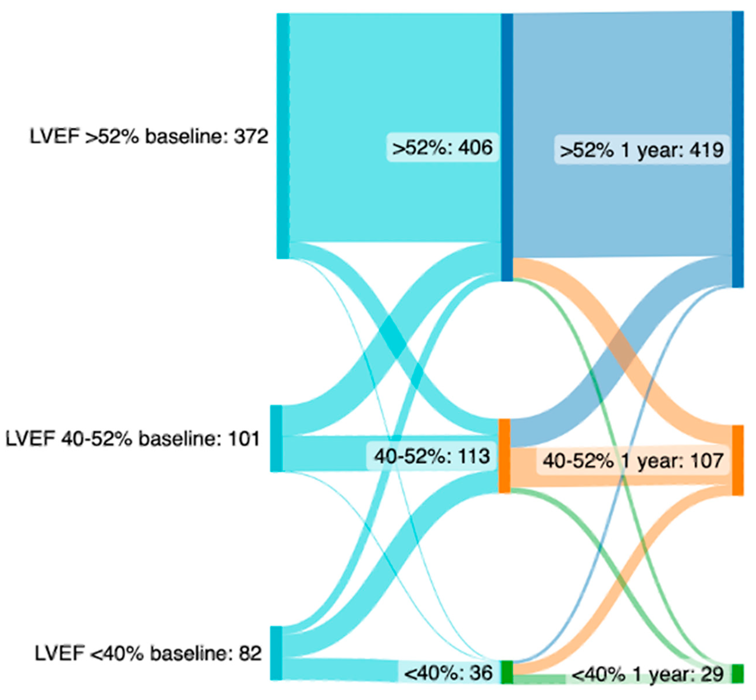
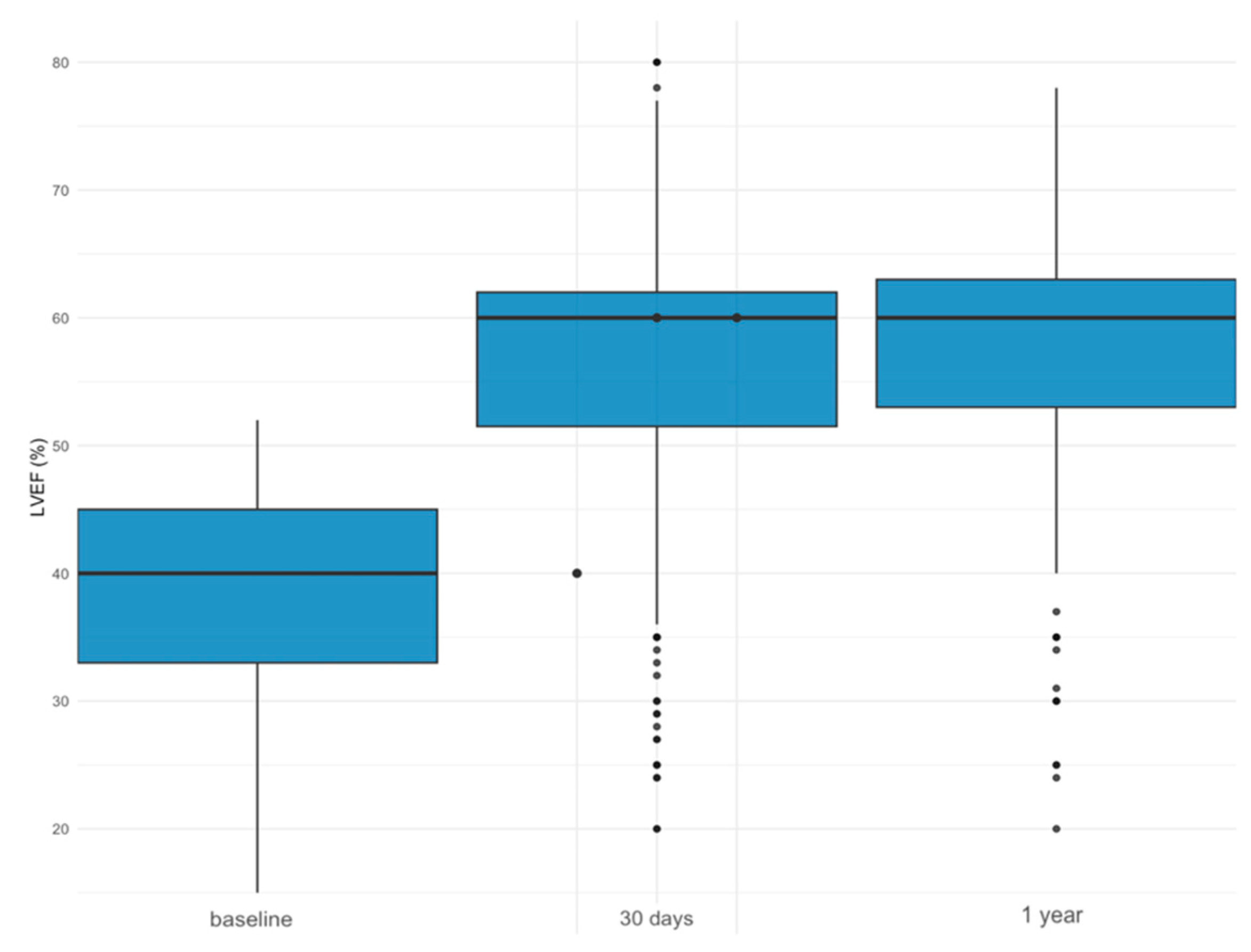
Among 1655 patients that underwent TAVR between September 2011 and April 2024, the TTE at baseline with valuable LVEF measurements was available in 1556 patients (94.0%). At baseline, 525 of 1556 patients (33.7%) were diagnosed with impaired LVEF. Out of these, follow-up TTE data including LVEF measurements were available in 384 (73.1%) cases.
Among patients with impaired LVEF at baseline, 303 (57.7%) patients had moderately reduced LVEF and 222 (42.3%) had severely reduced LVEF. Table 1 summarizes the baseline data for patients with preserved (>52%), moderately reduced (40–52%) and severely reduced (≤40%) LVEF. Table 2 shows interventional and echocardiographic findings. Compared to patients with preserved LVEF at baseline, patients with severely reduced LVEF were more often male (66.7% vs. 46.9%, p < 0.001); had more comorbidities such as coronary artery disease (CAD) (64.9% vs. 50.0%, p < 0.001), prior myocardial infarction (31.5% vs. 11.9%, p < 0.001) and diabetes (37.8% vs. 26.3%, p = 0.002) and had more often undergone interventions such as prior percutaneous coronary intervention (PCI) (38.3% vs. 30.2%, p < 0.001), cardiac surgery of any kind (15.8% vs. 7.5%, p < 0.001) or pacemaker implantation prior to TAVR (17.1% vs. 7.5%, p < 0.001). Patients with severely reduced LVEF were more frequently diagnosed with left ventricular hypertrophy (20.7% vs. 7.6%, p < 0.001). Additionally, they showed lower peak aortic valve gradients (49.0 vs. 70.0, p < 0.001), lower mean gradients (33 ± 13.8 vs. 45 ± 13.5, p = 0.001) and more atrial fibrillation (AF) (30.6% vs. 23.4%, p = 0.03). In terms of access site, no differences were found. In patients with moderately or severely reduced LVEF, valve sizes for TAVR were slightly larger than in patients with preserved LVEF (27.0 vs. 26.0, p < 0.001).

LVEF Improvement

Among 384 patients with moderately or severely reduced LVEF at baseline, 155 patients (40.4%) showed LVEF improvement after 30 days, while 229 (60.6%) patients showed no improvement. Mean LVEF improvement and decline in all patients was 7.8% (±9.67). Among the latter, 68 patients (26.6%) showed a decline in LVEF. Table 3 summarizes these groups’ baseline data, and Table 4 gives insights on echocardiographic and interventional findings. Patients with LVEF improvement compared to those without were more often female (47.7% vs. 32.3%, p = 0.003) and had less frequently undergone prior cardiac surgery of any kind (9.0% vs. 16.6%, p = 0.049). There was a difference in baseline LVEF between these groups, with baseline LVEF being significantly lower in patients that would show improvement after 30 days (38.0% vs. 42.0%, p = 0.002). Additionally, patients with LVEF improvement showed a smaller indexed aortic valve area (0.2 cm2/m2 vs. 0.3 cm2/m2, p = 0.03) and higher peak aortic valve gradients (60 mmHg vs. 53 mmHg, p = 0.011) and mean gradients (41.0 mmHg vs. 38.0 mmHg, p = 0.049). Right ventricular dysfunction was significantly less frequent in patients with LVEF improvement (14.2% vs. 31%, p < 0.001). Relevant medications are listed in the Supplementary Table S1.
Left ventricular hypertrophy at baseline was numerically, albeit insignificantly, less frequent in patients that showed improvement than in patients without improvement (16.7% vs. 20.9%, p = 0.37). After 30 days, left ventricular hypertrophy prevalence was declining in both groups but was significantly less frequent in patients with LVEF improvement (3.2% vs. 12.2%, p = 0.003). There was a difference regarding access site for TAVR, with transapical access being less frequent in patients with LVEF improvement (5.1% vs. 13.2%, p = 0.003). Multivariate linear regression for several factors such as age, sex, CAD, CKD, prior PCI, prior heart surgery, diabetes, hypertension, pacemaker implantation and cerebrovascular disease showed no predictive value for LVEF improvement at 30 days (Table 5).
The overall dynamics of LVEF change after TAVR are depicted in Figure 1. After 1 year, LVEF improvement had been maintained in the vast majority of patients (preserved LVEF at 30 days, 72%; after 1 year, 76%).

3.2. Outcomes after TAVR

Follow-up data were available in 88.9% of patients at 1 year and in 35.7% of patients at 5 years. Mortality after one year was lower in patients with preserved LVEF at baseline compared to patients with severely reduced LVEF (7.9% vs. 16.6%, p < 0.001, HR 0.45, 0.95 CI 0.30–0.66), as was mortality after five years (21.6% vs. 36.9%, p < 0.001, HR 0.52, 0.95 CI 0.40–0.67). There was no difference in the frequency of MACEs after five years (213 vs. 75 events, p = 0.76). The outcomes are depicted in Figure 2A–C.
Patients with LVEF improvement showed significantly lower mortality after one year (5.2% vs. 12.6%, p = 0.009, HR 2.68, 0.95 CI 1.23–5.85). There was a numerical, albeit insignificant, difference after five years (24.5% vs. 32.7%, p = 0.058, HR 1.45, 0.95 CI 0.98–2.13). Additionally, the combined endpoint of MACEs after five years occurred less frequently (109 vs. 306 events, p < 0.001). The outcomes are depicted in Figure 3A–C.

4. Discussion

In this study, we aimed to analyze the role of LVEF impairment and early improvement in LVEF in patients undergoing TAVR in a tertiary university center.
In the present study, patients showing improvements in LVEF were more often female than patients without improvement. Studies have shown that males generally suffer more frequently from ischemic heart disease than females, which might lead to a lower potential for LVEF recovery. In their systematic review, Kewcharoen et al. found that female gender is associated with a higher chance of LVEF recovery [14]. Other studies have also displayed gender-specific differences in left ventricular geometry as well as the extent of myocardial fibrosis [14,15], which may explain the differences in the reversal of left ventricular hypertrophy and left ventricular function after TAVR. Supporting these findings, Chen et al. showed that female patients have favorable reversed left ventricular remodeling, with earlier and greater LV mass regression [16]. These factors may contribute to higher chances of LVEF improvement in female patients.
Our study showed that in most patients with moderately or severely reduced LVEF, TAVR can lead to an improvement in LVEF, as depicted in Figure 4. This effect was constant in both patients with moderately reduced and those with severely reduced LVEF and was maintained after 1 year. Our findings underline the positive impact of TAVR on left ventricular dimensions, especially with left ventricular hypertrophy declining over the course of one year. It is known that high-grade aortic stenosis leads to chronic pressure overload due to high transaortic valve gradients, leading to pathologic remodeling in the form of concentric hypertrophy [17]. Correcting the high-pressure gradient using TAVR leads to reduced pressure overload for the left ventricle and might partly explain the improvement in left ventricular function that some, yet not all, patients achieve after TAVR [18]. Changes in left ventricular dimensions differed after TAVR, with the LVEDD being smaller and left ventricular hypertrophy being less frequent in patients with LVEF improvement. These changes support the recline of left ventricular remodeling after TAVR, contributing to the improvement of left ventricular function. TAVR aims to treat one of the causes of impaired EF. However, heart failure in patients with severe AS is not always caused by the stenosis alone. The vast majority of patients have concomitant cardiovascular diseases; therefore, medical heart failure treatment plays a major role in these patients as well. Especially younger patients with reduced ejection will benefit from long-term guideline-directed medical therapy and regular follow-up after TAVR to maintain or restore an impaired EF [19].
We found a difference in mortality after one year between patients with and without LVEF improvement, but only a numeric, statistically insignificant difference after 5 years. Contrary to our findings, Kolte et al. showed that an LVEF improvement of more than 10% is associated with decreased overall and cardiac death at 5 years [20]. We were not able to identify predictive factors associated with poor LVEF improvement, which may be explained by our sample size not being powered for this matter. However, the findings of Kuneman et al. demonstrated an association of CAD, prior myocardial infarction and permanent pacemaker implantation prior to TAVR with worse or missing LVEF improvement [21].
Additionally, our study revealed significant differences in mortality between patients with preserved LVEF at baseline compared to patients with impaired LVEF after one and five years. An explanation for this finding could be the fact that comorbidities such as CAD and prior cardiac surgery were more frequent in patients with impaired LVEF. Nevertheless, aortic valve stenosis itself negatively influences LV function and outcomes. Patients with an initial decline in their LVEF with severe aortic valve stenosis might benefit from earlier aortic valve replacement to prevent further deterioration of LV function [22]. Moreover, Dauerman and colleagues demonstrated that favorable 1-year outcomes were linked to early improvement in left ventricular ejection fraction (LVEF) [23].
In our study, the incidence of MACEs was less frequent in patients with significant LVEF improvement compared to patients without LVEF improvement. These findings are in contrast with previous research by Kuneman et al., who showed no difference in long-term event-free survival in patients with >5% improvement in LVEF compared to <5% improvement [21].
Our findings contribute to the current body of evidence on long-term outcomes after aortic valve replacement, particularly in the context of an increasing number of patients undergoing TAVR. We report the five main findings below.
First, patients with severely or moderately reduced LVEF at baseline were more often male and had a higher prevalence of comorbidities. They also presented more frequently with left ventricular hypertrophy, lower mean gradients and atrial fibrillation. Second, while 155 (40.4%) patients with severely or moderately reduced LVEF at baseline showed an improved LVEF after 30 days, 229 (60.6%) showed no improvement, and of these, 58 (25.3%) even showed a decline. Third, despite impaired LVEF function at baseline being more common in male patients, female patients more often had an LVEF improvement after 30 days (47.7% vs. 32.3%, p = 0.003). Fourth, patients with LVEF improvement at 30 days maintained their improvement after 1 year, highlighting a prolonged beneficial effect of TAVR on LVEF improvement. Lastly, we noted that improvement in LVEF was associated with improved mortality after one year and was linked to a reduction in MACEs after five years. Specifically, among the MACE endpoints, the occurrence of major bleeding and cardiovascular death appeared to be the most significantly associated with LVEF improvement.
Our study has several notable limitations. Firstly, the absence of uniformity in the laboratories conducting follow-up transthoracic echocardiographic studies is a limitation; however, our study represents real world data. Since our patients typically attended follow-up visits with external cardiologists, variations in the investigation protocols and results may arise based on the investigator. Additionally, given the retrospective nature of this data analysis, our study lacks the statistical power required to demonstrate clear differences in mortality outcomes. To ascertain the benefits of event-free survival conclusively, larger studies with a higher sample size may be necessary. Our study reflects real world data from a large tertiary university hospital over more than 10 years with consecutively enrolled patients in a prospective registry.

5. Conclusions

In conclusion, TAVR led to sustained LVEF improvement after 30 days and 1 year. Patients with preserved LVEF at baseline experienced better long-term outcomes. In addition, those who showed early improvement in LVEF post-TAVR had lower mortality rates after 1 year and a reduction in mortality and fewer cardiovascular events at 5 years. Further research is needed to elucidate the precise mechanisms impeding LVEF improvement.

Supplementary Materials

The following supporting information can be downloaded at: https://www.mdpi.com/article/10.3390/jcm13133639/s1, Table S1: Baseline medication before TAVR in patients with or without early LVEF-improvement.

Author Contributions

Conceptualization, M.W. and T.N.; Methodology, J.J.R.; Validation, J.B., G.L. and T.N.; Formal analysis, M.W.; Investigation, J.J.R. and T.S.; Resources, S.T., G.L., C.A.K. and T.N.; Data curation, S.T.; Writing—original draft, J.J.R.; Writing—review & editing, T.S., J.B. and T.N.; Visualization, J.J.R. and T.S.; Supervision, C.A.K. and T.N.; Funding acquisition, T.N. All authors have read and agreed to the published version of the manuscript.

Funding

Thomas Nestelberger has received research support from the Swiss National Science Foundation (P400PM_191037/1), the Max Cloëtta Foundation, the Margarete und Walter Lichtenstein-Stiftung (3MS1038) and the University Hospital Basel as well as speaker honoraria/consulting honoraria from Edwards Lifesciences, Siemens, Beckman Coulter, Bayer, Ortho Clinical Diagnostics and Orion Pharma, outside the submitted work.

Institutional Review Board Statement

The study was conducted according to the guidelines of the Declaration of Helsinki and approved by the Ethics Committee of Bern and Nordwestschweiz (protocol code 2021-01738 and date of approval for latest amendment 16 December 2023).

Informed Consent Statement

Informed consent was obtained from all subjects involved in the study. Written informed consent has been obtained from the patients to publish this paper.

Data Availability Statement

The data presented in this study are available on request from the corresponding author.

Conflicts of Interest

All other authors declare that they have no conflict of interest with this study. All authors critically reviewed the manuscript and approved the final version for submission. The manuscript and its contents have not been published previously and are not being considered for publications elsewhere in whole or in part in any language, including publicly accessible web sites or e-print servers. All other authors declare no conflicts of interest.

Abbreviations

ASAortic stenosis
CADCoronary artery disease
CABGCoronary arterial bypass graft
CCS-ScoreCanadian Cardiovascular Society-Score
CKDChronic kidney disease
CVDCerebrovascular disease
eGFREstimated glomerular filtration rate
ESCEuropean Society of Cardiology
EuroSCOREEuropean System for Cardiac Operative Risk Evaluation
ICDImplanted cardioverter defibrillator
LVEFLeft ventricular ejection fraction
LVEDDLeft ventricular end-diastolic diameter
LVESDLeft ventricular end-systolic diameter
NYHANew York Heart Association
PCIPercutaneous coronary intervention
STSSociety of Thoracic Surgeons
SAVRSurgical aortic valve replacement
TAVRTranscatheter aortic valve replacement
TTETransthoracic echocardiography
R2Coefficient of determination indicating correlation, where a value ≤ 0.35 indicates low or weak correlation

References

  1. Osnabrugge, R.L.J.; Mylotte, D.; Head, S.J.; Van Mieghem, N.M.; Nkomo, V.T.; LeReun, C.M.; Bogers, A.J.J.C.; Piazza, N.; Kappetein, A.P. Aortic Stenosis in the Elderly. J. Am. Coll. Cardiol. 2013, 62, 1002–1012. [Google Scholar] [CrossRef] [PubMed]
  2. Généreux, P.; Sharma, R.P.; Cubeddu, R.J.; Aaron, L.; Abdelfattah, O.M.; Koulogiannis, K.P.; Marcoff, L.; Naguib, M.; Kapadia, S.R.; Makkar, R.R.; et al. The Mortality Burden of Untreated Aortic Stenosis. J. Am. Coll. Cardiol. 2023, 82, 2101–2109. [Google Scholar] [CrossRef] [PubMed]
  3. Leon, M.B.; Smith, C.R.; Mack, M.; Miller, D.C.; Moses, J.W.; Svensson, L.G.; Tuzcu, E.M.; Webb, J.G.; Fontana, G.P.; Makkar, R.R.; et al. Transcatheter Aortic-Valve Implantation for Aortic Stenosis in Patients Who Cannot Undergo Surgery. N. Engl. J. Med. 2010, 363, 1597–1607. [Google Scholar] [CrossRef] [PubMed]
  4. Martine, G.; Hélène, E.; Bernard, I.; Patrick, D.-G.; Karine, C.; Jean, F.; Pascal, L.; Alain, L.; Michel, L.; Alain, P.; et al. Registry of Transcatheter Aortic-Valve Implantation in High-Risk Patients. New Engl. J. Med. 2012, 366, 1705–1715. [Google Scholar]
  5. Moat, N.E.; Ludman, P.; De Belder, M.A.; Bridgewater, B.; Cunningham, A.D.; Young, C.P.; Thomas, M.; Kovac, J.; Spyt, T.; MacCarthy, P.A.; et al. Long-Term Outcomes after Transcatheter Aortic Valve Implantation in High-Risk Patients with Severe Aortic Stenosis. J. Am. Coll. Cardiol. 2011, 58, 2130–2138. [Google Scholar] [CrossRef] [PubMed]
  6. Vahanian, A.; Beyersdorf, F.; Praz, F.; Milojevic, M.; Baldus, S.; Bauersachs, J.; Capodanno, D.; Conradi, L.; De Bonis, M.; De Paulis, R.; et al. 2021 ESC/EACTS Guidelines for the management of valvular heart disease: Developed by the Task Force for the management of valvular heart disease of the European Society of Cardiology (ESC) and the European Association for Cardio-Thoracic Surgery (EACTS). Rev. Esp. Cardiol. (Engl. Ed.) 2022, 75, 524. [Google Scholar] [CrossRef] [PubMed]
  7. Zheng, K.H.; Tzolos, E.; Dweck, M.R. Pathophysiology of aortic stenosis and future perspectives for medical therapy. Cardiol. Clin. 2020, 38, 1–2. [Google Scholar] [CrossRef] [PubMed]
  8. Vakili, B.A.; Okin, P.M.; Devereux, R.B. Prognostic implications of left ventricular hypertrophy. Am. Heart J. 2001, 141, 334–341. [Google Scholar] [CrossRef] [PubMed]
  9. Petrov, G.; Dworatzek, E.; Schulze, T.M.; Dandel, M.; Kararigas, G.; Mahmoodzadeh, S.; Knosalla, C.; Hetzer, R.; Regitz-Zagrosek, V. Maladaptive Remodeling Is Associated with Impaired Survival in Women but Not in Men after Aortic Valve Replacement. JACC Cardiovasc. Imaging 2014, 7, 1073–1080. [Google Scholar] [CrossRef]
  10. Elmariah, S.; Palacios, I.F.; McAndrew, T.; Hueter, I.; Inglessis, I.; Baker, J.N.; Kodali, S.; Leon, M.B.; Svensson, L.; Pibarot, P.; et al. Outcomes of Transcatheter and Surgical Aortic Valve Replacement in High-Risk Patients with Aortic Stenosis and Left Ventricular Dysfunction: Results from the Placement of Aortic Transcatheter Valves (PARTNER) Trial (Cohort A). Circ. Cardiovasc. Interv. 2013, 6, 604–614. [Google Scholar] [CrossRef]
  11. Attinger-Toller, A.; Ferrari, E.; Tueller, D.; Templin, C.; Muller, O.; Nietlispach, F.; Toggweiler, S.; Noble, S.; Roffi, M.; Jeger, R.; et al. Age-Related Outcomes after Transcatheter Aortic Valve Replacement. JACC Cardiovasc. Interv. 2021, 14, 952–960. [Google Scholar] [CrossRef] [PubMed]
  12. Wagener, M.; Reuthebuch, O.; Heg, D.; Tüller, D.; Ferrari, E.; Grünenfelder, J.; Huber, C.; Moarof, I.; Muller, O.; Nietlispach, F.; et al. Clinical Outcomes in High-Gradient, Classical Low-Flow, Low-Gradient, and Paradoxical Low-Flow, Low-Gradient Aortic Stenosis after Transcatheter Aortic Valve Implantation: A Report from the SwissTAVI Registry. JAHA 2023, 12, e029489. [Google Scholar] [CrossRef]
  13. Okuno, T.; Alaour, B.; Heg, D.; Tueller, D.; Pilgrim, T.; Muller, O.; Noble, S.; Jeger, R.; Reuthebuch, O.; Toggweiler, S.; et al. Long-Term Risk of Stroke after Transcatheter Aortic Valve Replacement: Insights from the SwissTAVI Registry. JACC Cardiovasc. Interv. 2023, 16, 2986–2996. [Google Scholar] [CrossRef] [PubMed]
  14. Kewcharoen, J.; Trongtorsak, A.; Thangjui, S.; Kanitsoraphan, C.; Prasitlumkum, N. Female Gender Is Associated with an Increased Left Ventricular Ejection Fraction Recovery in Patients with Heart Failure with Reduced Ejection Fraction. Med. Sci. 2022, 10, 21. [Google Scholar] [CrossRef]
  15. Tastet, L.; Kwiecinski, J.; Pibarot, P.; Capoulade, R.; Everett, R.J.; Newby, D.E.; Shen, M.; Guzzetti, E.; Arsenault, M.; Bédard, É.; et al. Sex-Related Differences in the Extent of Myocardial Fibrosis in Patients with Aortic Valve Stenosis. JACC Cardiovasc. Imaging 2020, 13, 699–711. [Google Scholar] [CrossRef] [PubMed]
  16. Chen, S.; Leu, H.; Chang, H.; Chen, I.; Chen, P.; Lin, S.; Chen, Y. Women had favourable reverse left ventricle remodelling after TAVR. Eur. J. Clin. Investig. 2020, 50, e13183. [Google Scholar] [CrossRef] [PubMed]
  17. Joseph, J.; Naqvi, S.Y.; Giri, J.; Goldberg, S. Aortic Stenosis: Pathophysiology, Diagnosis, and Therapy. Am. J. Med. 2017, 130, 253–263. [Google Scholar] [CrossRef]
  18. Granero, V.C.L.; Santos, S.F.; Fernández-Golfín, C.; Martín, M.P.; Faletra, F.F.; Swaans, M.J.; López-Fernández, T.; Mesa, D.; Monzonís, A.M.; Gómez, J.L.Z. Immediate improvement of left ventricular mechanics following transcatheter aortic valve replacement. Cardiol. J. 2018, 25, 487–494. [Google Scholar] [CrossRef] [PubMed]
  19. Russo, G.; Tang, G.H.; Sangiorgi, G.; Pedicino, D.; Enriquez-Sarano, M.; Maisano, F.; Taramasso, M. Lifetime Management of Aortic Stenosis: Transcatheter Versus Surgical Treatment for Young and Low-Risk Patients. Circ. Cardiovasc. Interv. 2022, 15, 915–927. [Google Scholar] [CrossRef]
  20. Kolte, D.; Bhardwaj, B.; Lu, M.; Alu, M.C.; Passeri, J.J.; Inglessis, I.; Vlahakes, G.J.; Garcia, S.; Cohen, D.J.; Lindman, B.R.; et al. Association Between Early Left Ventricular Ejection Fraction Improvement After Transcatheter Aortic Valve Replacement and 5-Year Clinical Outcomes. JAMA Cardiol. 2022, 7, 934. [Google Scholar] [CrossRef]
  21. Kuneman, J.H.; Butcher, S.C.; Singh, G.K.; Wang, X.; Hirasawa, K.; Van Der Kley, F.; Leon, M.B.; Knuuti, J.; Pibarot, P.; Ajmone Marsan, N.; et al. Prognostic Implications of Change in Left Ventricular Ejection Fraction after Transcatheter Aortic Valve Implantation. Am. J. Cardiol. 2022, 177, 90–99. [Google Scholar] [CrossRef] [PubMed]
  22. Généreux, P.; Pibarot, P.; Redfors, B.; Mack, M.J.; Makkar, R.R.; Jaber, W.A.; Svensson, L.G.; Kapadia, S.; Tuzcu, E.M.; Thourani, V.H.; et al. Staging classification of aortic stenosis based on the extent of cardiac damage. Eur. Heart J. 2017, 38, 3351–3358. [Google Scholar] [CrossRef] [PubMed]
  23. Dauerman, H.L.; Reardon, M.J.; Popma, J.J.; Little, S.H.; Cavalcante, J.L.; Adams, D.H.; Kleiman, N.S.; Oh, J.K. Early Recovery of Left Ventricular Systolic Function After CoreValve Transcatheter Aortic Valve Replacement. Circ. Cardiovasc. Interv. 2016, 9, e003425. [Google Scholar] [CrossRef] [PubMed]
Figure 1. Sankey plot depicting the course of LVEF in patients with severely reduced, moderately reduced and preserved LVEF at baseline (left), 30 days (middle) and after 1 year (right).
Jcm 13 03639 g001
Figure 2. (AC) Kaplan–Meier curves showing mortality after 1 year (A) and at 5 years (B) as well as MACEs at 5 years (C) in patients with preserved (LVEF > 52%), moderately reduced (40–52%) and severely reduced (LVEF < 40%) baseline LVEF.
Jcm 13 03639 g002
Figure 3. (AC) Kaplan–Meier curves showing mortality in patients with ≥10% and <10% LVEF improvement after one (A) and five (B) years, as well as MACEs at five years (C).
Jcm 13 03639 g003
Figure 4. Evolution of LVEF in patients with LVEF ≤ 52% at baseline, after 30 days and after 1 year.
Jcm 13 03639 g004
Table 1. Baseline characteristics of patients with preserved (>52%), moderately reduced (40–52%) and severely reduced (<40%) LVEF.
Severely Reduced LVEF (n = 222)Moderately Reduced LVEF (n = 303)Preserved LVEF (n = 1031)p
Sex
Female74 (33.3%)129 (42.6%)547 (53.1%)<0.001
Male148 (66.7%)174 (57.4%)484 (46.9%)
Age (years)82.8 [77.9, 87.0]83.5 [79.6, 87.4]82.5 [78.8, 85.9]0.065
BMI (kg/m2)25.1 [23.0, 28.5]25.6 [22.9, 28.7]26.3 [23.4, 29.6]0.013
EuroScore II4.9 [2.8, 8.6]3.2 [1.8, 5.4]1.9 [1.2, 3.5]<0.001
Society of Thoracic Surgeons (STS) Score 4.2 [2.6, 6.7]3.9 [2.5, 6.7]3.2 [2.2, 5.3]<0.001
Hypertension173 (77.9%)247 (81.5%)832 (80.7%)0.560
Diabetes84 (37.8%)80 (26.4%)271 (26.3%)0.002
Dyslipidemia117 (52.7%)181 (59.7%)628 (60.9%)0.077
Coronary artery disease (CAD)144 (64.9%)200 (66.0%)515 (50.0%)<0.001
Prior myocardial infarction (MI)70 (31.5%)66 (21.8%)123 (11.9%)<0.001
Prior percutaneous coronary intervention (PCI)85 (38.3%)136 (44.9%)311 (30.2%)<0.001
Prior aortic valvuloplasty15 (6.8%)10 (3.3%)28 (2.7%)0.011
Prior cardiac surgery35 (15.8%)46 (15.2%)77 (7.5%)<0.001
Prior implanted cardiac pacemaker38 (17.1%)39 (12.9%)83 (8.1%)<0.001
Implanted cardiac defibrillator (ICD)4 (1.8%)1 (0.3%)4 (0.4%)0.034
Atrial fibrillation (AF)68 (30.6%)85 (28.1%)241 (23.4%)0.037
Cerebrovascular disease (Stroke/TIA)27 (12.2%)38 (12.5%)128 (12.4%)0.991
Peripheral artery disease (PAD)46 (20.7%)60 (19.8%)160 (15.5%)0.066
Chronic kidney disease (CKD)170 (77.6%)217 (72.1%)609 (59.2%)<0.001
Dialysis10 (4.5%)9 (3.0%)18 (1.7%)0.038
COPD16 (7.2%)32 (10.6%)95 (9.2%)0.421
Abbreviations. BMI: body mass index; BSA: body surface area; STS: Society of Thoracic Surgeons; CKD: chronic kidney disease; GFR: glomerular filtration rate; CAD: coronary artery disease; PCI: prior percutaneous intervention; COPD: chronic obstructive pulmonary disease; ICD: implanted cardioverter defibrillator.
Table 2. Baseline echocardiographic and interventional findings of patients with preserved (>52%), moderately reduced (40–52%) and severely reduced (<40%) LVEF.
Severely Reduced LVEF (n = 222)Moderately Reduced LVEF (n = 303)Preserved LVEF (n = 1031)p
LVEF at baseline (%)31.0 [27.0, 35.0]45.0 [42.0, 50.0]60.0 [58.0, 65.0]<0.001
Left ventricular hypertrophy at baseline46 (20.7%)54 (17.8%)79 (7.6%)<0.001
Aortic valve area (cm2)0.7 [0.6, 0.8]0.7 [0.6, 0.9]0.8 [0.6, 0.9]<0.001
Indexed aortic valve area (cm2/m2)0.3 [0.2, 0.3]0.3 [0.2, 0.3]0.3 [0.2, 0.3]<0.001
Peak aortic valve gradient (mmHg)49.0 [36.0, 68.0]61.0 [44.0, 76.0]70.0 [54.0, 83.0]<0.001
Mean aortic valve gradient (mmHg)34.0 [25.0, 43.0]42.0 [34.0, 52.0]45.0 [40.0, 53.5]<0.001
Right ventricular dysfunction at baseline72 (35.6%)64 (23.5%)51 (5.6%)<0.001
Pulmonary hypertension at baseline46 (22.8%)54 (19.9%)79 (8.6%)<0.001
Left ventricular mass (g)228.0 [191.0, 256.5]204.0 [175.0, 238.0]185.0 [150.0, 223.0]<0.001
Indexed left ventricular mass (g/m2)123.5 [107.0, 141.0]116.5 [96.0, 139.0]104.0 [88.0, 122.0]<0.001
Left ventricular end-diastolic diameter (LVEDD, mm)54.0 [48.0, 58.0]48.0 [43.0, 53.0]44.0 [39.0, 48.0]<0.001
Left ventricular end-systolic diameter (LVESD, mm)45.0 [38.0, 49.0]35.0 [30.0, 40.0]28.0 [24.0, 33.0]<0.001
Valve prothesis size (mm)27.0 [26.0, 29.0]27.0 [25.0, 29.0]26.0 [25.0, 29.0]<0.001
Expandable valve type
Mechanical-expandable6 (2.7%)21 (6.9%)57 (5.6%)0.001
Self-expandable129 (58.4%)186 (61.4%)702 (68.4%)
Balloon-expandable86 (38.9%)96 (31.7%)268 (26.1%)
Vascular access for TAVR 0.377
Transfemoral access192 (86.5%)253 (83.5%)911 (88.4%)
Transapical access23 (10.4%)40 (13.2%)94 (9.2%)
Subclavian access6 (2.7%)8 (2.6%)24 (2.3%)
Direct aortic access1 (0.4%)2 (0.7%)2 (0.1%)
Abbreviations. AVA: aortic valve area; LVEF: left ventricular ejection fraction; LVEDD: left ventricular end-diastolic diameter; LVESD: left ventricular end-systolic diameter; Massindex: left ventricular mass index; AV max: maximum aortic valve pressure gradient; AV mean: mean aortic valve pressure gradient; hypertrophy: left ventricular hypertrophy.
Table 3. Baseline characteristics of patients showing significant LVEF improvement (≥10%) at 30-day follow-up (“Improvement”) vs. patients without significant (<10%) LVEF improvement (“No Improvement”).
Improvement (n = 155) No Improvement (n = 229)p
Sex
Female74 (47.7%)74 (32.3%)0.003
Male81 (52.3%)155 (67.7%)
Age82.5 (±6.6)82.1 (±6.9)0.496
BMI 25.5 [23.0, 28.4]26.4 [23.4, 29.1]0.316
EuroScore II 4.1 [2.0, 5.8]3.7 [2.0, 6.0]0.857
Society of Thoracic Surgeons (STS) Score 4.0 [2.6, 6.5]3.7 [2.4, 6.3]0.583
Hypertension 119 (76.8%)188 (82.1%)0.251
Diabetes 49 (31.6%)69 (30.1%)0.845
Dyslipidemia 78 (50.3%)132 (57.6%)0.190
Coronary artery disease (CAD) 95 (61.3%)155 (67.7%)0.238
Prior myocardial infarction (MI)40 (25.8%)52 (22.7%)0.564
Prior percutaneous coronary intervention (PCI) 61 (39.4%)101 (44.1%)0.413
Prior aortic valvuloplasty 3 (1.9%)15 (6.6%)0.064
Prior cardiac surgery14 (9.0%)38 (16.6%)0.049
Prior implanted cardiac pacemaker 19 (12.3%)41 (17.9%)0.176
Implanted cardiac defibrillator (ICD) 1 (0.6%)2 (0.9%)1.000
Atrial fibrillation (AF) 37 (23.9%)74 (32.3%)0.094
Cerebrovascular disease (Stroke/TIA) 19 (12.3%)23 (10.0%)0.606
Peripheral artery disease (PAD)24 (15.5%)46 (20.1%)0.312
Chronic kidney disease (CKD)105 (68.2%)168 (74.3%)0.233
Dialysis 3 (1.9%)11 (4.8%)0.233
COPD 9 (5.8%)20 (8.7%)0.385
Abbreviations. BMI: body mass index; BSA: body surface area; STS: Society of Thoracic Surgeons; CKD: chronic kidney disease; GFR: glomerular filtration rate; CAD: coronary artery disease; PCI: prior percutaneous intervention; COPD: chronic obstructive pulmonary disease; ICD: implanted cardioverter defibrillator.
Table 4. Baseline echocardiographic findings and interventional characteristics of patients with significant (≥10%) LVEF improvement at 30 days follow-up (“Improvement”) vs. patients without significant (<10%) LVEF improvement (“No Improvement”).
Improvement (n = 155) No Improvement (n = 229)p
LVEF at baseline (%)38.0 [30.0, 44.5]42.0 [35.0, 47.0]<0.001
Left ventricular hypertrophy at baseline 26 (16.7%)48 (20.96%)0.37
Aortic valve area (cm2)0.7 [0.5, 0.8]0.8 [0.6, 0.9]0.008
Indexed aortic valve area (cm2/m2)0.2 [0.2, 0.3]0.3 [0.2, 0.3]0.033
Peak aortic valve gradient (mmHg)60.0 [45.0, 78.0]53.0 [37.0, 71.0]0.011
Mean aortic valve gradient (mmHg)41.0 [31.0, 50.1]38.0 [27.0, 48.0]0.049
Right ventricular dysfunction at baseline22 (14.2%)71 (31.0%)<0.001
Pulmonary hypertension at baseline26 (18.6%)48 (23.9%)0.300
Left ventricular mass (g)225.5 [189.0, 256.0]218.0 [178.5, 250.5]0.428
Indexed left ventricular mass (g/m2)124.5 [112.2, 140.0]114.0 [98.0, 140.0]0.101
Left ventricular end-diastolic diameter (LVEDD, mm)50.0 [44.0, 54.2]50.0 [45.0, 56.0]0.242
Left ventricular end-systolic diameter (LVESD, mm)38.0 [33.0, 45.0]39.0 [32.0, 45.0]0.965
Valve prothesis size (mm)26.0 [25.0, 29.0]27.0 [26.0, 29.0]0.016
Expandable valve type 0.405
Mechanical-expandable8 (5.2%)13 (5.6%)
Self-expandable88 (56.8%)144 (62.9%)
Balloon-expandable59 (38.0%)72 (31.4%)
Vascular access for TAVR 0.042
Transfemoral access143 (92.3%)198 (86.5%)
Transapical access7 (4.5%)27 (11.8%)
Subclavian access5 (3.2%)3 (1.3%)
Direct aortic access01 (0.4%)
Abbreviations. AVA: aortic valve area; LVEF: left ventricular ejection fraction; LVEDD: left ventricular end-diastolic diameter; LVESD: left ventricular end-systolic diameter; Massindex: left ventricular mass index; AV max: maximum aortic valve pressure gradient; AV mean: mean aortic valve pressure gradient; hypertrophy: left ventricular hypertrophy.
Table 5. Multivariable logistic regression analysis for common comorbidities as predictors of LVEF improvement.
VariableR2p
Age0.060.64
Sex<0.0010.34
Arterial hypertension0.0080.07
Diabetes0.0020.31
CKD0.0030.29
CAD0.0010.49
Prior PCI0.0020.42
Prior heart surgery0.0030.28
Prior myocardial infarction<0.0050.75
Atrial fibrillation0.0020.49
Cerebrovascular disease0.0060.10
Implanted pacemaker0.0130.02
Disclaimer/Publisher’s Note: The statements, opinions and data contained in all publications are solely those of the individual author(s) and contributor(s) and not of MDPI and/or the editor(s). MDPI and/or the editor(s) disclaim responsibility for any injury to people or property resulting from any ideas, methods, instructions or products referred to in the content.

