
Eosinophilic Esophagitis—What Do We Know So Far?


Abstract
1. Introduction
2. Epidemiology
3. EoE Risk Factors
3.1. Genetic
3.2. Biological
3.3. Environmental
4. Etiology
5. Symptoms
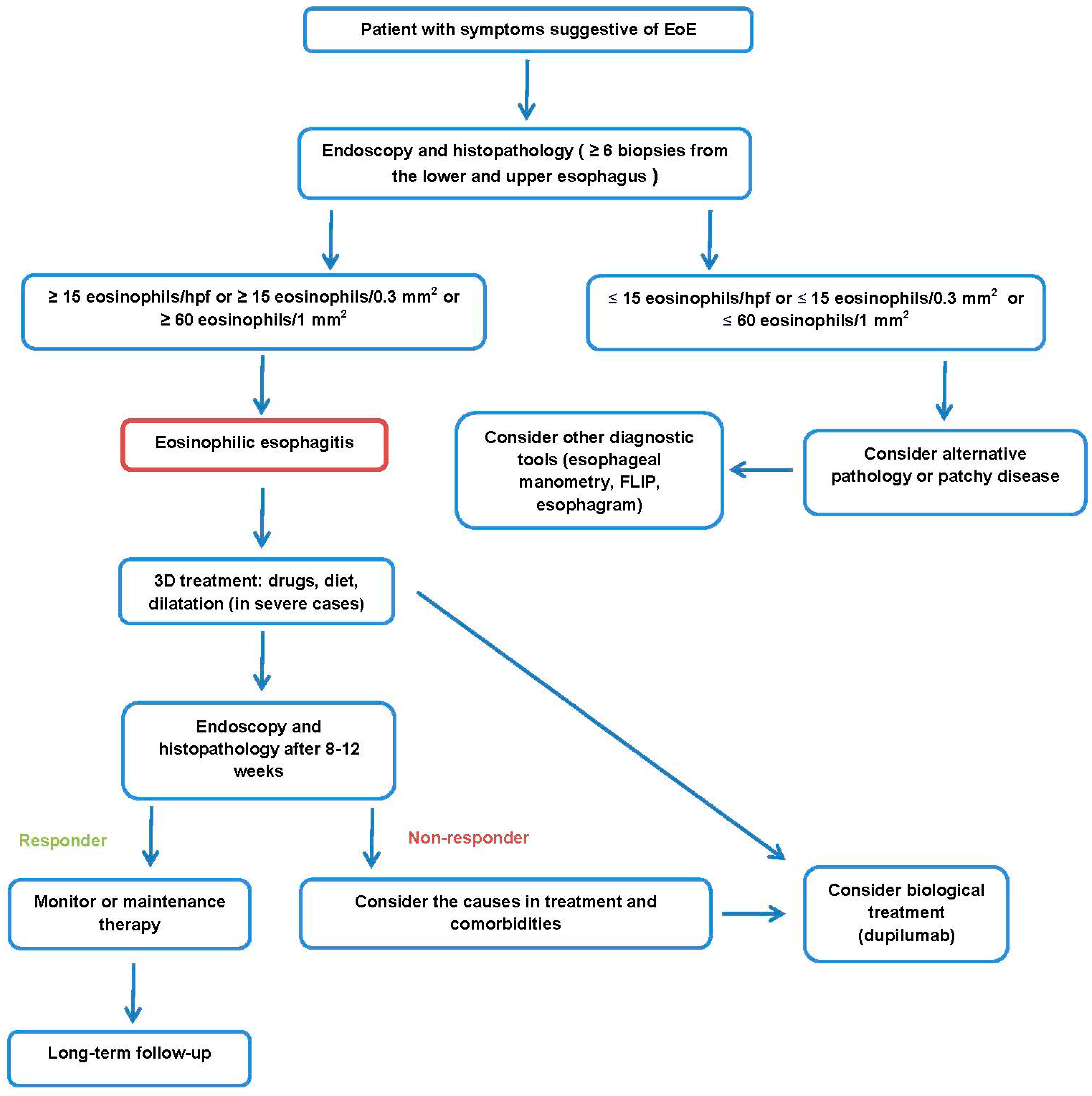
6. Diagnostics
6.1. Endoscopy and Histopathology
- Grade 0: none;
- Grade 1: subtle, circumferential ridges;
- Grade 2: distinct rings that do not impair the passage of a standard diagnostic adult endoscope;
- Grade 3: distinct rings that do not permit the passage of a diagnostic endoscope.
- Grade 0: none;
- Grade 1: involving <10% of the esophageal surface area;
- Grade 2: involving >10% of the esophageal surface area.
- Grade 0: absent;
- Grade 1: present.
- Grade 0: absent;
- Grade 1: loss of clarity of absence of vascular markings.
- Grade 0: absent;
- Grade 1: present.
6.2. Other Diagnostic Tools
6.3. Differential Diagnosis
7. Treatment
7.1. Dietary Treatment
7.2. Proton Pump Inhibitors
7.3. Swallowed Topical Corticosteroids
7.4. Biological Treatment
7.5. Complications Treatment
7.6. Long-Term Follow-Up

8. EoE Differences in Pediatric and Adolescent Patients
8.1. Symptoms
8.2. Diagnostics
8.3. Treatment
9. Conclusions
Author Contributions
Funding
Institutional Review Board Statement
Informed Consent Statement
Data Availability Statement
Conflicts of Interest
References
- Teele, R.; Katz, A.; Goldman, H.; Kettell, R. Radiographic features of eosinophilic gastroenteritis (allergic gastroenteropathy) of childhood. Am. J. Roentgenol. 1979, 132, 575–580. [Google Scholar] [CrossRef] [PubMed]
- Dobbins, J.W.; Sheahan, D.G.; Behar, J. Eosinophilic Gastroenteritis with Esophageal Involvement. Gastroenterology 1977, 72, 1312–1316. [Google Scholar] [CrossRef] [PubMed]
- Arias, Á.; Lucendo, A.J. Epidemiology and risk factors for eosinophilic esophagitis: Lessons for clinicians. Expert Rev. Gastroenterol. Hepatol. 2020, 14, 1069–1082. [Google Scholar] [CrossRef]
- Racca, F.; Pellegatta, G.; Cataldo, G.; Vespa, E.; Carlani, E.; Pelaia, C.; Paoletti, G.; Messina, M.R.; Nappi, E.; Canonica, G.W.; et al. Type 2 Inflammation in Eosinophilic Esophagitis: From Pathophysiology to Therapeutic Targets. Front. Physiol. 2022, 12, 815842. [Google Scholar] [CrossRef]
- Khokhar, D.; Marella, S.; Idelman, G.; Chang, J.W.; Chehade, M.; Hogan, S.P. Eosinophilic esophagitis: Immune mechanisms and therapeutic targets. Clin. Exp. Allergy 2022, 52, 1142–1156. [Google Scholar] [CrossRef]
- Arias, Á.; Lucendo, A.J. Molecular basis and cellular mechanisms of eosinophilic esophagitis for the clinical practice. Expert Rev. Gastroenterol. Hepatol. 2019, 13, 99–117. [Google Scholar] [CrossRef] [PubMed]
- Surdea-Blaga, T.; Popovici, E.; Stănculete, M.F.; Dumitrascu, D.L.; Scarpignato, C. Eosinophilic Esophagitis: Diagnosis and Current Management. J. Gastrointest. Liver Dis. 2020, 29, 85–97. [Google Scholar] [CrossRef]
- Ishimura, N.; Okimoto, E.; Shibagaki, K.; Nagano, N.; Ishihara, S. Similarity and difference in the characteristics of eosinophilic esophagitis between Western countries and Japan. Dig. Endosc. 2021, 33, 708–719. [Google Scholar] [CrossRef]
- Dellon, E.S.; Hirano, I. Epidemiology and Natural History of Eosinophilic Esophagitis. Gastroenterology 2018, 154, 319–332.e3. [Google Scholar] [CrossRef]
- Chang, J.W.; Haller, E.; Dellon, E.S. Dietary Management of Eosinophilic Esophagitis: Man Versus Food or Food Versus Man? Gastroenterol. Clin. N. Am. 2021, 50, 59–75. [Google Scholar] [CrossRef]
- Navarro, P.; Arias, Á.; Arias-González, L.; Laserna-Mendieta, E.J.; Ruiz-Ponce, M.; Lucendo, A.J. Systematic review with meta-analysis: The growing incidence and prevalence of eosinophilic oesophagitis in children and adults in population-based studies. Aliment. Pharmacol. Ther. 2019, 49, 1116–1125. [Google Scholar] [CrossRef] [PubMed]
- Lipowska, A.M.; Kavitt, R.T. Demographic Features of Eosinophilic Esophagitis. Gastrointest. Endosc. Clin. N. Am. 2018, 28, 27–33. [Google Scholar] [CrossRef] [PubMed]
- de Rooij, W.E.; Barendsen, M.E.; Warners, M.J.; van Rhijn, B.D.; Verheij, J.; Bruggink, A.H.; Bredenoord, A.J. Emerging incidence trends of eosinophilic esophagitis over 25 years: Results of a nationwide register-based pathology cohort. Neurogastroenterol. Motil. 2021, 33, e14072. [Google Scholar] [CrossRef]
- Schreiner, P.; Safroneeva, E.; Rossel, J.-B.; Limacher, A.; Saner, C.; Greuter, T.; Schoepfer, A.; Straumann, A.; Biedermann, L. Sex Impacts Disease Activity but Not Symptoms or Quality of Life in Adults with Eosinophilic Esophagitis. Clin. Gastroenterol. Hepatol. 2022, 20, 1729–1738.e1. [Google Scholar] [CrossRef] [PubMed]
- Shaheen, N.J.; Mukkada, V.; Eichinger, C.S.; Schofield, H.; Todorova, L.; Falk, G.W. Natural history of eosinophilic esophagitis: A systematic review of epidemiology and disease course. Dis. Esophagus 2018, 31, doy015. [Google Scholar] [CrossRef] [PubMed]
- Zdanowicz, K.; Kucharska, M.; Sobaniec-Lotowska, M.E.; Lebensztejn, D.M.; Daniluk, U. Eosinophilic Esophagitis in Children in North-Eastern Poland. J. Clin. Med. 2020, 9, 3869. [Google Scholar] [CrossRef]
- Altamimi, E.; Ahmad, B.; Abu-Aqoulah, A.; Rawabdeh, N. Clinico-pathological characteristics of eosinophilic esophagitis in Jordanian children. Gastroenterol. Rev. 2022, 16, 207–212. [Google Scholar] [CrossRef]
- Röjler, L.; Garber, J.J.; Roelstraete, B.; Walker, M.M.; Ludvigsson, J.F. Mortality in Eosinophilic Esophagitis—A nationwide, population-based matched cohort study from 2005 to 2017. Upsala J. Med. Sci. 2021, 126, e7688. [Google Scholar] [CrossRef]
- Suryawala, K.; Palle, S.; Altaf, M.A. Epidemiology, Clinical Presentation, and Seasonal Variation in the Diagnosis of Children with Eosinophilic Esophagitis in Oklahoma. South. Med. J. 2020, 113, 37–41. [Google Scholar] [CrossRef]
- Reed, C.C.; Iglesia, E.G.; Commins, S.P.; Dellon, E.S. Seasonal exacerbation of eosinophilic esophagitis histologic activity in adults and children implicates role of aeroallergens. Ann. Allergy Asthma Immunol. 2019, 122, 296–301. [Google Scholar] [CrossRef]
- Anderson, J.; Moonie, S.; Hogan, M.B.; Labus, B. A description of eosinophilic esophagitis in the Southwestern state of Nevada. Postgrad. Med. 2020, 132, 251–255. [Google Scholar] [CrossRef]
- Philpott, H.; Nandurkar, S.; Royce, S.G.; Thien, F.; Gibson, P.R. Risk factors for eosinophilic esophagitis. Clin. Exp. Allergy 2014, 44, 1012–1019. [Google Scholar] [CrossRef]
- Chandramouleeswaran, P.M.; Shen, D.; Lee, A.J.; Benitez, A.; Dods, K.; Gambanga, F.; Wilkins, B.J.; Merves, J.; Noah, Y.; Toltzis, S.; et al. Preferential Secretion of Thymic Stromal Lymphopoietin (TSLP) by Terminally Differentiated Esophageal Epithelial Cells: Relevance to Eosinophilic Esophagitis (EoE). PLoS ONE 2016, 11, e0150968. [Google Scholar] [CrossRef]
- Kottyan, L.; Rothenberg, M. Genetics of eosinophilic esophagitis. Mucosal Immunol. 2017, 10, 580–588. [Google Scholar] [CrossRef]
- Sherrill, J.D.; Gao, P.-S.; Stucke, E.M.; Blanchard, C.; Collins, M.H.; Putnam, P.E.; Franciosi, J.P.; Kushner, J.P.; Abonia, J.P.; Assa’Ad, A.H.; et al. Variants of thymic stromal lymphopoietin and its receptor associate with eosinophilic esophagitis. J. Allergy Clin. Immunol. 2010, 126, 160–165.e3. [Google Scholar] [CrossRef] [PubMed]
- Zhernov, Y.V.; Vysochanskaya, S.O.; Sukhov, V.A.; Zaostrovtseva, O.K.; Gorshenin, D.S.; Sidorova, E.A.; Mitrokhin, O.V. Molecular Mechanisms of Eosinophilic Esophagitis. Int. J. Mol. Sci. 2021, 22, 13183. [Google Scholar] [CrossRef] [PubMed]
- Lyles, J.; Rothenberg, M. Role of genetics, environment, and their interactions in the pathogenesis of eosinophilic esophagitis. Curr. Opin. Immunol. 2019, 60, 46–53. [Google Scholar] [CrossRef]
- Davis, B.P.; Stucke, E.M.; Khorki, M.E.; Litosh, V.; Rymer, J.K.; Rochman, M.; Travers, J.; Kottyan, L.; Rothenberg, M.E. Eosinophilic esophagitis–linked calpain 14 is an IL-13–induced protease that mediates esophageal epithelial barrier impairment. J. Clin. Investig. 2016, 1, e86355. [Google Scholar] [CrossRef] [PubMed]
- Lyles, J.L.; Martin, L.J.; Shoda, T.; Collins, M.H.; Trimarchi, M.P.; He, H.; Kottyan, L.C.; Mukkada, V.A.; Rothenberg, M.E. Very early onset eosinophilic esophagitis is common, responds to standard therapy, and demonstrates enrichment for CAPN14 genetic variants. J. Allergy Clin. Immunol. 2021, 147, 244–254.e6. [Google Scholar] [CrossRef]
- Litosh, V.A.; Rochman, M.; Rymer, J.K.; Porollo, A.; Kottyan, L.C.; Rothenberg, M.E. Calpain-14 and its association with eosinophilic esophagitis. J. Allergy Clin. Immunol. 2017, 139, 1762–1771.e7. [Google Scholar] [CrossRef]
- Jensen, E.T.; Dellon, E.S. Environmental factors and eosinophilic esophagitis. J. Allergy Clin. Immunol. 2018, 142, 32–40. [Google Scholar] [CrossRef] [PubMed]
- Shah, S.C.; Tepler, A.; Peek, R.M., Jr.; Colombel, J.-F.; Hirano, I.; Narula, N. Association between Helicobacter pylori Exposure and Decreased Odds of Eosinophilic Esophagitis—A Systematic Review and Meta-analysis. Clin. Gastroenterol. Hepatol. 2019, 17, 2185–2198.e3. [Google Scholar] [CrossRef] [PubMed]
- Asfari, M.M.; Kendrick, K.; Sarmini, M.T.; Uy, P.; Vega, K.J. Association of Eosinophilic Esophagitis and Human Immunodeficiency Virus. Dig. Dis. Sci. 2021, 66, 2669–2673. [Google Scholar] [CrossRef]
- Gomez-Aldana, A.J.; Jaramillo-Santos, M.; Delgado, A.F.; Jaramillo, C.; Lúquez-Mindiola, A. Eosinophilic esophagitis: Current concepts in diagnosis and treatment. World J. Gastroenterol. 2019, 25, 4598–4613. [Google Scholar] [CrossRef]
- Taverne-Ghadwal, L.; Kuhns, M.; Buhl, T.; Schulze, M.H.; Mbaitolum, W.J.; Kersch, L.; Weig, M.; Bader, O.; Groß, U. Epidemiology and Prevalence of Oral Candidiasis in HIV Patients from Chad in the Post-HAART Era. Front. Microbiol. 2022, 13, 844069. [Google Scholar] [CrossRef] [PubMed]
- Hughes, C.A.; Tseng, A.; Cooper, R. Managing drug interactions in HIV-infected adults with comorbid illness. Can. Med. Assoc. J. 2015, 187, 36–43. [Google Scholar] [CrossRef] [PubMed]
- Dellon, E.S.; Shaheen, O.; Koutlas, N.T.; Chang, A.O.; Martin, L.J.; Rothenberg, M.E.; Jensen, E.T. Early life factors are associated with risk for eosinophilic esophagitis diagnosed in adulthood. Dis. Esophagus 2021, 34, doaa074. [Google Scholar] [CrossRef]
- Ridolo, E.; Martignago, I.; Pellicelli, I.; Incorvaia, C. Assessing the Risk Factors for Refractory Eosinophilic Esophagitis in Children and Adults. Gastroenterol. Res. Pract. 2019, 2019, 1654543. [Google Scholar] [CrossRef]
- Chang, J.W.; Jensen, E.T.; Dellon, E.S. Nature with Nurture: The Role of Intrinsic Genetic and Extrinsic Environmental Factors on Eosinophilic Esophagitis. Curr. Allergy Asthma Rep. 2022, 22, 163–170. [Google Scholar] [CrossRef]
- Koutlas, N.T.; Eluri, S.; Rusin, S.; Perjar, I.; Hollyfield, J.; Woosley, J.T.; Shaheen, N.J.; Dellon, E.S. Impact of smoking, alcohol consumption, and NSAID use on risk for and phenotypes of eosinophilic esophagitis. Dis. Esophagus 2018, 31, 1–7. [Google Scholar] [CrossRef]
- Kanikowska, A.; Hryhorowicz, S.; Rychter, A.M.; Kucharski, M.A.; Zawada, A.; Iwanik, K.; Eder, P.; Słomski, R.; Dobrowolska, A.; Krela-Kaźmierczak, I. Immunogenetic, Molecular and Microbiotic Determinants of Eosinophilic Esophagitis and Clinical Practice—A New Perspective of an Old Disease. Int. J. Mol. Sci. 2021, 22, 10830. [Google Scholar] [CrossRef]
- Alhmoud, T.; Ghazaleh, S.; Ghanim, M.; Redfern, R.E. The Risk of Esophageal Food Impaction in Eosinophilic Esophagitis Patients: The Role of Clinical and Socioeconomic Factors. Clin. Exp. Gastroenterol. 2022, 15, 153–161. [Google Scholar] [CrossRef]
- Wilson, J.M.; Li, R.-C.; McGowan, E.C. The Role of Food Allergy in Eosinophilic Esophagitis. J. Asthma Allergy 2020, 13, 679–688. [Google Scholar] [CrossRef] [PubMed]
- Guajardo, J.R.; Zegarra-Bustamante, M.A.; Brooks, E.G. Does Aeroallergen Sensitization Cause or Contribute to Eosinophilic Esophagitis? Clin. Rev. Allergy Immunol. 2018, 55, 65–69. [Google Scholar] [CrossRef]
- Hernandez, P.V.; Amer, S.; Lam-Himlin, D.M.; DiSantis, D.J.; Menias, C.O.; Horsley-Silva, J.L. Eosinophilic esophagitis: Imaging features with endoscopic and pathologic correlation. Abdom. Radiol. 2020, 45, 591–600. [Google Scholar] [CrossRef]
- Visaggi, P.; Ghisa, M.; Marabotto, E.; Venturini, A.; Donati, D.S.; Bellini, M.; Savarino, V.; de Bortoli, N.; Savarino, E. Esophageal dysmotility in patients with eosinophilic esophagitis: Pathogenesis, assessment tools, manometric characteristics, and clinical implications. Esophagus 2023, 20, 29–38. [Google Scholar] [CrossRef]
- Dhar, A.; Haboubi, H.N.; Attwood, S.E.; Auth, M.K.H.; Dunn, J.M.; Sweis, R.; Morris, D.; Epstein, J.; Novelli, M.R.; Hunter, H.; et al. British Society of Gastroenterology (BSG) and British Society of Paediatric Gastroenterology, Hepatology and Nutrition (BSPGHAN) joint consensus guidelines on the diagnosis and management of eosinophilic oesophagitis in children and adults. Gut 2022, 71, 1459–1487. [Google Scholar] [CrossRef] [PubMed]
- Holvoet, S.; Doucet-Ladevèze, R.; Perrot, M.; Barretto, C.; Nutten, S.; Blanchard, C. Beneficial effect of Lactococcus lactis NCC 2287 in a murine model of eosinophilic esophagitis. Allergy 2016, 71, 1753–1761. [Google Scholar] [CrossRef] [PubMed]
- Lim, A.H.; Wong, S.; Nguyen, N.Q. Eosinophilic Esophagitis and IgG4: Is There a Relationship? Dig. Dis. Sci. 2021, 66, 4099–4108. [Google Scholar] [CrossRef]
- Pope, A.E.; Stanzione, N.; Naini, B.V.; Garcia-Lloret, M.; Ghassemi, K.A.; Marcus, E.A.; Martin, M.G.; Wozniak, L.J. Esophageal IgG4: Clinical, Endoscopic, and Histologic Correlations in Eosinophilic Esophagitis. J. Pediatr. Gastroenterol. Nutr. 2019, 68, 689–694. [Google Scholar] [CrossRef]
- Weidlich, S.; Nennstiel, S.; Jesinghaus, M.; Brockow, K.; Slotta-Huspenina, J.; Bajbouj, M.; Schmid, R.M.; Schlag, C. IgG4 is Elevated in Eosinophilic Esophagitis but Not in Gastroesophageal Reflux Disease Patients. J. Clin. Gastroenterol. 2020, 54, 43–49. [Google Scholar] [CrossRef]
- Almeida, K.A.; Andrade, E.D.Q.; Burns, G.; Hoedt, E.C.; Mattes, J.; Keely, S.; Collison, A. The microbiota in eosinophilic esophagitis: A systematic review. J. Gastroenterol. Hepatol. 2022, 37, 1673–1684. [Google Scholar] [CrossRef] [PubMed]
- Rodríguez-Sánchez, J.; Barrio, J.; Castillejo, O.N.; Valdivieso-Cortazar, E.; Pérez-Martínez, I.; Boumidi, A.; Olmos-Jérez, J.A.; Payeras-Llodra, G.; Alcaide-Suarez, N.; Ruiz-Rebollo, L.; et al. The Endoscopic Reference Score shows modest accuracy to predict either clinical or histological activity in adult patients with eosinophilic oesophagitis. Aliment. Pharmacol. Ther. 2017, 45, 300–309. [Google Scholar] [CrossRef]
- Schreiner, P.; Biedermann, L.; Greuter, T.; Wright, B.L.; Straumann, A. How to approach adult patients with asymptomatic esophageal eosinophilia. Dis. Esophagus 2021, 34, doaa105. [Google Scholar] [CrossRef] [PubMed]
- Suzuki, Y.; Iizuka, T.; Hosoi, A.; Kikuchi, D.; Okamura, T.; Ochiai, Y.; Hayasaka, J.; Dan, N.; Mitsunaga, Y.; Tanaka, M.; et al. Clinicopathological Differences between Eosinophilic Esophagitis and Asymptomatic Esophageal Eosinophilia. Intern. Med. 2022, 61, 1319–1327. [Google Scholar] [CrossRef]
- Fujiwara, Y. Symptom-based diagnostic approach for eosinophilic esophagitis. J. Gastroenterol. 2020, 55, 833–845. [Google Scholar] [CrossRef]
- Visaggi, P.; Savarino, E.; Sciume, G.; Di Chio, T.; Bronzini, F.; Tolone, S.; Frazzoni, M.; Pugno, C.; Ghisa, M.; Bertani, L.; et al. Eosinophilic esophagitis: Clinical, endoscopic, histologic and therapeutic differences and similarities between children and adults. Ther. Adv. Gastroenterol. 2021, 14, 1756284820980860. [Google Scholar] [CrossRef]
- Dellon, E.S.; Liacouras, C.A.; Molina-Infante, J.; Furuta, G.T.; Spergel, J.M.; Zevit, N.; Spechler, S.J.; Attwood, S.E.; Straumann, A.; Aceves, S.S.; et al. Updated International Consensus Diagnostic Criteria for Eosinophilic Esophagitis: Proceedings of the AGREE Conference. Gastroenterology 2018, 155, 1022–1033.e10. [Google Scholar] [CrossRef] [PubMed]
- Rubinstein, E.; Rosen, R.L. Respiratory symptoms associated with eosinophilic esophagitis. Pediatr. Pulmonol. 2018, 53, 1587–1591. [Google Scholar] [CrossRef]
- Arratibel, P.; Gil-Lasa, I.; Cobian, J.; Izagirre-Arostegi, A.; Arzallus, T.; Etxart, A.; Sarasqueta, C.; Zubiaurre, L.; Bujanda, L. Incidence and evolution of foreign body impaction in the upper gastrointestinal tract and its relationship with eosionophilic oesophagitis. Gastroenterol. Y Hepatol. 2022, 45, 274–281. [Google Scholar] [CrossRef]
- Alexander, J.A. Endoscopic and Radiologic Findings in Eosinophilic Esophagitis. Gastrointest. Endosc. Clin. N. Am. 2018, 28, 47–57. [Google Scholar] [CrossRef]
- Samadi, F.; Levine, M.S.; Rubesin, S.E.; Katzka, D.A.; Laufer, I. Feline Esophagus and Gastroesophageal Reflux. Am. J. Roentgenol. 2010, 194, 972–976. [Google Scholar] [CrossRef]
- Hirano, I.; Moy, N.; Heckman, M.G.; Thomas, C.S.; Gonsalves, N.; Achem, S.R. Endoscopic assessment of the oesophageal features of eosinophilic oesophagitis: Validation of a novel classification and grading system. Gut 2013, 62, 489–495. [Google Scholar] [CrossRef] [PubMed]
- Dellon, E.S.; Cotton, C.C.; Gebhart, J.H.; Higgins, L.L.; Beitia, R.; Woosley, J.T.; Shaheen, N.J. Accuracy of the Eosinophilic Esophagitis Endoscopic Reference Score in Diagnosis and Determining Response to Treatment. Clin. Gastroenterol. Hepatol. 2016, 14, 31–39. [Google Scholar] [CrossRef] [PubMed]
- Yoon, H.J.; Youn, Y.H.; Park, J.C.; Park, H. Reversibility of Endoscopic Features after Treatment for Eosinophilic Esophagitis. Yonsei Med. J. 2021, 62, 487–493. [Google Scholar] [CrossRef] [PubMed]
- Navarro, P.; Laserna-Mendieta, E.J.; Casabona, S.; Savarino, E.; Pérez-Fernández, M.T.; Ghisa, M.; Pérez-Martínez, I.; Guagnozzi, D.; Perelló, A.; Guardiola-Arévalo, A.; et al. Accurate and timely diagnosis of Eosinophilic Esophagitis improves over time in Europe. An analysis of the EoE CONNECT Registry. United Eur. Gastroenterol. J. 2022, 10, 507–517. [Google Scholar] [CrossRef] [PubMed]
- Tourlamain, G.; Garcia-Puig, R.; Gutiérrez-Junquera, C.; Papadopoulou, A.; Roma, E.; Kalach, N.; Oudshoorn, J.; Sokollik, C.; Karolewska-Bochenek, K.; Oliva, S.; et al. Differences in Management of Eosinophilic Esophagitis in Europe: An Assessment of Current Practice. J. Pediatr. Gastroenterol. Nutr. 2020, 71, 83–90. [Google Scholar] [CrossRef] [PubMed]
- Richter, J.E. Endoscopic Treatment of Eosinophilic Esophagitis. Gastrointest. Endosc. Clin. N. Am. 2018, 28, 97–110. [Google Scholar] [CrossRef]
- Donnan, E.N.; Pandolfino, J.E. Applying the Functional Luminal Imaging Probe to Esophageal Disorders. Curr. Gastroenterol. Rep. 2020, 22, 10. [Google Scholar] [CrossRef]
- Hirano, I.; Pandolfino, J.E.; Boeckxstaens, G.E. Functional Lumen Imaging Probe for the Management of Esophageal Disorders: Expert Review from the Clinical Practice Updates Committee of the AGA Institute. Clin. Gastroenterol. Hepatol. 2017, 15, 325–334. [Google Scholar] [CrossRef]
- Savarino, E.; di Pietro, M.; Bredenoord, A.J.; Carlson, D.A.; Clarke, J.O.; Khan, A.; Vela, M.F.; Yadlapati, R.; Pohl, D.; Pandolfino, J.E.; et al. Use of the Functional Lumen Imaging Probe in Clinical Esophagology. Am. J. Gastroenterol. 2020, 115, 1786–1796. [Google Scholar] [CrossRef]
- Vendrami, C.L.; Kelahan, L.; Escobar, D.J.; Goodhartz, L.; Hammond, N.; Nikolaidis, P.; Yang, G.-Y.; Hirano, I.; Miller, F.H. Imaging Findings of Eosinophilic Gastrointestinal Diseases in Adults. Curr. Probl. Diagn. Radiol. 2023, 52, 139–147. [Google Scholar] [CrossRef]
- Uchida, A.M.; Ro, G.; Garber, J.J.; Peterson, K.A.; Round, J.L. Models and Tools for Investigating Eosinophilic Esophagitis at the Bench. Front. Immunol. 2022, 13, 943518. [Google Scholar] [CrossRef]
- Votto, M.; De Filippo, M.; Castagnoli, R.; Delle Cave, F.; Giffoni, F.; Santi, V.; Vergani, M.; Caffarelli, C.; De Amici, M.; Marseglia, G.L.; et al. Non-invasive biomarkers of eosinophilic esophagitis. Acta Biomed. 2021, 92, e2021530. [Google Scholar] [CrossRef]
- Kuźmiński, A.; Rosada, T.; Przybyszewska, J.; Ukleja-Sokołowska, N.; Bartuzi, Z. Eosinophilic gastroenteritis—A manifestation of an allergic disease in the gastrointestinal tract? Part 1. Epidemiology and diagnosis. Gastroenterol. Rev. 2022, 17, 1–6. [Google Scholar] [CrossRef]
- Molina-Infante, J.; Lucendo, A.J. Dietary therapy for eosinophilic esophagitis. J. Allergy Clin. Immunol. 2018, 142, 41–47. [Google Scholar] [CrossRef] [PubMed]
- Sher, E.R.; Ross, J.A.; Weine, D.M.; Arjun, A.C. Current and emerging therapies for eosinophilic esophagitis. Allergy Asthma Proc. 2022, 43, 178–186. [Google Scholar] [CrossRef] [PubMed]
- Molina-Infante, J. Nutritional and Psychological Considerations for Dietary Therapy in Eosinophilic Esophagitis. Nutrients 2022, 14, 1588. [Google Scholar] [CrossRef]
- Greuter, T.; Alexander, J.A.; Straumann, A.; Katzka, D.A. Diagnostic and Therapeutic Long-term Management of Eosinophilic Esophagitis—Current Concepts and Perspectives for Steroid Use. Clin. Transl. Gastroenterol. 2018, 9, e212. [Google Scholar] [CrossRef]
- Andrist, C.M.; Jörg, L.; Greuter, T.; Gschwend, A.; Straumann, A.; Helbling, A. Delayed hypersensitivity reaction to orodispersible budesonide in a case with eosinophilic esophagitis. BMC Gastroenterol. 2020, 20, 419. [Google Scholar] [CrossRef]
- Dellon, E.S.; Woosley, J.T.; Arrington, A.; McGee, S.J.; Covington, J.; Moist, S.E.; Gebhart, J.H.; Tylicki, A.E.; Shoyoye, S.O.; Martin, C.F.; et al. Efficacy of Budesonide vs Fluticasone for Initial Treatment of Eosinophilic Esophagitis in a Randomized Controlled Trial. Gastroenterology 2019, 157, 65–73.e5. [Google Scholar] [CrossRef]
- Dellon, E.S.; Lucendo, A.J.; Schlag, C.; Schoepfer, A.M.; Falk, G.W.; Eagle, G.; Nezamis, J.; Comer, G.M.; Knoop, K.; Hirano, I. Fluticasone Propionate Orally Disintegrating Tablet (APT-1011) for Eosinophilic Esophagitis: Randomized Controlled Trial. Clin. Gastroenterol. Hepatol. 2022, 20, 2485–2494.e15. [Google Scholar] [CrossRef] [PubMed]
- Dellon, E.S.; Katzka, D.A.; Collins, M.H.; Hamdani, M.; Gupta, S.K.; Hirano, I.; Kagalwalla, A.; Lewis, J.; Markowitz, J.; Nurko, S.; et al. Budesonide Oral Suspension Improves Symptomatic, Endoscopic, and Histologic Parameters Compared with Placebo in Patients with Eosinophilic Esophagitis. Gastroenterology 2017, 152, 776–786.e5. [Google Scholar] [CrossRef]
- Lipka, S.; Kumar, A.; Miladinovic, B.; Richter, J.E. Systematic review with network meta-analysis: Comparative effectiveness of topical steroids vs. PPIs for the treatment of the spectrum of eosinophilic oesophagitis. Aliment. Pharmacol. Ther. 2016, 43, 663–673. [Google Scholar] [CrossRef]
- Dellon, E.S.; Spergel, J.M. Biologics in eosinophilic gastrointestinal diseases. Ann. Allergy Asthma Immunol. 2023, 130, 21–27. [Google Scholar] [CrossRef] [PubMed]
- Eskian, M.; Khorasanizadeh, M.; Assa’Ad, A.H.; Rezaei, N. Monoclonal Antibodies for Treatment of Eosinophilic Esophagitis. Clin. Rev. Allergy Immunol. 2018, 55, 88–98. [Google Scholar] [CrossRef] [PubMed]
- Straumann, A.; Conus, S.; Grzonka, P.; Kita, H.; Kephart, G.; Bussmann, C.; Beglinger, C.; Smith, D.A.; Patel, J.; Byrne, M.; et al. Anti-interleukin-5 antibody treatment (mepolizumab) in active eosinophilic oesophagitis: A randomised, placebo-controlled, double-blind trial. Gut 2010, 59, 21–30. [Google Scholar] [CrossRef]
- Spergel, J.M.; Rothenberg, M.E.; Collins, M.H.; Furuta, G.T.; Markowitz, J.E.; Fuchs, G.; O’Gorman, M.A.; Abonia, J.P.; Young, J.; Henkel, T.; et al. Reslizumab in children and adolescents with eosinophilic esophagitis: Results of a double-blind, randomized, placebo-controlled trial. J. Allergy Clin. Immunol. 2012, 129, 456–463.e3. [Google Scholar] [CrossRef]
- Rothenberg, M.E.; Wen, T.; Greenberg, A.; Alpan, O.; Enav, B.; Hirano, I.; Nadeau, K.; Kaiser, S.; Peters, T.; Perez, A.; et al. Intravenous anti–IL-13 mAb QAX576 for the treatment of eosinophilic esophagitis. J. Allergy Clin. Immunol. 2015, 135, 500–507. [Google Scholar] [CrossRef] [PubMed]
- Harb, H.; Chatila, T.A. Mechanisms of Dupilumab. Clin. Exp. Allergy 2020, 50, 5–14. [Google Scholar] [CrossRef]
- Al-Horani, R.A.; Chiles, R. First Therapeutic Approval for Eosinophilic Esophagitis. Gastroenterol. Insights 2022, 13, 24. [Google Scholar] [CrossRef] [PubMed]
- Dellon, E.S.; Rothenberg, M.E.; Collins, M.H.; Hirano, I.; Chehade, M.; Bredenoord, A.J.; Lucendo, A.J.; Spergel, J.M.; Aceves, S.; Sun, X.; et al. Dupilumab in Adults and Adolescents with Eosinophilic Esophagitis. N. Engl. J. Med. 2022, 387, 2317–2330. [Google Scholar] [CrossRef] [PubMed]
- Aceves, S.S.; Dellon, E.S.; Greenhawt, M.; Hirano, I.; Liacouras, C.A.; Spergel, J.M. Clinical guidance for the use of dupilumab in eosinophilic esophagitis: A yardstick. Ann. Allergy Asthma Immunol. 2022, 130, 371–378. [Google Scholar] [CrossRef] [PubMed]
- Clayton, F.; Fang, J.C.; Gleich, G.J.; Lucendo, A.J.; Olalla, J.M.; Vinson, L.A.; Lowichik, A.; Chen, X.; Emerson, L.; Cox, K.; et al. Eosinophilic Esophagitis in Adults Is Associated with IgG4 and Not Mediated by IgE. Gastroenterology 2014, 147, 602–609. [Google Scholar] [CrossRef]
- Lam, A.Y.; Ma, C.; Lee, J.K.; Bredenoord, A.J. Eosinophilic esophagitis: New molecules, better life? Curr. Opin. Pharmacol. 2022, 63, 102183. [Google Scholar] [CrossRef]
- Young, E.; Philpott, H. Pathophysiology of Dysphagia in Eosinophilic Esophagitis: Causes, Consequences, and Management. Dig. Dis. Sci. 2022, 67, 1101–1115. [Google Scholar] [CrossRef]
- Moole, H.; Jacob, K.; Duvvuri, A.; Moole, V.; Dharmapuri, S.; Boddireddy, R.; Uppu, A.; Puli, S.R. Role of endoscopic esophageal dilation in managing eosinophilic esophagitis: A systematic review and meta-analysis. Medicine 2017, 96, e5877. [Google Scholar] [CrossRef]
- Podboy, A.J.; Lavey, C.; Mara, K.; Geno, D.; Khana, S.; Ravi, K.; Katzka, D.; Alexander, J. Eosinophilic Esophagitis Is Rarely Continually Symptomatic 10 Years after an Initial Treatment Course in Adults. Dig. Dis. Sci. 2019, 64, 3568–3578. [Google Scholar] [CrossRef]
- De Matteis, A.; Pagliaro, G.; Corleto, V.D.; Pacchiarotti, C.; Di Giulio, E.; Villa, M.P.; Parisi, P.; Vassallo, F.; Ziparo, C.; Di Nardo, G. Eosinophilic Esophagitis in Children: Clinical Findings and Diagnostic Approach. Curr. Pediatr. Rev. 2020, 16, 206–214. [Google Scholar] [CrossRef]
- Barni, S.; Arasi, S.; Mastrorilli, C.; Pecoraro, L.; Giovannini, M.; Mori, F.; Liotti, L.; Saretta, F.; Castagnoli, R.; Caminiti, L.; et al. Pediatric eosinophilic esophagitis: A review for the clinician. Ital. J. Pediatr. 2021, 47, 230. [Google Scholar] [CrossRef]
- Kim, H.P.; Vance, R.B.; Shaheen, N.J.; Dellon, E.S. The Prevalence and Diagnostic Utility of Endoscopic Features of Eosinophilic Esophagitis: A Meta-analysis. Clin. Gastroenterol. Hepatol. 2012, 10, 988–996.e5. [Google Scholar] [CrossRef] [PubMed]
- Wechsler, J.B.; Bolton, S.M.; Amsden, K.; Wershil, B.K.; Hirano, I.; Kagalwalla, A.F. Eosinophilic Esophagitis Reference Score Accurately Identifies Disease Activity and Treatment Effects in Children. Clin. Gastroenterol. Hepatol. 2018, 16, 1056–1063. [Google Scholar] [CrossRef] [PubMed]
- Spergel, J.M.; Brown-Whitehorn, T.A.; Muir, A.; Liacouras, C.A. Medical algorithm: Diagnosis and treatment of eosinophilic esophagitis in children. Allergy 2020, 75, 1522–1524. [Google Scholar] [CrossRef] [PubMed]
- Hirano, I.; Dellon, E.S.; Gupta, S.K.; Katzka, D.A.; Collins, M.H.; Wojtowicz, A.M.; Terreri, B.; Zhang, W.; Boules, M.; Bhatia, S.; et al. Safety of an investigational formulation of budesonide (budesonide oral suspension) for eosinophilic oesophagitis: An integrated safety analysis of six phase 1–3 clinical trials. Aliment. Pharmacol. Ther. 2023; online ahead of print. [Google Scholar] [CrossRef]
- Munoz-Osores, E.; Maldonado-Campos, I.; Olivares-Labbe, M.T.; Villarroel, L.; Gana, J.C. Corticosteroids for Eosinophilic Esophagitis in Children: A Meta-analysis. Pediatrics 2020, 146, e20200874. [Google Scholar] [CrossRef]
- Oliva, S.; Rossetti, D.; Papoff, P.; Tiberti, A.; Mallardo, S.; Volpe, D.; Ruggiero, C.; Russo, G.; Vezzoli, D.; Isoldi, S.; et al. A 12-Week Maintenance Therapy with a New Prepared Viscous Budesonide in Pediatric Eosinophilic Esophagitis. Dig. Dis. Sci. 2019, 64, 1571–1578. [Google Scholar] [CrossRef]
- Carr, S.; Chan, E.S.; Watson, W. Eosinophilic esophagitis. Allergy Asthma Clin. Immunol. 2018, 14 (Suppl. 2), 58. [Google Scholar] [CrossRef] [PubMed]
- Sindher, S.B.; Barshow, S.; Tirumalasetty, J.; Arasi, S.; Atkins, D.; Bauer, M.; Bégin, P.; Collins, M.H.; Deschildre, A.; Doyle, A.D.; et al. The role of biologics in pediatric food allergy and eosinophilic gastrointestinal disorders. J. Allergy Clin. Immunol. 2023, 151, 595–606. [Google Scholar] [CrossRef]
- Junquera, C.G.; Fernández, S.F.; Domínguez-Ortega, G.; Miravet, V.V.; Puig, R.G.; Romero, R.G.; de Valderrama, A.F.; Rivas, R.A.; de Trastornos Gastrointestinales, G.D.T.; Vicente, C.A.; et al. Recomendaciones para el diagnóstico y manejo práctico de la esofagitis eosinofílica pediátrica: Diagnóstico y tratamiento de la esofagitis eosinofílica pediátrica. An. Pediatr. 2020, 92, 379. [Google Scholar]
- Lucendo, A.J.; Molina-Infante, J.; Arias, Á.; Von Arnim, U.; Bredenoord, A.J.; Bussmann, C.; Dias, J.A.; Bove, M.; González-Cervera, J.; Larsson, H.; et al. Guidelines on eosinophilic esophagitis: Evidence-based statements and recommendations for diagnosis and management in children and adults. United Eur. Gastroenterol. J. 2017, 5, 335–358. [Google Scholar] [CrossRef]

| EREFS Features (%) | Rodríguez-Sánchez et al. (2017) [53] | Dellon et al. (2016) [64] | ||||
|---|---|---|---|---|---|---|
| Pre-treatment (n = 67) | Post-treatment (n = 117) | p | Pre-treatment (n = 67) | Post-treatment (n = 67) | p | |
| Fixed rings (G0) * | 29.9 | 35.6 | 0.40 | 22 | 46 | 0.001 |
| Fixed rings (G1) | 37.3 | 39.3 | 0.78 | 40 | 43 | |
| Fixed rings (G2) | 28.4 | 19.7 | 0.17 | 30 | 11 | |
| Fixed rings (G3) | 6.3 | 5.1 | 0.80 | 8 | 0 | |
| Exudates (G0) | 56.7 | 62.4 | 0.44 | 51 | 76 | 0.005 |
| Exudates (G1) | 26.9 | 26.5 | 0.95 | 28 | 18 | |
| Exudates (G2) | 20.9 | 11.1 | 0.07 | 21 | 6 | |
| Furrows (G0) | 22.4 | 41 | 0.01 | 11 | 52 | |
| Furrows (G1) | 77.6 | 59 | 70 | 42 | ||
| Edema (G0) | 14.9 | 25.6 | 0.09 | 37 | 72 | <0.001 |
| Edema (G1) | 85.1 | 74.4 | 63 | 28 | ||
| Stricture (G0) | 83.6 | 79.5 | 0.66 | 76 | 75 | 0.84 |
| Stricture (G1) | 16.4 | 20.5 | 24 | 25 | ||
| Study Treatment | Dose | Age, Y, Mean ± SD | Baseline EREFS ± SD | Baseline Peak Eosinophil Count, eos/hpf ± SD | History of Atopy (%) | Duration (Weeks) | EREFS Mean Change ± SD | % Histologic Response * | Ref. |
|---|---|---|---|---|---|---|---|---|---|
| Fluticasone propionate (n = 22) | 1.5 mg twice daily | 41.3 ± 12.2 | 4.2 ± 1.8 | 69.2 ± 33.3 | 20 (91) | 12 | −2.9 ± 1.92 | 86 | [82] |
| Fluticasone propionate (n = 20) | 3 mg twice daily | 36.8 ± 9.2 | 3.9 ± 1.7 | 55.1 ± 21.3 | 18 (90) | 12 | −2.2 ± 1.84 | 80 | [82] |
| Fluticasone propionate (n = 21) | 1.5 mg once daily | 36.8 ± 11.7 | 4.7 ± 1.4 | 56.2 ± 25.9 | 20 (95) | 12 | −2.4 ± 1.85 | 48 | [82] |
| Fluticasone (n = 64) | 880 mcg twice daily | 39.0 ± 14.5 | 4.7 ± 1.9 | 72.5 ± 59.1 | 50 (78) | 8 | −1.9 ± 2.0 | 64 | [81] |
| Budesonide (n = 65) | 1 mg twice daily | 36.2 ± 19.1 | 4.7 ± 1.8 | 74.1 ± 48.2 | 46 (71) | 8 | −2.6 ± 1.8 | 71 | [81] |
| Budesonide (n = 51) | 2 mg twice daily | 22.3 ± 7.9 | 7.7 ± 3.5 | 156.3 ± 97.6 | 12 | −3.8 ± 3.9 | 39 | [83] |
| Association | Year | First Line Therapy | PPI Dose | PPI Duration | OVB Dose | FP Dose | STC Duration | Systemic Steroids |
|---|---|---|---|---|---|---|---|---|
| BSG and BSPGHAN [47] | 2022 | PPI/diet/STC | - | 8–12 weeks | Requires approval from local authorities for adolescents; for children, given in age-appropriate, viscous formulations and volume | - | 12–24 weeks | Not recommended |
| SEGHNP [109] | 2020 | PPI/diet/STC | 1–2 mg/kg b.i.d. (max 40 mg) | - | 0.5 mg/dose (2 doses a day) <10 years | 400 μg/dose (2 doses a day) <10 years | - | Consider only in emergency patients with severe dysphagia and significant weight loss or severe stenosis |
| 1 mg/dose (2 doses a day) >10 years | 800 μg/dose (2 doses a day) >10 years | |||||||
| UEG, EAACI ESPGHAN, and EUREOS [110] | 2017 | PPI/diet/STC | 1–2 mg/kg b.i.d. | - | - | - | - | Not recommended |
Disclaimer/Publisher’s Note: The statements, opinions and data contained in all publications are solely those of the individual author(s) and contributor(s) and not of MDPI and/or the editor(s). MDPI and/or the editor(s) disclaim responsibility for any injury to people or property resulting from any ideas, methods, instructions or products referred to in the content. |
© 2023 by the authors. Licensee MDPI, Basel, Switzerland. This article is an open access article distributed under the terms and conditions of the Creative Commons Attribution (CC BY) license (https://creativecommons.org/licenses/by/4.0/).
Share and Cite
Wąsik, J.; Małecka-Wojciesko, E. Eosinophilic Esophagitis—What Do We Know So Far? J. Clin. Med. 2023, 12, 2259. https://doi.org/10.3390/jcm12062259
Wąsik J, Małecka-Wojciesko E. Eosinophilic Esophagitis—What Do We Know So Far? Journal of Clinical Medicine. 2023; 12(6):2259. https://doi.org/10.3390/jcm12062259
Chicago/Turabian StyleWąsik, Jakub, and Ewa Małecka-Wojciesko. 2023. "Eosinophilic Esophagitis—What Do We Know So Far?" Journal of Clinical Medicine 12, no. 6: 2259. https://doi.org/10.3390/jcm12062259
APA StyleWąsik, J., & Małecka-Wojciesko, E. (2023). Eosinophilic Esophagitis—What Do We Know So Far? Journal of Clinical Medicine, 12(6), 2259. https://doi.org/10.3390/jcm12062259