Article

Point-of-Care Ultrasonography in a Pulmonary Hypertension Clinic: A Randomized Pilot Study

1
Assuta Medical Center, Ha-Refu’a St 7, Ashdod 7747629, Israel
2
Faculty of Health Sciences, Ben-Gurion University, Beer-Sheva 8410501, Israel
3
Department of Obstetrics and Gynecology, Hadassah Mount Scopus, Jerusalem 9765422, Israel
4
Division of Pulmonary, Critical Care, and Sleep Medicine, Mount Sinai Hospital, Icahn School of Medicine at Mount Sinai, New York, NY 10029, USA
5
Division of Medicine, Soroka University Medical Center, Ben-Gurion University of the Negev, Beer-Sheva 8410101, Israel
6
Department of Medicine, University of Toronto, Toronto, ON M5S 1A8, Canada
7
Respirology, Pulmonary Hypertension Program, University Health Network, Toronto, ON M5G 2C4, Canada
8
Medical Intensive Care Unit, Soroka University Medical Center, Ben-Gurion University of the Negev, Beer-Sheva 8410101, Israel
*
Author to whom correspondence should be addressed.
J. Clin. Med. 2023, 12(5), 1752; https://doi.org/10.3390/jcm12051752
Submission received: 9 January 2023 / Revised: 3 February 2023 / Accepted: 17 February 2023 / Published: 22 February 2023
(This article belongs to the Section Intensive Care)

Abstract

:
Pulmonary arterial hypertension (PAH) is a rare condition with the potential to progress to right heart failure. Point-of-Care Ultrasonography (POCUS), used and interpreted in real time at the bedside to further the cardiopulmonary assessment, has the potential to improve the longitudinal care of PAH patients in the ambulatory setting. Patients from PAH clinics at two academic medical centers were randomized to either a POCUS assessment cohort or non-POCUS standard care (ClinicalTrials.gov identifier NCT05332847). The POCUS group received blinded heart, lung, and vascular ultrasound assessments. Thirty-six patients were randomized to the study and followed over time. Mean age was 65 in both groups and majority female (76.5% and 88.9% females in POCUS and control, respectively). Median time for POCUS assessment was 11 min (range 8–16). There were significantly more changes in management in the POCUS group than control (73% vs. 27%, p-value < 0.001). Multivariate analysis revealed that management changes were more likely to occur with a POCUS assessment, with an odds ratio (OR) of 12 when POCUS was added to physical exam vs. OR of 4.6 compared to physical examination alone (p < 0.001). POCUS in the PAH clinic is feasible and, when combined with physical examination, increases the number of findings and results in changes in management without significantly prolonging visit encounters. POCUS may help support clinical evaluation and decision making in ambulatory PAH clinics.

1. Introduction

Pulmonary arterial hypertension (PAH) is an uncommon, progressive, and lethal disease that can eventually lead to right heart failure (RHF) [1]. The pathophysiological changes in the pulmonary vasculature which occur in advanced PAH ultimately lead to functional changes in the right ventricle (RV) [2,3]. An increase in RV afterload eventually results in the disruption of normal ventriculoatrial coupling, pressure and/or volume overload, RV dysfunction, and finally ventricular failure [4,5]. Changes in the RV include wall hypertrophy, chamber dilatation, and bowing of the interventricular septum towards the left ventricle (LV) [6,7]. Point-of-Care Ultrasonography (POCUS) is a rapidly evolving bedside modality in which ultrasonography is utilized as a part of the physical examination and the patients’ clinical assessment and is interpreted in real time at the bedside [8]. During POCUS, the cardiovascular system and the lungs are quickly examined by the clinician, and the information obtained is immediately integrated into the assessment process, therefore directly influencing treatment decisions in the moment [9,10]. Randomized trials and observational data have previously demonstrated the ability of a POCUS assessment to significantly shorten the time to definitive treatment in inpatients with dyspnea [11,12]. Recent data also suggest that individual physical examination findings alone have inadequate diagnostic utility in patients with pulmonary hypertension (PH) [13]. Formal echocardiography, interpreted by a cardiologist, is an essential modality for PAH patient management, including the assessment of disease severity, prognosis, and treatment [3,14,15]. However, a transthoracic echocardiogram (TTE) is not available routinely and expeditiously in an outpatient clinic, official results can be delayed, and obtaining a TTE often necessitates additional contacts with the medical system on the part of the patient. A full formal echocardiogram, if integrated into a clinic visit, extends the duration of the patient visit and is resource-intensive.
POCUS can be used to identify alterations in RV morphology, size, and function, and can provide a more accurate assessment of a patient’s volume status. It has the power to estimate the right ventricular systolic pressure (RVSP) as a surrogate for pulmonary artery pressure, and the presence of a pericardial effusion [11,13]. Furthermore, POCUS provides real-time information on the LV wall size and function, and can establish the diagnosis of valvulopathies [2,9,14,16]. Lung ultrasound can evaluate the presence of an increase in interstitial lung water, pleural effusion, atelectasis, or consolidation [17]. More advanced echocardiographic variables, including the RV/LV basal diameter/area ratio, flattening of the interventricular septum, and the tricuspid annular plane systolic excursion (TAPSE), are all measurable using POCUS at the bedside and are all endorsed in the most recent comprehensive PH European Respiratory Society (ERS) guidelines [18]. These bedside, immediate, additive data may assist clinicians with the identification of PAH progression, complications, and meaningful clinical changes in real time [19], with the potential over a prolonged period of care in the ambulatory setting to shorten time to treatment escalation, hospitalization, or referral to lung transplantation. While thoracic ultrasound has been well studied in the inpatient and intensive care settings and has been shown to change clinical management [20], data extending to the outpatient setting are lacking.
This study sought to evaluate the potential clinical benefits of the addition of a POCUS assessment during an ambulatory PH clinic encounter, conducted by a non-cardiologist as part of the routine clinical patient evaluation, compared to standard clinic practice without ultrasonography. To date, no other study has evaluated the utility of a thoracic cardiopulmonary POCUS assessment in a PH clinic or PAH patient population.

2. Materials and Methods

We conducted a randomized controlled pilot study to assess the feasibility and utility of the integration of POCUS into the physical assessment of PH clinic outpatients in two academic centers. This study was registered at ClinicalTrials.gov (ClinicalTrials.gov identifier NCT05332847). Patients were recruited from the PH clinics at the Soroka University Medical Center and Barzilai University Medical Center between January 2018 and March 2019. Patients were included if they were diagnosed with group 1 PAH, as defined by the ERS clinical classification of PH [3], including right heart catheterization, and provided informed consent. Patients were excluded if they were diagnosed with a World Health Organization group 2–5 PH disease, had congenital heart disease, liver cirrhosis, or suspected pulmonary venous occlusive disease.
On enrollment, randomization of patients was performed 1:1 (using ClinStat) to either the POCUS assessment group or control group. Both groups underwent the same clinical assessment according to clinic protocols and were evaluated by the same pulmonary hypertension specialist (A.A.) to reduce variability. Every patient was scheduled for a follow-up appointment every three months as part of usual PAH ambulatory clinical care. The study protocol was approved by each participating center’s research ethics board (BRZ 0106-18, SOR0327-16). Clinic visit evaluations for all patients in both groups were standardized (Figure 1a). Each visit included a history, physical and laboratory evaluation, BNP level, 6 min walk test, and a quality-of-life assessment using emPHasis 10, a well-validated health-related quality-of-life questionnaire in PAH [3,21].
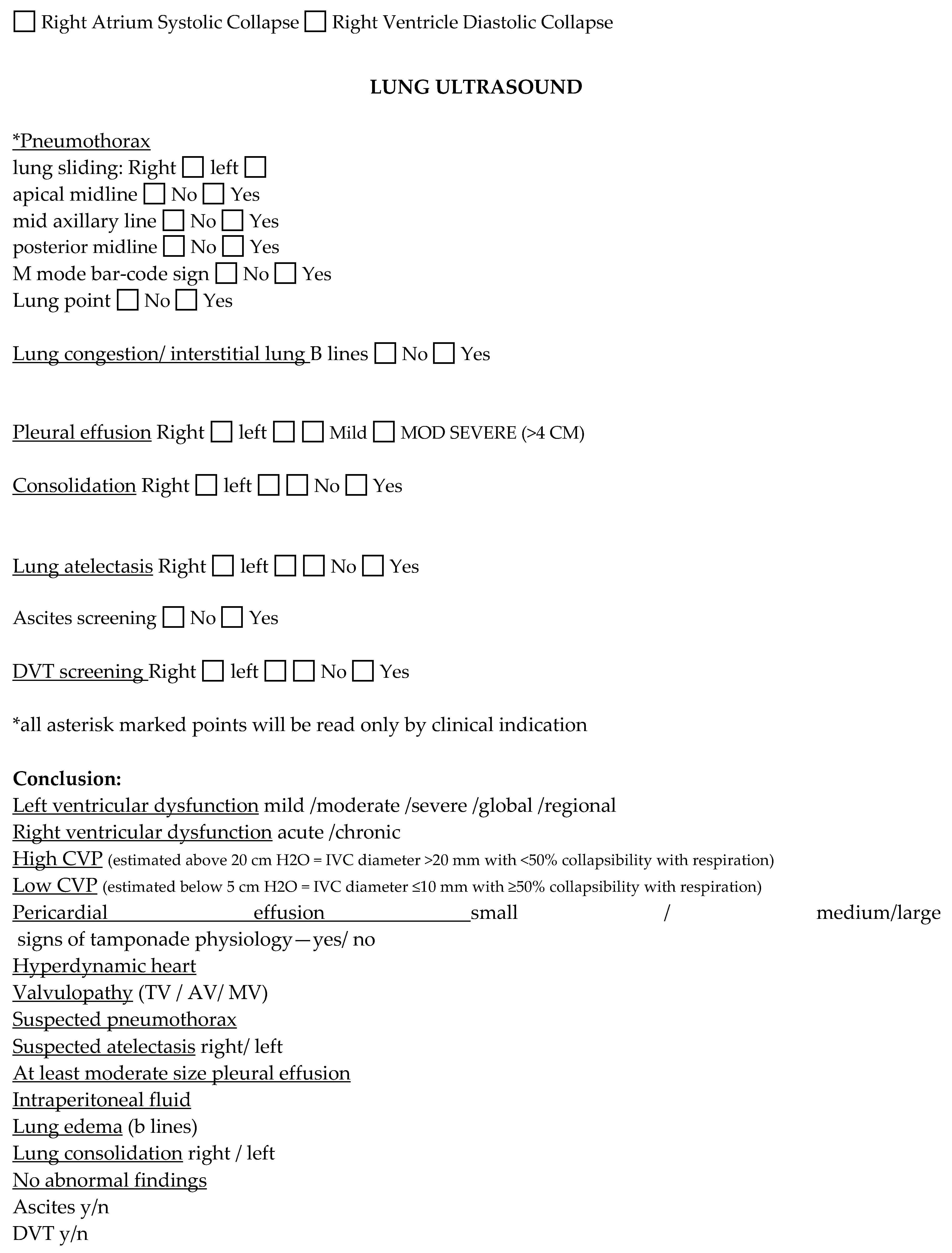
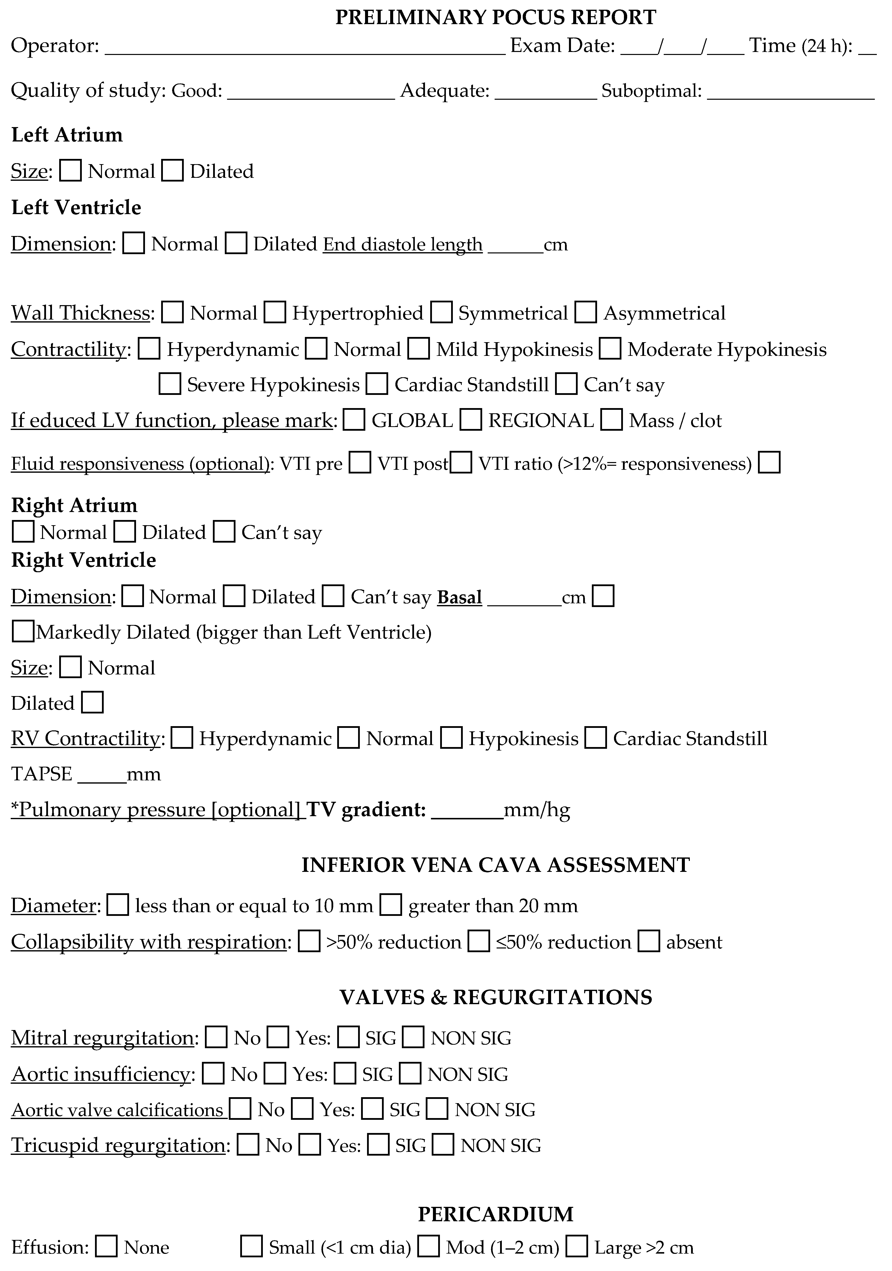
All patients in the intervention group underwent a standardized bedside-focused sonographic assessment of the heart, lungs, abdomen, and inferior vena cava (IVC) in every clinic visit during the period of study. The POCUS operator was blinded and not privy to the patients’ medical records, nor any previous assessments, and did not provide any diagnostic or treatment recommendations at any time. The exam was conducted either at the beginning or after the regular patient assessment. Views obtained included 2-dimensional parasternal long and short axis, subcostal long and IVC, apical 5, 4, and 2 chamber views. As this exam was POCUS-centered and not a formal cardiac echocardiographic study, the use of protocolized routine Doppler and size measurements were minimized, and instead a global assessment for chamber function and size was obtained. When specific pathologies were identified, more relevant advanced measurements were taken at the discretion of the scanning POCUS clinician, and the patient was referred for a formal TTE. For example, when a calcified aortic valve was detected, and aortic stenosis was suspected, a continuous pulse-wave Doppler for maximal velocity measurement was conducted. Standard measurements were determined for various indications, including left ventricle wall thickness, chamber size, IVC collapsibility index, RVSP, and size of pericardial effusion when found. TAPSE, which has been shown to correlate with survival in PH [22], was calculated when indicated. Lung ultrasound was focused on the detection of pulmonary congestion (screening and counting B-lines on both lungs, midclavicular line and midaxillary line), pleural effusion, lung atelectasis, lung consolidations (mid and posterior axillary lines), and sliding of pleura for pneumothorax (midclavicular line). Doppler for deep vein thrombosis (DVT) was performed only when suggested by the clinical context.
All POCUS exams were documented in a standardized form (Appendix A) which was made available to the treating pulmonologist in real time and was documented in the medical record (Figure 1a). The treating clinician was not present for the ultrasonographic portion, and the pulmonologist’s exposure to the POCUS case report was the only differing variable between the two study arms. All POCUS studies were performed by a physician (L.F) with six years’ experience in point-of-care ultrasound as a trained operator, with the performance of over 200 heart and lung exams. The POCUS study was designed to take 10 min. Two ultrasound machines were used: GE Vivid S70 with Cardiac Sector Probe M5Sc-D and ESaote Italy/MyLab 5/cardiac probe PA121, Naples, Italy. A cardiac probe, 3 MHZ, was used for the cardiac exam. The same probe was used for the lung ultrasound exam. A linear probe, 9 MHZ, was used for DVT screening when relevant.
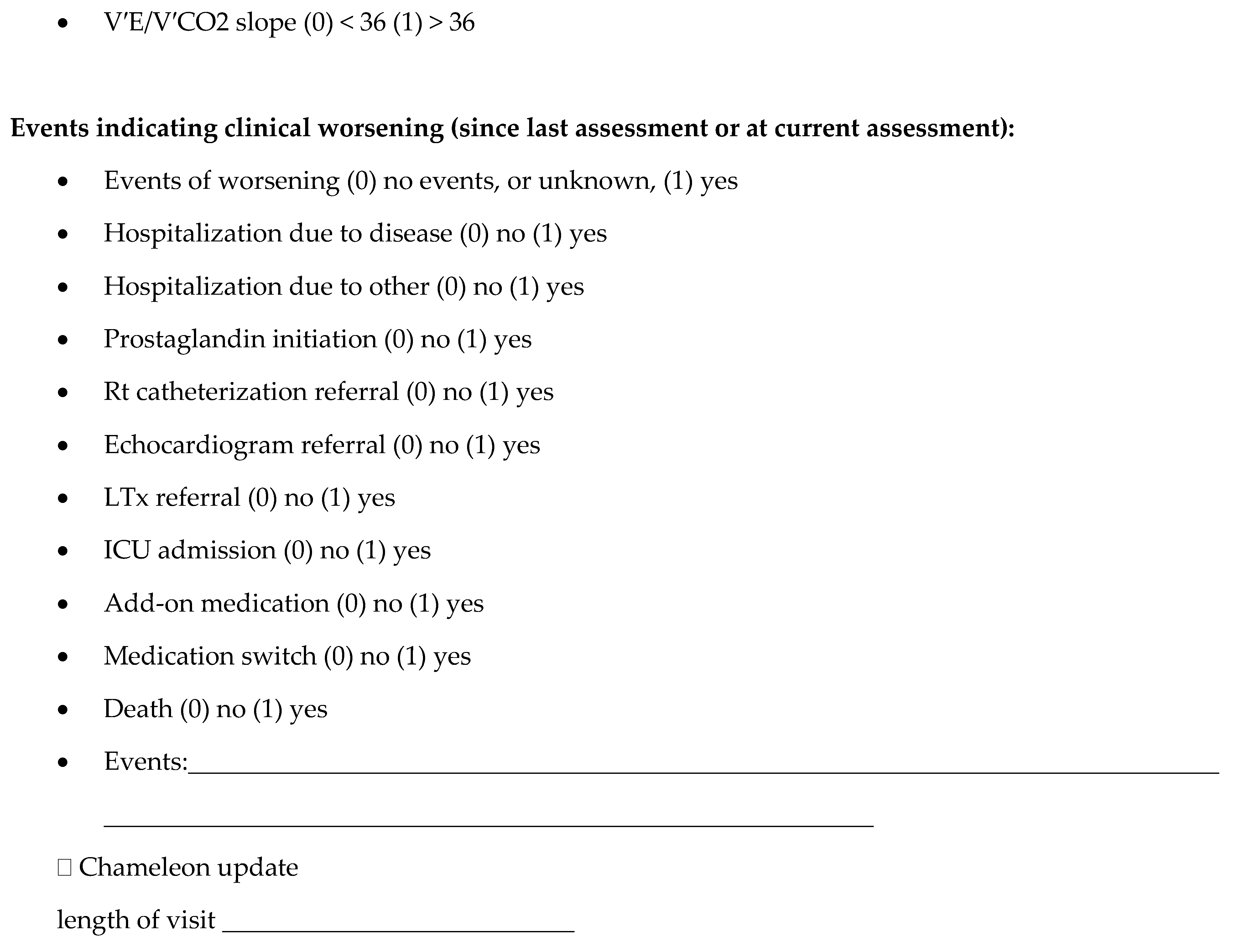
The primary outcome was set as change in management to evaluate the association between the POCUS assessment and any changes in clinical management during the ambulatory visit. Management change was defined as any treatment adjustment, including medication changes, hospitalizations, referral for new diagnostic studies or procedures, and other further investigations. We manually counted each management change by reviewing electronic medical records, physician’s notes, and medical orders, and documented the number of changes per visit in each group. Secondary outcomes included six-minute walk test (6MWT) distance, New York Heart Association (NYHA) class, Brain Natriuretic Peptide (BNP) levels, hospitalization rate, ICU admission, quality-of-life assessed by the emPHasis-10 questionnaire, and the length of the clinic visit as measured from arrival to departure from clinic. POCUS evaluation, lab results, six-minute walk test, and medical decisions were documented in the Case Report Form (Appendix B) and the patients’ medical record.
Baseline characteristics of the study population were examined across allocated intervention groups. Data are reported as percentages for categorical variables and means + standard deviations or medians (interquartile ranges) for continuous variables, according to the variable’s distribution. Data were compared across intervention groups, at baseline and follow-up, using Chi-square statistical test for categorical variables and Mann–Whitney or independent T-test for continuous variables, according to the variable’s distribution. To further assess for the primary outcome, multivariate logistic regression was performed to control for potential confounders and other possible influential variables. The multivariable models also specifically included the possible interaction between abnormal physical examination and abnormal POCUS examination as two dichotomous variables. The physical examination in the model was marked as normal or abnormal when one or more of the following was detected: increased jugular vein distention, peripheral or pulmonary edema, hepatomegaly, or ascites. In the statistical model, the abnormal physical exam was marked as “yes” or “no” and was entered to the logistic regression model as such. Convenient sample size for a pilot feasibility study was set to thirty patients. Significance levels were set to a two-sided p-value of <0.05. Statistical tests were performed using SPSS v. 26 (IBM, Armonk, NY, USA).

3. Results

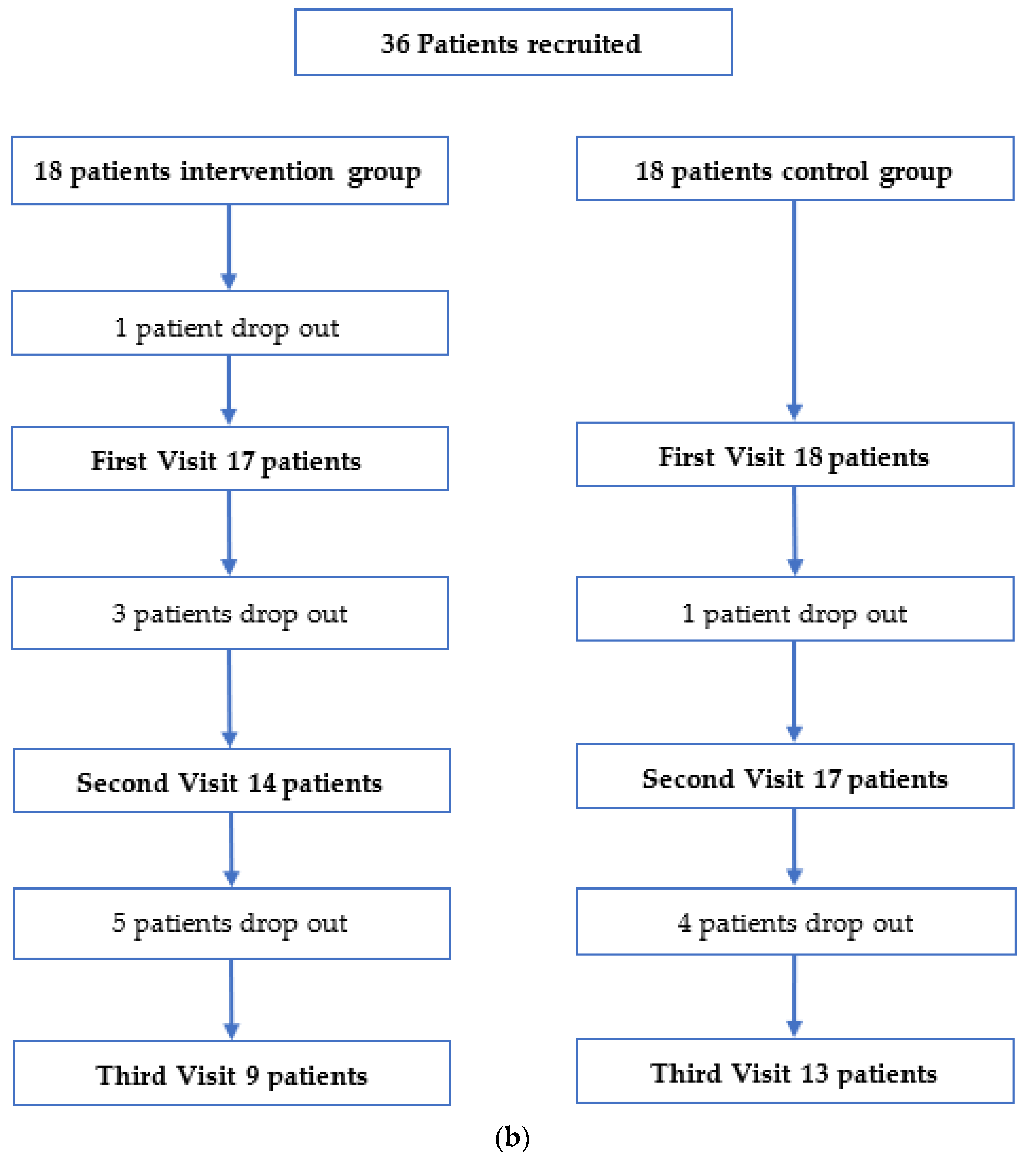
In this 13-month prospective study period, we enrolled 36 patients from 2 academic PH clinics. Eighteen were randomized to the intervention group and eighteen to the control group. Twenty-two patients from both groups completed three follow-up visits each (Figure 1b). Two patients declined to participate in the study and the remaining patients that dropped out of the study did so due to the physical distance of their homes from the PAH clinic. Table 1 summarizes demographic and clinical characteristics at enrollment. There was no crossover of patients between study groups. Demographics and health status was similar between groups. At enrollment, the intervention POCUS group had a lower median NYHA score (NYHA class 2 (1–3) compared to NYHA class 3 (2–4) in the control group, p = 0.05) (Table 1). Despite this, 6-MWT, BNP measurements, signs of RV failure, and EmPHasis-10 scores were similar between cohorts. The median time for POCUS assessment was 11 min (range 8–16 min) across ambulatory visits in the intervention arm, however there was no significant difference between the median total visit time between the intervention and control arm (29 vs. 30 min, p = 0.105).
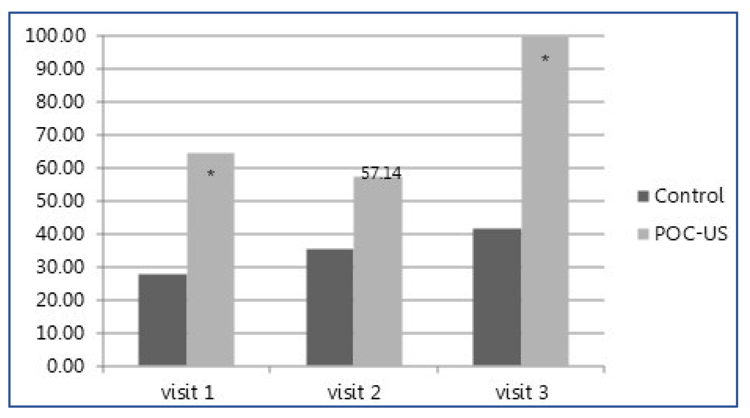
More management changes overall, as well as more total absolute number of visits with management changes, occurred in the POCUS group compared to the control (48 (73%) vs. 18 (27%) p < 0.001, and 32 (74.4%) vs. 17 (34.7%), respectively, p < 0.001) (Table 2). The total number of changes in clinical management on a per-visit basis was significantly higher in the POCUS intervention group (1.2 vs. 0.37 p < 0.001, Table 2). Figure 2 shows the number of changes for each of the three clinic visits, shown as a percentage of total patients exposed to any management change. For all documented visits across the duration of the study follow-up, the number of changes was significantly higher in the POCUS group (visit 1, 64.71% vs. 27.78 %; visit 2, 57.14 % vs. 35.29%; and visit 3, 100 % vs. 41.67%, respectively). Across the study period, of the 17 patients in the intervention group, 14 (82%) had any change in management, while in the control group, 11 out of 18 patients (61%) had similar changes recorded. In the POCUS group, across 43 total outpatient visits, 33% of encounters included a medication change (n = 14), while 72% included a diagnostic change (n = 31), a statistically significant difference compared to in the control group, where only 17.6% of visits included a medication-related change and 15.7% of visits included a diagnostic change (p value = 0.094 for medication changes and 0.005 for non-medication-related changes).
In the multivariable regression models, the POCUS intervention study arm and the abnormal physical examination were the only variables that were statistically significant for influencing management change (Table 3). In the fully adjusted multivariable model, POCUS remained significantly associated with management change with an OR of 11.98 (3.59–40.01, p < 0.001), while abnormal physical examination had an OR of 4.66 (1.34–16.22, p-value = 0.01) (Table 3). There was no significant interaction between physical examination abnormality and POCUS intervention (p > 0.1). The inclusion of an abnormal physical examination to the model increased the association of POCUS and management changes dramatically.
Analysis of secondary outcomes revealed no significant differences between study groups. Hospitalization rate (6% in POCUS group vs. 4.7% in control, p-value 0.79), new PH-specific medication prescriptions (28% in POCUS group vs. 32.6% in the control group, p-value 0.778), ICU admission rate (2% in the POCUS group vs. 0 in the control group, p-value 1), EmPHasis-10 score (23% in the POCUS group vs. 25% in the control group, p value = 0.93), and PH-specific therapy medication changes (2% in the POCUS group vs. 4.8% in the control group, p-value 0.398) were all statistically unchanged between the two cohorts (Appendix C).
Table 4 summarizes 26 of the relevant significant POCUS findings in our study. The most common findings were “unexpected normal” RVSP/central venous pressure (CVP), where elevation had been anticipated, and LV hypertrophy. Table 5 presents case series examples of some POCUS findings from the intervention arm and the management changes and interventions that followed.

4. Discussion

We present, for the first time in a pilot randomized control trial, that the integration of POCUS as an adjunct to the physical examination of the heart and lungs during an outpatient visit in patients with PAH in the ambulatory setting is feasible, rapid, and leads to significantly more management changes and treatment adjustments compared to the standard patient assessment.
Technological advances have improved the portability and lowered the price of ultrasound systems, bringing this modality to the patients’ bedside in the ambulatory clinic. POCUS has infiltrated almost all hospital wards, from the emergency departments to the intensive care units [8]. POCUS training has been incorporated into medical educational curricula, with the standardization of teaching, at the novice level, how to integrate this modality into daily practice across disciplines [23]. Previous studies have shown that POCUS can be used as a tool to affect decision making in the clinical setting and improve patient management and time to definitive treatment [11,12,24,25]. Recently, the international multi-center Ultra Man study showed that the use of thoracic ultrasound at the bedside in an inpatient population led to frequent changes in clinical impressions and subsequent meaningful management changes in nearly half of the patients evaluated with POCUS [20]. POCUS has been less utilized outside the hospital in ambulatory environments, with minimal reports of POCUS assessment in the pre-hospital setting [26,27,28], and few descriptions of utility in outpatient pulmonary hypertension ambulatory clinics. Samant et al. described the use of portable handheld ultrasound to measure right atrial pressures in an outpatient PH clinic and demonstrated a correlation with BNP and clinical worsening [29].
Our study looked to examine the feasibility and impact of introducing a comprehensive cardiopulmonary POCUS assessment into a PAH clinic as an additional tool for making quick, effective, and potentially cost-effective therapeutic decisions. We demonstrated in a blinded, randomized fashion that POCUS assessment is feasible in the ambulatory PH setting and has the potential to significantly and meaningfully affect patient care, with an increase in total management changes and in the frequency of such changes, as compared to traditional care sans POCUS. These changes encompassed both meaningful diagnostic and medication-related alterations in care. Other downstream clinical outcomes including rate of hospitalization due to PAH exacerbations, referral to lung transplantation, and death during the study period did not differ between groups, likely as a result of the small sample size and short interval follow-up time. This study was designed to serve as a feasibility trial to demonstrate proof of concept and serve as a foundation for future investigations and was not designed or powered to determine changes in these potentially more meaningful clinical outcomes.
In an additional multivariate analysis, our findings were reinforced with the demonstration that the only independent factors associated directly with PAH management changes were the findings in both the physical examination and POCUS (Table 3). The statistical significance of the relationship between POCUS and clinical management changes was even stronger when physical examination was combined with POCUS. We did not find any management changes independently associated with BNP, 6 min walk, or EmPHasis-10 questionnaire (Table 3). Examples of POCUS findings that influenced management are described in Table 5. For example, POCUS assessment of Patient 12 during visit three revealed pulmonary B lines, indicating pulmonary edema. In response to this, the pulmonologist added a diuretic drug and ordered a formal TTE, which would not have occurred without the additional clinical information POCUS provided. In another example, POCUS of Patient 23 diagnosed a high CVP, an elevated RVSP, and a restrictive diastolic pattern on ultrasound, prompting the addition of a diuretic and a formal re-evaluation of cardiac and LV function. Patient 24 on visit three was diagnosed with LVH and multiple B lines reflecting lung congestion, and in response to these findings, the treating physician ordered a new right heart catheterization.
Our results demonstrate meaningful clinical changes when outpatient PH clinics incorporate POCUS assessment into the routine patient evaluation. The integration of this ultrasound modality in the clinic is feasible to upgrade the physical exam, known to perform poorly in PH [13], in the diagnosis of pathology that could not be found by the conventional examination (Table 4 and Table 5). This clinical benefit came without an increase in the total time of the outpatient clinic encounter, as the POCUS assessments took only 10 min to perform in a comprehensive manner. POCUS assessment is viable in the outpatient clinic and has potential in the hands of a trainer operator to decrease time to appropriate treatment, and potentially more meaningful downstream clinical outcomes.
This study was designed as a hypothesis-generating feasibility study and was not powered to evaluate clinical outcomes such as improved functional status or survival, nor whether they were related to the management changes recorded. To assess these clinical outcomes and causality, a prospective controlled study with a larger enrollment and longer follow-up is needed. Our study serves as the foundation and suggestion for this future work. In addition, our study did not have a protocolized treatment algorithm that was based on the POCUS findings. Rather, it was designed to serve as a real-world surrogate, where the clinician reacted to the POCUS findings using their own clinical judgment. Only one treating pulmonologist and one trained POCUS operator were involved in the study, reducing the risk of inter-operatory variability, albeit potentially limiting the generalizability of the findings. However, with the advent of POCUS across medical disciplines and the growth of critical care echocardiography [30], these skills are expanding beyond the first wave of highly trained operators into routine clinical practice. In future studies, the feasibility of pulmonary hypertension specialists in performing their own POCUS studies should be evaluated. In this regard, the training of physicians to conduct and become proficient in POCUS also needs to be considered and prioritized. While this may take time in an ambulatory clinic, brief training sessions have been shown to meaningfully improve the skills of novice operators in both the short and long term [31,32] and there continues to be significant interest in ultrasound education across medical training [33,34]. Hopefully, our results support the notion that this time investment is worthwhile, with the strong potential to be meaningfully beneficial to patient care. It is our position, supported by the literature, that the real-time POCUS assessment is important to patients’ management by making new diagnoses that prompt management changes, shortening time to definitive treatment [11,12,20,24,35]. Our pilot study results support and hopefully will serve to inform the conduct of a larger multicenter prospective trial to evaluate the role of POCUS in improving patient outcomes in the ambulatory setting in patients with PH.

5. Conclusions

POCUS integrated into the physical examination during routine PAH clinic encounters increases the number and rate of management changes compared to standard usual care without ultrasonography and is feasible without sacrificing ambulatory clinic time in patient visits. POCUS is a valid diagnostic tool that may help the treating physician at the bedside and support a clinical cardiopulmonary evaluation in patients with pulmonary hypertension in the outpatient environment. Further, prospective, larger studies are needed to confirm if POCUS assessment in this setting affects downstream clinical patient outcomes.

Author Contributions

A.A.: conception and design; data collection; analysis and interpretation of data; first draft of the article; revision of the article critically for important intellectual content. A.B.L.S.: first draft of the article; revision of the article critically for important intellectual content. A.H.K.: interpretation of data; revision of the article critically for important intellectual content. J.T.: interpretation of data; revision of the article critically for important intellectual content. D.S.: data collection; revision of the article critically for important intellectual content. G.T.: data collection, interpretation of data; revision of the article for critically for important intellectual content. M.A.-S.: data collection, interpretation of data; revision of the article critically for important intellectual content. J.G.: interpretation of data; revision of the article critically for important intellectual content. L.F.: conception and design; interpretation of data; revision of the article critically for important intellectual content. All authors have read and agreed to the published version of the manuscript.

Funding

This research received no external funding.

Institutional Review Board Statement

The study was conducted in accordance with the Declaration of Helsinki, and approved by the Institutional Review Boards of Soroka University Medical Center and Barzilai University Medical Center (SOR0327-16, BRZ 0106-18,) for studies involving humans.

Informed Consent Statement

All subjects gave their informed consent for inclusion before they participated in the study.

Data Availability Statement

The data presented in this study are available on request from the corresponding author. The data are not publicly available due to privacy restrictions.

Conflicts of Interest

None of the authors has any relevant potential conflict of interest.

Appendix A. POCUS Examination Protocol
Jcm 12 01752 i001Jcm 12 01752 i002

Appendix B. Case Report Form

Jcm 12 01752 i003Jcm 12 01752 i004Jcm 12 01752 i005

Appendix C. Other Secondary Outcomes

NYHA (Mean ± SD)
[median]
POCUSControlp-Value
Visit 12.4 (±0.6) [2]2.8 (±0.4) [3]0.05
Visit 22.1 (±0.6) [2]2.8 (±0.5) [3]0.012
Visit 32.4 (±0.7) [3]2.9 (±0.3) [3]0.05
6 MWT (meter)
(Mean ± SD) [median]
POCUSControlp-Value
Visit 1396 (±134) [423]334 (±98) [333]0.084
Visit 2373 (±171) [433]373 (±95) [361]0.184
Visit 3334 (±165) [371]353 (±77) [364]0.8
BNP (Mean ± SD) [median]POCUSControlp-Value
Visit 1757.9 (±1420) [141]690 (±979) [285]0.495
Visit 2734.5 (±1373) [158]517 (±1049) [147]0.817
Visit 3519 (±873) [257]730 (±1180) [273]0.849
emPHasis-10
(Mean ± SD) [median]
POCUSControlp-Value
Visit 120.8 (±12.8) [24]25.5 (±11) [23]0.248
Visit 223.9 (±13.4) [28]23.7 (±8.4) [24]0.878
Visit 330.3 (±15) [34]24.1 (±10) [24.5]0.227
Events of worseningPOCUSControlp-Value
Visit 1---
Visit 22 (14.3%)4 (23.5%)0.664
Visit 31 (11.1%)2 (16.7%)1
Prostaglandin
initiation n (%)
POCUSControlp-Value
Visit 11 (5.9%)00.486
Visit 200-
Visit 300-
Transplantation
referral n (%)
POCUSControlp-Value
Visit 11 (5.9%)1 (5.6%)1
Visit 201 (5.9%)1
Visit 31 (11.1%)00.429
Add-on PH medication n (%)POCUSControlp-Value
Visit 15 (29.4%)6 (33.3%)1
Visit 25 (35.7%)5 (29.4%)1
Visit 34 (44.4%)3 (25%)0.397
Medication switch n (%)POCUSControlp-Value
Visit 12 (11.8%)00.229
Visit 200-
Visit 301 (8.3%)1
Management change n (%)POCUSControlp-Value
Visit 111 (64.7%)5 (27.8%)0.044
Visit 28 (57.1%)6 (35.3%)0.289
Visit 39 (100%)5 (41.7%)0.003
NYHA change n (%) visit 2POCUS, n = 14 (%)Control, n = 17 (%)p-Value
increased1 (7.1%)4 (23.5%)0.428
decreased5 (35.7%)4 (23.5%)
NYHA change n (%) visit 3POCUS, n = 9 (%)Control, n = 13 (%)p-Value
increased2 (22.2%)4 (36.4%)0.79
decreased1 (11.1%)1 (9.1%)

References

  1. Galie, N.; McLaughlin, V.V.; Rubin, L.J.; Simonneau, G. An overview of the 6th World Symposium on Pulmonary Hypertension. Eur. Respir. J. 2019, 53, 1802148. [Google Scholar] [CrossRef] [Green Version]
  2. McLaughlin, V.V.; Archer, S.L.; Badesch, D.B.; Barst, R.J.; Farber, H.W.; Lindner, J.R.; Mathier, M.A.; McGoon, M.D.; Park, M.H.; Rosenson, R.S.; et al. ACCF/AHA 2009 expert consensus document on pulmonary hypertension: A report of the American College of Cardiology Foundation Task Force on Expert Consensus Documents and the American Heart Association: Developed in collaboration with the American College of Chest Physicians, American Thoracic Society, Inc., and the Pulmonary Hypertension Association. Circulation 2009, 119, 2250–2294. [Google Scholar]
  3. Galie, N.; Humbert, M.; Vachiery, J.L.; Gibbs, S.; Lang, I.; Torbicki, A.; Simonneau, G.; Peacock, A.; Noordegraaf, A.V.; Beghetti, M.; et al. 2015 ESC/ERS Guidelines for the diagnosis and treatment of pulmonary hypertension: The Joint Task Force for the Diagnosis and Treatment of Pulmonary Hypertension of the European Society of Cardiology (ESC) and the European Respiratory Society (ERS): Endorsed by: Association for European Paediatric and Congenital Cardiology (AEPC), International Society for Heart and Lung Transplantation (ISHLT). Eur. Heart J. 2016, 37, 67–119. [Google Scholar]
  4. D'Alto, M.; Romeo, E.; Argiento, P.; Di Salvo, G.; Badagliacca, R.; Cirillo, A.P.; Kaemmerer, H.; Bossone, E.; Naeije, R. Pulmonary arterial hypertension: The key role of echocardiography. Echocardiography 2015, 32 (Suppl. 1), S23–S37. [Google Scholar] [CrossRef]
  5. Blumberg, F.C.; Arzt, M.; Lange, T.; Schroll, S.; Pfeifer, M.; Wensel, R. Impact of right ventricular reserve on exercise capacity and survival in patients with pulmonary hypertension. Eur. J. Heart Fail. 2013, 15, 771–775. [Google Scholar] [CrossRef]
  6. Hoeper, M.M.; Granton, J. Intensive care unit management of patients with severe pulmonary hypertension and right heart failure. Am. J. Respir. Crit. Care Med. 2011, 184, 1114–1124. [Google Scholar] [CrossRef]
  7. Grignola, J.C.; Gines, F.; Bia, D.; Armentano, R. Improved right ventricular-vascular coupling during active pulmonary hypertension. Int. J. Cardiol. 2007, 115, 171–182. [Google Scholar] [CrossRef]
  8. Moore, C.L.; Copel, J.A. Point-of-care ultrasonography. N. Engl. J. Med. 2011, 364, 749–757. [Google Scholar] [CrossRef] [Green Version]
  9. Kobal, S.L.; Atar, S.; Siegel, R.J. Hand-carried ultrasound improves the bedside cardiovascular examination. Chest 2004, 126, 693–701. [Google Scholar] [CrossRef]
  10. Labovitz, A.J.; Noble, V.E.; Bierig, M.; Goldstein, S.A.; Jones, R.; Kort, S.; Porter, T.R.; Spencer, K.T.; Tayal, V.S.; Wei, K. Focused cardiac ultrasound in the emergent setting: A consensus statement of the American Society of Echocardiography and American College of Emergency Physicians. J. Am. Soc. Echocardiogr. 2010, 23, 1225–1230. [Google Scholar] [CrossRef]
  11. Ben-Baruch Golan, Y.; Sadeh, R.; Mizrakli, Y.; Shafat, T.; Sagy, I.; Slutsky, T.; Kobal, S.; Novack, V.; Fuchs, L. Early Point-of-Care Ultrasound Assessment for Medical Patients Reduces Time to Appropriate Treatment: A Pilot Randomized Controlled Trial. Ultrasound Med. Biol. 2020, 46, 1908–1915. [Google Scholar] [CrossRef]
  12. Koh, Y.; Chua, M.T.; Ho, W.H.; Lee, C.; Chan, G.W.H.; Kuan, W.S. Assessment of dyspneic patients in the emergency department using point-of-care lung and cardiac ultrasonography—A prospective observational study. J. Thorac. Dis. 2018, 10, 6221. [Google Scholar] [CrossRef]
  13. Braganza, M.; Shaw, J.; Solverson, K.; Vis, D.; Janovcik, J.; Varughese, R.A.; Thakrar, M.V.; Hirani, N.; Helmersen, D.; Weatherald, J. A Prospective Evaluation of the Diagnostic Accuracy of the Physical Examination for Pulmonary Hypertension. Chest 2019, 155, 982–990. [Google Scholar] [CrossRef]
  14. McLaughlin, V.V.; Shah, S.J.; Souza, R.; Humbert, M. Management of pulmonary arterial hypertension. J. Am. Coll. Cardiol. 2015, 65, 1976–1997. [Google Scholar] [CrossRef] [Green Version]
  15. Delcroix, M.; Howard, L. Pulmonary arterial hypertension: The burden of disease and impact on quality of life. Eur. Respir. Rev. 2015, 24, 621–629. [Google Scholar] [CrossRef] [Green Version]
  16. Simonneau, G.; Gatzoulis, M.A.; Adatia, I.; Celermajer, D.; Denton, C.; Ghofrani, A.; Sanchez, M.A.G.; Kumar, R.K.; Landzberg, M.; Machado, R.F.; et al. Updated Clinical Classification of Pulmonary Hypertension. J. Am. Coll. Cardiol. 2013, 62 (Suppl. 25), D34–D41. [Google Scholar] [CrossRef] [Green Version]
  17. Ashton-Cleary, D.T. Is thoracic ultrasound a viable alternative to conventional imaging in the critical care setting? Br. J. Anaesth. 2013, 111, 152–160. [Google Scholar] [CrossRef] [Green Version]
  18. Humbert, M.; Kovacs, G.; Hoeper, M.M.; Badagliacca, R.; Berger, R.M.F.; Brida, M.; Carlsen, J.; Coats, A.J.S.; Escribano-Subias, P.; Ferrari, P.; et al. 2022 ESC/ERS Guidelines for the diagnosis and treatment of pulmonary hypertension: Developed by the task force for the diagnosis and treatment of pulmonary hypertension of the European Society of Cardiology (ESC) and the European Respiratory Society (ERS). Endorsed by the International Society for Heart and Lung Transplantation (ISHLT) and the European Reference Network on rare respiratory diseases (ERN-LUNG). Eur. Heart J. 2022, 43, 3618–3731. [Google Scholar]
  19. Howard, L.S.; Grapsa, J.; Dawson, D.; Bellamy, M.; Chambers, J.B.; Masani, N.D.; Nihoyannopoulos, P.; Gibbs, J.S.R. Echocardiographic assessment of pulmonary hypertension: Standard operating procedure. Eur. Respir. Rev. 2012, 21, 239–248. [Google Scholar] [CrossRef] [Green Version]
  20. Heldeweg, M.L.A.; Matta, J.E.L.; Pisani, L.M.; Slot, S.M.; Haaksma, M.E.; Smit, J.M.; Mousa, A.M.; Magnesa, G.; Massaro, F.; Touw, H.R.W.M.; et al. The Impact of Thoracic Ultrasound on Clinical Management of Critically Ill Patients (UltraMan): An International Prospective Observational Study. Crit. Care Med. 2022. [Google Scholar] [CrossRef]
  21. Mathai, S.C.; Puhan, M.A.; Lam, D.; Wise, R.A. The minimal important difference in the 6-minute walk test for patients with pulmonary arterial hypertension. Am. J. Respir. Crit. Care Med. 2012, 186, 428–433. [Google Scholar] [CrossRef] [Green Version]
  22. Mazurek, J.A.; Vaidya, A.; Mathai, S.C.; Roberts, J.D.; Forfia, P.R. Follow-up tricuspid annular plane systolic excursion predicts survival in pulmonary arterial hypertension. Pulm. Circ. 2017, 7, 361–371. [Google Scholar] [CrossRef] [Green Version]
  23. Solomon, S.D.; Saldana, F. Point-of-Care Ultrasound in Medical Education—Stop Listening and Look. N. Engl. J. Med. 2014, 370, 1083–1085. [Google Scholar] [CrossRef] [Green Version]
  24. Lait, D.; Friedman, Z.; Mizrakli, Y.; Fuchs, L. The effect of point of care ultrasonography before urgent orthopedic surgery on anesthetic management—A prospective, pilot study. J. Clin. Anesth. 2020, 66, 109902. [Google Scholar] [CrossRef]
  25. Lichter, Y.; Topilsky, Y.; Taieb, P.; Banai, A.; Hochstadt, A.; Merdler, I.; Oz, A.G.; Vine, J.; Goren, O.; Cohen, B.; et al. Lung ultrasound predicts clinical course and outcomes in COVID-19 patients. Intensive Care Med. 2020, 46, 1873–1883. [Google Scholar] [CrossRef]
  26. Gundersen, G.H.; Norekval, T.M.; Haug, H.H.; Skjetne, K.; Kleinau, J.O.; Graven, T.; Dalen, H. Adding point of care ultrasound to assess volume status in heart failure patients in a nurse-led outpatient clinic. A randomised study. Heart 2016, 102, 29–34. [Google Scholar] [CrossRef]
  27. Hamill, C.; Ellis, P.K.; Johnston, P.C. Point of Care Thyroid Ultrasound (POCUS) in Endocrine Outpatients: A Pilot Study. Ulster Med. J. 2020, 89, 21–24. [Google Scholar]
  28. Niblock, F.; Byun, H.; Jabbarpour, Y. Point-of-Care Ultrasound Use by Primary Care Physicians. J. Am. Board Fam. Med. 2021, 34, 859–860. [Google Scholar] [CrossRef]
  29. Samant, S.; Tran, H.V.; Uddo, R.B.; Deboisblanc, B.P.; Saketkoo, L.A.; Saito, S.; Helmcke, F.R.; Lammi, M.R. Use of Handheld Ultrasound to Estimate Right Atrial Pressure in a Pulmonary Hypertension Clinic. Ann. Am. Thorac. Soc. 2022, 19, 179–185. [Google Scholar] [CrossRef]
  30. Vieillard-Baron, A.; Millington, S.J.; Sanfilippo, F.; Chew, M.; Diaz-Gomez, J.; McLean, A.; Pinsky, M.R.; Pulido, J.; Mayo, P.; Fletcher, N. A decade of progress in critical care echocardiography: A narrative review. Intensiv. Care Med. 2019, 45, 770–788. [Google Scholar] [CrossRef]
  31. Tuvali, O.; Sadeh, R.; Kobal, S.; Yarza, S.; Golan, Y.; Fuchs, L. The long-term effect of short point of care ultrasound course on physicians’ daily practice. PLOS ONE 2020, 15, e0242084. [Google Scholar] [CrossRef]
  32. Clay, R.D.; Lee, E.C.; Kurtzman, M.F.; Dversdal, R.K. Teaching the internist to see: Effectiveness of a 1-day workshop in bedside ultrasound for internal medicine residents. Crit. Ultrasound J. 2016, 8, 11. [Google Scholar] [CrossRef] [Green Version]
  33. Hoppmann, R.A.; Mladenovic, J.; Melniker, L.; Badea, R.; Blaivas, M.; Montorfano, M.; Abuhamad, A.; Noble, V.; Hussain, A.; Prosen, G.; et al. International consensus conference recommendations on ultrasound education for undergraduate medical students. Ultrasound J. 2022, 14, 31. [Google Scholar] [CrossRef]
  34. Lopresti, C.M.; Schnobrich, D.J.; Dversdal, R.K.; Schembri, F. A road map for point-of-care ultrasound training in internal medicine residency. Ultrasound J. 2019, 11, 10. [Google Scholar] [CrossRef] [Green Version]
  35. Zanobetti, M.; Scorpiniti, M.; Gigli, C.; Nazerian, P.; Vanni, S.; Innocenti, F.; Stefanone, V.T.; Savinelli, C.; Coppa, A.; Bigiarini, S.; et al. Point-of-Care Ultrasonography for Evaluation of Acute Dyspnea in the ED. Chest 2017, 151, 1295–1301. [Google Scholar] [CrossRef]
Figure 1. (a). Study flow diagram: 6MW—6 min walk test; BNP—brain natriuretic peptide. (b). Patient recruitment.
Jcm 12 01752 g001aJcm 12 01752 g001b
Figure 2. Management change rate per visit. Management change by treatment group presented as percentage of patients exposed to a change. * refers to a statistically significant difference, p value < 0.05.
Jcm 12 01752 g002
Table 1. Baseline characteristics.
VariablePOCUS (n = 17)Control (n = 18)p-Value
Sex (female) n (%)13 (76.5%)16 (88.9%)0.402
Age (years) mean ± SD65 ± 1465 ± 160.934
BMI (weight) mean ± SD29 (±6.3)28.7 ± 10.90.666
Duration of disease (month) median (IQR)33 (8, 39)21 (5, 30)0.105
Left ventricular dysfunction n (%)2 (11.8%)00.229
Lung disease n (%)2 (11.8%)00.402
Chronic kidney disease n (%)1 (5.9%)1 (5.6%)1
Chronic liver disease n (%)1 (5.9%)00.486
Malignancy n (%)
PAH Diagnosis:
1 (5.9%)1 (5.6%)1
Connective tissue disease2 (11.8%)7 (38.9%)0.121
IPAH14 (82.4%)9 (50%)0.044
FPAH02 (11.1%)0.486
Drug and toxins exposure1 (5.9%)00.486
HIV00NA
PH medicationsNo2 (11.8%)3 (16.7%)0.194
Yes7 (41.2%)11 (61.1%)0.862
>1 PH drug8 (47.1%)4 (22.2%)0.862
Diuretics8 (47.1%)9 (50%)0.862
NYHA Class11(5.9%)00.05
28 (47.1%)4 (22.2%)0.05
38 (47.1%)14 (77.8%)0.05
Symptoms on enrolmentNone6 (35.3%)5 (27.8%)0.948
Orthopnea1 (5.9%)00.948
Chest pain3 (17.6%)00.948
Palpitations000.948
Syncope000.948
Edema1 (5.9%)2 (11.1%)0.948
Increased abdominal girth000.948
Constitutional symptoms000.948
Dyspnea6 (35.3%)11 (61.1%)0.948
Right heart failure5 (31.3%)7 (38.9%)0.642
BNP mean ± SD758 ± 1420.3847 ± 1064.30.369
6 Min walk test mean ± SD374 ± 166334 ± 980.179
EmPHasis-10 evaluation median (IQR)19 (8, 26)26 (18, 33)0.143
Abbreviations: BMI, body mass index; CTD, connective tissue disease; IPAH, idiopathic pulmonary artery hypertension; FPAH, familial pulmonary arterial hypertension; HIV, human immunodeficiency virus; NYHA, New York Heart Association; BNP, brain natriuretic peptide.
Table 2. Total management changes.
VariablePOCUSControlp Value
Total PH outpatient visits43491
Visits with management changes (%)32 (74.4%)17 (34.7%)<0.001
Total management changes4818<0.001
Average management changes per visit1.20.37<0.001
Table 3. Models for management change rate factors among two study cohorts.
ModelOdds RatioConfidence Interval 95%p-Value
LowerUpper
Model 1
Age0.990.961.020.51
Sex (male)1.70.456.30.43
POCUS group6.022.315.9<0.001
Model 2
Age0.990.961.020.47
Sex (male)1.80.457.030.40
Symptoms1.120.961.300.14
BMI0.990.941.040.99
POCUS group8.72.9225.97<0.001
Model 3
Age0.980.951.010.23
Sex (male)2.340.569.890.24
Symptoms1.170.961.300.16
BMI0.970.921.030.42
Physical examination4.661.3416.220.01
POCUS group11.983.5940.02<0.001
Table 4. POCUS findings.
POCUS FindingsNumber (%)
Total n = 26
Suspected aortic stenosis1 (3.8)
Right ventricular hypertrophy1 (3.8)
Left ventricular hypertrophy5 (19.3)
“Unexpectedly normal” RVSP6 (23.2)
Pericardial effusion1 (3.8)
Pulmonary congestion4 (15.4)
D-shaped septum2 (7.7)
Arrhythmia1 (3.8)
Elevated central venous pressure3 (11.5)
Restrictive diastolic pattern2 (7.7)
Table 5. Selected examples of management changes following POCUS findings.
Patient Code/VisitPOCUS FindingsManagement Change
Patient 1/Visit 2Calcified aortic valve
Suspected aortic stenosis
A new formal TTE study was ordered
Patient 10/Visit 1Signs of LVH, small pericardial effusion
B-lines suggestive of pulmonary congestion
Diuretics were added
Patient 12/Visit 3B lines suggestive of pulmonary congestionDiuretics were added
A new formal Echo study was ordered
Patient 21/Visit 1Surprisingly normal POCUS study with low CVPDiuretics were stopped
A new right catheterization was scheduled
Patient 22/Visit 1Small left ventricle
Low CVP
Diuretics dosage was reduced
Chest computed tomography study ordered
Full pulmonary function test ordered
A new right catheterization was scheduled
Patient 23/Visit 2High CVP
Elevated RVSP (58 mm Hg)
Restrictive diastolic pattern
A new formal TTE to assess suspected diastolic left ventricular dysfunction was ordered
Patient 24/visit 2B lines suggestive of lung congestionDiuretic dosage increased
Patient 24/visit 3LVH
B lines suggestive of lung congestion
A new right catheterization was scheduled
Patient 30/visit 1Elevated CVP
Elevated RVSP (55)
D-shaped Septum
A new formal TTE study was ordered
Disclaimer/Publisher’s Note: The statements, opinions and data contained in all publications are solely those of the individual author(s) and contributor(s) and not of MDPI and/or the editor(s). MDPI and/or the editor(s) disclaim responsibility for any injury to people or property resulting from any ideas, methods, instructions or products referred to in the content.