Review

A Longer Tpeak-Tend Interval Is Associated with a Higher Risk of Death: A Meta-Analysis

by
Cathrin Caroline Braun
1,
Matthias Daniel Zink
2,
Sophie Gozdowsky
3,
Julie Martha Hoffmann
1,
Nadine Hochhausen
1,
Anna Bettina Röhl
1,
Stefan Kurt Beckers
1,4 and
Felix Kork
1,*
1
Department of Anesthesiology, Medical Faculty, RWTH Aachen University, Pauwelsstraße 30, 52074 Aachen, Germany
2
Department of Cardiology, Medical Faculty, RWTH Aachen University, Pauwelsstraße 30, 52074 Aachen, Germany
3
Medical Management, Emergency Medical Service, Berlin Fire Brigade, 10150 Berlin, Germany
4
Aachen Institute of Emergency Medicine and Civil Security, Medical Faculty, RWTH Aachen University, Pauwelsstraße 30, 52074 Aachen, Germany
*
Author to whom correspondence should be addressed.
J. Clin. Med. 2023, 12(3), 992; https://doi.org/10.3390/jcm12030992
Submission received: 12 December 2022 / Revised: 15 January 2023 / Accepted: 19 January 2023 / Published: 28 January 2023
(This article belongs to the Section Cardiology)

Abstract

:
A noninvasive tool for cardiovascular risk stratification has not yet been established in the clinical routine analysis. Previous studies suggest a prolonged Tpeak-Tend interval (the interval from the peak to the end of the T-wave) to be predictive of death. This meta-analysis was designed to systematically evaluate the association of the Tpeak-Tend interval with mortality outcomes. Medline (via PubMed), Embase and the Cochrane Library were searched from 1 January 2008 to 21 July 2020 for articles reporting the ascertainment of the Tpeak-Tend interval and observation of all-cause-mortality. The search yielded 1920 citations, of which 133 full-texts were retrieved and 29 observational studies involving 23,114 patients met the final criteria. All-cause deaths had longer Tpeak-Tend intervals compared to survivors by a standardized mean difference of 0.41 (95% CI 0.23–0.58) and patients with a long Tpeak-Tend interval had a higher risk of all-cause death compared to patients with a short Tpeak-Tend interval by an overall odds ratio of 2.33 (95% CI 1.57–3.45). Heart rate correction, electrocardiographic (ECG) measurement methods and the selection of ECG leads were major sources of heterogeneity. Subgroup analyses revealed that heart rate correction did not affect the association of the Tpeak-Tend interval with mortality outcomes, whereas this finding was not evident in all measurement methods. The Tpeak-Tend interval was found to be significantly associated with all-cause mortality. Further studies are warranted to confirm the prognostic value of the Tpeak-Tend interval.

1. Introduction

Cardiovascular diseases (CVDs) affect the entire arterial circulation, while coronary heart disease (CHD) is the leading cause of death [1]. The prevalence of CHD is considered 6.7% in adults aged ≥ 20 years in the United States and around 35% of patients die as a result of a coronary event in a given year [2]. Despite an overall decline in CHD and CVD mortality rates over the last decades, this trend does not apply to all ethnicities, socioeconomic groups or geographic regions [2,3,4]. Adaptions in medical treatments and risk factors noticeably reduced deaths, however, risk stratification remains difficult [5].
Traditional electrocardiographic (ECG) findings of abnormal ventricular repolarization have been established as prognostic risk markers for cardiac events, i.e., arrhythmias as well as cardiac and all-cause death [6,7,8]. Whereas the QT interval reflects the total duration of ventricular depolarization and repolarization, the interval from the peak to the end of the T wave (Tpeak-Tend interval) supposedly correlates with the dispersion of the repolarization process [9].
Previous studies suggest a prolonged Tpeak-Tend interval to be predictive of death. Tse et al. (2017) already performed meta-analyses of these investigations by pooling together the odds or hazard ratios regarding arrhythmic and mortality outcomes [10]. However, several substantial shortcomings were debated controversially, such as an insufficient consideration of differing ECG measurement approaches or the heart rate correction of the Tpeak-Tend interval [11,12]. Additionally, the inclusion of six studies examining the risk in patients with Brugada syndrome is likely to have biased the results based on congenital repolarization disorders. Compared to the meta-analysis of Tse et al. we excluded patients with Brugada syndrome and focused primarily on the predictive value of prolonged Tpeak-Tend intervals for all-cause death. In addition, several subsequent studies have not been taken into consideration so far. Individual studies identified a prolonged Tpeak-Tend interval to be a surrogate marker for diffuse myocardial injury and myocardial damage without ST-elevation (NSTEMI) [13,14], which may potentially lead to arrhythmias and death [15,16,17]. Therefore, we conducted a meta-analysis investigating the association of the Tpeak-Tend interval with all-cause mortality, troponin elevation and the incidence of NSTEMI.

2. Materials and Methods

This meta-analysis was performed in accordance with current Cochrane standards [18] and reported based on the Preferred Reporting Items for Systematic Reviews and Meta-Analyses (PRISMA) statement [19,20]. Methodological details of the analysis were specified in advance and the resulting study protocol was registered at PROSPERO (CRD42020131622).

2.1. Eligibility Criteria and Search Strategy

Before the search was initiated, a precise clinical research question was developed using the population, intervention, comparisons and outcomes (PICO) format [21]. Potential studies were required to address the following study-specific elements of interest: Individuals of any age receiving an ECG recording with ascertainment of the Tpeak-Tend interval and observation of all-cause mortality, troponin elevation or NSTEMI as outcome.
We performed a systematic literature search of studies published between 1 January 2008 and 21 July 2020 in MEDLINE (via PubMed), Embase and the Cochrane Library. As standard of care rapidly improves over time (specifically for this analysis with regards to precision of measurement and patients’ outcome), we restricted the literature search to twelve years into the past from the time it was conducted. The search terms used to identify relevant studies were the following: “Tpeak-Tend” OR “Tpeak-Tend” OR “Tp-Te” OR “Tp-Te” OR “Tpeak-end” OR “Tp-e” OR “T(peak)-T(end)” OR “T wave peak-to-end” OR “T peak-T end“ OR “TPEc“ OR “T-peak to T-end“ OR “Tpeak-to-Tend“ OR “T-Wave Peak to T-Wave End” OR “TpTe”. The search for keywords was limited to titles and abstracts, but no limits were applied for language. In the online supplement, the full electronic search strategy for MEDLINE via PubMed is presented (Resource S1). A manually screening of the reference lists of articles to identify additional matching studies complemented the electronic search.

2.2. Study Selection Process

The findings were imported into a reference managing software (Endnote for Windows, version X9.3.3, Clarivate Analytics, Philadelphia, PA, USA) and duplicates were removed. All reports of retrospective and prospective observational studies (cohort studies, case control studies and cross sectional studies) in humans were included if the Tpeak-Tend interval duration was determined and either all-cause mortality, troponin elevation or NSTEMI were reported as endpoint and odds ratio (OR) or hazard ratio (HR) including the 95% confidence intervals (CIs), ROC curves or raw data necessary to calculate these were described. Both conference abstracts and journal articles were eligible for inclusion. Potential sample overlap was handled as follows: (1) if there was a corresponding article to a conference abstract that likely included the same or parts of the same sample population reported in the conference abstract (based on the time of publication and reported recruitment period), both the conference abstract and the journal article are reported but only the data from the journal article was introduced in the analysis; (2) if there was only a conference abstract without a corresponding journal article, we included the data of the conference abstract. Studies published in other languages than English that could not be translated by us or known associates, as well as review articles, letters and comments were excluded.
Screening of abstracts was performed independently by two members of the study team (CCB, SG; interrater agreement Cohen’s kappa 0.72) and relevant full-texts were then assessed for eligibility twice. Any disagreement between the reviewers was solved by full-text retrieval of the respective study to avoid that any potentially eligible investigation was skipped in the full-text review.

2.3. Data Extraction and Management

Data from each study were extracted using a prespecified and piloted spreadsheet in Microsoft Excel. Each article was given an internal record identifier and the spreadsheet was structured as followed: the first section contained general information about the article including type of work (journal article or conference abstract) and the study design of the study. This section was followed by publication details including the first author’s surname, the year of publication as well as the country the study was conducted in. Going further into detail, in the next section we documented extracted data on the population that was investigated in the article, including sample size, number of patients and controls, sex distribution of the population, age distribution of the population and whether the investigated population suffered from a certain condition that characterizes this population (e.g., transcatheter aortic valve replacement patients). The following section contained characteristics regarding the ECG measurement conducted in the study, how was the Tpeak-Tend interval defined, what method was used to measure the Tpeak-Tend interval, in which leads was this measured, and whether reported interval lengths were heart rate corrected. After these basic study characteristics, the data extraction sheet continued with three respective sections for each investigated outcome endpoint: All-cause death, NSTEMI, and troponin change. For all-cause death, we extracted time of follow-up reported, number of survivors and deaths with respective mean Tpeak-Tend intervals, number of survivors and deaths with short or long Tpeak-Tend interval including the Tpeak-Tend cutoff lengths, reported odds and hazard ratios, whether these were derived from univariate or multivariate analyses, whether an receiver operating characteristic analysis was conducted with respective cutoff, sensitivity and specificity, positive predictive value and negative predictive value. Sections for endpoints NSTEMI and troponin change were similar.
Data extraction was conducted by one reviewer (CCB) and checked afterwards by a second reviewer (FK). Discrepancies were resolved by discussion between both reviewers. If one of the reviewer suspected data needed for meta-analysis to be available although not reported in the publication, corresponding authors of potentially eligible and included studies were contacted via e-mail and asked whether they could provide the unreported data (summary or individual de-identified data) for the purpose to be included in this meta-analysis. For each request, a minimum response time of 30 days was granted.

2.4. Risk of Bias Assessment

We evaluated the quality of primary studies applying an adapted version of the Newcastle-Ottawa Quality Assessment Scale for observational studies [22]. A quality score, ranging from 0–9 stars for cohort studies and 0–8 stars for case-control studies and cross-sectional studies, was calculated based on three major domains: selection of study groups, comparability of study groups and assessment of outcome or exposure. Age and sex of the study participants were defined as the most important covariates regarding comparability. An adapted form of the scale by Herzog et al. was used to provide quality assessment for one cross-sectional study [23]. The evaluation was completed by one reviewer (CCB) and was then verified by another team member (FK). Quality scores indicating poorer methodological quality were not considered as an exclusion criterion.

2.5. Assessment of Heterogeneity and Data Synthesis

All analyses were conducted using the meta-package [24] for R 3.5.2 (www.r-project.org, accessed on 11 December 2022) for MacOS. Heterogeneity was assessed using the I2 statistic from chi-squared test. Due to a priori assumption of high clinical heterogeneity across trials due to different study populations, ECG leads used in measurements and types of death subsumed in all-cause mortality, we always used the appropriate random effects models. For comparing Tpeak-Tend intervals (milliseconds) in all-cause deaths and survivors, a standardized mean difference estimate was used to adjust for different types of Tpeak-Tend measurement methods (tangent method, tail method, Marquette 12SL algorithm, other computer-based measurements). To compare all-cause death in patients with longer versus shorter Tpeak-Tend intervals, unadjusted odds ratios using the DerSimonian-Laird method were applied. For studies reporting areas under the receiver operating characteristic (AUROC) curve with sensitivity, specificity and corresponding cutoff, the confusion matrix was back-calculated and a summary ROC curve was estimated using the “diagmeta” package for R. Subgroup analyses and sensitivity analyses were conducted post-hoc induced by the reported data of the original studies. Subgrouping was performed for groups that may present differences in the associations of the Tpeak-Tend interval with the outcome all-cause death: heart rate correction vs. no heart rate correction and different technical measurement methods of the Tpeak-Tend interval. Sensitivity analyses were conducted to account for possible bias by study design (prospective studies only) and study quality (Newcastle-Ottawa Quality Assessment Scale score greater 6).

3. Results

3.1. Description of Studies

The search and selection process of studies is presented in Figure 1.
The searching of Medline, Embase and the Cochrane Library provided a total of 1920 records. After deleting 829 duplicates, the title and abstract of a total of 1091 records were screened for eligibility. Of these, 958 records were non-compliant with the inclusion criteria and were therefore discarded and the remaining 133 records underwent full-text review. No additional studies could be identified via the examination of the reference lists. One study was excluded from the analysis, because the study population overlapped to a large extent with another study included in the analysis. Finally, 29 studies involving a total of 23,114 patients met the inclusion criteria. Table 1 provides an overview of the characteristics of the included studies; the quality assessment of the studies is presented in Table S1. Of the 29 studies included, 29 reported the primary endpoint all-cause mortality and none reported the secondary endpoints increase in troponin or NSTEMI.
All included studies were observational studies published in English, originating mainly from Turkey (n = 6), the USA (n = 6), and Italy (n = 5). Among them were prospective (n = 15) and retrospective (n = 8) cohort studies, as well as case-control studies (n = 5) and one cross-sectional study (n = 1). The publication years ranged from 2009 to 2020, the majority was published in 2018 (n = 6). The most frequently examined populations were patients with cardiomyopathy/heart failure (n = 8), ST-segment elevation myocardial infarction (STEMI) (n = 6), cardiovascular diseases (n = 6) and other diseases (n = 7). Furthermore, two studies assessed the Tpeak-Tend interval in the general population. The ascertainment of the Tpeak-Tend interval presented as follows: 11 studies used precordial leads and again nine studies of these focused on leads V4-V6. Three studies assessed a combination of limb and precordial leads, two studies assessed leads with limited ST-segment deviation and five studies measured the Tpeak-Tend interval in all 12 leads. Additionally, five studies reported to have chosen the lead with the longest Tpeak-Tend interval. Studies determined the Tpeak-Tend interval applying four different methods: The tangent method (n = 6), the tail method (n = 4), an automatic measurement with the GE Healthcare Marquette 12SL algorithm (n = 5) and an automatic measurement using different software (n = 3). Furthermore, one study compared the tangent and tail method. Six studies did not report the leads for measuring the Tpeak-Tend interval and 11 studies did not report the method of measurement.

3.2. Association of Tpeak-Tend Interval with Mortality

The Tpeak-Tend Interval in Comparison between Survivors and Non-Survivors

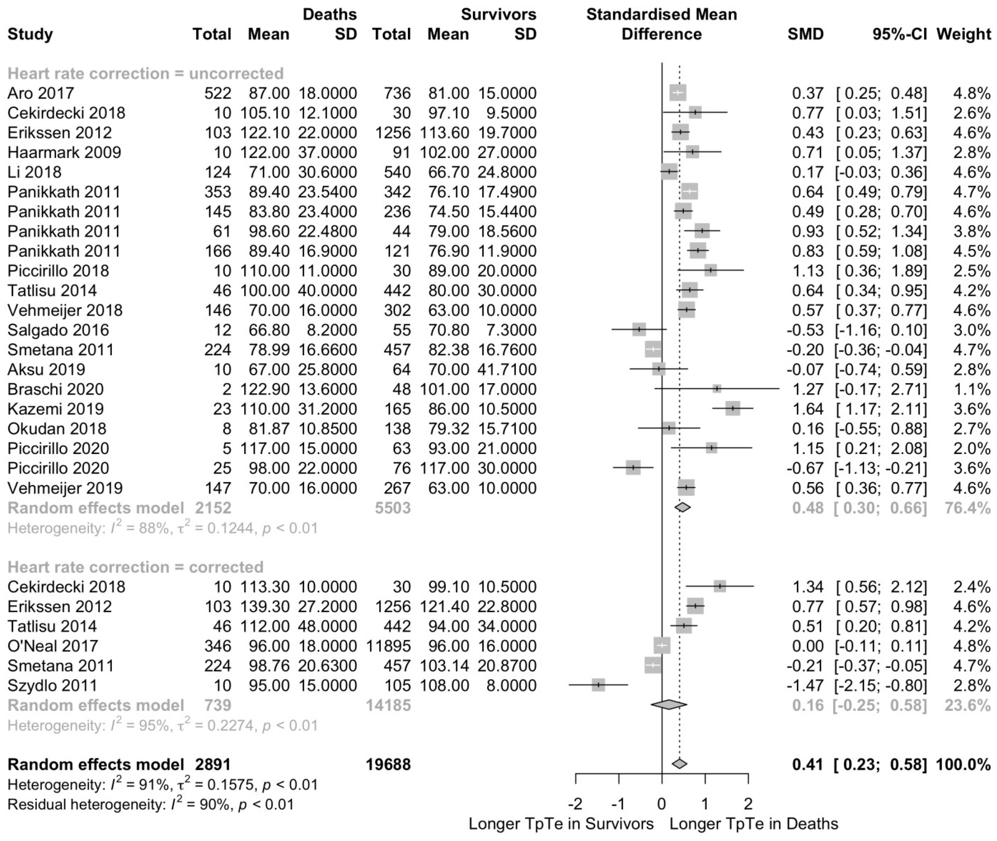
The Tpeak-Tend interval was available for 27 patient populations in 16 articles and five conference abstracts, reporting data for 20,011 patients with 2508 all-cause deaths. We contacted eight investigators for additional data on the Tpeak-Tend interval, but only two authors provided the requested data. Cardiovascular diseases and STEMI represented the most frequent patient populations in this analysis with seven and six populations, respectively. Overall effect sizes for mortality outcomes are presented in Figure 2. The estimated overall standardized mean difference was 0.41 (95% CI 0.23–0.58; p < 0.0001; I2 = 91%; p < 0.01), indicating a statistically significant longer Tpeak-Tend interval in all-cause deaths with a moderate effect size.
A subgroup analysis was performed comparing 19 studies reporting an uncorrected Tpeak-Tend interval in 7655 patients versus six studies reporting a heart rate-corrected Tpeak-Tend interval in 14,924 patients. In both subgroups, all-cause deaths had a longer Tpeak-Tend interval: All-cause deaths had a longer uncorrected Tpeak-Tend interval by a standardized mean difference of 0.48 (95% CI 0.30–0.66; p < 0.0001; I2 = 88%; p < 0.01; Figure 2) and in tendency longer heart rate corrected Tpeak-Tend interval by 0.16 (95% CI −0.25–0.58; p = 0.45; I2 = 95%; p < 0.01; Figure 2). However, standardized mean difference did not differ when comparing both subgroups (p = 0.17).
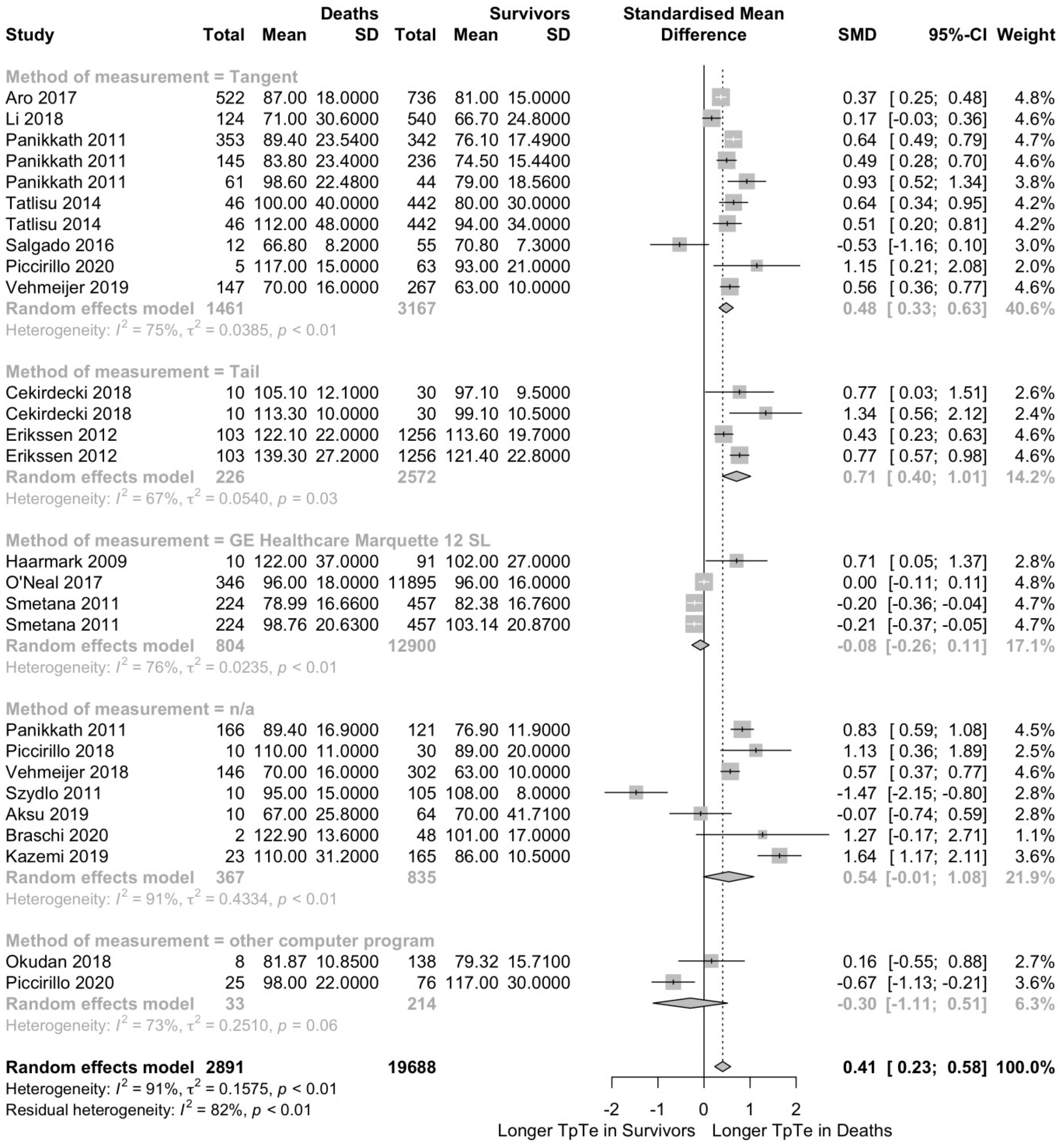
A second subgroup analysis was conducted to determine the effect of the measurement method. All-cause deaths had a longer Tpeak-Tend interval in subgroups reporting the tangent method (SMD 0.48; 95% CI 0.33–0.63; p < 0.001; I2 = 75%; p < 0.01; Figure 3) and the tail method (SMD 0.71; 95% CI 0.40–1.01; p < 0.001; I2 = 67%; p = 0.03; Figure 3). This finding did not apply for subgroups using the GE Healthcare Marquette 12SL algorithm (SMD −0.08; 95% CI −0.26–0.11; p = 0.42; I2 = 76%; p < 0.01; Figure 3) or other computer programs (SMD −0.30; 95% CI −1.11–0.51; p = 0.47; I2 = 73%; p < 0.06; Figure 3) and in the subgroup without specification of the measurement method (SMD 0.54; 95% CI −0.01–1.08; p = 0.05; I2 = 91%; p < 0.01; Figure 3).
To account for possible bias by study design or study quality, we conducted two post-hoc sensitivity analyses. Both a sensitivity analysis restricted to 11 studies with a prospective cohort study design including 17,063 patients, and a sensitivity analysis restricted to 12 studies with a Newcastle-Ottawa Quality Assessment Scale score greater six, including 19,842 patients, confirmed the finding. In both prospective cohort studies and studies with a Newcastle-Ottawa Quality Assessment Scale score greater than six, all-cause deaths had a longer Tpeak-Tend interval of 0.38 (95% CI 0.08–0.67; p = 0.0141; I2 = 92 %, p < 0.001; Figure S1) and 0.50 (95% CI 0.33–0.67; p < 0.001; I2 = 87%, p < 0.01; Figure S2), respectively.

3.3. Unadjusted Odds of Death in Patients with Longer vs. Shorter Tpeak-Tend Intervals

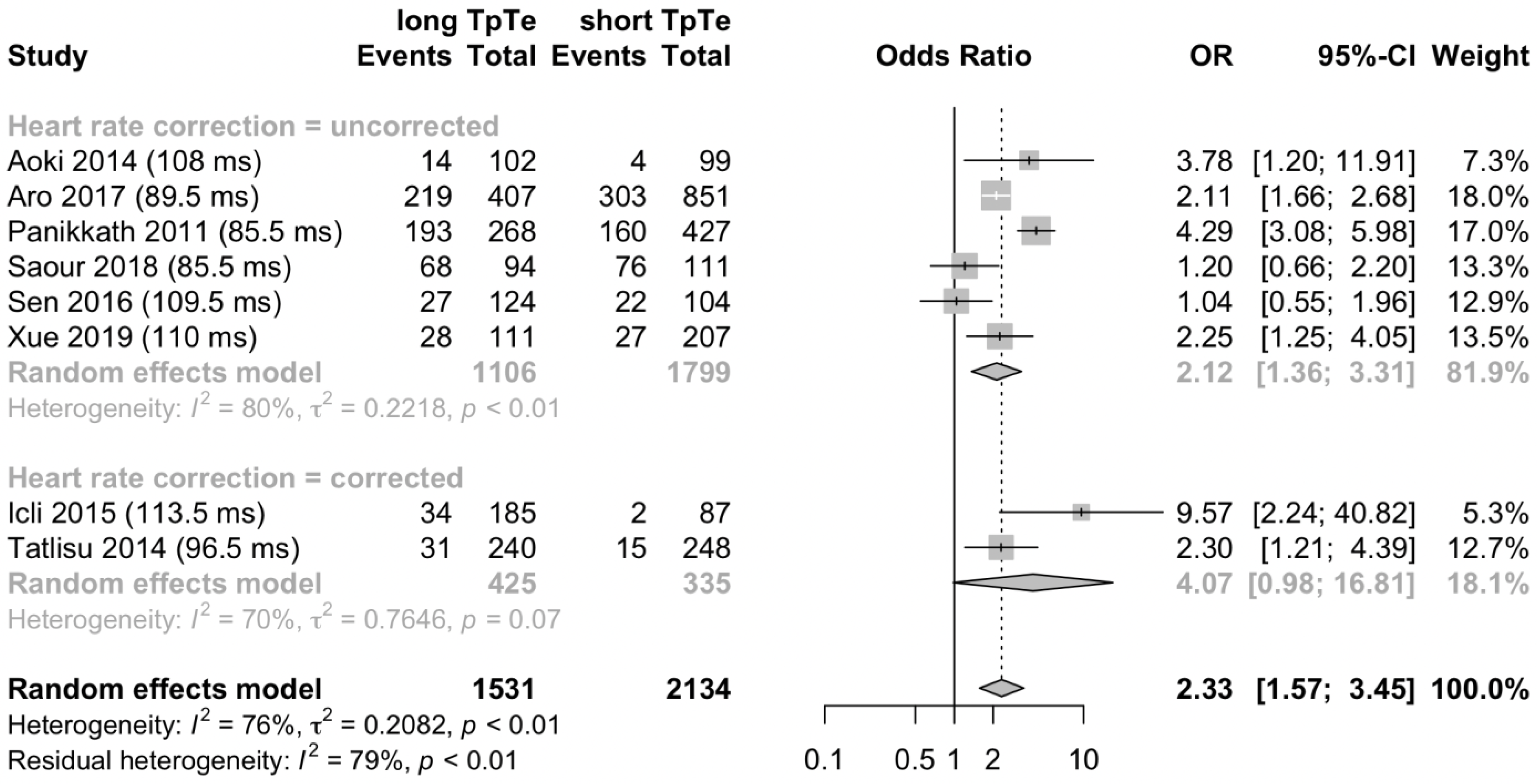
A fourfold table with long and short Tpeak-Tend interval versus all-cause deaths and survivors was available for eight patient populations in seven articles and one conference abstract, reporting data for 3665 patients and 1223 all-cause deaths. We contacted 11 authors for additional outcome data for this analysis and received the requested data from one author. The most frequent patient population analyzed was heart failure. The random effects model for this analysis is presented in Figure 4. A combined analysis revealed an overall odds ratio of 2.33 (95% CI 1.57–3.45; p < 0.001; I2 = 76%; p < 0.01); indicating that a long Tpeak-Tend interval was significantly associated with all-cause death.
A subgroup analysis was performed comparing six studies reporting mortality data associated with an uncorrected Tpeak-Tend interval and two studies reporting mortality data associated with a heart rate-corrected Tpeak-Tend interval. The subgroups included 2905 patients and 760 patients, respectively. In the pooled analysis, in both subgroups, a long Tpeak-Tend interval was associated with an increased risk of all-cause death: A long uncorrected Tpeak-Tend interval was associated with all-cause death by an odds ratio of 2.12 (95% CI 1.36–3.31; p < 0.01; I2 = 80%; p < 0.01; Figure 4) and in tendency long heart rate-corrected Tpeak-Tend interval by 4.07 (95% CI 0.98–16.81; p = 0.05 I2 = 70%; p = 0.07; Figure 4). However, standardized mean difference did not differ when comparing both subgroups (p = 0.39).
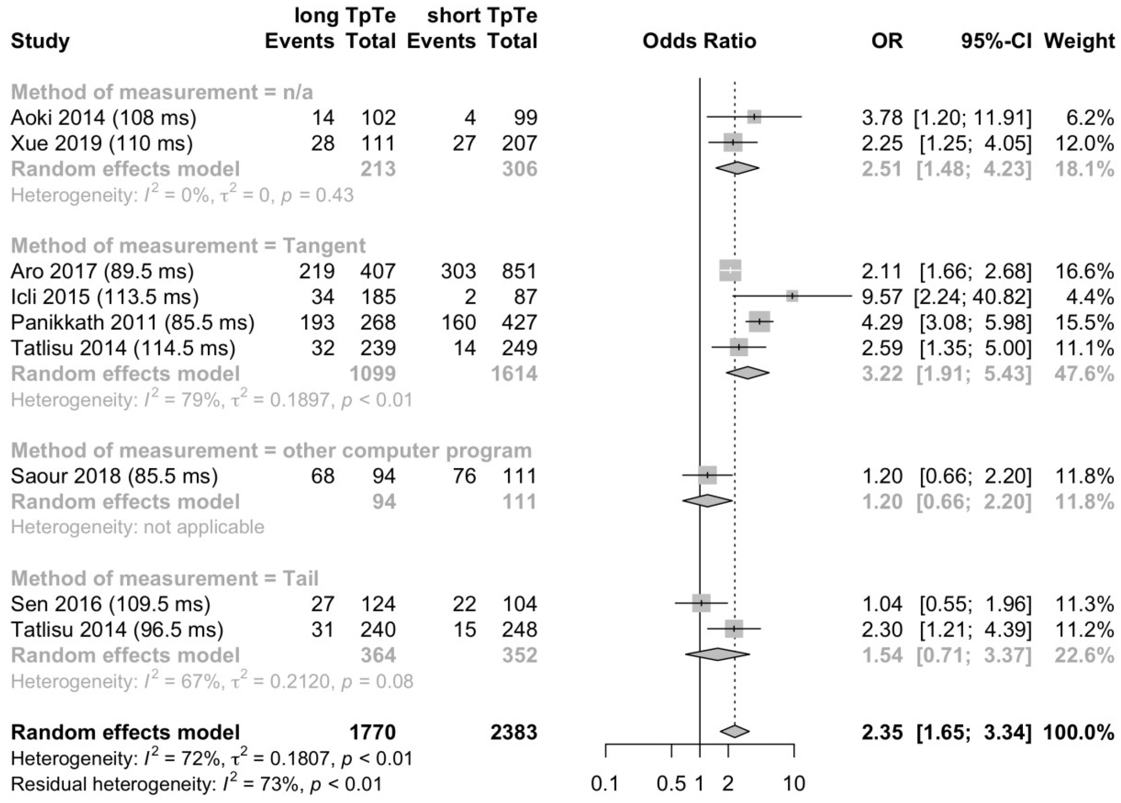
A second subgroup analysis was conducted to determine the effect of the measurement method. A long Tpeak-Tend interval was associated with all-cause death in the subgroup reporting the tangent method (OR 3.22; 95% CI 1.91–5.43; p < 0.001; I2 = 79%; p < 0.01; Figure 5) and in the subgroup without specification of the measurement method (OR 2.51; 95% CI 1.48–4.23; p < 0.01; I2 = 0%; p = 0.43; Figure 5). This finding did not apply for subgroups reporting the tail method (OR 1.54; 95% CI 0.71–3.37; p = 0.28; I2 = 67%; p = 0.08; Figure 5) and other computing programs (OR 1.20; 95% CI 0.66–2.20; Figure 5).
To account for possible bias by study design or study quality, we conducted two post-hoc sensitivity analyses. Both a sensitivity analyses restricted to four studies with a prospective cohort study design including 1235 patients, and a sensitivity analyses restricted to six studies with a Newcastle-Ottawa Quality Assessment Scale score greater six, including 3146 patients, confirmed this finding. In both prospective cohort studies and studies with a Newcastle-Ottawa Quality Assessment Scale score greater than six, a long Tpeak-Tend interval was associated with an increased risk of all-cause death of 1.95 (95% CI 1.20–3.15; p = 0.0066; I2 = 46%; p = 0.14; Figure S3) and 2.25 (95% CI 1.39–3.65; p = 0.0010;I2 = 82%; p < 0.01; Figure S4), respectively.

3.4. Predicting Death with Tpeak-Tend Interval

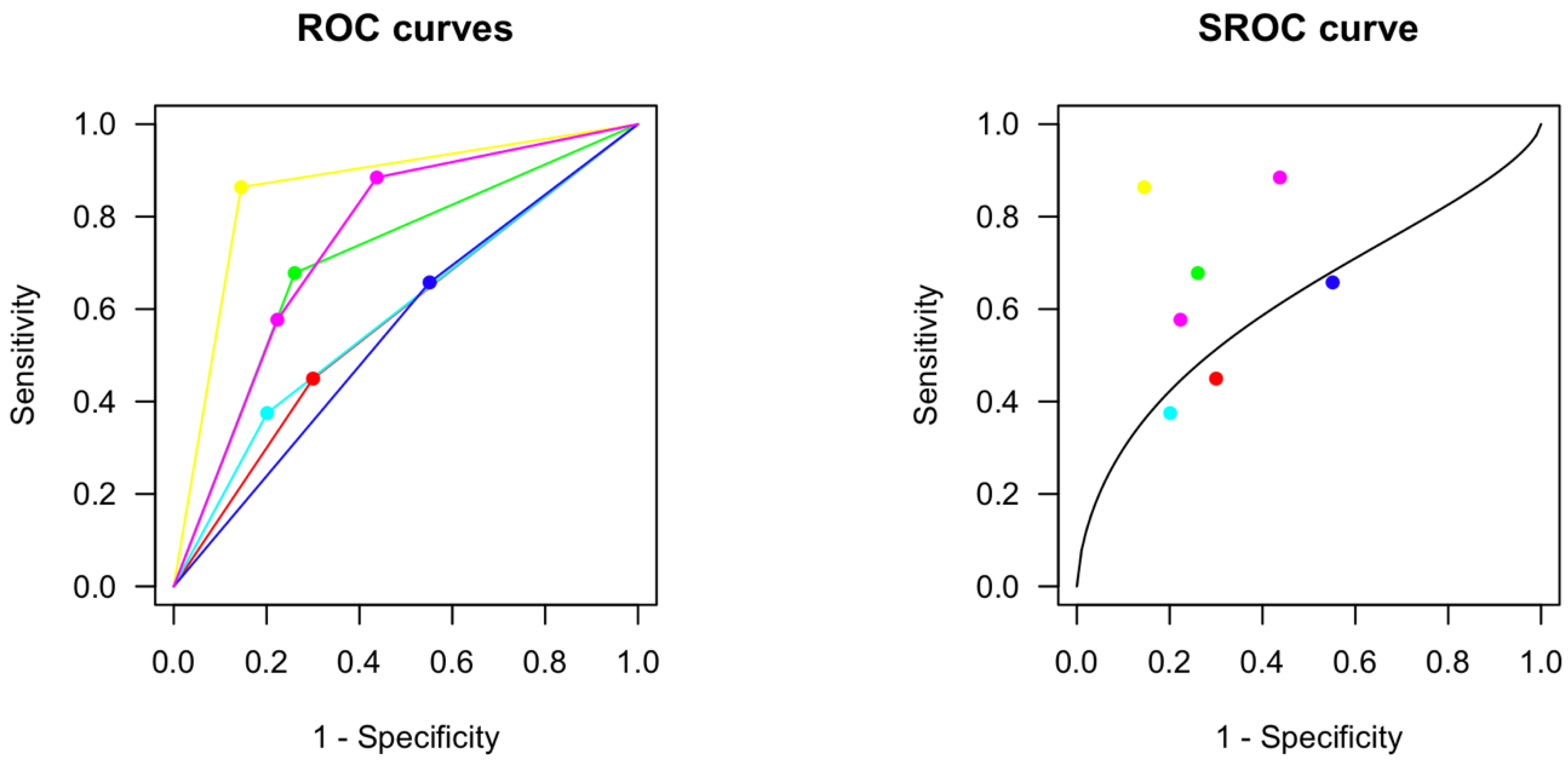
A total of six studies including 3951 patients reported AUCs for receiver operating characteristics (ROC) with corresponding cutoffs, sensitivity and specificity, analyzing predictability of all-cause death on the basis of the Tpeak-Tend interval (Table 2).
One study reported AUROC, sensitivity and specificity for two different cutoffs in the same population. Meta-analysis of these studies estimated a summary ROC curve with an AUC of 0.65 and estimated an optimal cutoff at 98.8 ms (Figure 6). At the optimal cutoff, sensitivity would be 0.61 (95%CI 0.37–0.81) and specificity 0.61 (95%CI 0.41–0.77).

3.5. Secondary Endpoints

No study reported the predefined secondary endpoints increase in troponin and NSTEMI with regard to the Tpeak-Tend interval. During full-text review, several studies were identified as likely to have these data but none of the three authors contacted for data on increase in troponin and one contacted for NSTEMI responded to our request.

4. Discussion

In this meta-analysis, we analyzed 29 studies to determine the association of the Tpeak-Tend interval with mortality outcomes. There is strong evidence of an association considering our main findings. All-cause deaths had longer Tpeak-Tend intervals compared to survivors by a standardized mean difference of 0.41 (95% CI 0.23–0.58) and patients with a long Tpeak-Tend interval had a higher risk of all-cause death compared to patients with a short Tpeak-Tend interval by an overall odds ratio of 2.33 (95% CI 1.57–3.45). Our subgroup analyses also suggest that heart rate-correction of the Tpeak-Tend interval did not affect the association of Tpeak-Tend interval with mortality outcomes, whereas this finding did only apply to the two most common ECG methods of measurement, the tangent and the tail method. In a summary AUROC analysis based on studies analyzing the predictability of the Tpeak-Tend interval for all-cause mortality, we found a summary AUROC of 0.65 and an optimal cutoff at 99 ms.
Considering that 21 studies reported the duration of the Tpeak-Tend interval for all-cause deaths and survivors, while eight studies reported the number of all-cause deaths and survivors with short and long Tpeak-Tend interval based on their respective cutoff, we performed two different analyses to include all available data on the association of the Tpeak-Tend interval with all-cause death. Both calculated summary statistics indicated a significant association and, therefore, strengthen the results of our meta-analysis.
We know of one former meta-analysis investigating the Tpeak-Tend interval in different patient populations with regard to mortality outcomes [10]. Since then, 14 studies have been published and the inclusion of these is keeping our meta-analysis up to date. Similar to Tse et al., we included studies regardless of the patient population and, therefore, accepted a wide variation in study populations. While Tse et al. found higher Tpeak-Tend intervals in a subgroup of three studies to be associated with 4.5 higher odds of all-cause death, our analysis showed higher odds of 2.3 in eight more recent studies. However, the exclusion of patients with Brugada syndrome most likely reduced bias in our results. Brugada syndrome, characterized by depolarization and repolarization heterogeneities [55,56], is predisposing particularly symptomatic patients to ventricular tachycardia and fibrillation and sudden cardiac death [57,58]. However, other covariates besides the congenital dispersion of repolarization, such as the occurrence of a syncope [59], might impact the likelihood of developing adverse events and bias the association of the Tpeak-Tend interval and all-cause death.
Further sources of heterogeneity of the included studies were present in our meta-analysis including different study designs, methods and leads of ECG measurement or whether a heart rate-correction was implemented. Based on the a priori assumption of these limitations and their discussion following the publication of the former meta-analysis [11], we conducted the desired subgroup analyses on the method of measurement and heart rate-correction. Unfortunately, high variability in the ECG leads used for measuring the Tpeak-Tend interval did not allow for a subgroup analysis of this feature, instead we analyzed the standardized mean difference to account for this heterogeneity. To account for possible introduction of bias by study design (prospective vs. retrospective) or study quality, we conducted two sensitivity analyses. All sensitivity analyses, including only prospective cohort studies or including only high-quality studies confirmed our results and rendered the introduction of bias to our results by study design or study quality unlikely.
The Tpeak-Tend interval can be manually measured using two methods. For the ‘tangent method’, it is defined as the time from the peak of the T wave and the intersection between the tangent at the steepest point of the T-wave downslope and the isoelectric line [60]. For the ‘tail method’, it is defined as the time from the peak of the T wave to the point where the wave reaches the isoelectric line [61]. Both definitions could be found repeatedly in various studies, but there is no agreement on the optimal method of measurement. Tatlisu et al. found the tail method to be a better predictor of mortality in STEMI patients compared to the tangent method [51], while Rosenthal et al. found both methods to be similarly predictive for mortality and rather recommend an automated measurement method, such as the GE Healthcare Marquette 12 SL/QT Guard plus analysis program (12 SL), considering its higher reliability and reproducibility [62]. The measurement of high-precision digital ECGs could possibly narrow measurement inaccuracies and measurement heterogeneity in the future [12]. Additionally, a consistency regarding the measurement of more complex T wave morphologies still needs to be established.
The heart rate dependence of the Tpeak-Tend interval is controversial. Increasing heart rate is principally known to shorten the QT interval [63]. However, previous analyses suggested the Tpeak-Tend interval to be only minimally or even not at all dependent on the heart rate [64,65]. Several of the included studies in this meta-analysis still performed a heart rate-correction mainly using the method described by Bazett and colleagues [66]. At high heart rates, the Bazett formula overcorrects the QT interval and leads to artificially prolonged QTc intervals [63]. Therefore, the heart rate-correction by Fridericia and colleagues [67] has become the preferable method by now [63,68,69]. However, all approaches using a formula are subject to limitations since their derivation is bound to specific patient populations and the dependence of the QT interval on heart rate can be altered by different diseases and drugs [63]. An alternative approach is to determine the heart rate dependence for each individual whilst taking into account biological differences [63,68,70,71]. Future studies will possibly clarify the heart rate dependence of the Tpeak-Tend interval and examine the feasibility of subject-specific correction for routine clinical analysis.
On the basis of included studies analyzing the predictability of the Tpeak-Tend interval, the Tpeak-Tend interval can be used as a prognostic tool to a limited extent only. Very few studies provided AUROC data and the available data was highly heterogeneous. Heterogeneity in the predictive accuracy was possibly due to differences in the study populations, methodological approaches and outcome measures. The analysis of AUROC data would potentially be more promising regarding the prediction of NSTEMI and troponin increase on the basis of the Tpeak-Tend interval. Compared to death, both entities are rather direct consequences to repolarization disorders and arrhythmogenic events, possibly increasing the predictive value of future AUROC analyses.
This meta-analysis presents a great body of evidence supporting the association between the Tpeak-Tend interval and all-cause death and confirms the value of the Tpeak-Tend interval for risk stratification. Considering that the included data was very heterogeneous in several aspects and consistency in the measurement of the Tpeak-Tend interval is still not established, further studies are needed to confirm our summary findings.
The QT interval is subject to beat-to-beat variability and heart rate changes [63,72]. Especially in patients with atrial fibrillation and bundle branch block, the QT interval varies greatly based on different cycle lengths [73,74]. However, the results of our meta-analysis indicate that the predictability of all-cause death is lower when the Tpeak-Tend interval is corrected for heart rate. This might support the aforementioned consideration of the Tpeak-Tend interval as a heart rate-independent and steady marker of repolarization disorders. A prospective longitudinal analysis is desirable to clarify the heart rate dependence of the Tpeak-Tend interval. To account for the respiration-induced beat-to-beat variability of the Tpeak-Tend interval, a measurement of an averaged Tpeak-Tend interval from a large number of consecutive beats could be implemented [11].
All-cause death as the primary endpoint of this meta-analysis included fulminant courses only. However, NSTEMI and troponin elevation and their association with the Tpeak-Tend interval as a surrogate marker for repolarization disorders are also of great interest. We assumed to obtain enough data for a secondary endpoint analysis, but so far this association has been investigated barely to not at all. We are currently planning a retrospective analysis of several thousand pre-clinical ECGs from patients with acute coronary syndrome in order to evaluate the association between the Tpeak-Tend interval and the incidence of NSTEMI or troponin increase. Moreover, the change of the Tpeak-Tend interval over time could also be of great interest for future clinical analysis. The association of the Tpeak-Tend interval and delta troponin or decreasing troponin after reperfusion therapy might be an interesting addition for future studies.
In conclusion, all-cause deaths had longer Tpeak-Tend intervals compared to survivors and patients with a long Tpeak-Tend interval had a higher risk of all-cause death compared to patients with a short Tpeak-Tend interval. Based on the few studies analyzing the predictability of the Tpeak-Tend interval, the prognostic value of the Tpeak-Tend interval is limited. Consequently, further large-scale prospective and consistently performed studies are required.

Supplementary Materials

The following supporting information can be downloaded at: https://www.mdpi.com/article/10.3390/jcm12030992/s1, Resource S1: Display of the full electronic search strategy for MEDLINE via PubMed, Table S1: Quality Assessment of the included Studies, Resource S2: Detailed Quality Assessment Method used for Table S1, Figure S1: Tpeak-Tend interval is longer in patients who died compared to survivors in prospective cohort studies; Figure S2: Tpeak-Tend interval is longer in patients who died compared to survivors in studies with a Newcastle-Ottawa-Scale greater than six. Figure S3: A long Tpeak-Tend interval is associated with all-cause in-hospital death in prospectice cohort studies only. Figure S4: A long Tpeak-Tend interval is associated with all-cause in-hospital death in studies with an Newcastle-Ottawa-Scale greater than 6.

Author Contributions

C.C.B. and F.K. designed the research. C.C.B. performed data collection. C.C.B., S.G. and J.M.H. were responsible for the screening process of studies. C.C.B. and F.K. were responsible for data analysis. C.C.B. wrote the manuscript. F.K., M.D.Z., N.H., A.B.R. and S.K.B. interpreted the data and revised the manuscript substantively. All authors gave the final approval of the submitted version to be published and all authors agreed to be accountable for all aspects of the work in ensuring that questions related to the accuracy or integrity of any part of the work are appropriately investigated and resolved. All authors have read and agreed to the published version of the manuscript.

Funding

This research received no external funding.

Institutional Review Board Statement

The manuscript does not contain clinical studies or patient data.

Informed Consent Statement

Not applicable.

Data Availability Statement

The data presented in this study are available in the Figures and Tables presented in this article and the Supplementary Materials.

Conflicts of Interest

The authors declare no conflict of interest.

References

  1. Townsend, N.; Wilson, L.; Bhatnagar, P.; Wickramasinghe, K.; Rayner, M.; Nichols, M. Cardiovascular disease in Europe: Epidemiological update 2016. Eur. Heart J. 2016, 37, 3232–3245. [Google Scholar] [CrossRef]
  2. Virani, S.S.; Alonso, A.; Benjamin, E.J.; Bittencourt, M.S.; Callaway, C.W.; Carson, A.P.; Chamberlain, A.M.; Chang, A.R.; Cheng, S.; Delling, F.N.; et al. Heart Disease and Stroke Statistics—2020 Update: A Report from the American Heart Association. Circulation 2020, 141, e139–e596. [Google Scholar] [CrossRef]
  3. Rosamond, W.D.; Chambless, L.E.; Folsom, A.R.; Cooper, L.S.; Conwill, D.E.; Clegg, L.; Wang, C.-H.; Heiss, G. Trends in the Incidence of Myocardial Infarction and in Mortality Due to Coronary Heart Disease, 1987 to 1994. N. Engl. J. Med. 1998, 339, 861–867. [Google Scholar] [CrossRef]
  4. Benjamin, E.J.; Muntner, P.; Alonso, A.; Bittencourt, M.S.; Callaway, C.W.; Carson, A.P.; Chamberlain, A.M.; Chang, A.R.; Cheng, S.; Das, S.R.; et al. Heart disease and stroke statistics—2019 update: A report from the American heart association. Circulation 2019, 139, e56–e528. [Google Scholar] [CrossRef]
  5. Ford, E.S.; Ajani, U.A.; Croft, J.B.; Critchley, J.A.; Labarthe, D.R.; Kottke, T.E.; Giles, W.H.; Capewell, S. Explaining the Decrease in U.S. Deaths from Coronary Disease, 1980–2000. N. Engl. J. Med. 2007, 356, 2388–2398. [Google Scholar] [CrossRef]
  6. Surawicz, B. The Qt Interval and Cardiac Arrhythmias. Annu. Rev. Med. 1987, 38, 81–90. [Google Scholar] [CrossRef]
  7. Elming, H.; Holm, E.A.; Jun, L.; Torp-Pedersen, C.; Køber, L.; Kircshoff, M.; Malik, M.; Camm, J. The prognostic value of the QT interval and QT interval dispersion in all-cause and cardiac mortality and morbidity in a population of Danish citizens. Eur. Heart J. 1998, 19, 1391–1400. [Google Scholar] [CrossRef]
  8. Tse, G.; Yan, B.P. Traditional and novel electrocardiographic conduction and repolarization markers of sudden cardiac death. EP Eur. 2017, 19, 712–721. [Google Scholar] [CrossRef]
  9. Arteyeva, N.V.; Goshka, S.L.; Sedova, K.A.; Bernikova, O.G.; Azarov, J.E. What does the Tpeak-Tend interval reflect? An experimental and model study. J. Electrocardiol. 2013, 46, 296.e1–296.e8. [Google Scholar] [CrossRef]
  10. Tse, G.; Gong, M.; Wong, W.T.J.; Georgopoulos, S.; Letsas, K.P.; Vassiliou, V.; Chan, Y.S.; Yan, B.P.; Wong, S.H.; Wu, W.K.; et al. The T peak—T end interval as an electrocardiographic risk marker of arrhythmic and mortality outcomes: A systematic review and meta-analysis. Heart Rhythm. 2017, 14, 1131–1137. [Google Scholar] [CrossRef]
  11. Malik, M.; Huikuri, H.; Lombardi, F.; Schmidt, G.; Zabel, M.; on behalf of e-Rhythm Study Group of EHRA. Conundrum of the Tpeak-Tend interval. J. Cardiovasc. Electrophysiol. 2018, 29, 767–770. [Google Scholar] [CrossRef]
  12. Malik, M.; Huikuri, H.V.; Lombardi, F.; Schmidt, G.; Verrier, R.L.; Zabel, M. Is the Tpeak-Tend interval as a measure of repolarization heterogeneity dead or just seriously wounded? Heart Rhythm 2019, 16, 952–953. [Google Scholar] [CrossRef] [Green Version]
  13. Akilli, N.B.; Akinci, E.; Akilli, H.; Dundar, Z.D.; Koylu, R.; Polat, M.; Cander, B. A new marker for myocardial injury in carbon monoxide poisoning: T peak–T end. Am. J. Emerg. Med. 2013, 31, 1651–1655. [Google Scholar] [CrossRef]
  14. Ozyurt, A.; Karpuz, D.; Yucel, A.; Tosun, M.D.; Kibar, A.E.; Hallioglu, O. Effects of Acute Carbon Monoxide Poisoning on ECG and Echocardiographic Parameters in Children. Cardiovasc. Toxicol. 2017, 17, 326–334. [Google Scholar] [CrossRef]
  15. Azarov, J.E.; Demidova, M.M.; Koul, S.; van der Pals, J.; Erlinge, D.; Platonov, P.G. Progressive increase of the Tpeak-Tend interval is associated with ischaemia-induced ventricular fibrillation in a porcine myocardial infarction model. Europace 2018, 20, 880–886. [Google Scholar] [CrossRef]
  16. Demidova, M.; Carlson, J.; Erlinge, D.; Azarov, J.; Platonov, P. Prolonged Tpeak-Tend interval is associated with ventricular fibrillation during reperfusion in ST-elevation myocardial infarction. Int. J. Cardiol. 2019, 280, 80–83. [Google Scholar] [CrossRef]
  17. Wang, X.; Zhang, L.; Gao, C.; Zhu, J.; Yang, X. Tpeak-Tend/QT interval predicts ST-segment resolution and major adverse cardiac events in acute ST-segment elevation myocardial infarction patients undergoing percutaneous coronary intervention. Medicine 2018, 97, e12943. [Google Scholar] [CrossRef]
  18. Higgins, J.; Thomas, J. Cochrane Handbook for Systematic Reviews of Interventions, 2nd ed.; Higgins, J.P., Thomas, J., Chandler, J., Cumpston, M., Li, T., Page, M.J., Welch, V.A., Eds.; John Wiley and Sons, Ltd.: Chichester, UK, 2019; pp. 143–176. [Google Scholar]
  19. Moher, D.; Liberati, A.; Tetzlaff, J.; Altman, D.G.; PRISMA Group. Preferred reporting items for systematic reviews and meta-analyses: The PRISMA statement. PLoS Med. 2009, 6, e1000097. [Google Scholar] [CrossRef] [Green Version]
  20. Liberati, A.; Altman, D.G.; Tetzlaff, J.; Mulrow, C.; Gøtzsche, P.C.; Ioannidis, J.P.A.; Clarke, M.; Devereaux, P.J.; Kleijnen, J.; Moher, D. The PRISMA Statement for Reporting Systematic Reviews and Meta-Analyses of Studies That Evaluate Health Care Interventions: Explanation and Elaboration. PLoS Med. 2009, 6, e1000100. [Google Scholar] [CrossRef]
  21. Needleman, I.G. A guide to systematic reviews. J. Clin. Periodontol. 2002, 29, 6–9. [Google Scholar] [CrossRef]
  22. Wells, G.A.; Shea, B.; O’Connell, D.; Peterson, J.; Welch, V.; Losos, M.; Tugwell, P. The Newcastle-Ottawa Scale (NOS) for Assessing the Quality of Nonrandomized Studies in Meta-Analysis. Available online: https://web.archive.org/web/20210716121605id_/http://www3.med.unipmn.it/dispense_ebm/2009-2010/Corso%20Perfezionamento%20EBM_Faggiano/NOS_oxford.pdf (accessed on 11 December 2022).
  23. Herzog, R.; Álvarez-Pasquin, M.J.; Díaz, C.; Del Barrio, J.L.; Estrada, J.M.; Gil, Á. Are healthcare workers’ intentions to vaccinate related to their knowledge, beliefs and attitudes? A systematic review. BMC Public Health 2013, 13, 154. [Google Scholar] [CrossRef] [Green Version]
  24. Schwarzer, G. Meta: An R Package for Meta-Analysis. R News 2007, 7, 40–45. [Google Scholar]
  25. Aksu, E. Relationship of fragmented qrs with mortality in hemodialysis patients. Anatol. J. Cardiol. 2019, 22, 32–33. [Google Scholar]
  26. Aoki, S.; Sakakibara, M.; Yamagichi, S.; Iwakawa, N.; Takeuchi, S.; Kitagawa, K.; Ito, T.; Jinno, Y.; Okumura, T.; Murohara, T. Prognostic value of Tpeak-Tend interval in patients with acute heart failure syndrome. Eur. Heart J. 2014, 35, 678. [Google Scholar] [CrossRef]
  27. Schlatzer, C.; Schwarz, E.I.; Sievi, N.A.; Clarenbach, C.F.; Gaisl, T.; Haegeli, L.M.; Duru, F.; Stradling, J.R.; Kohler, M. Intrathoracic pressure swings induced by simulated obstructive sleep apnoea promote arrhythmias in paroxysmal atrial fibrillation. Europace 2016, 18, 64–70. [Google Scholar] [CrossRef]
  28. Bombelli, M.; Maloberti, A.; Raina, L.; Facchetti, R.; Boggioni, I.; Pizzala, D.P.; Cuspidi, C.; Mancia, G.; Grassi, G. Prognostic relevance of electrocardiographic Tpeak-Tend interval in the general and in the hypertensive population: Data from the Pressioni Arteriose Monitorate E Loro Associazioni study. J. Hypertens. 2016, 34, 1823–1830. [Google Scholar] [CrossRef]
  29. Braschi, A.; Frasheri, A.; Lombardo, R.M.; Abrignani, M.G.; Lo Presti, R.; Vinci, D.; Traina, M. Association between Tpeak-Tend/QT and major adverse cardiovascular events in patients with Takotsubo syndrome. Acta Cardiol. 2021, 76, 732–738. [Google Scholar] [CrossRef]
  30. Cekirdekci, E.I.; Bugan, B. Can abnormal dispersion of ventricular repolarization be a predictor of mortality in arrhythmogenic right ventricular cardiomyopathy: The importance of Tp-e interval. Ann. Noninvasive Electrocardiol. 2019, 24, e12619. [Google Scholar] [CrossRef] [Green Version]
  31. Erikssen, G.; Liestøl, K.; Gullestad, L.; Haugaa, K.H.; Bendz, B.; Amlie, J.P. The terminal part of the QT interval (T peak to T end): A predictor of mortality after acute myocardial infarction. Ann. Noninvasive Electrocardiol. 2012, 17, 85–94. [Google Scholar] [CrossRef]
  32. Haarmark, C.; Hansen, P.R.; Vedel-Larsen, E.; Pedersen, S.H.; Graff, C.; Andersen, M.P.; Toft, E.; Wang, F.; Struijk, J.J.; Kanters, J.K. The prognostic value of the Tpeak-Tend interval in patients undergoing primary percutaneous coronary intervention for ST-segment elevation myocardial infarction. J. Electrocardiol. 2009, 42, 555–560. [Google Scholar] [CrossRef]
  33. Icli, A.; Kayrak, M.; Akilli, H.; Aribas, A.; Coskun, M.; Ozer, S.F.; Ozdemir, K. Prognostic value of Tpeak-Tend interval in patients with acute pulmonary embolism. BMC Cardiovasc. Disord. 2015, 15, 99. [Google Scholar] [CrossRef] [Green Version]
  34. Kazemi, B.; Hajizadeh, R.; Ranjbar, A.; Sohrabi, B.; Vaezi, H. Evaluation of Tpeak to end/QT and Tpeak to end/QTc ratios in patients with STEMI undergoing percutaneous intervention vs. thrombolytic therapy. J. Electrocardiol. 2020, 58, 160–164. [Google Scholar] [CrossRef]
  35. Li, J.; Wyrsch, D.; Heg, D.; Stoller, M.; Zanchin, T.; Perrin, T.; Windecker, S.; Räber, L.; Roten, L. Electrocardiographic predictors of mortality in patients after percutaneous coronary interventions–a nested case–control study. Acta Cardiol. 2019, 74, 341–349. [Google Scholar] [CrossRef]
  36. Morin, D.P.; Saad, M.N.; Shams, O.F.; Owen, J.S.; Xue, J.Q.; Abi-Samra, F.M.; Khatib, S.; Nelson-Twakor, O.S.; Milani, R.V. Relationships between the T-peak to T-end interval, ventricular tachyarrhythmia, and death in left ventricular systolic dysfunction. Europace 2012, 14, 1172–1179. [Google Scholar] [CrossRef]
  37. Okudan, Y.E.; Bagci, A.; Isik, I.B.; Aksoy, F.; Ozturk, F.; Varol, E. Relationship between Tp-E, Tp-E/QT and mortality/morbidity in patients presenting with acute anterior myocardial infarction. Anatol. J. Cardiol. 2018, 20, 123–124. [Google Scholar]
  38. O’Neal, W.T.; Singleton, M.J.; Roberts, J.D.; Tereshchenko, L.G.; Sotoodehnia, N.; Chen, L.Y.; Marcus, G.M.; Soliman, E.Z. Association between QT-Interval Components and Sudden Cardiac Death: The ARIC Study (Atherosclerosis Risk in Communities). Circ. Arrhythm. Electrophysiol. 2017, 10, e005485. [Google Scholar] [CrossRef]
  39. Panikkath, R.; Teodorescu, C.; Reinier, K.; Uy-Evanado, A.; Navarro, J.; Chugh, H.; Mariani, R.; Gunson, K.; Jui, J.; Chugh, S. When the qt interval fails due to intraventricular conduction delay, prolonged tpeak-tend interval predicts risk of sudden death. Heart Rhythm 2011, 8, S348. [Google Scholar] [CrossRef]
  40. Panikkath, R.; Teodorescu, C.; Reinier, K.; Uy-Evanado, A.; Navarro, J.; Chugh, H.; Mariani, R.; Gunson, K.; Jui, J.; Chugh, S.S. Prolonged tpeak to tend interval, novel predictor of sudden death is unaffected by QT prolonging drugs. Heart Rhythm 2011, 8, S269. [Google Scholar] [CrossRef]
  41. Piccirillo, G.; Moscucci, F.; Mastropietri, F.; Di Iorio, C.; Mariani, M.V.; Fabietti, M.; Stricchiola, G.M.; Parrotta, I.; Sardella, G.; Mancone, M.; et al. Possible predictive role of electrical risk score on transcatheter aortic valve replacement outcomes in older patients: Preliminary data. Clin. Interv. Aging 2018, 13, 1657–1667. [Google Scholar] [CrossRef] [Green Version]
  42. Piccirillo, G.; Moscucci, F.; Fabietti, M.; Di Iorio, C.; Mastropietri, F.; Sabatino, T.; Crapanzano, D.; Bertani, G.; Zaccagnini, G.; Lospinuso, I.; et al. Age, gender and drug therapy influences on Tpeak-tend interval and on electrical risk score. J. Electrocardiol. 2020, 59, 88–92. [Google Scholar] [CrossRef]
  43. Piccirillo, G.; Moscucci, F.; Mariani, M.V.; Di Iorio, C.; Fabietti, M.; Mastropietri, F.; Crapanzano, D.; Bertani, G.; Sabatino, T.; Zaccagnini, G.; et al. Hospital mortality in decompensated heart failure. A pilot study. J. Electrocardiol. 2020, 61, 147–152. [Google Scholar] [CrossRef]
  44. Piccirillo, G.; Moscucci, F.; Bertani, G.; Lospinuso, I.; Mastropietri, F.; Fabietti, M.; Sabatino, T.; Zaccagnini, G.; Crapanzano, D.; Di Diego, I.; et al. Short-Period Temporal Dispersion Repolarization Markers Predict 30-Days Mortality in Decompensated Heart Failure. J. Clin. Med. 2020, 9, 1879. [Google Scholar] [CrossRef]
  45. Rosenthal, T.M.; Stahls, P.F.; Abi Samra, F.M.; Bernard, M.L.; Khatib, S.; Polin, G.M.; Xue, J.Q.; Morin, D.P. T-peak to T-end interval for prediction of ventricular tachyarrhythmia and mortality in a primary prevention population with systolic cardiomyopathy. Heart Rhythm 2015, 12, 1789–1797. [Google Scholar] [CrossRef]
  46. Salgado, A.A.; Barbosa, P.R.B.; Ferreira, A.G.; de Souza, C.A.; Reis, S.; Terra, C. Prognostic value of a new marker of ventricular repolarization in cirrhotic patients. Arq. Bras. Cardiol. 2016, 107, 523–531. [Google Scholar] [CrossRef]
  47. Saour, B.M.; Wang, J.H.; Lavelle, M.P.; Mathew, R.O.; Sidhu, M.S.; Boden, W.E.; Sacco, J.D.; Costanzo, E.J.; Hossain, M.A.; Vachharanji, T.; et al. TpTe and TpTe/QT: Novel markers to predict sudden cardiac death in ESRD? Braz. J. Nephrol. 2019, 41, 38–47. [Google Scholar] [CrossRef] [Green Version]
  48. Sen, O.; Yilmaz, S.; Sen, F.; Balci, K.G.; Akboga, M.K.; Yayla, C.; Özeke, O. T-peak to T-end Interval Predicts Appropriate Shocks in Patients with Heart Failure Undergoing Implantable Cardioverter Defibrillator Implantation for Primary Prophylaxis. Ann. Noninvasive Electrocardiol. 2016. [Google Scholar] [CrossRef]
  49. Smetana, P.; Schmidt, A.; Zabel, M.; Hnatkova, K.; Franz, M.; Huber, K.; Malik, M. Assessment of repolarization heterogeneity for prediction of mortality in cardiovascular disease: Peak to the end of the T wave interval and nondipolar repolarization components. J. Electrocardiol. 2011, 44, 301–308. [Google Scholar] [CrossRef]
  50. Szydlo, K.; Wita, K.; Trusz-Gluza, M. Early and late repolarization duration and variability in patients after anterior myocardial infarction with malignant ventricular arrhythmias: The interlead differences in Holter recordings. Europace 2011, 13, i24. [Google Scholar] [CrossRef]
  51. Tatlisu, M.A.; Özcan, K.S.; Güngör, B.; Ekmekçi, A.; Çekirdekçi, E.I.; Aruǧarslan, E.; Çinar, T.; Zengin, A.; Karaca, M.; Eren, M.; et al. Can the T-peak to T-end interval be a predictor of mortality in patients with ST-elevation myocardial infarction? Coron. Artery Dis. 2014, 25, 399–404. [Google Scholar] [CrossRef]
  52. Vehmeijer, J.T.; Koyak, Z.; Mulder, B.; De Groot, J.R. The peak-to-end interval of the T-wave: A risk factor for sudden cardiac death in adults with congenital heart disease. Heart Rhythm 2018, 15, S50. [Google Scholar]
  53. Vehmeijer, J.T.; Koyak, Z.; Vink, A.S.; Budts, W.; Harris, L.; Silversides, C.K.; Oechslin, E.N.; Zwinderman, A.H.; Mulder, B.J.M.; de Groot, J.R. Prolonged Tpeak-Tend interval is a risk factor for sudden cardiac death in adults with congenital heart disease. Congenit. Heart Dis. 2019, 14, 952–957. [Google Scholar] [CrossRef] [Green Version]
  54. Xue, C.; Hua, W.; Cai, C.; Ding, L.G.; Niu, H.X.; Fan, X.H.; Liu, Z.M.; Gu, M.; Zhao, Y.Z.; Zhang, S. Predictive value of Tpeak-Tend interval for ventricular arrhythmia and mortality in heart failure patients with an implantable cardioverter-defibrillator: A cohort study. Medicine 2019, 98, e18080. [Google Scholar] [CrossRef]
  55. Li, K.H.C.; Lee, S.; Yin, C.; Liu, T.; Ngarmukos, T.; Conte, G.; Yan, G.-X.; Sy, R.W.; Letsas, K.P.; Tse, G. Brugada syndrome: A comprehensive review of pathophysiological mechanisms and risk stratification strategies. IJC Heart Vasc. 2020, 26, 100468. [Google Scholar] [CrossRef]
  56. Wilde, A.A.; Postema, P.G.; Di Diego, J.M.; Viskin, S.; Morita, H.; Fish, J.M.; Antzelevitch, C. The pathophysiological mechanism underlying Brugada syndrome: Depolarization versus repolarization. J. Mol. Cell. Cardiol. 2010, 49, 543–553. [Google Scholar] [CrossRef]
  57. Brugada, J.; Brugada, R.; Antzelevitch, C.; Towbin, J.; Nademanee, K.; Brugada, P. Long-Term Follow-Up of Individuals with the Electrocardiographic Pattern of Right Bundle-Branch Block and ST-Segment Elevation in Precordial Leads V1 to V3. Circulation 2002, 105, 73–78. [Google Scholar] [CrossRef] [Green Version]
  58. Eckardt, L.; Probst, V.; Smits, J.P.P.; Bahr, E.S.; Wolpert, C.; Schimpf, R.; Wichter, T.; Boisseau, P.; Heinecke, A.; Breithardt, G.; et al. Long-Term Prognosis of Individuals with Right Precordial ST-Segment–Elevation Brugada Syndrome. Circulation 2005, 111, 257–263. [Google Scholar] [CrossRef]
  59. Okamura, H.; Kamakura, T.; Morita, H.; Tokioka, K.; Nakajima, I.; Wada, M.; Ishibashi, K.; Miyamoto, K.; Noda, T.; Aiba, T.; et al. Risk Stratification in Patients with Brugada Syndrome Without Previous Cardiac Arrest. Circ. J. 2015, 79, 310–317. [Google Scholar] [CrossRef] [Green Version]
  60. Charbit, B.; Samain, E.; Merckx, P.; Funck-Brentano, C. QT interval measurement: Evaluation of automatic QTc measurement and new simple method to calculate and interpret corrected QT interval. Anesthesiology 2006, 104, 255–260. [Google Scholar] [CrossRef]
  61. Salles, G.F.; Cardoso, C.R.; Leocadio, S.M.; Muxfeldt, E.S. Recent Ventricular Repolarization Markers in Resistant Hypertension: Are They Different from the Traditional QT Interval? Am. J. Hypertens. 2008, 21, 47–53. [Google Scholar] [CrossRef] [Green Version]
  62. Rosenthal, T.M.; Masvidal, D.; Samra, F.M.A.; Bernard, M.L.; Khatib, S.; Polin, G.M.; Rogers, P.A.; Xue, J.Q.; Morin, D.P. Optimal method of measuring the T-peak to T-end interval for risk stratification in primary prevention. Europace 2018, 20, 698–705. [Google Scholar] [CrossRef]
  63. Davey, P. How to correct the QT interval for the effects of heart rate in clinical studies. J. Pharmacol. Toxicol. Methods 2002, 48, 3–9. [Google Scholar] [CrossRef]
  64. Hnatkova, K.; Johannesen, L.; Vicente, J.; Malik, M. Heart rate dependency of JT interval sections. J. Electrocardiol. 2017, 50, 814–824. [Google Scholar] [CrossRef]
  65. Andersen, M.P.; Xue, J.Q.; Graff, C.; Kanters, J.; Toft, E.; Struijk, J. New descriptors of T-wave morphology are independent of heart rate. J. Electrocardiol. 2008, 41, 557–561. [Google Scholar] [CrossRef]
  66. Bazett, H.C. An Analysis of the Time-Relations of Electrocardiograms. Ann. Noninvasive Electrocardiol. 1997, 2, 177–194. [Google Scholar] [CrossRef]
  67. Fridericia, L.S. The Duration of Systole in an Electrocardiogram in Normal Humans and in Patients with Heart Disease. Ann. Noninvasive Electrocardiol. 2003, 8, 343–351. [Google Scholar] [CrossRef]
  68. Vandenberk, B.; Vandael, E.; Robyns, T.; Vandenberghe, J.; Garweg, C.; Foulon, V.; Ector, J.; Willems, R. Which QT Correction Formulae to Use for QT Monitoring? J. Am. Heart Assoc. 2016, 5, e003264. [Google Scholar] [CrossRef]
  69. Aytemir, K.; Maarouf, N.; Gallagher, M.M.; Yap, Y.G.; Waktare, J.E.; Malik, M. Comparison of formulae for heart rate correction of QT interval in exercise electrocardiograms. Pacing Clin. Electrophysiol. 1999, 22, 1397–1401. [Google Scholar] [CrossRef]
  70. Davey, P. A new physiological method for heart rate correction of the QT interval. Heart 1999, 82, 183–186. [Google Scholar] [CrossRef] [Green Version]
  71. Malik, M.; Färbom, P.; Batchvarov, V.; Hnatkova, K.; Camm, A.J. Relation between QT and RR intervals is highly individual among healthy subjects: Implications for heart rate correction of the QT interval. Heart 2002, 87, 220–228. [Google Scholar] [CrossRef] [Green Version]
  72. Baumert, M.; Porta, A.; Vos, M.A.; Malik, M.; Couderc, J.-P.; Laguna, P.; Piccirillo, G.; Smith, G.L.; Tereshchenko, L.G.; Volders, P.G. QT interval variability in body surface ECG: Measurement, physiological basis, and clinical value: Position statement and consensus guidance endorsed by the European Heart Rhythm Association jointly with the ESC Working Group on Cardiac Cellular Electrophysiology. Europace 2016, 18, 925–944. [Google Scholar] [CrossRef]
  73. Haigney, M.C.; Zareba, W.; Gentlesk, P.J.; Goldstein, R.E.; Illovsky, M.; McNitt, S.; Andrews, M.L.; Moss, A.J.; the MADIT II Investigators. QT interval variability and spontaneous ventricular tachycardia or fibrillation in the Multicenter Automatic Defibrillator Implantation Trial (MADIT) II patients. J. Am. Coll. Cardiol. 2004, 44, 1481–1487. [Google Scholar] [CrossRef] [Green Version]
  74. Talbot, S. QT interval in right and left bundle-branch block. Br. Heart J. 1973, 35, 288–291. [Google Scholar] [CrossRef]
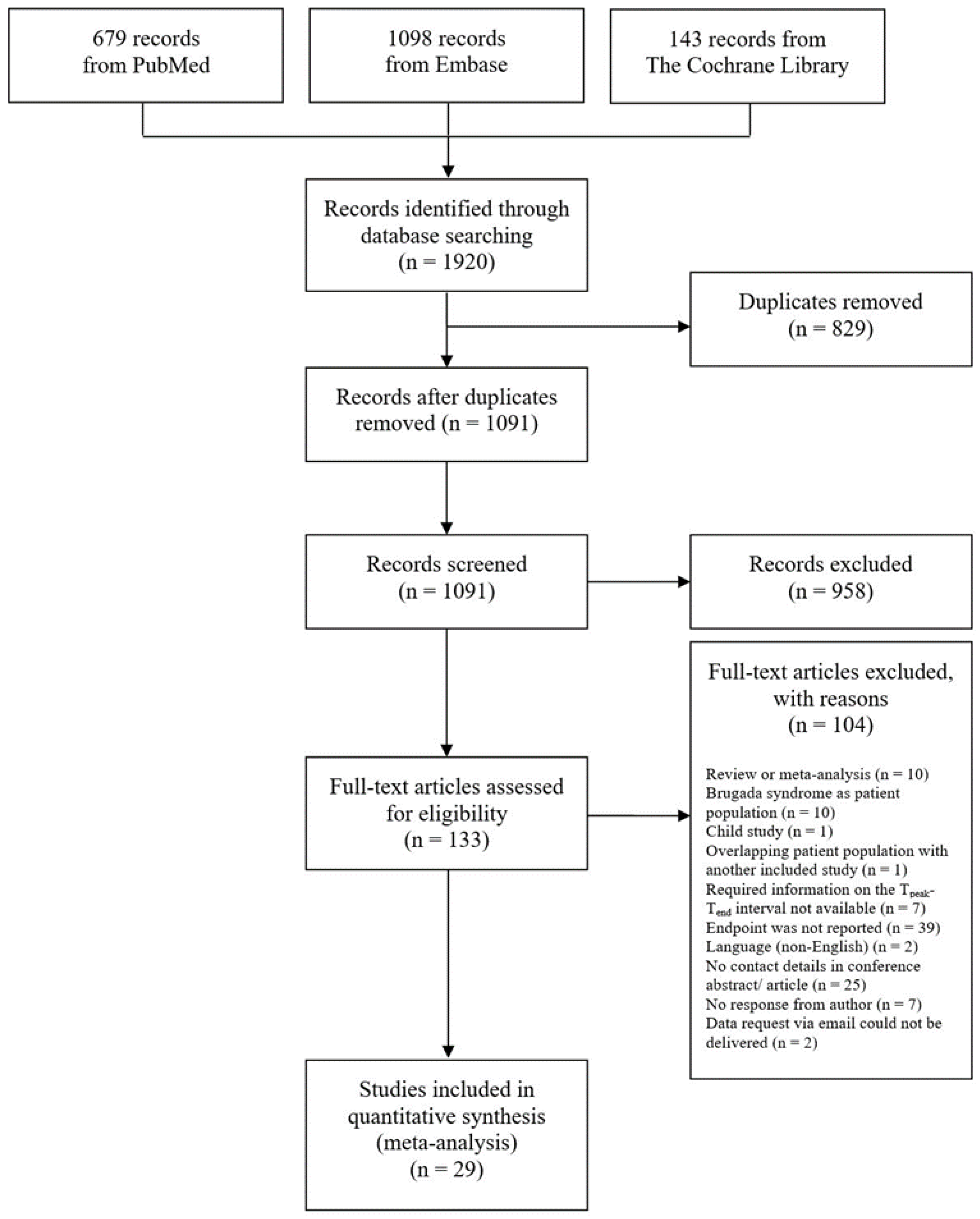
Figure 1. Flow chart of the search strategy and study selection process.
Jcm 12 00992 g001
Figure 2. Tpeak-Tend interval is longer in patients who died compared to survivors. The forest plot shows the data from 20,011 patients from 21 studies, comparing the Tpeak-Tend interval in deaths and survivors. Patients who died had a longer Tpeak-Tend interval by a standard mean difference (SMD) of 0.41. TpTe = Tpeak-Tend interval; SD [25,27,29,30,31,32,34,35,37,39,40,41,42,43,46,49,51,52,53].
Jcm 12 00992 g002
Figure 3. Tpeak-Tend interval is longer in patients who died compared to survivors. This forest plot presents the same studies as Figure 2 but categorized by the ECG measuring method to determine the Tpeak-Tend interval [25,27,29,30,31,32,34,35,37,38,39,40,41,42,43,46,49,50,51,52,53].
Jcm 12 00992 g003
Figure 4. A long Tpeak-Tend interval is associated with all-cause death. The forest plot shows the data from 3665 patients from eight studies, comparing long and short Tpeak-Tend interval in deaths and the total number of patients with the respective Tpeak-Tend interval. Classification in long and short Tpeak-Tend interval was adopted from studies according to the reported cut-off value. A long Tpeak-Tend interval is associated with death by an odds ratio (OR) of 2.33. TpTe = Tpeak-Tend interval; OR = odds ratio; CI = confidence interval [26,27,33,39,47,48,51,54].
Jcm 12 00992 g004
Figure 5. A long Tpeak-Tend interval is associated with all-cause death. This forest plot presents the same studies as Figure 4 but categorized by the ECG measuring method to determine the Tpeak-Tend interval [26,27,33,39,47,48,51,54].
Jcm 12 00992 g005
Figure 6. Estimated receiver operating characteristics of the seven reported AUROCS with corresponding sensitivity and specificity (left) and the estimated summary ROC (SROC) curve with an AUC of 0.65 (right). (Bombelli (2016) [28] (red); Cekridecki (2018) [30] (yellow); Erikssen (2012) [31] (green); Morin (2012) [36] (light blue); Rosenthal (2015) [45] (dark blue); Salgado (2016) [46] (pink)).
Jcm 12 00992 g006
Table 1. Study characteristics.
First Author YearLocationStudy DesignPathologySample Size (n)Women
(n, [%])
Age (Years)Leads for Measuring Tpeak-Tend IntervalMethod for Measuring Tpeak-Tend IntervalHeart-Rate Correction of Tpeak-Tend Interval?Follow-Up Duration (Months)Endpoint
Aksu, E.
2019 [25]
TurkeyRetrospective cohort study *Chronic hemodialysis patients7437 (50)n/an/an/aUncorrected12–15 Cardiac death
Aoki, S.
2014 [26]
JapanProspective cohort study *Acute heart failure syndrome201115 (57.2)78.4 ± n/an/an/aUncorrected161 ± n/aCardiac death
Aro, A. L.
2017 [27]
USA, FinlandCase control studySCD1258409 (32.5)65.6 ± 12.8 aV5Tangent methodUncorrectedn/aSCD
Bombelli, M.
2016 [28]
ItalyCross-sectional studyNew onset hypertension1853928 (50)50.4 ± 13.5V5Tail methodHeart-rate- corrected192 ± n/aAll-cause mortality + cardiovascular mortality
Braschi, A.
2020 [29]
ItalyRetrospective cohort studyPatients with Takotsubo syndome50n/a66.2 ± 9.9Precordial lead with the longest Tpeak-Tend intervaln/aUncorrectedn/a (during hospitalization)All-cause mortality
Cekirdecki, E. I.
2019 [30]
TurkeyRetrospective cohort studyArrhythmo-genic right ventricular cardiomyo-pathy40 d10 (25)34.0 ± 11.53 cII, V2, V5 (lead with the longest Tpeak-Tend)Tail methodHeart-rate- corrected + uncorrected9.5 ± 30.5 All-cause mortality
Erikssen, G.
2012 [31]
NorwayProspective cohort studySTEMI and NSTEMI patients who underwent PCI1359 e839 (61.7)65.5 ± 12.5 aPrecordial lead with the longest Tpeak-Tend intervalTail methodHeart-rate- corrected + uncorrected15.6 ± n/aAll-cause mortality
Haarmark, C.
2009 [32]
DenmarkProspective cohort studyPatients with STEMI undergoing PCI10127 (26.7)62 ± n/aNon-infarct-related leads (ST-segment deviations below 0.055 mV at the J-point in the pre-PCI ECG)
V5, V4, V6, II, III, and I (in descending order)
GE Healthcare Marquette 12SLUncorrected22.5 ± 6.9All-cause mortality
Icli, A.
2015 [33]
TurkeyRetrospective cohort studyAcute pulmonary embolism272119 (43.8)63.1 ± 16.8V5 or, if V5 was not suitable, V4 and V6 in that order were usedTangent methodCorrected1.0 ± n/aAll-cause mortality
Kazemi, B.
2019 [34]
IranProspective cohort studySTEMI patients undergoing primary PCI or thrombo-lytic therapy188116 (61.7)85.97 ± 9.93Leads without ST-segment elevationn/aUncorrectedn/a (hospitalization period)Cardiac death
Li, J.
2019 [35]
SwitzerlandNested case control studyPatients after PCI644152 (23.6)68.5 ± 12.3II or V5 when a parameter was not measurable in lead IITangent methodUncorrectedn/a (within 1 year)All-cause mortality
Morin, D. P.
2012 [36]
USARetrospective cohort studyPatients with an implanted ICD and LVEF ≤ 35%32783 (25.4)67 ± 11V2-V5GE Healthcare Marquette 12SLHeart-rate- corrected + uncorrected30 ± 13 All-cause mortality
Okudan, Y. E.
2018 [37]
TurkeyRetrospective cohort study *Patients with acute anterior MI146n/an/aPrecordial leadsBitRule programmeUncorrectedn/a (first month and first year MACE)Cardiovascular mortality
O’Neal, W. T.
2017 [38]
USAProspective cohort studyGeneral Population (participants from ARIC study)12,2416781 (55.4)54 ± 5.7Median value of all 12 leadsGE Healthcare Marquette 12SLHeart-rate-corrected273.6 ± 34.8 cSCD
Panikkath, R.
2011 [39]
USACase control studySCD from out-of-hospital cardiac arrests695116 (16.7)66.6 ± 14.4V5 or, if this lead was not suitable, leads V4 and V6 in that order were usedTangent methodUncorrectedn/aSCD
SCD with normal QTcn/an/a
SCD with intraventri-cular conduction delay17 (2.4)72.3 ± 13.9
Panikkath, R.
2011 [40]
USANested case-control study *QT prolonging drugs28793 (32.4)65.5 ± 13.3 aV5n/aUncorrectedn/aSCD
Piccirillo, G.
2018 [41]
ItalyProsepctive cohort studyTranscatheter aortic valve replacement patients4017 (42.5)81.0 ± 7.0n/an/aUncorrected12.0 ± n/aAll-cause mortality + cardiovascular mortality
Piccirillo, G.
2020 [42]
ItalyProspective cohort studyLow SCD risk-out-patients with asymp-tomatic and treated car-diovascular risk factors
- elderly subgroup
(>60 years)
6840 (58.8)73.82 ± 7.26n/aTangent methodUncorrected27.6 ± 6All-cause mortality
Piccirillo, G.
2020 [43]
ItalyProspective cohort studyPatients with decompensated CHF10147 (46.5)83 ± 11 an/aSoftware by Berger et al. Uncorrectedn/a (during hospitalization)All-cause mortality
Piccirillo, G. f
2020 [44]
ItalyProspective cohort studyPatients with decompensated CHF11354 (47.8)82.7 ± 10.3n/aSoftware by Berger et al.Uncorrected1 ± n/aAll-cause mortality
Rosenthal, T. M.
2015 [45]
USAProspective cohort studySystolic cardiomyopathy (Patients with an implanted ICD and LVEF ≤ 35%)30582 (26.9)70 ± 11V2-V5 (values of Tpeak are averaged to obtain global Tpeak)GE Healthcare Marquette 12SLHeart-rate- corrected + uncorrected49 ± 21 All-cause mortality
Salgado, A. A.
2016 [46]
BrazilProspective cohort studyLiver Cirrhosis6732 (47.8)54.0 ± 1.9 aAll leadsTangent methodUncorrected9.7 ± 6.8 cAll-cause mortality
Saour, B. M.
2019 [47]
USARetrospective cohort studyEnd stage renal disease2051 (0.5)66.6 ± 12.3V5 or, if V5 was not inter-pretable, V4 and then V6 were usedDifference of QT interval and QRS complex Uncorrected42 ± n/aAll-cause mortality + SCD +
Non-SCD
Sen, Ö.
2016 [48]
TurkeyProspective cohort studyHeart failure patients undergoing ICD implantation22856 (24.6)59.3 ± 12.3Precordial lead with the longest Tpeak-Tend intervalTail methodUncorrected22.3 ± 7.7 All-cause mortality
Smetana, P.
2011 [49]
England, AustriaRetrospective cohort studyMale US veterans with cardio-vascular disease6810 (0)61.05 ± 10.25V4–V6GE Healthcare Marquette 12SLHeart-rate- corrected + uncorrected87.6 ± 44.4All-cause mortality
Szydlo, K.
2011 [50]
PolandProspective cohort study *Patients with anterior MI treated with primary PCI11528 (24.3)58.43 ± 11.21 an/an/aHeart-rate-correctedn/a (within 36 months)Cardiac death
Tatlisu, M. A.
2014 [51]
TurkeyProspective cohort studySTEMI undergoing primary PCI48879 (16.2)55.6 ± 11.2 aLeads without ST-segment elevation; the longest Tpeak-Tend interval was chosenTail method/ Tangent methodHeart-rate- corrected + uncorrected21 ± 10.2 All-cause mortality
Vehmeijer, J. T.
2018 [52]
NetherlandsProspective cohort study *Adults with congenital heart disease448152 (33.9) b35.9 ± 16.2 b,cOne T-wave of each ECG leadn/aUncorrectedn/aSCD
Vehmeijer, J. T.
2019 [53]
NetherlandsCase control studyAdults with congenital heart disease414147 (35.5)35.9 ± 15.3 a,cOne T-wave of each ECG leadTangent methodUncorrectedn/aSCD
Xue, C.
2019 [54]
ChinaProspective cohort studyHeart failure patients with an implantable cardioverter-defibrillator 31879 (24.8)57.59 ± 11.36 Median value of all 12 leadsn/aUncorrected32.12 ± 25.07All-cause mortality
a According to the method by Altman et al. (2000): Combined means and standard deviations of two study populations into one group. b According to the method by Altman et al. (2000): Assumed combined statistics because cases and controls were matched for age and sex. c According to the method by Wan et al. (2014): Estimated means and standard deviations from sample size, median and interquartile range. d We included 40 patients of the entire population of 105 patients, because mortality outcomes were reported for this subgroup only. e We included 1359 patients of the entire population of 1384 patients, because ECG data was available for this subgroup only. f This study was excluded from the analysis, because the study population overlaps to a large extent with the study listed above. * conference abstract; n/a: not applicable; SCD: sudden cardiac death; STEMI: ST elevation myocardial infarction; NSTEMI: non-ST-elevation myocardial infarction; PCI: percutaneous coronary intervention; ICD: implanted cardiac defibrillator; LVEF: left ventricular ejection fraction; MACE: major adverse cardiovascular events; CHF: chronic heart failure.
Table 2. List of studies reporting area under the receiver operating characteristics (AUROC) curves to predict all-cause death by Tpeak-Tend interval.
Study (Year)Cutoff (ms)AUROCSensitivitySpecificity
Bombelli (2016) [28]1210.590.450.70
Cekridecki (2018) [30]1070.890.900.88
Erikssen (2012) [31]1320.770.680.74
Morin (2012) [36]126,70.600.370.80
Rosenthal (2015) [45]1040.580.660.45
Salgado (2016) [46]500.690.900.57
Salgado (2016) [46]600.760.600.79
Disclaimer/Publisher’s Note: The statements, opinions and data contained in all publications are solely those of the individual author(s) and contributor(s) and not of MDPI and/or the editor(s). MDPI and/or the editor(s) disclaim responsibility for any injury to people or property resulting from any ideas, methods, instructions or products referred to in the content.