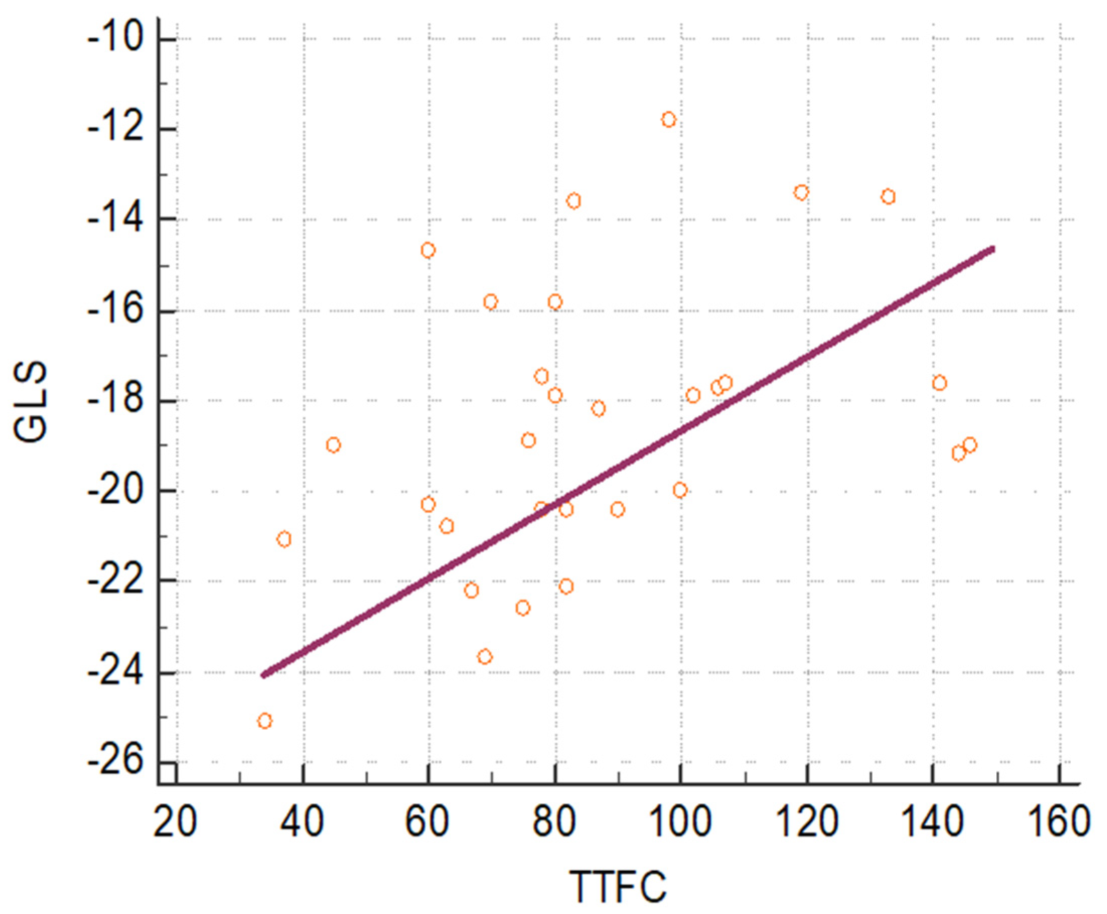
Longitudinal Strain Analysis and Correlation with TIMI Frame Count in Patients with Ischemia with No Obstructive Coronary Artery (INOCA) and Microvascular Angina (MVA)
Abstract
1. Introduction
2. Materials and Methods
3. Results
4. Discussion
5. Limitations of the Study
6. Conclusions
Author Contributions
Funding
Institutional Review Board Statement
Informed Consent Statement
Data Availability Statement
Acknowledgments
Conflicts of Interest
References
- Padro, T.; Manfrini, O.; Bugiardini, R.; Canty, J.; Cenko, E.; De Luca, G.; Duncker, D.J.; Eringa, E.C.; Koller, A.; Tousoulis, D.; et al. ESC Working Group on Coronary pathophysiology and Microcirculation position paper on coronary microvascular dysfunction in cardiovascular disease. Cardiovasc Res. 2020, 116, 741–755. [Google Scholar] [CrossRef]
- Lanza, G.A.; Crea, F. Primary coronary microvascular dysfunction: Clinical presentation, pathophysiology and management. Circulation 2010, 121, 2317–2325. [Google Scholar] [CrossRef] [PubMed]
- Taqueti, V.R.; Shaw, L.J.; Cook, N.R.; Murthy, V.L.; Shah, N.R.; Foster, C.R.; Hainer, J.; Blankstein, R.; Dorbala, S.; Di Carli, M.F. Risk in women relative to men referred for coronary angiography is associated with severely impaired coronary flow reserve, not obstructive disease. Circulation 2017, 135, 566–577. [Google Scholar] [CrossRef]
- Nugent, L.; Mehta, P.K.; Bairey Merz, C.N. Gender and microvascular angina. J. Thromb. Thrombolysis 2011, 31, 37–46. [Google Scholar] [CrossRef]
- Camici, P.G.; Crea, F. Coronary microvascular dysfunction. N. Engl. J. Med. 2007, 356, 830–840. [Google Scholar] [CrossRef] [PubMed]
- Sucato, V.; Madaudo, C.; Galassi, A.R. Classification, Diagnosis, and Treatment of Coronary Microvascular Dysfunction. J. Clin. Med. 2022, 11, 4610. [Google Scholar] [CrossRef] [PubMed]
- Sucato, V.; Novo, G.; Saladino, A.; Rubino, M.; Caronna, N.; Luparelli, M.; D’Agostino, A.; Novo, S.; Evola, S.; Galassi, A.R. Ischemia in patients with no obstructive coronary artery disease: Classification, diagnosis and treatment of coronary microvascular dysfunction. Coron. Artery Dis. 2020, 31, 472–476. [Google Scholar] [CrossRef] [PubMed]
- Gibson, C.M.; Cannon, C.P.; Daley, W.L.; Dodge, J.T., Jr.; Alexander, B., Jr.; Marble, S.J.; McCabe, C.H.; Raymond, L.; Fortin, T.; Poole, W.K.; et al. TIMI frame count: A quantitative method of assessing coronary artery flow. Circulation 1996, 93, 879–888. [Google Scholar] [CrossRef] [PubMed]
- Sara, J.D.; Widmer, R.J.; Matsuzawa, Y.; Lennon, R.J.; Lerman, L.O.; Lerman, A. Prevalence of Coronary Microvascular Dysfunction Among Patients with Chest Pain and Nonobstructive Coronary Artery Disease. J. Am. Collage Cardiol. 2015, 11, 1445–1453. [Google Scholar] [CrossRef] [PubMed]
- Masi, S.; Rizzoni, D.; Taddei, S.; Widmer, R.J.; Montezano, A.C.; Lüscher, T.F.; Schiffrin, E.L.; Touyz, R.M.; Paneni, F.; Lerman, A.; et al. Assessment and pathophysiology of microvascular disease: Recent progress and clinical implications. Eur. Heart J. 2020, 42, 2590–2604. [Google Scholar] [CrossRef]
- Merz, C.B.; Kelsey, S.F.; Pepine, C.J.; Reichek, N.; E Reis, S.; Rogers, W.J.; Sharaf, B.L.; Sopko, G. The Women’s Ischemia Syndrome Evaluation (WISE) Study: Protocol design, methodology and feasibility report. J. Am. Coll. Cardiol. 1999, 33, 1453–1461. [Google Scholar] [CrossRef] [PubMed]
- Cannon, R.O.; Epstein, S.E. ‘Microvascular angina’ as a cause of chest pain with angiographically normal coronary arteries. Am. J. Cardiol. 1988, 61, 1338–1343. [Google Scholar] [CrossRef]
- Brainin, P.; Frestad, D.; Prescott, E. The prognostic value of coronary endothelial and microvascular dysfunction in subjects with normal or non-obstructive coronary artery disease: A systematic review and meta-analysis. Int. J. Cardiol. 2018, 254, 1–9. [Google Scholar] [CrossRef] [PubMed]
- Pepine, C.J.; Ferdinand, K.C.; Shaw, L.J.; Light-McGroary, K.A.; Shah, R.U.; Gulati, M.; Duvernoy, C.; Walsh, M.N.; Bairey Merz, C.N.; ACC CVD in Women Committee. Emergence of nonobstructive coronary artery disease: A women’s problem and need for change in definition on angiography. J. Am. Collage Cardiol. 2015, 66, 1918–1933. [Google Scholar] [CrossRef] [PubMed]
- Gulati, M.; Cooper-DeHoff, R.M.; McClure, C.; Johnson, B.D.; Shaw, L.J.; Handberg, E.M.; Zineh, I.; Kelsey, S.F.; Arnsdorf, M.F.; Black, H.R.; et al. Adverse cardiovascular outcomes in women with nonobstructive coronary artery disease: A report from the Women’s Ischemia Evaluation Study and the St James Women Take Heart Project. Arch. Intern. Med. 2009, 169, 843–850. [Google Scholar] [CrossRef]
- Stehouwer, C.D.A. Microvascular dysfunction and hyperglicemia: A vicious cycle with widespread consequences. Diabetes 2018, 67, 1729–1741. [Google Scholar] [CrossRef]
- Sucato, V.; Evola, S.; Quagliana, A.; Novo, G.; Andolina, G.; Assennato, P.; Novo, S. Comparison of coronary artery flow impairment in diabetic and hypertensive patients with stable microvascular angina. Eur. Rev. Med. Pharmacol. Sci. 2014, 18, 3687–3689. [Google Scholar]
- Potier, L.; Chequer, R.; Roussel, R.; Mohammedi, K.; Sismail, S.; Hartemann, A.; Amouyal, C.; Marre, M.; Le Guludec, D.; Hyafil, F. Relationship between cardiac microvascular dysfunction measured with 82 Rubidium PET and albuminuria in patients with diabetes mellitus. Cardiovasc. Diabetol. 2018, 17, 11. [Google Scholar] [CrossRef]
- Yokoyama, I.; Momomura, S.I.; Ohtake, T.; Yonekura, K.; Nishikawa, J.; Sasaki, Y.; Omata, M. Reduced myocardial flow reserve in non-insulin-dependent diabetes mellitus. J. Am. Coll. Cardiol. 1997, 30, 1472–1477. [Google Scholar] [CrossRef]
- Martin, J.W.; Briesmiester, K.; Bargardi, A.; Muzik, O.; Mosca, L.; Duvernoy, C.S. Weight changes and obesity predict impaired resting and endothelium-dependent myocardial blood flow in postmenopausal women. Clin. Cardiol. 2005, 28, 13–18. [Google Scholar] [CrossRef]
- Bairey Merz, C.N.; Shaw, L.J.; Reis, S.E.; Bittner, V.; Kelsey, S.F.; Olson, M.; Johnson, B.D.; Pepine, C.J.; Mankad, S.; Sharaf, B.L.; et al. Insight from the NHLBI-sponsored Women’s Ischemia Syndrome Evaluation (WISE) Study: Part I: Gender differences in traditional and novel risk factors, symptom evaluation, and gender-optimized diagnostic strategies. J. Am. Collage Cardiol. 2006, 47, S4–S20. [Google Scholar]
- Reis, S.E.; Holubkov, R.; Smith, A.C.; Kelsey, S.F.; Sharaf, B.L.; Reichek, N.; Rogers, W.J.; Merz, C.N.B.; Sopko, G.; Pepine, C.J.; et al. Coronary microvascular dysfunction is higly prevalent in women with chest pain in the absence of coronary artery disease: Results of the NHLBI WISE study. Am. Heart J. 2001, 141, 735–741. [Google Scholar] [CrossRef] [PubMed]
- Sucato, V.; Novo, S.; Rubino, M.; D’Agostino, A.; Evola, S.; Novo, G. Prognosis in patients with microvascular angina: A clinical follow-up. J. Cardiovasc. Med. 2019, 20, 794–795. [Google Scholar] [CrossRef] [PubMed]
- Xu, X.; Zhou, J.; Zhang, Y.; Li, Q.; Guo, L.; Mao, Y.; He, L. Evaluate the Correlation between the TIMI Frame Count, IMR, and CFR in Coronary Microvascular Disease. J. Interv. Cardiol. 2022, 2022, 6361398. [Google Scholar] [CrossRef] [PubMed]

| Comparison of Baseline Echocardiographic Data in the INOCA and MVA and Control Group (Mean Values = M and Standard Deviation = SD) | |||||
|---|---|---|---|---|---|
| INOCA and MVA Group (n = 85) | Control Group (n = 70) | p-Value | |||
| M | SD | M | SD | ||
| Left Ventricular Ejection Fraction (%) | 56.15 | 5.34 | 58.55 | 5.7 | 0.13 |
| Inter Ventricular Septum (mm) | 9.72 | 1.51 | 9.45 | 1.15 | 0.5 |
| End-diastolic diameter (mm) | 45.66 | 5.4 | 43.1 | 4.13 | 0.08 |
| End-diastolic volume (mL) | 86.71 | 19.31 | 96.8 | 20.59 | 0.08 |
| Left atrial volume (mL) | 50.20 | 27.4 | 44.20 | 9.59 | 0.35 |
| E wave (m/s) | 0.64 | 0.16 | 0.66 | 0.16 | 0.66 |
| A wave (m/s) | 0.69 | 0.27 | 0.60 | 0.16 | 0.18 |
| E/A ratio | 0.95 | 0.30 | 1.13 | 0.41 | 0.07 |
| Deceleration time (ms) | 219.81 | 61.32 | 197.90 | 41.32 | 0.16 |
| septal e’ (m/s) | 0.07 | 0.02 | 0.08 | 0.03 | 0.15 |
| septal a’ (m/s) | 0.09 | 0.02 | 0.11 | 0.01 | f |
| septal s’ (m/s) | 0.07 | 0.02 | 0.07 | 0.01 | 0.9 |
| lateral e’ (m/s) | 0.10 | 0.03 | 0.12 | 0.03 | 0.02 |
| lateral a’ (m/s) | 0.11 | 0.02 | 0.13 | 0.03 | 0.006 |
| lateral s’ (m/s) | 0.09 | 0.03 | 0.09 | 0.02 | 0.9 |
| E/e’ ratio (m/s) | 8.01 | 4.01 | 7.34 | 2.86 | 0.52 |
| Longitudinal Strain in INOCA and MVA Patients and Control Group (Mean Values M with Standard Deviation SD) | |||||
|---|---|---|---|---|---|
| INOCA and MVA Group (n = 85) | Control Group (n = 70) | p-Value | |||
| M | SD | M | SD | ||
| Basal Anterior Septum | −16.8 | 4.78 | −17.85 | 3.23 | 0.48 |
| Mid Anterior Septum | −18.6 | 6.59 | −22.6 | 3.07 | 0.03 |
| Apical Anterior Septum | −17.9 | 6.51 | −23.3 | 4.86 | 0.016 |
| Basal infero-lateral wall | −17.22 | 4.41 | −21.95 | 2.29 | 0.0006 |
| Mid infero-lateral wall | −17.5 | 2.72 | −21.35 | 2.56 | 0.0007 |
| Apical infero-lateral wall | −18.5 | 5.24 | −20.37 | 3.64 | 0.264 |
| Longitudinal strain APLAX (apical long axis view) | −17.22 | 3.53 | −20.69 | 1.76 | 0.001 |
| Basal Antero-lateral wall | −18.2 | 5.77 | −19.20 | 3.59 | 0.563 |
| Mid Antero-lateral wall | −16.7 | 6.29 | −18.95 | 2.86 | 0.185 |
| Apical Antero-lateral wall | −16.22 | 6.92 | −18.55 | 4.74 | 0.286 |
| Basal Inferior Septum | −15.2 | 4.08 | −15.84 | 3.42 | 0.653 |
| Mid Inferior Septum | −18.4 | 5.66 | −20.20 | 3.58 | 0.295 |
| Apical Inferior Septum | −18 | 6.48 | −23.35 | 4.7 | 0.015 |
| Longitudinal strain A4C (Apical Four Chamber View) | −16.77 | 3.71 | −18.91 | 2.35 | 0.0634 |
| Basal Anterior Wall | −16.3 | 5.12 | −16.40 | 4.71 | 0.96 |
| Mid Anterior Wall | −15.2 | 5.14 | −20.80 | 4.81 | 0.0065 |
| Apical Anterior Wall | −13.11 | 7.47 | −19.16 | 5.89 | 0.022 |
| Basal Inferior Wall | −17.1 | 3.31 | −20.74 | 3.07 | 0.006 |
| Mid Inferior Wall | −20.6 | 3.02 | −21.95 | 4.06 | 0.204 |
| Apical Inferior Wall | −19 | 5.39 | −21.74 | 5.66 | 0.215 |
| Longitudinal strain A2C (Apical Two Chamber View) | −16.8 | 3.37 | −19.50 | 2.47 | 0.019 |
| Global Longitudinal Strain (GLS) | −16.77 | 2.82 | −19.64 | 1.91 | 0.003 |
Disclaimer/Publisher’s Note: The statements, opinions and data contained in all publications are solely those of the individual author(s) and contributor(s) and not of MDPI and/or the editor(s). MDPI and/or the editor(s) disclaim responsibility for any injury to people or property resulting from any ideas, methods, instructions or products referred to in the content. |
© 2023 by the authors. Licensee MDPI, Basel, Switzerland. This article is an open access article distributed under the terms and conditions of the Creative Commons Attribution (CC BY) license (https://creativecommons.org/licenses/by/4.0/).
Share and Cite
Sucato, V.; Novo, G.; Madaudo, C.; Di Fazio, L.; Vadalà, G.; Caronna, N.; D’Agostino, A.; Evola, S.; Tuttolomondo, A.; Galassi, A.R. Longitudinal Strain Analysis and Correlation with TIMI Frame Count in Patients with Ischemia with No Obstructive Coronary Artery (INOCA) and Microvascular Angina (MVA). J. Clin. Med. 2023, 12, 819. https://doi.org/10.3390/jcm12030819
Sucato V, Novo G, Madaudo C, Di Fazio L, Vadalà G, Caronna N, D’Agostino A, Evola S, Tuttolomondo A, Galassi AR. Longitudinal Strain Analysis and Correlation with TIMI Frame Count in Patients with Ischemia with No Obstructive Coronary Artery (INOCA) and Microvascular Angina (MVA). Journal of Clinical Medicine. 2023; 12(3):819. https://doi.org/10.3390/jcm12030819
Chicago/Turabian StyleSucato, Vincenzo, Giuseppina Novo, Cristina Madaudo, Luca Di Fazio, Giuseppe Vadalà, Nicola Caronna, Alessandro D’Agostino, Salvatore Evola, Antonino Tuttolomondo, and Alfredo Ruggero Galassi. 2023. "Longitudinal Strain Analysis and Correlation with TIMI Frame Count in Patients with Ischemia with No Obstructive Coronary Artery (INOCA) and Microvascular Angina (MVA)" Journal of Clinical Medicine 12, no. 3: 819. https://doi.org/10.3390/jcm12030819
APA StyleSucato, V., Novo, G., Madaudo, C., Di Fazio, L., Vadalà, G., Caronna, N., D’Agostino, A., Evola, S., Tuttolomondo, A., & Galassi, A. R. (2023). Longitudinal Strain Analysis and Correlation with TIMI Frame Count in Patients with Ischemia with No Obstructive Coronary Artery (INOCA) and Microvascular Angina (MVA). Journal of Clinical Medicine, 12(3), 819. https://doi.org/10.3390/jcm12030819


