Article

Pharmacogenomic Considerations for Anticoagulant Prescription in Patients with Hereditary Haemorrhagic Telangiectasia

by
Sarah C. McCarley
1,
Daniel A. Murphy
2,3,
Jack Thompson
1 and
Claire L. Shovlin
1,3,4,*
1
National Heart and Lung Institute, Imperial College London, London W12 0NN, UK
2
Pharmacy Department, Imperial College Healthcare NHS Trust, London W2 1NY, UK
3
Social, Genetic and Environmental Determinants of Health Theme, NIHR Imperial Biomedical Research Centre, London W2 1NY, UK
4
Specialist Medicine, Hammersmith Hospital, Imperial College Healthcare NHS Trust, London W12 0HS, UK
*
Author to whom correspondence should be addressed.
J. Clin. Med. 2023, 12(24), 7710; https://doi.org/10.3390/jcm12247710
Submission received: 15 November 2023 / Revised: 10 December 2023 / Accepted: 12 December 2023 / Published: 15 December 2023

Abstract

:
Hereditary haemorrhagic telangiectasia (HHT) is a vascular dysplasia that commonly results in bleeding but with frequent indications for therapeutic anticoagulation. Our aims were to advance the understanding of drug-specific intolerance and evaluate if there was an indication for pharmacogenomic testing. Genes encoding proteins involved in the absorption, distribution, metabolism, and excretion of warfarin, heparin, and direct oral anticoagulants (DOACs) apixaban, rivaroxaban, edoxaban, and dabigatran were identified and examined. Linkage disequilibrium with HHT genes was excluded, before variants within these genes were examined following whole genome sequencing of general and HHT populations. The 44 genes identified included 5/17 actionable pharmacogenes with guidelines. The 76,156 participants in the Genome Aggregation Database v3.1.2 had 28,446 variants, including 9668 missense substitutions and 1076 predicted loss-of-function (frameshift, nonsense, and consensus splice site) variants, i.e., approximately 1 in 7.9 individuals had a missense substitution, and 1 in 71 had a loss-of-function variant. Focusing on the 17 genes relevant to usually preferred DOACs, similar variant profiles were identified in HHT patients. With HHT patients at particular risk of haemorrhage when undergoing anticoagulant treatment, we explore how pre-emptive pharmacogenomic testing, alongside HHT gene testing, may prove beneficial in reducing the risk of bleeding and conclude that HHT patients are well placed to be at the vanguard of personalised prescribing.

1. Introduction

Hereditary haemorrhagic telangiectasia (HHT) is an autosomal dominant multisystem vascular dysplasia arising from a single heterozygous loss-of-function variant (“mutation”), usually in ENG, ACVRL1, or SMAD4 [1,2,3,4,5]. As recently reviewed [6,7,8,9], patients develop internal, visceral arteriovenous malformations (AVMs) and smaller telangiectasia that bleed recurrently. International consensus is available to guide clinical management [7,8,10,11]. Initial guidance was through the generation of consensus clinical diagnostic criteria (the Curaçao Criteria) where the presence of three criteria from spontaneous recurrent nosebleeds, mucocutaneous telangiectasia, visceral involvement, and family history can be used to define definite clinical HHT [10,11,12]. These criteria are less helpful in children where there are fewer clinical features [9,13,14,15], and conversely, it is possible to overdiagnose HHT if based on nosebleeds, telangiectasia, and family history alone [16,17]. The 2020 Second International Guidelines [7] recommended obtaining a genetic diagnosis of the HHT-causative mutation to facilitate targeted screening for internal AVMs. HHT gene testing pathways are now in place in multiple countries worldwide [7,8,18,19,20,21,22,23,24,25] and facilitate the diagnosis of HHT, targeted AVM screening programmes, and direction of SMAD4 families to SMAD4-specific preventative measures [26,27,28,29,30,31].
Haemorrhage and anemia are hallmarks of HHT. Recurrent bleeds from nasal and/or gastrointestinal telangiectasia in HHT commonly result in anaemia and dependence on oral iron, intravenous iron, and/or red cell transfusions [7,8,32,33], with anaemia exacerbated by additional aetiologies [34,35,36,37]. Despite this, there are frequent indications for therapeutic anticoagulation. One major indication is venous thromboembolism (VTE), which is more common in HHT than in the general population [38,39,40] and may also be precipitated by therapies to treat HHT bleeding [41,42]. A further common indication is atrial fibrillation, which complicates high cardiac output states in the setting of HHT hepatic AVMs, particularly when anaemia develops [43]. Left atrial appendage closure is increasingly performed for HHT patients unable to tolerate the usually recommended anticoagulation [44,45,46]. In addition to treatment (therapeutic) doses, anticoagulants are often indicated at lower (prophylactic) doses, for example, peri-operatively or in hospitalized, acutely ill medical patients [47,48].
Previous observational data in HHT-affected individuals have indicated marked extremes in tolerance of anticoagulant therapies. HHT is the second most common heritable bleeding disorder after von Willebrand’s disease (HHT 11–17 per 100,000 [49,50,51,52]; VWD 109–2200 per 100,000 [53]) as it is more common than the haemophilias [54]. Given that up to 10,000 HHT patients in Europe are estimated to require anticoagulation [55], it is surprising that a modest number of small retrospective cohort studies (Supplementary Table S1) represent the bulk of our knowledge on the safety and tolerability of different anticoagulant drugs in HHT [39,40,55,56,57,58,59,60]. Incidence of major bleeding episodes necessitating the discontinuation of anticoagulation ranged from 21.6 to 50.1 per 100 patients per year across studies [39,58]. For example, a large retrospective cohort study surveying 126 patients on the French national HHT registry found that just over a third of HHT patients prematurely discontinued anticoagulation in the first three months of treatment [60]. Reasons for discontinuation included mucosal bleeding, major bleeding events, and corresponding increases in red cell transfusions and/or hospitalisation [60].
Until recently, it was not possible to directly compare anticoagulant agents in HHT as no single study included the full range of low molecular weight heparins (LMWH), Vitamin K antagonists such as warfarin/acenocoumarol, and direct oral anticoagulants (DOACs) in any significant numbers (Supplementary Table S1). In part, this reflected the reluctance of major HHT centres to prescribe DOACs where there is less opportunity to reverse, and no evidence of tolerance in HHT, in contrast to heparin and warfarin [56,57]. For instance, in early data from the Hospital Italiano de Buenos Aires, no DOAC use was reported in their HHT registry [61], while across Europe, only 32 DOAC treatment episodes were identified by the European Reference Network [55]. Comparing these small numbers to historical online patient survey data [57] and expert opinion led the Second International Guidelines Committee to suggest LMWH and warfarin over DOACs in 2020 [7]. More recently, US data provided sufficient numbers to enable direct comparisons between anticoagulant agents in HHT patients for the first time [59]. This study found that the rates of dose-reduction or premature treatment discontinuation due to bleeding were similar in those episodes involving warfarin (16/35 [46%]), heparin-based anticoagulation (LMWH or fondaparinux, 14/27 [48%]), DOACs (11/25 [44%]), and multiple agents simultaneously (18/41 [44%]), noting that anticoagulation discontinuation rates were potentially higher than reported in some earlier studies [40,55,56]. In view of variability, missing data, and some analyses that combined prophylactic (low dose) and therapeutic episodes (Supplementary Table S1), despite identifying 356 anticoagulant treatment episodes in HHT across 13 cohort studies and 64 case reports, the authors of a 2023 “scoping review” did not feel they could make over-arching conclusions on the optimal anticoagulation agent in HHT [62].
What these studies do show is that while more people with HHT tolerate therapeutic anticoagulation with no discernible adverse consequences [39,40,55,56,57,58,59,60,61,62], a significant proportion of patients on heparin, Vitamin K antagonists, and DOACs such as apixaban, dabigatran, edoxaban, and rivaroxaban, have serious exacerbation in their bleeding diathesis and have to discontinue therapy [39,40,55,56,57,58,59,60,61,62]. Crucially, several series demonstrate that tolerance differs between different anticoagulant agents in the same patient [55,57].
There is no evidence in man that a specific HHT causal genotype is associated with tolerance of individual anticoagulant agents [55] or, indeed, a higher or lower overall HHT bleeding risk [4,63]. This is supported by recent data from mice that indicate that in both major HHT genotypes (ACVRL1 and ENG), haemostasis is impaired to a similar degree, though through different mechanisms [64]. Thus, simple HHT diagnosis with or without HHT gene testing does not allow prediction of who is at higher risk of bleeding, or higher risk of bleeding on prescription of anticoagulants. Beyond HHT-causal genes, however, whole genome sequencing (WGS) data in 104 HHT patients recruited to the 100,000 Genomes Project demonstrated that patients with greater haemorrhagic severity had more deleterious variants in genes encoding platelet and coagulation cascade-related proteins [63].
These observations prompted us to extend genomic analyses to additional genes that may modify the HHT bleeding phenotype, focusing on commonly prescribed drugs. The field of pharmacogenomics examines the impact of variation in the genome on drug pharmacology and offers the potential to reduce adverse events and improve drug efficacy [65]. Loss and gain-of-function alleles in multiple genes have been shown to affect drug pharmacokinetics (how the body handles drugs) and pharmacodynamics (how drugs affect the body) [66]. Recent guidelines for the general population detail multiple genes with DNA variants that have sufficient clinical impact to merit changes to the drug or dose [67,68,69]. In the general population, a major goal is to reduce adverse events and hospital admissions, and for anticoagulants, the greatest concern is major bleeding events [70]. These concerns are exacerbated for people with HHT, who are already prone to bleeds due to abnormal vascular structures. Separately, there are pharmacogenomic considerations for efficacy, in other words, preventing pathological thromboses. While that is less of a focus for the current study, the higher VTE rates in HHT mean that efficacy considerations within what may be a narrower therapeutic window are also important.
Therefore, we considered pharmacogenomic considerations of anticoagulant therapies to be particularly relevant for people with HHT. Our first goal was to provide evidence to support or refute individual drug-specific intolerance so that a single failed anticoagulation episode does not prevent future use of other anticoagulants. A second goal, focusing on countries where gene testing is implemented in HHT diagnostics, was to evaluate if there could be an indication for diagnostic pharmacogenomic testing in HHT, using linkage disequilibrium and pharmacogenetic DNA variant prevalence in general and HHT populations. We specifically focused on the newer DOACs that are favoured in general population guidelines and by patients but where more extreme haemorrhagic responses have been described in HHT [55].

2. Materials and Methods

2.1. Gene Identification

A literature search was conducted to reconstruct the warfarin, heparin, and DOAC biochemical pathways and underlying genes. The search was conducted using the Google Scholar and Pub Med databases. The search included key phrases such as “Anticoagulants AND Pharmacogenetics”, “Pharmacogenetics AND Warfarin”, “Warfarin metabolism”, “Warfarin biochemical pathway”, “Pharmacogenetics AND Heparin”, “Heparin metabolism”, “Heparin biochemical pathway”, “Pharmacogenetics AND DOACs”, “DOACs metabolism”, and “DOACs biochemical pathway”. Articles on each anticoagulant biochemical pathway and anticoagulant pharmacogenetics were selected. The Data Supplement provides a detailed appreciation of gene involvement in the anticoagulants’ biochemical pathways.
In a separate study (Murphy et al., manuscript in preparation), the 17 genes with actionable guidance in clinical practice were generated by making use of the Dutch Pharmacogenetics Working Group [71] and the Clinical Pharmacogenetics Implementation Consortium [72] clinical guidelines, each derived from data within the Pharmacogenomics Knowledgebase (PharmGKB) [73].

2.2. Gene Location and Variant Identification

Gene positions on the Genome Reference Consortium human(GRCh) build 38 [74] were identified using the University of California Santa Cruz (UCSC) Genome Browser [75]. RefSeq [76] was used to obtain further metrics for each gene and major transcripts.
Data from 76,156 participants in the Genome Aggregation Database (gnomAD) v3.1.2 [77] were used to estimate general population variant prevalence, specifically for predicted loss-of-function (pLOF) variants (nonsense, frameshift, and consensus splice site variants), and separately for missense variants that result in an amino acid substitution.
Anonymised outputs from the Genomics England internal bioinformatics pipelines and analyses were examined in the Genomics England Research Environment [78] using Participant Explorer and Interactive Variant Analysis (IVA) v2.2.3. Data were approved for export through the Research Environment AirLock under subproject RR42 (HHT-Gene-Stop).

2.3. Data Analysis

A dataset was constructed to visualise the variation in the pharmacogenes using publicly available databases. GRCh38 [74] and MANE Project v1.2 [79] were used to obtain the length of each gene, the length of the coding region in nucleotides, and the number of exons. Within gnomAD 3.1.2 [77], for each pharmacogene, we extracted the total number of missense and pLOF variants (separately and combined), the total number of individuals reported each missense or pLOF variant, and the allele frequency of each variant. The number of gene variants per coding region nucleotide was calculated.
Data from HHT patients recruited to the 100,000 Genomes Project [78] were used to construct a dataset containing the total number of variants and pLOF variants in the HHT cohort for each of the DOAC pharmacogenes. We then calculated the number of variants per [HHT] individual, pLOF variants per [HHT] individual, variants in an individual per coding sequence nucleotide, and the ratio of the number of variants in an HHT individual to the number of variants in the general population per coding sequence for each of the DOAC pharmacogenes.
Descriptive, comparative, and relationship statistics were generated using GraphPad Prism 9.5.1 (GraphPad Software, San Diego, CA, USA) and STATA version 15 (StataCorp, College Station, TX, USA). For continuous data, two group comparisons were using Mann–Whitney. Normality testing and graphical generation were performed using GraphPad Prism 9.5.1 (GraphPad Software, San Diego, CA, USA).

3. Results

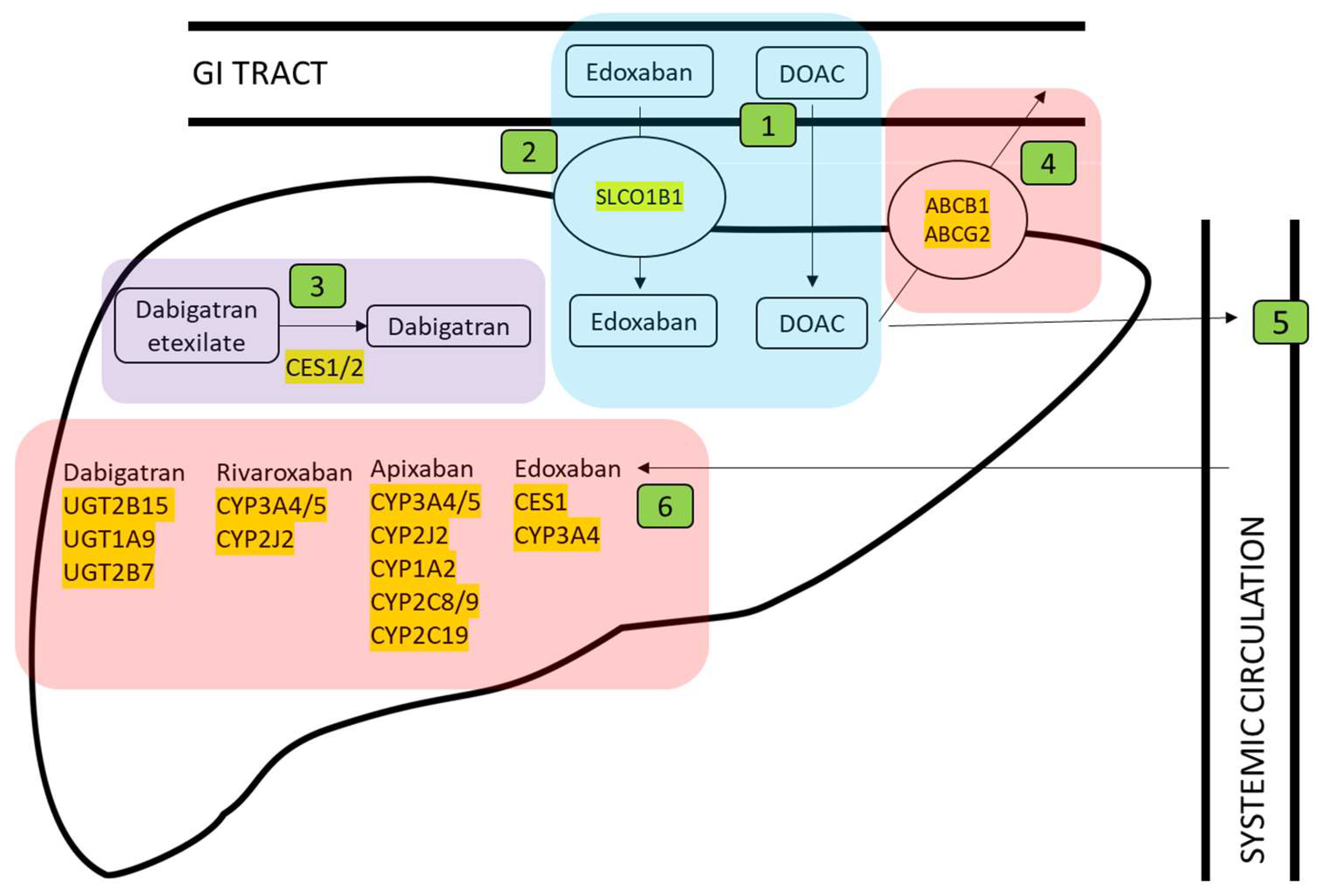
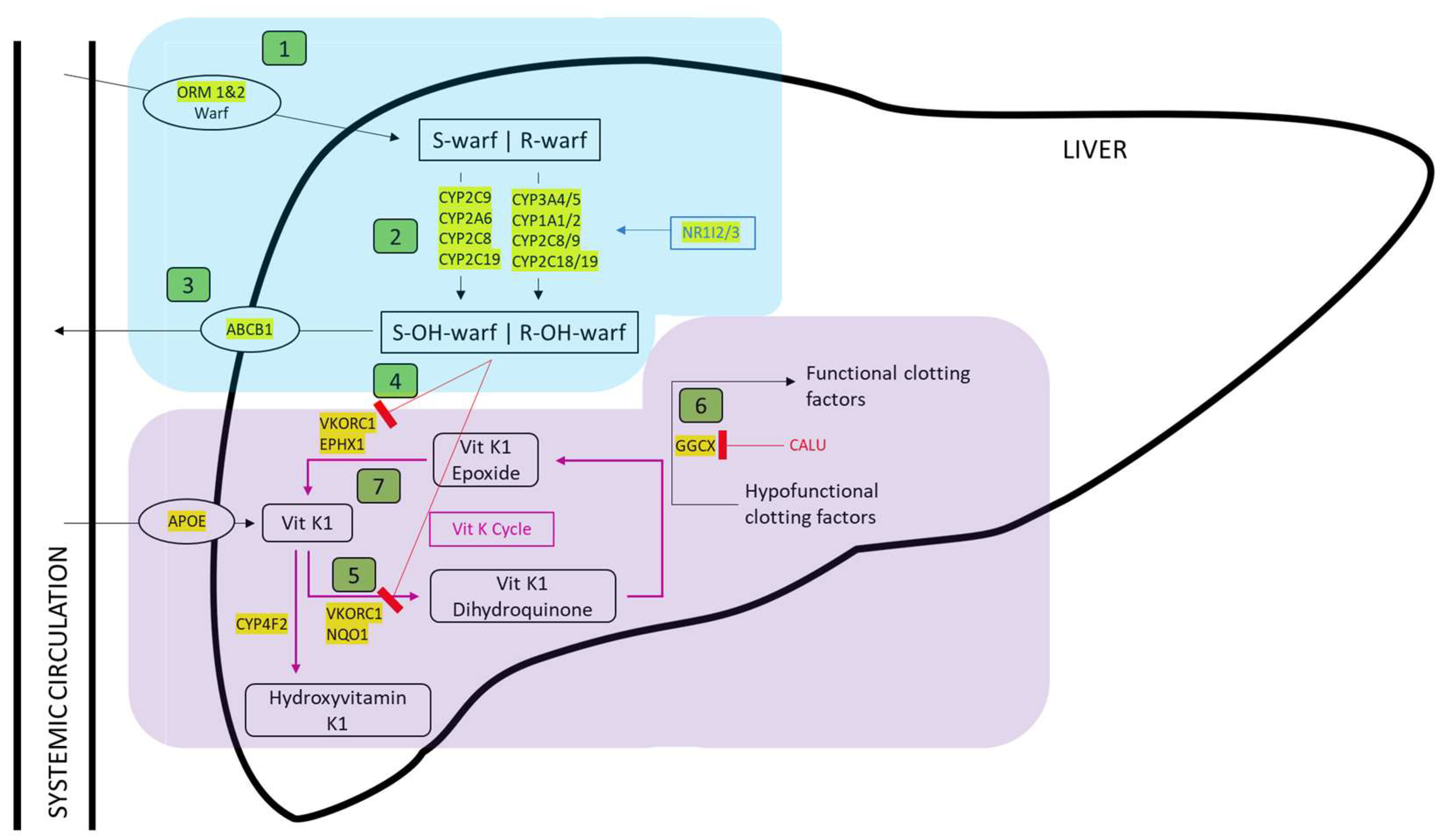
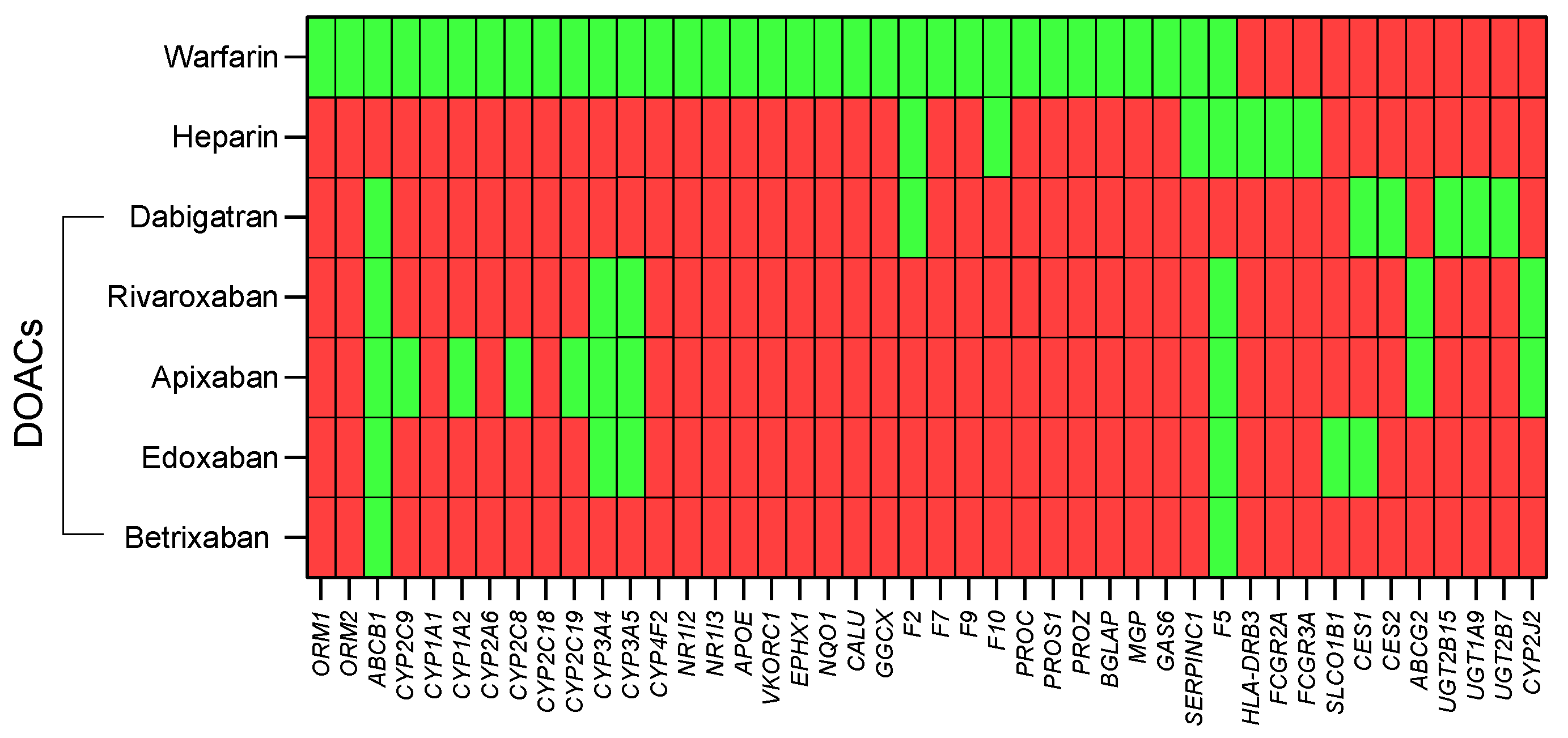
3.1. Identification of Genes Involved in Anticoagulant Metabolism

In total, 44 genes were identified where encoded proteins impacted the pharmacokinetics or pharmacodynamics of warfarin (Figure 1), heparin (Figure 2), or direct oral anticoagulants dabigatran, rivaroxaban, apixaban, edoxaban, and betrixaban (Figure 3).
The full list of 44 pharmacogenes is provided in Supplementary Table S2. As shown in Figure 4, individual genes are relevant to more than one drug.

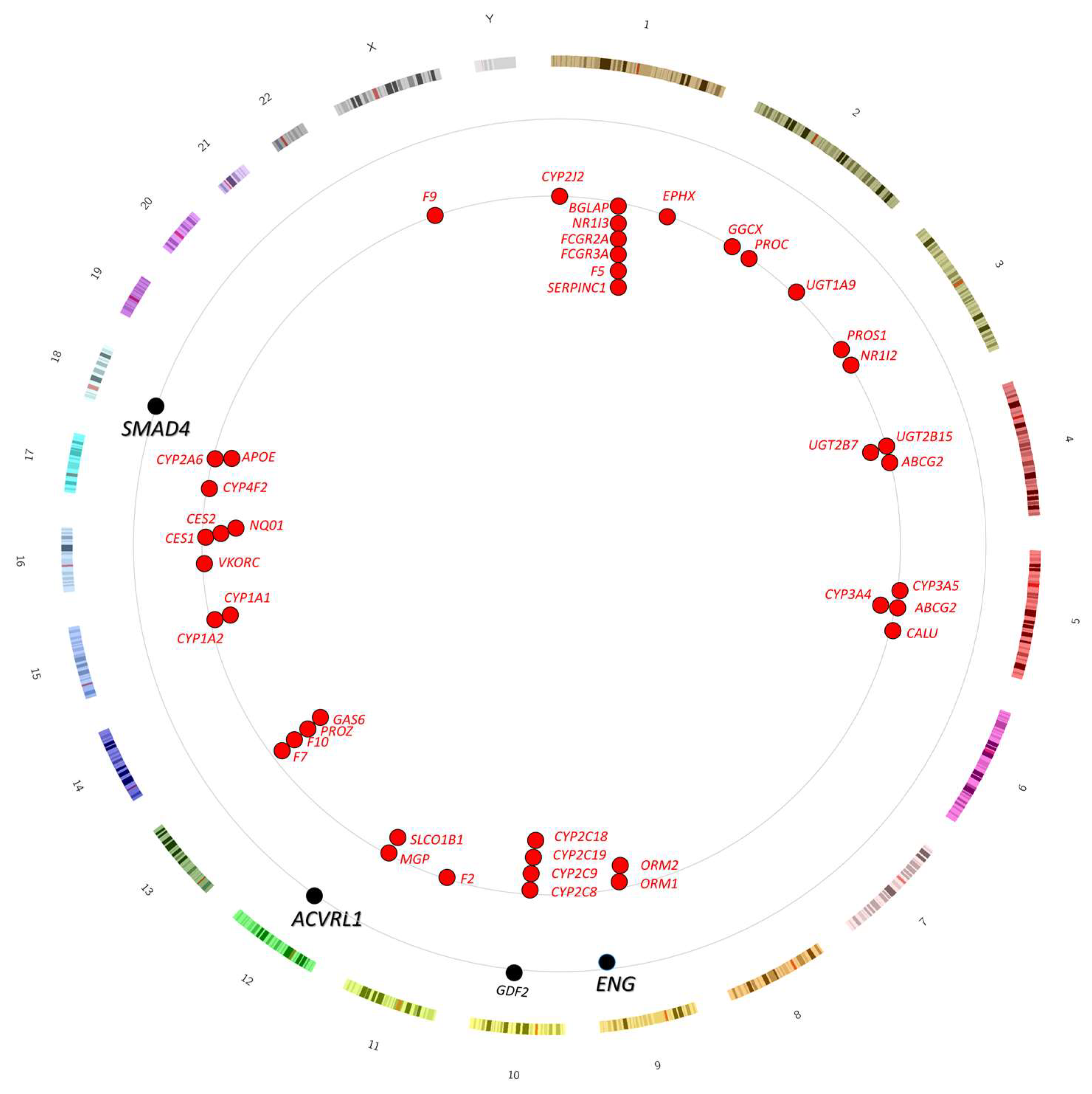
3.2. Genomic Location of Genes Involved in Anticoagulant Metabolism

None of the 44 pharmacogenes genes are located within 1.5 Mb of the major HHT genes ACVRL1, ENG, or SMAD4 (Figure 5, Supplementary Table S2). ENG (GRCh38 chr9: 127,815,016–127,854,658) was the closest to one of the pharmacogenes with ORM2 sited 13.5 Mb away at chr9:114,329,869–114,333,251. However, ORM2 was almost adjacent (only 75 kb, i.e., 0.075 Mb distant) to the D9S59 locus that was unlinked to ENG in one of the original HHT families used to identify ENG as a HHT-causative gene [80]. We concluded that neither HHT gene testing nor familial responses would be likely to predict pharmacogenomic responses to anticoagulants in HHT families.

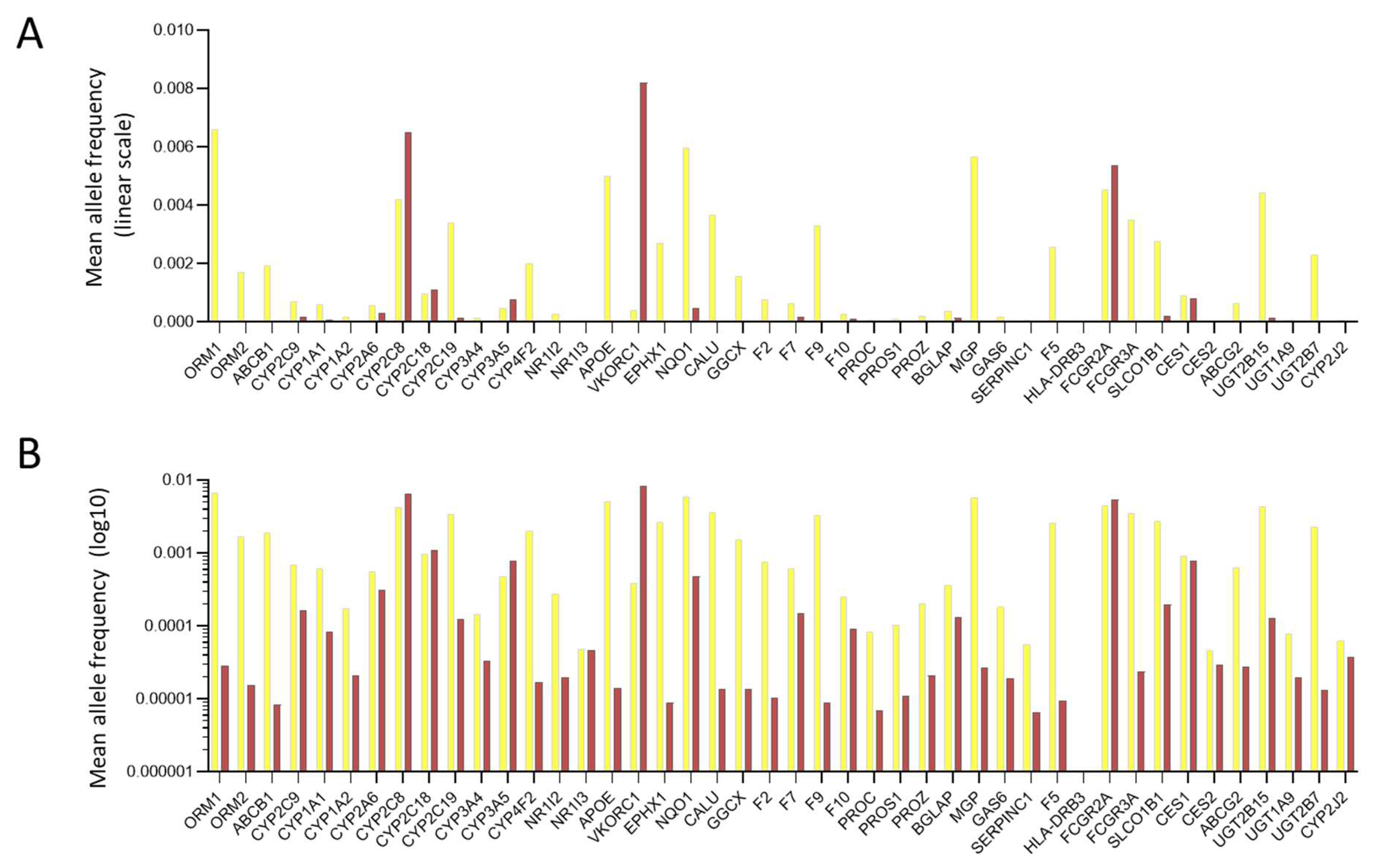
3.3. General Population Variant Burdens

With reference to Figure 1, Figure 2 and Figure 3 predicted loss-of-function (pLOF) alleles, including frameshift indels, nonsense substitutions, splice site, and some missense alleles, would predict higher plasma drug levels for two already actionable pharmacogenes (VKORC1 for warfarin and SLCO1B1 for heparin). In addition, for at least 11 further genes, pLOF variants would predict higher plasma drug levels. We, therefore, tested the frequency of pLOF loss-of-function allelic variants.
First, the number of variants within the 44 identified pharmacogenes was examined in the 76,156 participants in gnomAD v3.1.2 [77]. Recruited participants were from diverse backgrounds, including 39,345 Europeans, 20,744 Africans/African Americans, 7647 Latino/Admixed Americans, and 5023 individuals from South or East Asia. In total, there were 9668 different missense substitutions of an amino acid that may be silent but may cause loss-of-function (as for many of the HHT-causal variants in ACVRL1, ENG, and SMAD4) or more rarely, gain-of-function. Separately, there were 1076 different pLOF variants, i.e., frameshift, nonsense, and consensus splice site variants. Variant allele frequencies are displayed in Figure 6 across all genes and were seen in all ethnicities. Importantly, within the gnomAD sample of 76,156 people, this approximately translated to 1 in 7.9 individuals having a missense substitution and 1 in 71 a predicted loss of function allele.
Adjusting for gene length, VKORC1, an actionable gene for warfarin prescription [71,72,73], had the greatest number of missense and pLOF variants per coding region nucleotide (nt) at 0.34 missense variants per nucleotide and 0.045 pLOF variants per nucleotide. F9 encoding Factor 9 had the fewest at 0.051 missense variants/nt and 0.014 pLOF variants/nt. The number of pLOF variants per coding region nucleotide passed normality testing using all four of Anderson–Darling, D’Agostino and Pearson, Shapiro–Wilk, and Kolmogorov-Smirnov tests (all p values > 0.09) in support of random origin and maintenance. Missense variants did not pass normality testing using any of the four tests (all p values > 0.097, with VKORC1 and F9 being outliers).

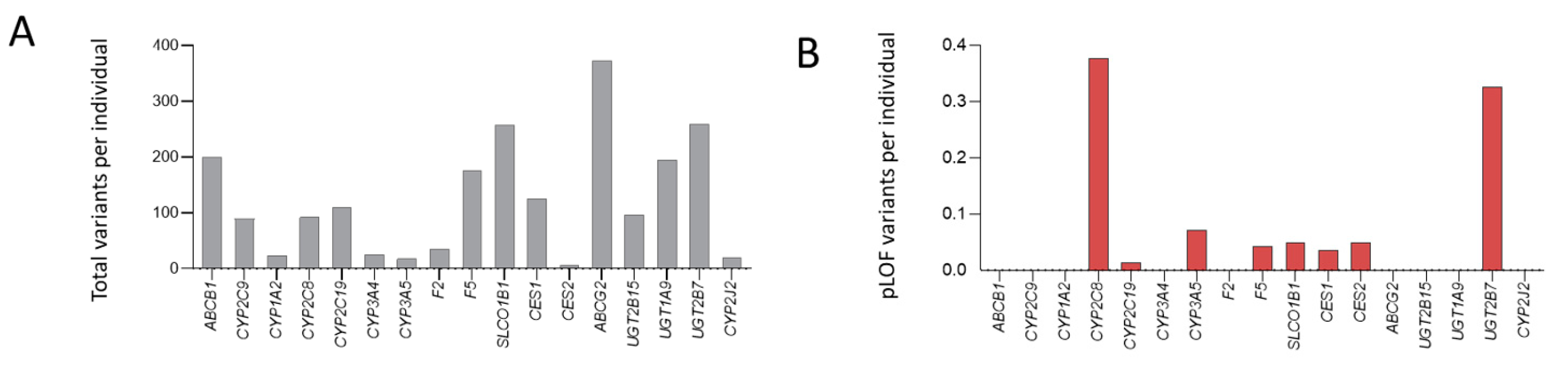
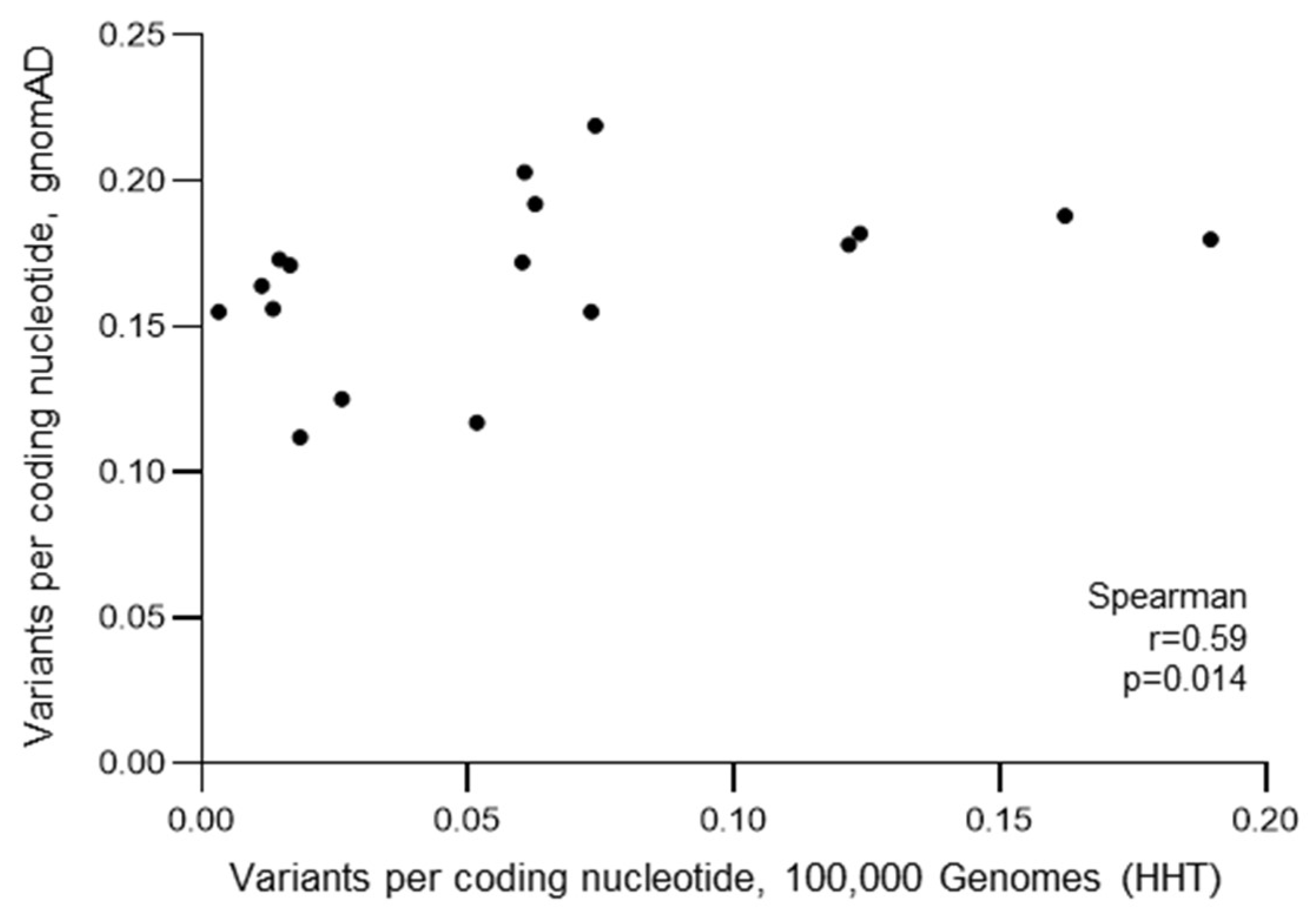
3.4. HHT Population Variant Burdens

While there was no reason to expect HHT patients to have differing proportions of variants in these genes, we took the opportunity to examine variant frequencies in the 100,000 Genomes Project-recruited HHT population. Mindful of the change in general medical practice away from warfarin and heparin requiring efficacy assessments and/or injections, towards direct oral anticoagulants that do not, we focused on the 17 genes relevant to DOAC mechanisms of action. As shown in Figure 7, within the modestly sized (N = 141) HHT population recruited to the 100,000 Genomes Project, variants were identified in all genes, with pLOF variants identified in eight genes.
Despite the differing methods of ascertainment, genome alignments, and stringency metrics, there was a direct correlation between the number of variants per coding sequence nucleotide in the HHT cohort and that identified in the gnomAD general population. In other words, the number of variants per coding sequence nucleotide in the HHT cohort increased as the number of variants per coding sequence nucleotide in the general population increased (p = 0.014, Figure 8). Further, for pLOF variants where the impact would be confidently predicted, there were between 0 and 53 per gene across the 141 HHT patients, representing an average of 0–0.38 per patient per gene. Overall, across all 17 genes implicated in DOAC mechanisms of action, the mean number of pLOF variants per HHT patient recruited to the 100,000 Genomes Project was 0.96 (standard deviation 0.11).

4. Discussion

We have shown that across a series of 44 genes where gene products influence pharmacokinetics or pharmacodynamics of major, currently used anticoagulants, at least 1 in 7 individuals can be expected to have a missense substitution, and more than 1 in 70 to have a loss-of-function variant. This includeds drug metabolism pharmacogenes in which heterozygous loss-of-function alleles, along with other pharmacogenes-encoding drug transporters and receptors, predict higher drug levels. The variants were also identified in people with HHT, where there is a narrower therapeutic window due to abnormal vasculature. Thus, while it is not possible to predict bleeding tendency based on familial HHT gene variants or phenotypes, knowledge of pharmacogenetic variants may allow predictions facilitating individualised anticoagulant prescriptions. Conversely, for non-genotyped populations, chance differences in the prevalence of these variants could result in skewed results of less general applicability across HHT than previously thought.
The main study limitations were the absence of functional data in the participants with the gene variants and the lack of ethnic diversity in the HHT patients recruited to the 100,000 Genomes Project (though more ethnically diverse genome datasets were examined through gnomAD). However, the main study strength is to alert the field of the presence of these variants and their potential importance to prescribing.
In terms of implications for practice, the first element is relevance to prescribing anticoagulants in HHT without pharmacogenomic data, as this is the current situation. Previous work has shown marked variability in anticoagulant tolerance in terms of bleeding in HHT [39,40,55,56,57,58,59,60,61,62], and the current findings are in keeping with this. Previous work has also shown that individuals with HHT who are unable to tolerate one particular anticoagulant due to excessive bleeding are able to tolerate other agents [55,57]. Again, the current findings are in keeping with this. Given the narrow therapeutic window and tendency to higher thrombotic rates in HHT [38,39,40], there is value in being able to monitor efficacy directly through laboratory assays (e.g., international normalized ratio (INR) for warfarin [81] or activated partial thromboplastin time (APTT) for heparin [82]) to ensure the patient is anticoagulated to the correct degree. For warfarin, different loading regimens can be employed, and a more conservative low-dose approach, such as the Crowther protocol [83], may be preferred above a rapid loading schedule, such as recommended by Tait [84].
Where pharmacogenetic testing is available, validated variants can be translated into personalised prescribing by employing internationally recognised guidelines such as those found on PharmGKB [73]. That said, of the anticoagulants examined in the current manuscript, only warfarin has clinically established pharmacogenomic prescribing guidelines [85,86,87,88]. Even amongst these well-established warfarin guidelines, there is a lack of consensus with differing approaches to dose alterations arising from clinically actionable pharmacogenomic variants. Notably, with alternatives such as efficacy monitoring through the INR [81] and wider use of DOACs [89,90], there may not be a push to harmonise the warfarin guidance.
For DOACs, which are now the main oral anticoagulants in clinical use [91,92], the data are in the arena of “newly discovered variants”, where PharmGKB recommends determining the level of evidence for the impact a variant would have on whether a dosing amendment is needed [93]. The effect of a loss-of-function variant can be inferred using the principles from the American College of Medical Genetics and Association for Molecular Pathology [94] for “very important pharmacogenes” in which the drug–gene association has been strongly established. However, for variants where drug–gene associations have not been made, studies examining the association between pharmacogenetic variants and drug response would need to be performed. This is the case for all DOACs where pharmacogenomic testing is not yet used in clinical practice [95].
There is a trend towards greater adoption of pharmacogenomic testing in mainstream clinical practice to improve safety and efficacy. For example, England’s National Health Service commissions a limited number of individual drug–gene pairs [96], and the goal is to move towards pre-emptive panel-based testing across a wide range of drug–gene pairs [97], providing clinically actionable genetic information at the point of prescribing. The recent multicentre randomised control trial, PREPARE, demonstrated the success of this approach by testing for 50 variants in 12 pharmacogenes resulting in a significantly lower number of adverse drug events [68]. Subsequently, the PROGRESS programme is assessing the feasibility of introducing NHS-wide genetic testing to guide prescription in common practice [98,99]. Although economic panel-based arrays are proposed as the first step to introduce pharmacogenomics into common practice [65,96,97,98,99], as shown by our study, whole genome sequencing facilitates the identification of novel clinically relevant loss-of-function variants, which would otherwise go undetected. As such, as sequencing costs fall, this approach may become more commonplace.
It must be recognised that pharmacogenomics will be implemented at different rates in different healthcare structures. So, taken together, what conclusions can be drawn from the current HHT study? First, as guideline-emphasised, therapeutic anticoagulation is not contraindicated in HHT [7]. If no personalised genomic data are available, drug treatment can still be personalised using demographic and clinical data, remembering the HHT-specific observational studies summarised in Supplementary Table S1. Given that DOACs have so many advantages for easier prescription (though they are more difficult to monitor and reverse), this is the area where pharmacogenomics may be of most potential importance to HHT and other states with narrower therapeutic windows for anticoagulation. For people with HHT, the significant bleeding risk posed by anticoagulation, and their higher incidence of VTE, together with nosebleeds providing less severe manifestations of excessive bleeding, represent a strong argument in favour of further study in the population.
In order to address some of the study limitations, future work could use functional data to validate pharmacogene variants. In addition, associations between pharmacogene variants and drug response could be measured. Although prospective clinical trials may not be ethical in this population, retrospective analyses could be performed; alternatively, pharmacogenomic data from unaffected individuals could be extrapolated to HHT patients. It will also be important to educate patients about the benefits of pharmacogenomic testing to increase awareness and acceptance of this approach, and of anticoagulation when clinically indicated. Ultimately, since most indications for anticoagulants are in the emergency setting, prior understanding of individual drug risk profiles would enhance patient safety.

5. Conclusions

High proportions of the HHT population carry DNA variants that predict particular anticoagulants will carry a higher risk of haemorrhage. In view of their narrow therapeutic window and the usually urgent nature of anticoagulant prescribing, we encourage the development of pre-emptive pharmacogenomic testing alongside HHT gene testing. More generally, the HHT population is well placed to be at the vanguard of personalised prescribing.

Supplementary Materials

The following supporting information can be downloaded at https://www.mdpi.com/2077-0383/12/24/7710/s1: Supplementary Methods; Supplementary Table S1: Previous HHT Anticoagulation Studies; Supplementary Table S2: Pharmacogenes and location; Supplementary References [39,40,55,56,57,58,59,60,100,101,102,103,104,105,106,107,108,109,110,111,112,113,114,115,116,117,118,119,120,121,122,123,124].

Author Contributions

Conceptualization, C.L.S. and D.A.M.; methodology, S.C.M., D.A.M., J.T. and C.L.S.; formal analysis, S.C.M., D.A.M., J.T. and C.L.S.; investigation, C.L.S. resources, S.C.M., D.A.M., J.T. and C.L.S.; data curation, C.L.S.; writing—original draft preparation, S.C.M.; writing—review and editing, S.C.M., D.A.M., J.T. and C.L.S.; visualization, S.C.M., J.T. and C.L.S.; project administration, C.L.S.; funding acquisition, C.L.S. All authors have read and agreed to the published version of the manuscript.

Funding

Funding support was obtained from the NIHR Imperial Biomedical Research Centre. Part of the research was made possible through access to the data and findings generated by the 100,000 Genomes Project. The 100,000 Genomes Project is managed by Genomics England Limited (a wholly owned company of the Department of Health and Social Care). The 100,000 Genomes Project is funded by the National Institute for Health Research (NIHR) and NHS England. The Wellcome Trust, Cancer Research UK, and the Medical Research Council have also funded research infrastructure. The 100,000 Genomes Project uses data provided by patients and collected by the National Health Service as part of their care and support. Some HHT patients were recruited to the 100,000 Genomes Project through the National Institute for Health and Care Research (NIHR) Imperial Clinical Research Facility (CRF).

Institutional Review Board Statement

The study was conducted in accordance with the Declaration of Helsinki. Ethical approvals required for the 100,000 Genomes Project analyses were provided by the Health Research Authority (HRA) Committee East England-Cambridge South (REC Ref 14/EE/1112). All participants provided written consent. Obtaining ethics approval was not required for data collection and analysis of the Genome Aggregation Database (gnomAD) v3.1.2.

Informed Consent Statement

Informed consent was obtained from all subjects involved in the study.

Data Availability Statement

Data supporting reported results can be found in the Data Supplement. Primary data from the 100,000 Genomes Project, which are held in a secure Research Environment, are available to registered users. Please see https://www.genomicsengland.co.uk/research/academic (accessed on 11 December 2023) for further information.

Acknowledgments

We thank the patients for their involvement in the 100,000 Genomes Project and the investigators whose work is cited in this manuscript.

Conflicts of Interest

The authors declare no conflict of interest. The funders had no role in the design of the study, in the collection, analyses, or interpretation of data, in the writing of the manuscript, or in the decision to publish the results. The views expressed are those of the authors and not necessarily those of funders, the NHS, the NIHR, or the Department of Health and Social Care.

Correction Statement

This article has been republished with a minor correction (the spelling error in "Envionmental" has changed to "Environmental") to the existing affiliation information. This change does not affect the scientific content of the article.

References

  1. McAllister, K.A.; Grogg, K.M.; Johnson, D.W.; Gallione, C.J.; Baldwin, M.A.; Jackson, C.E.; Helmbold, E.A.; Markel, D.S.; McKinnon, W.C.; Murrel, J.; et al. Endoglin, a TGF-β binding protein of endothelial cells, is the gene for hereditary haemorrhagic telangiectasia type 1. Nat. Genet. 1994, 8, 345–351. [Google Scholar] [CrossRef]
  2. Johnson, D.W.; Berg, J.N.; Baldwin, M.A.; Gallione, C.J.; Marondel, I.; Yoon, S.-J.; Stenzel, T.T.; Speer, M.; Pericak-Vance, M.A.; Diamond, A.; et al. Mutations in the activin receptor–like kinase 1 gene in hereditary haemorrhagic telangiectasia type 2. Nat. Genet. 1996, 13, 189–195. [Google Scholar] [CrossRef] [PubMed]
  3. Gallione, C.J.; Repetto, G.M.; Legius, E.; Rustgi, A.K.; Schelley, S.L.; Tejpar, S.; Mitchell, G.; Drouin, E.; Westermann, C.J.; Marchuk, D.A. A combined syndrome of juvenile polyposis and hereditary haemorrhagic telangiectasia associated with mutations in MADH4 (SMAD4). Lancet 2004, 363, 852–859. [Google Scholar] [CrossRef] [PubMed]
  4. Shovlin, C.L.; Simeoni, I.; Downes, K.; Frazer, Z.C.; Megy, K.; Bernabeu-Herrero, M.E.; Shurr, A.; Brimley, J.; Patel, D.; Kell, L.; et al. Mutational and phenotypic characterization of hereditary hemorrhagic telangiectasia. Blood 2020, 136, 1907–1918. [Google Scholar] [CrossRef] [PubMed]
  5. Xiao, S.; Kai, Z.; Murphy, D.; Li, D.; Patel, D.; Bielowka, A.M.; Bernabeu-Herrero, M.E.; Abdulmogith, A.; Mumford, A.D.; Westbury, S.K.; et al. Functional filter for whole-genome sequencing data identifies HHT and stress-associated non-coding SMAD4 polyadenylation site variants >5 kb from coding DNA. Am. J. Hum. Genet. 2023, 110, 1903–1918. [Google Scholar] [CrossRef] [PubMed]
  6. VASCERN Orphanet Definition of Hereditary Hemorrhagic Telangiectasia. 2019. Available online: https://www.orpha.net/consor/www/cgi-bin/OC_Exp.php?lng=EN&Expert=774 (accessed on 11 December 2023).
  7. Faughnan, M.E.; Mager, J.J.; Hetts, S.W.; Palda, V.A.; Lang-Robertson, K.; Buscarini, E.; Deslandres, E.; Kasthuri, R.S.; Lausman, A.; Poetker, D.; et al. Second International Guidelines for the Diagnosis and Management of Hereditary Hemorrhagic Telangiectasia. Ann. Intern. Med. 2020, 173, 989–1001. [Google Scholar] [CrossRef] [PubMed]
  8. Shovlin, C.L.; Buscarini, E.; Sabbà, C.; Mager, H.J.; Kjeldsen, A.D.; Pagella, F.; Sure, U.; Ugolini, S.; Torring, P.M.; Suppressa, P.; et al. The European Rare Disease Network for HHT Frameworks for management of hereditary haemorrhagic telangiectasia in general and speciality care. Eur. J. Med. Genet. 2022, 65, 104370. [Google Scholar] [CrossRef] [PubMed]
  9. Danesino, C.; Cantarini, C.; Olivieri, C. Hereditary Hemorrhagic Telangiectasia in Pediatric Age: Focus on Genetics and Diagnosis. Pediatr. Rep. 2023, 15, 129–142. [Google Scholar] [CrossRef]
  10. Shovlin, C.L.; Guttmacher, A.E.; Buscarini, E.; Faughnan, M.E.; Hyland, R.H.; Westermann, C.J.; Kjeldsen, A.D.; Plauchu, H. Diagnostic criteria for hereditary hemorrhagic telangiectasia (Rendu-Osler-Weber syndrome). Am. J. Med. Genet. 2000, 91, 66–67. [Google Scholar] [CrossRef]
  11. Faughnan, M.E.; Palda, V.A.; Garcia-Tsao, G.; Geisthoff, U.W.; McDonald, J.; Proctor, D.D.; Spears, J.; Brown, D.H.; Buscarini, E.; Chesnutt, M.S.; et al. International guidelines for the diagnosis and management of hereditary haemorrhagic telangiectasia. J. Med. Genet. 2011, 48, 73–87. [Google Scholar] [CrossRef]
  12. Van Gent, M.W.; Velthuis, S.; Post, M.C.; Snijder, R.J.; Westermann, C.J.; Letteboer, T.G.; Mager, J.J. Hereditary hemorrhagic telangiectasia: How accurate are the clinical criteria? Am. J. Med. Genet. A 2013, 161, 461–466. [Google Scholar] [CrossRef]
  13. Pollak, M.; Gatt, D.; Shaw, M.; Hewko, S.L.; Lamanna, A.; Santos, S.; Ratjen, F. Longitudinal Assessment of Curaçao Criteria in Children with Hereditary Hemorrhagic Telangiectasia. J. Pediatr. 2023, 263, 113665. [Google Scholar] [CrossRef]
  14. Pahl, K.S.; Choudhury, A.; Wusik, K.; Hammill, A.; White, A.; Henderson, K.; Pollak, J.; Kasthuri, R.S. Applicability of the Curaçao Criteria for the Diagnosis of Hereditary Hemorrhagic Telangiectasia in the Pediatric Population. J. Pediatr. 2018, 197, 207–213. [Google Scholar] [CrossRef]
  15. Gonzalez, C.D.; Cipriano, S.D.; Topham, C.A.; Stevenson, D.A.; Whitehead, K.J.; Vanderhooft, S.; Presson, A.P.; McDonald, J. Localization and age distribution of telangiectases in children and adolescents with hereditary hemorrhagic telangiectasia: A retrospective cohort study. J. Am. Acad. Dermatol. 2019, 81, 950–955. [Google Scholar] [CrossRef] [PubMed]
  16. McDonald, J.; Kornish, J.; Stevenson, D.A.; Hanson-Kahn, A.; Balch, H.; James, J.; Naik, H.; Whitehead, K.J. Frequency of epistaxis and telangiectasia in patients with hereditary hemorrhagic telangiectasia (HHT) in comparison with the general population: Curaçao diagnostic criteria revisited. Genet Med. 2023, 25, 100865. [Google Scholar] [CrossRef] [PubMed]
  17. Shovlin, C.L.; Almaghlouth, F.I.; Alsafi, A.; Coote, N.; Rennie, C.; Wallace, G.M.; Govani, F.S.; Research Consortium, G.E. Updates on diagnostic criteria for hereditary haemorrhagic telangiectasia in the light of whole genome sequencing of ‘gene-negative’ individuals recruited to the 100,000 Genomes Project. J. Med. Genet. 2023, 16, jmg-2023-109195. Available online: https://jmg.bmj.com/content/early/2023/08/15/jmg-2023-109195 (accessed on 11 December 2023).
  18. Baysal, M.; Demir, S.; Ümit, E.G.; Gürkan, H.; Baş, V.; Karaman Gülsaran, S.; Demirci, U.; Kırkızlar, H.O.; Demir, A.M. Genetic Diagnosis of Hereditary Hemorrhagic Telangiectasia: Four Novel Pathogenic Variations in Turkish Patients. Balkan Med. J. 2019, 37, 43–46. [Google Scholar] [PubMed]
  19. Zhao, Y.; Zhang, Y.; Wang, X.; Zhang, L. Variant analysis in Chinese families with hereditary hemorrhagic telangiectasia. Mol. Genet. Genomic. Med. 2019, 7, e893. [Google Scholar] [CrossRef] [PubMed]
  20. Koenighofer, M.; Parzefall, T.; Frohne, A.; Allen, M.; Unterberger, U.; Laccone, F.; Schoefer, C.; Frei, K.; Lucas, T. Spectrum of Novel Hereditary Hemorrhagic Telangiectasia Variants in an Austrian Patient Cohort. Clin. Exp. Otorhinolaryngol. 2019, 12, 405–411. [Google Scholar] [CrossRef] [PubMed]
  21. Mutize, T.T.; Seedat, R.Y.; Ploos van Amstel, J.K.; Mager, J.J.; Brown, S.C.; Gebremariam, F.; Coetzee, M.J. The clinical and genetic features of hereditary haemorrhagic telangiectasia (HHT) in central South Africa-three novel pathogenic variants. Mol. Biol. Rep. 2020, 47, 9967–9972. [Google Scholar] [CrossRef]
  22. Gil, R.; Añón, S.; Salazar-Mendiguchía, J.; Riera-Mestre, A. RiHHTa Investigators of the Rare Diseases Working Group from the Spanish Society of Internal Medicine. Current HHT genetic overview in Spain and its phenotypic correlation: Data from RiHHTa registry. Orphanet J. Rare Dis. 2020, 15, 138. [Google Scholar]
  23. Kitayama, K.; Ishiguro, T.; Komiyama, M.; Morisaki, T.; Morisaki, H.; Minase, G.; Hamanaka, K.; Miyatake, S.; Matsumoto, N.; Kato, M.; et al. Mutational and clinical spectrum of Japanese patients with hereditary hemorrhagic telangiectasia. BMC Med. Genomics. 2021, 14, 288. [Google Scholar] [CrossRef]
  24. Major, T.; Bereczky, Z.; Gindele, R.; Balogh, G.; Rácz, B.; Bora, L.; Kézsmárki, Z.; Brúgós, B.; Pfliegler, G. Current Status of Clinical and Genetic Screening of Hereditary Hemorrhagic Telangiectasia Families in Hungary. J. Clin. Med. 2021, 10, 3774. [Google Scholar] [CrossRef]
  25. Kim, B.G.; Jung, J.H.; Kim, M.J.; Moon, E.H.; Oh, J.H.; Park, J.W.; Cha, H.E.; Kim, J.H.; Kim, Y.J.; Chung, J.W.; et al. Genetic Variants and Clinical Phenotypes in Korean Patients with Hereditary Hemorrhagic Telangiectasia. Clin. Exp. Otorhinolaryngol. 2021, 14, 399–406. [Google Scholar] [CrossRef]
  26. Heald, B.; Rigelsky, C.; Moran, R.; LaGuardia, L.; O’Malley, M.; Burke, C.A.; Zahka, K. Prevalence of thoracic aortopathy in patients with juvenile Polyposis Syndrome-Hereditary Hemorrhagic Telangiectasia due to SMAD4. Am. J. Med. Genet. A 2015, 167, 1758–1762. [Google Scholar] [CrossRef] [PubMed]
  27. Vorselaars, V.M.M.; Diederik, A.; Prabhudesai, V.; Velthuis, S.; Vos, J.A.; Snijder, R.J.; Westermann, C.J.J.; Mulder, B.J.; Ploos van Amstel, J.K.; Mager, J.J.; et al. SMAD4 gene mutation increases the risk of aortic dilation in patients with hereditary haemorrhagic telangiectasia. Int. J. Cardiol. 2017, 245, 114–118. [Google Scholar] [CrossRef] [PubMed]
  28. McDonald, N.M.; Ramos, G.P.; Sweetser, S. SMAD4 mutation and the combined juvenile polyposis and hereditary hemorrhage telangiectasia syndrome: A single center experience. Int. J. Color. Dis. 2020, 35, 1963–1965. [Google Scholar] [CrossRef] [PubMed]
  29. Jelsig, A.M.; Kjeldsen, A.; Christensen, L.L.; Bertelsen, B.; Karstensen, J.G.; Brusgaard, K.; Torring, P.M. Hereditary haemorrhagic telangiectasia in Danish patients with pathogenic variants in SMAD4: A nationwide study. J. Med. Genet. 2023, 60, 464–468. [Google Scholar] [CrossRef] [PubMed]
  30. Boland, C.R.; Idos, G.E.; Durno, C.; Giardiello, F.M.; Anderson, J.C.; Burke, C.A.; Dominitz, J.A.; Gross, S.; Gupta, S.; Jacobson, B.C.; et al. Diagnosis and management of cancer risk in the gastrointestinal hamartomatous polyposis syndromes: Recommendations from the U.S. Multi-Society Task Force on Colorectal Cancer. Gastrointest Endosc. 2022, 95, 1025–1047. [Google Scholar] [CrossRef] [PubMed]
  31. Matsumoto, T.; Umeno, J.; Jimbo, K.; Arai, M.; Iwama, I.; Kashida, H.; Kudo, T.; Koizumi, K.; Sato, Y.; Sekine, S.; et al. Clinical Guidelines for Diagnosis and Management of Juvenile Polyposis Syndrome in Children and Adults-Secondary Publication. J. Anus Rectum Colon 2023, 7, 115–125. [Google Scholar] [CrossRef] [PubMed]
  32. Kjeldsen, A.D.; Kjeldsen, J. Gastrointestinal bleeding in patients with hereditary hemorrhagic telangiectasia. Am. J. Gastroenterol. 2000, 95, 415–418. [Google Scholar] [CrossRef] [PubMed]
  33. Finnamore, H.; Le Couteur, J.; Hickson, M.; Busbridge, M.; Whelan, K.; Shovlin, C.L. Hemorrhage-adjusted iron requirements, hematinics and hepcidin define hereditary hemorrhagic telangiectasia as a model of hemorrhagic iron deficiency. PLoS ONE 2013, 8, e76516. [Google Scholar] [CrossRef] [PubMed]
  34. Cherif, H.; Karlsson, T. Combination treatment with an erythropoiesis-stimulating agent and intravenous iron alleviates anaemia in patients with hereditary haemorrhagic telangiectasia. Ups J. Med. Sci. 2014, 119, 350–353. [Google Scholar] [CrossRef] [PubMed]
  35. Thielemans, L.; Layton, D.M.; Shovlin, C.L. Low serum haptoglobin and blood films suggest intravascular hemolysis contributes to severe anemia in hereditary hemorrhagic telangiectasia. Haematologica 2019, 104, e127–e130. [Google Scholar] [CrossRef] [PubMed]
  36. Zarka, J.; Jeong, K.; Yabes, J.G.; Ragni, M.V. Prevalence and risk factors for bleeding in hereditary hemorrhagic telangiectasia: A National Inpatient Sample study. Blood Adv. 2023, 7, 5843–5850. [Google Scholar] [CrossRef]
  37. Hvelplund, T.; Lange, B.; Bird, S.D.; Korsholm, M.; Kjeldsen, A.D. A retrospective cohort study on European Reference Network for Rare Vascular Diseases 5 outcome measures for Hereditary Haemorrhagic Telangiectasia in Denmark. Orphanet J. Rare Dis. 2022, 17, 8. [Google Scholar] [CrossRef]
  38. Livesey, J.A.; Manning, R.A.; Meek, J.H.; Jackson, J.E.; Kulinskaya, E.; Laffan, M.A.; Shovlin, C.L. Low serum iron levels are associated with elevated plasma levels of coagulation factor VIII and pulmonary emboli/deep venous thromboses in replicate cohorts of patients with hereditary haemorrhagic telangiectasia. Thorax 2012, 67, 328–333. [Google Scholar] [CrossRef]
  39. Riera-Mestre, A.; Mora-Luján, J.M.; Trujillo-Santos, J.; Del Toro, J.; Nieto, J.A.; Pedrajas, J.M.; López-Reyes, R.; Soler, S.; Ballaz, A.; Cerdà, P.; et al. Natural history of patients with venous thromboembolism and hereditary hemorrhagic telangiectasia. Findings from the RIETE registry. Orphanet J. Rare Dis. 2019, 14, 196. [Google Scholar] [CrossRef]
  40. Tentoni, N.; Lapidus, M.I.; Peuchot, V.A.; Vazquez, F.J.; Serra, M.M. Bleeding events during anticoagulation in patients with hereditary hemorrhagic telangiectasia. Thromb. Res. 2021, 197, 109–111. [Google Scholar] [CrossRef]
  41. Penaloza, A.; Vekemans, M.C.; Lambert, C.; Hermans, C. Deep vein thrombosis induced by thalidomide to control epistaxis secondary to hereditary haemorrhagic telangiectasia. Blood Coagul. Fibrinolysis 2011, 22, 616–618. [Google Scholar] [CrossRef]
  42. Maestraggi, Q.; Bouattour, M.; Toquet, S.; Jaussaud, R.; Kianmanesh, R.; Durand, F.; Servettaz, A. Bevacizumab to Treat Cholangiopathy in Hereditary Hemorrhagic Telangiectasia: Be Cautious: A Case Report. Medicine 2015, 94, e1966. [Google Scholar] [CrossRef] [PubMed]
  43. Buscarini, E.; Leandro, G.; Conte, D.; Danesino, C.; Daina, E.; Manfredi, G.; Zambelli, A. Natural history and outcome of hepatic vascular malformations in a large cohort of patients with hereditary hemorrhagic teleangiectasia. Dig Dis Sci. 2011, 56, 2166–2178. [Google Scholar] [CrossRef]
  44. Velthuis, S.; Swaans, M.J.; Mager, J.J.; Rensing, B.J.; Boersma, L.V.; Post, M.C. Left atrial appendage closure for stroke prevention in patients with atrial fibrillation and hereditary hemorrhagic telangiectasia. Case Rep. Cardiol. 2012, 2012, 646505. [Google Scholar] [CrossRef] [PubMed]
  45. Pepe, M.; Suppressa, P.; Giuliano, A.F.; Nestola, P.L.; Bortone, A.S.; DECillis, E.; Acquaviva, T.; Forleo, C.; Moscarelli, M.; Lenato, G.M.; et al. Safety of reduced or absent antithrombotic therapy after left atrial appendage closure in patients affected by hereditary hemorrhagic telangiectasia and atrial fibrillation. Minerva Cardiol. Angiol. 2022, 70, 537–544. [Google Scholar] [CrossRef] [PubMed]
  46. Cepas-Guillen, P.L.; López-Mínguez, J.R.; García, J.C.N.; Nombela-Franco, L.; Benito-González, T.; Cruz-González, I.; Freixa, X. Left Atrial Appendage Occlusion in Hereditary Haemorrhagic Telangiectasia Patients (Rendu Osler Syndrome) with Non-Valvular Atrial Fibrillation: Prevention of Cardioembolic Events While Avoiding the Long-Term Risks of Oral Anticoagulation. Cardiovasc. Revasc. Med. 2022, 43, 140–142. [Google Scholar] [CrossRef] [PubMed]
  47. Ducloy-Bouthors, A.S.; Baldini, A.; Abdul-Kadir, R.; Nizard, J. ESA VTE Guidelines Task Force. European guidelines on perioperative venous thromboembolism prophylaxis: Surgery during pregnancy and the immediate postpartum period. Eur. J. Anaesthesiol. 2018, 35, 130–133. [Google Scholar] [CrossRef]
  48. Spyropoulos, A.C.; Levy, J.H.; Ageno, W.; Connors, J.M.; Hunt, B.J.; Iba, T.; Levi, M.; Samama, C.M.; Thachil, J.; Giannis, D.; et al. Scientific and Standardization Committee communication: Clinical guidance on the diagnosis, prevention, and treatment of venous thromboembolism in hospitalized patients with COVID-19. J. Thromb. Haemost. 2020, 18, 1859–1865. [Google Scholar] [CrossRef]
  49. Plauchu, H.; de Chadarévian, J.P.; Bideau, A.; Robert, J.M. Age-related clinical profile of hereditary hemorrhagic telangiectasia in an epidemiologically recruited population. Am. J. Med. Genet. 1989, 32, 291–297. [Google Scholar] [CrossRef]
  50. Kjeldsen, A.D.; Vase, P.; Green, A. Hereditary haemorrhagic telangiectasia: A population-based study of prevalence and mortality in Danish patients. J. Intern. Med. 1999, 245, 31–39. [Google Scholar] [CrossRef]
  51. Dakeishi, M.; Shioya, T.; Wada, Y.; Shindo, T.; Otaka, K.; Manabe, M.; Nozaki, J.; Inoue, S.; Koizumi, A. Genetic epidemiology of hereditary hemorrhagic telangiectasia in a local community in the northern part of Japan. Hum. Mutat. 2002, 19, 140–148. [Google Scholar] [CrossRef]
  52. Donaldson, J.W.; McKeever, T.M.; Hall, I.P.; Hubbard, R.B.; Fogarty, A.W. The UK prevalence of hereditary haemorrhagic telangiectasia and its association with sex, socioeconomic status and region of residence: A population-based study. Thorax 2014, 69, 161–167. [Google Scholar] [CrossRef]
  53. Du, P.; Bergamasco, A.; Moride, Y.; Truong Berthoz, F.; Özen, G.; Tzivelekis, S. Von Willebrand Disease Epidemiology, Burden of Illness and Management: A Systematic Review. J. Blood Med. 2023, 14, 189–208. [Google Scholar] [CrossRef] [PubMed]
  54. Iorio, A.; Stonebraker, J.S.; Chambost, H.; Makris, M.; Coffin, D.; Herr, C.; Germini, F. Data and Demographics Committee of the World Federation of Hemophilia. Establishing the Prevalence and Prevalence at Birth of Hemophilia in Males: A Meta-analytic Approach Using National Registries. Ann. Intern. Med. 2019, 171, 540–546. [Google Scholar] [CrossRef] [PubMed]
  55. Shovlin, C.L.; Millar, C.M.; Droege, F.; Kjeldsen, A.; Manfredi, G.; Suppressa, P.; Ugolini, S.; Coote, N.; Fialla, A.D.; Geisthoff, U.; et al. Safety of direct oral anticoagulants in patients with hereditary hemorrhagic telangiectasia. Orphanet J. Rare Dis. 2019, 14, 210. [Google Scholar] [CrossRef] [PubMed]
  56. Edwards, C.P.; Shehata, N.; Faughnan, M.E. Hereditary hemorrhagic telangiectasia patients can tolerate anticoagulation. Ann. Hematol. 2012, 91, 1959–1968. [Google Scholar] [CrossRef] [PubMed]
  57. Devlin, H.L.; Hosman, A.E.; Shovlin, C.L. Antiplatelet and anticoagulant agents in hereditary hemorrhagic telangiectasia. N. Engl. J. Med. 2013, 368, 876–878. [Google Scholar] [CrossRef] [PubMed]
  58. Gaetani, E.; Agostini, F.; Giarretta, I.; Porfidia, A.; Di Martino, L.; Gasbarrini, A.; Pola, R.; on behalf of the Multidisciplinary Gemelli Hospital Group for HHT. Antithrombotic Therapy in Hereditary Hemorrhagic Telangiectasia: Real-World Data from the Gemelli Hospital HHT Registry. J. Clin. Med. 2020, 9, 1699. [Google Scholar] [CrossRef] [PubMed]
  59. Virk, Z.M.; Zhang, E.; Rodriguez-Lopez, J.; Witkin, A.; Wong, A.K.; Luther, J.; Lin, A.E.; Ning, M.; Grabowski, E.; Holbrook, E.H.; et al. Safety, tolerability, and effectiveness of anticoagulation and antiplatelet therapy in hereditary hemorrhagic telangiectasia. J. Thromb. Haemostasis. 2023, 21, 26–36. [Google Scholar] [CrossRef]
  60. Grobost, V.; Hammi, S.; Pereira, B.; Guilhem, A.; Duffau, P.; Seguier, J.; Parrot, A.; Gautier, G.; Alric, L.; Kerjouan, M.; et al. Antiplatelet and anticoagulant therapies in hereditary hemorrhagic telangiectasia: A large French cohort study (RETROPLACOTEL). Thromb. Res. 2023, 229, 107–113. [Google Scholar] [CrossRef]
  61. Serra, M.M.; Elizondo, C.M.; Alonso, M.; Peuchot, V.; Vazquez, F.J. Incidence of Thromboembolic Disease in Hereditary Hemorrhagic Telangiectasia (Osler Weber Rendu Syndrome) Abstract. Blood 2018, 132 (Suppl. S1), 4967. [Google Scholar] [CrossRef]
  62. Zhang, E.; Virk, Z.M.; Rodriguez-Lopez, J.; Al-Samkari, H. Anticoagulation and antiplatelet therapy in hereditary hemorrhagic telangiectasia: A scoping review. Thromb. Res. 2023, 226, 150–155. [Google Scholar] [CrossRef] [PubMed]
  63. Joyce, K.E.; Onabanjo, E.; Brownlow, S.; Nur, F.; Olupona, K.; Fakayode, K.; Sroya, M.; Thomas, G.A.; Ferguson, T.; Redhead, J.; et al. Whole genome sequences discriminate hereditary hemorrhagic telangiectasia phenotypes by non-HHT deleterious DNA variation. Blood Adv. 2022, 6, 3956–3969. [Google Scholar] [CrossRef] [PubMed]
  64. Egido-Turrión, C.; Rossi, E.; Ollauri-Ibáñez, C.; Pérez-García, M.L.; Sevilla, M.A.; Bastida, J.M.; González-Porras, J.R.; Rodríguez-Barbero, A.; Bernabeu, C.; Lopez-Novoa, J.M.; et al. Functional Alterations Involved in Increased Bleeding in Hereditary Hemorrhagic Telangiectasia Mouse Models. Front. Med. 2022, 9, 871903. [Google Scholar] [CrossRef] [PubMed]
  65. Pirmohammed, M.; O’Donoghue, D.; Turner, R.; Magavern, E.; Roebuck, D.; Ross, P.; Keavney, B.; Shovlin, C.; Popoola, J.; Nasser, S.; et al. Personalised Prescribing. Using Pharmacogenomics to Improve Patient Outcomes. A Report from the Royal College of Physicians and British Pharmacological Society Joint Working Party. Available online: https://www.rcp.ac.uk/projects/outputs/personalised-prescribing-using-pharmacogenomics-improve-patient-outcomes (accessed on 24 March 2022).
  66. Franconi, F.; Campesi, I. Pharmacogenomics, pharmacokinetics and pharmacodynamics: Interaction with biological differences between men and women. Br. J. Pharmacol. 2014, 171, 580–594. [Google Scholar] [CrossRef] [PubMed]
  67. Kabbani, D.; Akika, R.; Wahid, A.; Daly, A.K.; Cascorbi, I.; Zgheib, N.K. Pharmacogenomics in practice: A review and implementation guide. Front Pharmacol. 2023, 14, 1189976. [Google Scholar] [CrossRef]
  68. Swen, J.J.; van der Wouden, C.H.; Manson, L.E.; Abdullah-Koolmees, H.; Blagec, K.; Blagus, T.; Böhringer, S.; Cambon-Thomsen, A.; Cecchin, E.; Cheung, K.C.; et al. A 12-gene pharmacogenetic panel to prevent adverse drug reactions: An open-label, multicentre, controlled, cluster-randomised crossover implementation study. Lancet 2023, 401, 347–356, Erratum in Lancet 2023, 402, 692. [Google Scholar] [CrossRef]
  69. European Medicines Agency. Good Pharmacogenomic Practice-Scientific Guideline. Available online: https://www.ema.europa.eu/en/good-pharmacogenomic-practice-scientific-guideline (accessed on 19 March 2018).
  70. NHS England. Community Pharmacy Oral Anticoagulant Safety Audit 2021/22. Available online: https://www.england.nhs.uk/long-read/community-pharmacy-oral-anticoagulant-safety-audit-2021-22/ (accessed on 27 March 2023).
  71. Beunk, L.; Nijenhuis, M.; Soree, B.; de Boer-Veger, N.J.; Buunk, A.M.; Guchelaar, H.J.; Houwink, E.J.F.; Risselada, A.; Rongen, G.A.P.J.M.; van Schaik, R.H.N.; et al. Dutch Pharmacogenetics Working Group (DPWG) guideline for the gene-drug interaction between CYP2D6, CYP3A4 and CYP1A2 and antipsychotics. Eur. J. Hum. Genet. 2023; epub ahead of print. [Google Scholar] [CrossRef]
  72. Relling, M.V.; Klein, T.E. CPIC: Clinical Pharmacogenetics Implementation Consortium of the Pharmacogenomics Research Network. Clin. Pharmacol. Ther. 2011, 89, 464–467. [Google Scholar] [CrossRef]
  73. Hewett, M.; Oliver, D.E.; Rubin, D.L.; Easton, K.L.; Stuart, J.M.; Altman, R.B.; Klein, T.E. PharmGKB: The Pharmacogenetics Knowledge Base. Nucleic Acids Res. 2002, 30, 163–165. [Google Scholar] [CrossRef]
  74. National Center for Biotechnology Information, U.S. National Library of Medicine: Genome Reference Consortium Human Build 38. 2023. Available online: https://www.ncbi.nlm.nih.gov/datasets/genome/GCF_000001405.26/ (accessed on 11 December 2023).
  75. Nassar, L.R.; Barber, G.P.; Benet-Pagès, A.; Casper, J.; Clawson, H.; Diekhans, M.; Fischer, C.; Gonzalez, J.N.; Hinrichs, A.S.; Lee, B.T.; et al. The UCSC Genome Browser database: 2023 update. Nucleic Acids Res. 2023, 51, D1188–D1195. [Google Scholar] [CrossRef]
  76. Sayers, E.W.; Bolton, E.E.; Brister, J.R.; Canese, K.; Chan, J.; Comeau, D.C.; Farrell, C.M.; Feldgarden, M.; Fine, A.M.; Funk, K.; et al. Database resources of the National Center for Biotechnology Information in 2023. Nucleic Acids Res. 2023, 51, D29–D38. [Google Scholar] [CrossRef]
  77. Karczewski, K.J.; Francioli, L.C.; Tiao, G.; Cummings, B.B.; Alföldi, J.; Wang, Q.; Collins, R.L.; Laricchia, K.M.; Ganna, A.; Birnbaum, D.P.; et al. The mutational constraint spectrum quantified from variation in 141,456 humans. Nature 2020, 581, 434–443. [Google Scholar] [CrossRef] [PubMed]
  78. The National Genomics Research and Healthcare Knowledgebase v5; Genomics England: London, UK, 2019. Available online: https://figshare.com/articles/dataset/GenomicEnglandProtocol_pdf/4530893 (accessed on 11 December 2023).
  79. Morales, J.; Pujar, S.; Loveland, J.E.; Astashyn, A.; Bennett, R.; Berry, A.; Cox, E.; Davidson, C.; Ermolaeva, O.; Farrell, C.M.; et al. A joint NCBI and EMBL-EBI transcript set for clinical genomics and research. Nature 2022, 604, 310–315. [Google Scholar] [CrossRef]
  80. Shovlin, C.L.; Hughes, J.M.; Tuddenham, E.G.; Temperley, I.; Perembelon, Y.F.; Scott, J.; Seidman, C.E.; Seidman, J.G. A gene for hereditary haemorrhagic telangiectasia maps to chromosome 9q3. Nat. Genet. 1994, 6, 205–209. [Google Scholar] [CrossRef] [PubMed]
  81. American Society of Hematology. To Test or Not to Test: Is Pharmacogenomic Testing for Warfarin Valuable? Available online: https://ashpublications.org/ashclinicalnews/news/2776/To-Test-or-Not-to-Test-Is-Pharmacogenomic-Testing (accessed on 30 December 2021).
  82. Thomas, O.; Lybeck, E.; Strandberg, K.; Tynngård, N.; Schött, U. Monitoring low molecular weight heparins at therapeutic levels: Dose-responses of, and correlations and differences between aPTT, anti-factor Xa and thrombin generation assays. PLoS ONE 2015, 10, e0116835. [Google Scholar] [CrossRef] [PubMed]
  83. Crowther, M.A.; Ginsberg, J.B.; Kearon, C.; Harrison, L.; Johnson, J.; Massicotte, M.P.; Hirsh, J. A randomized trial comparing 5-mg and 10-mg warfarin loading doses. Arch Intern Med. 1999, 159, 46–48. [Google Scholar] [CrossRef] [PubMed]
  84. Tait, R.C.; Sefcick, A. A warfarin induction regimen for out-patient anticoagulation in patients with atrial fibrillation. Br. J. Haematol. 1998, 101, 450–454. [Google Scholar] [CrossRef]
  85. Flockhart, D.A.; O’Kane, D.; Williams, M.S.; Watson, M.S.; Flockhart, D.A.; Gage, B.; Gandolfi, R.; King, R.; Lyon, E.; Nussbaum, R.; et al. Pharmacogenetic testing of CYP2C9 and VKORC1 alleles for warfarin. Genet Med. 2008, 10, 139–150. [Google Scholar] [CrossRef]
  86. Johnson, J.A.; Gong, L.; Whirl-Carrillo, M.; Gage, B.F.; Scott, S.A.; Stein, C.M.; Anderson, J.L.; Kimmel, S.E.; Lee, M.T.; Pirmohamed, M.; et al. Clinical Pharmacogenetics Implementation Consortium Guidelines for CYP2C9 and VKORC1 genotypes and warfarin dosing. Clin. Pharmacol. Ther. 2011, 90, 625–629. [Google Scholar] [CrossRef]
  87. Kim, S.; Yun, Y.M.; Chae, H.J.; Cho, H.J.; Ji, M.; Kim, I.S.; Wee, K.A.; Lee, W.; Song, S.H.; Woo, H.I.; et al. Clinical Pharmacogenetic Testing and Application: Laboratory Medicine Clinical Practice Guidelines. Ann. Lab. Med. 2017, 37, 180–193. [Google Scholar] [CrossRef]
  88. Pratt, V.M.; Cavallari, L.H.; Del Tredici, A.L.; Hachad, H.; Ji, Y.; Kalman, L.V.; Ly, R.C.; Moyer, A.M.; Scott, S.A.; Whirl-Carrillo, M.; et al. Recommendations for Clinical Warfarin Genotyping Allele Selection: A Report of the Association for Molecular Pathology and the College of American Pathologists. J. Mol. Diagn. 2020, 22, 847–859. [Google Scholar] [CrossRef] [PubMed]
  89. Giugliano, R.P.; Morrow, D.A.; Patel, M.R.; Wallentin, L.; Alexander, J.H.; Cecilia Bahit, M.; Benz, A.P.; Bohula, E.A.; Chao, T.F.; Dyal, L.; et al. Direct Oral Anticoagulants Versus Warfarin in Patients with Atrial Fibrillation: Patient-Level Network Meta-Analyses of Randomized Clinical Trials With Interaction Testing by Age and Sex. Circulation 2022, 145, 242–255. [Google Scholar]
  90. National Institute for Health and Care Ecxcellence. Oral Anticoagulants. Available online: https://cks.nice.org.uk/topics/deep-vein-thrombosis/prescribing-information/oral-anticoagulants/ (accessed on 11 December 2023).
  91. Ho, K.H.; Van Hove, M.; Leng, G. Trends in anticoagulant prescribing: A review of local policies in English primary care. BMC Health Serv Res. 2020, 20, 279. [Google Scholar] [CrossRef] [PubMed]
  92. Afzal, S.; Zaidi, S.T.R.; Merchant, H.A.; Babar, Z.U.D.; Hasan, S.S. Prescribing trends of oral anticoagulants in England over the last decade: A focus on new and old drugs and adverse events reporting. J. Thromb. Thrombolysis. 2021, 52, 646–653. [Google Scholar] [CrossRef]
  93. PharmGKB Clinical Annotation Levels of Evidence. 2023. Available online: https://www.pharmgkb.org/page/clinAnnLevels (accessed on 11 December 2023).
  94. Richards, S.; Aziz, N.; Bale, S.; Bick, D.; Das, S.; Gastier-Foster, J.; Grody, W.W.; Hegde, M.; Lyon, E.; Spector, E.; et al. Standards and guidelines for the interpretation of sequence variants: A joint consensus recommendation of the American College of Medical Genetics and Genomics and the Association for Molecular Pathology. Genet. Med. 2015, 17, 405–424. [Google Scholar] [CrossRef] [PubMed]
  95. Raymond, J.; Imbert, L.; Cousin, T.; Duflot, T.; Varin, R.; Wils, J.; Lamoureux, F. Pharmacogenetics of Direct Oral Anticoagulants: A Systematic Review. J. Pers. Med. 2021, 11, 37. [Google Scholar] [CrossRef]
  96. NHS England. National Genomic Test Directory. Available online: https://www.england.nhs.uk/publication/national-genomic-test-directories/ (accessed on 20 September 2023).
  97. NHS England. Accelerating Genomic Medicine in the NHS. Available online: https://www.england.nhs.uk/long-read/accelerating-genomic-medicine-in-the-nhs/ (accessed on 31 October 2022).
  98. NHS North West Genomic Medicine Service Alliance. Spotlight: PROGRESS Project. 2023. Available online: https://www.nw-gmsa.nhs.uk/about-us/our-projects/spotlight#:~:text=PROGRESS%20is%20the%20first%20study,being%20prescribed%20a%20new%20medicine (accessed on 11 December 2023).
  99. Connelly, D. Pharmacogenomic Testing Pilot to Start in General Practice from June 2023. The Pharmaceutical Journal. 18 May 2023. Available online: https://pharmaceutical-journal.com/article/news/pharmacogenomic-testing-pilot-to-start-in-general-practice-from-june-2023 (accessed on 11 December 2023).
  100. Nakagawa, T.; Kishino, S.; Itoh, S.; Sugawara, M.; Miyazaki, K. Differential binding of disopyramide and warfarin enantiomers to human alpha(1)-acid glycoprotein variants. Br. J. Clin. Pharmacol. 2003, 56, 664–669. [Google Scholar] [CrossRef]
  101. Otagiri, M.; Maruyama, T.; Imai, T.; Suenaga, A.; Imamura, Y. A comparative study of the interaction of warfarin with human α1-acid glycoprotein and human albumin. J. Pharm. Pharmacol. 1987, 39, 416–420. [Google Scholar] [CrossRef]
  102. Wadelius, M.; Pirmohamed, M. Pharmacogenetics of warfarin: Current status and future challenges. Pharmacogenomics J. 2007, 7, 99–111. [Google Scholar] [CrossRef]
  103. Wadelius, M.; Sörlin, K.; Wallerman, O.; Karlsson, J.; Yue, Q.Y.; Magnusson, P.K.; Wadelius, C.; Melhus, H. Warfarin sensitivity related to CYP2C9, CYP3A5, ABCB1 (MDR1) and other factors. Pharmacogenomics J. 2003, 4, 40–48. [Google Scholar] [CrossRef]
  104. Assenat, E.; Gerbal-Chaloin, S.; Larrey, D.; Saric, J.; Fabre, J.M.; Maurel, P.; Vilarem, M.J.; Pascussi, J.M. Interleukin 1beta inhibits CAR-induced expression of hepatic genes involved in drug and bilirubin clearance. Hepatology 2004, 40, 951–960. [Google Scholar] [CrossRef]
  105. Lehmann, J.M.; McKee, D.D.; Watson, M.A.; Willson, T.M.; Moore, J.T.; Kliewer, S.A. The human orphan nuclear receptor PXR is activated by compounds that regulate CYP3A4 gene expression and cause drug interactions. J. Clin. Investig. 1998, 102, 1016–1023. [Google Scholar] [CrossRef] [PubMed]
  106. Chen, Y.; Ferguson, S.S.; Negishi, M.; Goldstein, J.A. Induction of human CYP2C9 by rifampicin, hyperforin, and phenobarbital is mediated by the pregnane X receptor. J. Pharmacol. Exp. Ther. 2004, 308, 495–501. [Google Scholar] [CrossRef] [PubMed]
  107. Jonas, D.E.; McLeod, H.L. Genetic and clinical factors relating to warfarin dosing. Trends Pharmacol. Sci. 2009, 30, 375–386. [Google Scholar] [CrossRef] [PubMed]
  108. Moualla, H.; Garcia, D. Vitamin K Antagonists—Current Concepts and Challenges. Thromb Res. 2011, 128, 210–215. [Google Scholar] [CrossRef] [PubMed]
  109. Kohlmeier, M.; Salomon, A.; Saupe, J.; Shearer, M.J. Transport of vitamin K to bone in humans. J. Nutr. 1996, 126, 1192S–1196S. [Google Scholar] [CrossRef] [PubMed]
  110. Yu, W.Y.; Sun, X.; Wadelius, M.; Huang, L.; Peng, C.; Ma, W.L.; Yang, G.P. Influence of APOE Gene Polymorphism on Interindividual and Interethnic Warfarin Dosage Requirement: A Systematic Review and Meta-Analysis. Cardiovasc. Ther. 2016, 34, 297–307. [Google Scholar] [CrossRef]
  111. Tian, L.; Xiao, P.; Zhou, B.; Chen, Y.; Kang, L.; Wang, Q.; Lin, J.; Son, M.; Wu, Q. Influence of NQO1 Polymorphisms on Warfarin Maintenance Dose: A Systematic Review and Meta-Analysis (rs1800566 and rs10517). Cardiovasc Ther. 2021, 2021, 5534946. [Google Scholar] [CrossRef] [PubMed]
  112. Mishima, E.; Ito, J.; Wu, Z.; Nakamura, T.; Wahida, A.; Doll, S.; Tonnus, W.; Nepachalovich, P.; Eggenhofer, E.; Aldrovandi, M.; et al. A non-canonical vitamin K cycle is a potent ferroptosis suppressor. Nature 2022, 608, 778–783. [Google Scholar] [CrossRef]
  113. McDonald, M.G.; Rieder, M.J.; Nakano, M.; Hsia, C.K.; Rettie, A.E. CYP4F2 Is a Vitamin K1 Oxidase: An Explanation for Altered Warfarin Dose in Carriers of the V433M Variant. Mol. Pharmacol. 2009, 75, 1337–1346. [Google Scholar] [CrossRef]
  114. Danziger, J. Vitamin K-dependent proteins, warfarin, and vascular calcification. Clin. J. Am. Soc. Nephrol. 2008, 3, 1504–1510. [Google Scholar] [CrossRef] [PubMed]
  115. Vecsler, M.; Loebstein, R.; Almog, S.; Kurnik, D.; Goldman, B.; Halkin, H.; Gak, E. Combined genetic profiles of components and regulators of the vitamin K-dependent γ-carboxylation system affect individual sensitivity to warfarin. Thromb Haemost. 2006, 95, 205–211. [Google Scholar] [CrossRef] [PubMed]
  116. Loebstein, R.; Vecsler, M.; Kurnik, D.; Austerweil, N.; Gak, E.; Halkin, H.; Almog, S. Common Genetic Variants of Microsomal Epoxide Hydrolase Affect Warfarin Dose Requirements Beyond the Effect of Cytochrome P450 2C9. Clin. Pharmacol. Ther. 2005, 77, 365–372. [Google Scholar] [CrossRef] [PubMed]
  117. Larsen, T.B.; Lassen, J.F.; Dahler-Eriksen, B.S.; Petersen, P.H.; Brandslund, I. Effect of anticoagulant therapy on the hypercoagulable state in patients carrying the factor V Arg506Gln mutation. Thromb Res. 1998, 92, 157–162. [Google Scholar] [CrossRef] [PubMed]
  118. Franchini, M.; Liumbruno, G.M.; Bonfanti, C.; Lippi, G. The evolution of anticoagulant therapy. Blood Transfus. 2016, 14, 175–184. [Google Scholar] [PubMed]
  119. Oduah, E.I.; Linhardt, R.J.; Sharfstein, S.T. Heparin: Past, Present, and Future. Pharmaceuticals 2016, 9, 38. [Google Scholar] [CrossRef] [PubMed]
  120. Alridha, A.M.A.; Al-Gburi, K.M.; Abbood, S.K. A review of pharmacogenetics of anticoagulant therapy: Heparins, rivaroxaban, apixaban, and dabigatran. Med. J. Babylon. 2023, 19, 332–340. [Google Scholar] [CrossRef]
  121. Vandell, A.G.; Lee, J.; Shi, M.; Rubets, I.; Brown, K.S.; Walker, J.R. An integrated pharmacokinetic/pharmacogenomic analysis of ABCB1 and SLCO1B1 polymorphisms on edoxaban exposure. Pharmacogenomics J. 2016, 18, 153–159. [Google Scholar] [CrossRef]
  122. Shnayder, N.A.; Petrova, M.M.; Shesternya, P.A.; Savinova, A.V.; Bochanova, E.N.; Zimnitskaya, O.V.; Pozhilenkova, E.A.; Nasyrova, R.F. Using Pharmacogenetics of Direct Oral Anticoagulants to Predict Changes in Their Pharmacokinetics and the Risk of Adverse Drug Reactions. Biomedicines 2021, 9, 451. [Google Scholar] [CrossRef]
  123. Merali, Z.; Ross, S.; Paré, G. The pharmacogenetics of carboxylesterases: CES1 and CES2 genetic variants and their clinical effect. Drug Metabol. Drug Interact. 2014, 29, 143–151. [Google Scholar] [CrossRef]
  124. Tseng, A.S.; Patel, R.D.; Quist, H.E.; Kekic, A.; Maddux, J.T.; Grilli, C.B.; Shamoun, F.E. Clinical Review of the Pharmacogenomics of Direct Oral Anticoagulants. Cardiovasc Drugs Ther. 2018, 32, 121–126. [Google Scholar] [CrossRef] [PubMed]
Figure 1. Mechanism of action of warfarin relevant to pharmacogene identification (further details provided in the Data Supplement). Blue box highlights the pharmacokinetic warfarin (warf.) metabolic pathway; purple box the Vitamin K cycles and pharmacodynamic warfarin pathway. Genes encoding participating proteins are highlighted in yellow. Warfarin exerts its anticoagulant effect by inhibiting the functioning of the VKOR enzyme, which results in a reduction in coagulation factors. In detail, warfarin is ① transported to the liver, where it is absorbed and ② metabolised to active metabolites. At this point, some of the drug is ③ eliminated. The remaining active metabolites interfere with the Vitamin K cycle by ④ inhibiting vitamin K epoxide reductase encoded by VKOR. Vitamin K1 is ⑤ reduced to Vitamin K1 dihydroquinone, which is an essential cofactor to γ-glutamyl carboxylase. Γ-glutamyl carboxylase ⑥ carboxylates multiple proteins involved in the clotting cascade, including Factor (F)II, FVII, FIX, FX, Protein C, Protein S and Protein Z (encoded by PROC, PROS1 and PROZ respectively); proteins involved in bone and tissue modulation such as osteocalcin (encoded by BGLAP); circulating matrix Gla protein (encoded by MGP), and apoptosis-related Gas6 (encoded by GAS6). The Vitamin K-independent cycle is not included in the diagram. ⑦ The Vitamin K cycle is completed by VKOR and Epoxide Hydrolase 1 (EPHX1) which reduce Vitamin K epoxide to Vitamin K.
Jcm 12 07710 g001
Figure 2. Mechanism of action of heparin relevant to pharmacogene identification (further details provided in the Data Supplement). The coagulation cascade intrinsic (black) and extrinsic (blue) pathways are indicated. Genes encoding participating proteins are highlighted in yellow. ① The conversion of fibrinogen to fibrin is reduced by ② heparin, which inhibits Factors Xa and IIa. Low molecular weight heparin and unfractionated heparin bind to antithrombin III (AT-III encoded by SERPINC1 and ③ produced in the liver) and ④ enhance inhibition of FXa and FXa plus FIIa, respectively. LMWH: low molecular weight heparin; UFH: unfractionated heparin.
Jcm 12 07710 g002
Figure 3. Mechanism of action of direct oral anticoagulants (DOACs) relevant to pharmacogene identification (further details provided in the Data Supplement). DOACs circulating in the blood exert anticoagulant effects by directly inhibiting coagulation factors. Blue box highlights ① DOAC uptake from the gastrointestinal tract following oral ingestion (note: most DOACs are absorbed directly from the gastrointestinal tract into the liver) and ② Edoxaban uptake, which is separately facilitated by an organic anion transporter protein. Purple box highlights ③ activation of dabigatran. Pink boxes highlight DOAC elimination via ④ ATP-binding cassette (ABC) efflux transporters. The remaining DOAC ⑤ circulates in the blood and exerts its anticoagulant effect by directly inhibiting coagulation factors. Eventually, all DOACs will be eliminated via the liver (⑥ highlighted in pink) or kidney.
Jcm 12 07710 g003
Figure 4. Heat map of the 44 identified pharmacogenes by involvement in mechanisms of action for warfarin, heparin, or selected DOACs (dabigatran, rivaroxaban, apixaban, edoxaban, and betrixaban). Green indicates gene product involved in the mechanism of action; red indicates not involved.
Jcm 12 07710 g004
Figure 5. Circus ideogram indicating loci for the HHT genes and the 44 pharmacogenes identified for warfarin, heparin, or DOACs, and the HHT genes. Chromosomes 1–22, X and Y are displayed as an outer ring, HHT genes on the middle ring (black symbols/text), and pharmacogenes on the inner ring (red symbols/text). For precise pharmacogene locations, see Supplementary Table S2.
Jcm 12 07710 g005
Figure 6. General population variant data in the pharmacogenes. Numeric data were extracted from gnomAD 3.1.2 [77] and have been plotted (A) on a linear scale to emphasise relative frequencies; (B) on a logarithmic scale to emphasise where pLOF variants are present. Yellow bars indicate missense variants; maroon bars indicate predicted loss-of-function (pLOF) variants.
Jcm 12 07710 g006
Figure 7. HHT patient variant prevalence in the 17 pharmacogenes for DOACs. Variant prevalence data was ascertained through the 141 HHT patients recruited to the 100,000 Genomes Project, ref. [78] expressed as variants per individual, and plotted for (A) total and (B) pLOF variants.
Jcm 12 07710 g007
Figure 8. Correlation between variant numbers ascertained in the general population through the Genome Aggregation Database (gnomAD) v3.1.2 (general population) [77] and the hereditary haemorrhagic telangiectasia (HHT) population recruited to the 100,000 Genomes Project [78]. Numeric data were extracted from the respective sources, and variants per coding nucleotide calculated and plotted as described in the Methods.
Jcm 12 07710 g008
Disclaimer/Publisher’s Note: The statements, opinions and data contained in all publications are solely those of the individual author(s) and contributor(s) and not of MDPI and/or the editor(s). MDPI and/or the editor(s) disclaim responsibility for any injury to people or property resulting from any ideas, methods, instructions or products referred to in the content.

