

Are Rotations and Translations of Head Posture Related to Gait and Jump Parameters?

 ,
,  , , ,
, , ,  and
and 
Abstract
1. Introduction
2. Materials and Methods
2.1. Outcome Measures
2.1.1. Posture Measurement
- Cranio-vertebral angle (CVA), which is the acute angle that is formed between a straight line that connects the spinous process of C7 to the tragus of the ear, and the horizontal line that passes through the spinous process of C7.
- Anterior head translation, which is the movement of the head anteriorly.
- Lateral head translation in the coronal plane, which is movement of the entire head to either side.
- Lateral head side bending, which is bending the head towards either side.
2.1.2. Gait and Jump Parameters
- 1—
- Spatiotemporal Parameters-Global Analysis
- Cadence (steps/min): number of steps taken by the participant in one minute;
- Speed (m/s): average walking speed;
- Stride length (m): average value of distances between each initial contact and the next one of the same sides;
- % Stride length (% height): Stride length normalized over the height of the subject.
- 2—
- Stance Phase
- This includes all the steps taken by the patient during the trial showcased by initial contact and toe off (thus showcasing the symmetry between right and left steps, and the symmetry between steps taken on each side, respectively.
- 3—
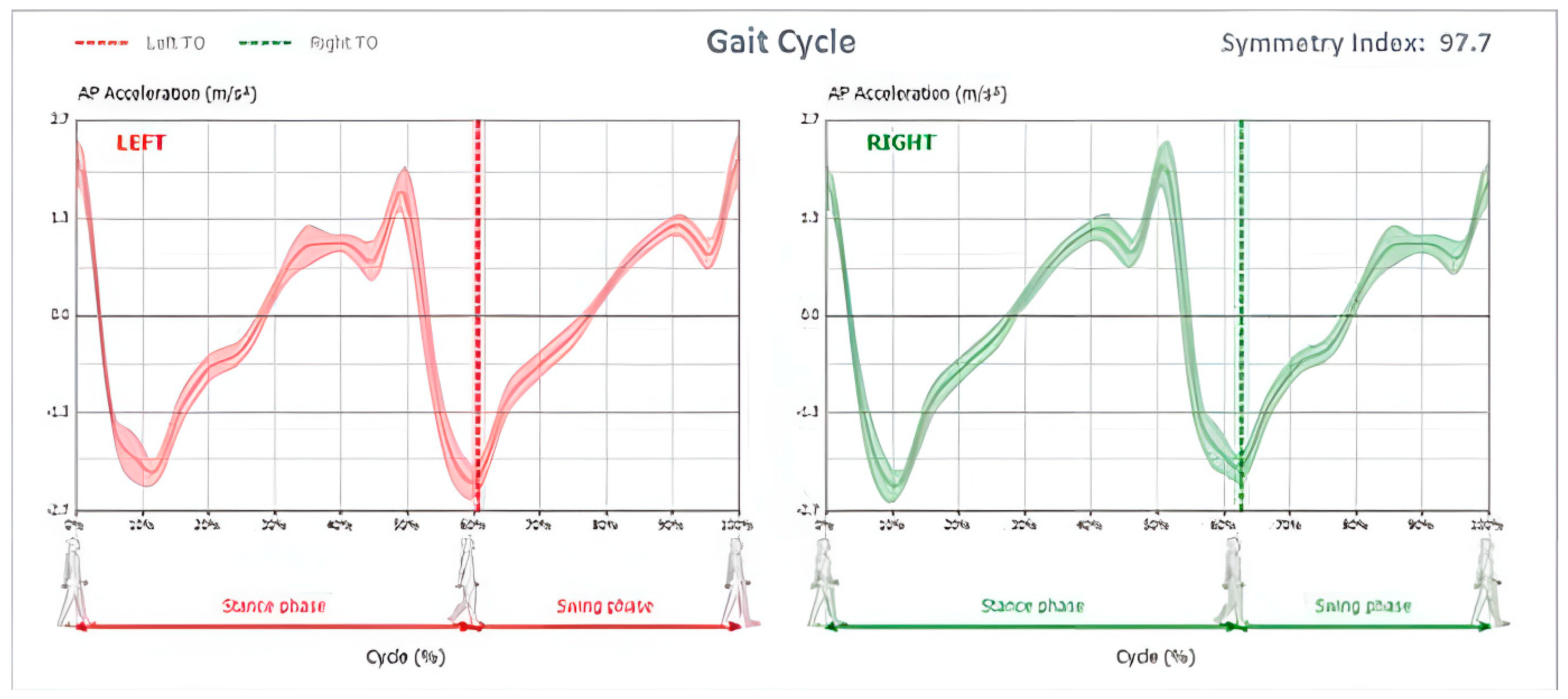
- Gait Cycle
- Different graphs representing left and right gait cycles starting with stance phase, shown via initial contact (0%), and the next initial contact on the same foot (100%), in addition to toe off (represented using a dotted line) to signal the start of the stride phase;
- Symmetry index, which is the percentage of symmetry between the curve of anterior/posterior acceleration during left and right gait cycles, the maximum value of 100 represents ideal symmetry throughout walking, this is displayed in Figure 1.
- 4—
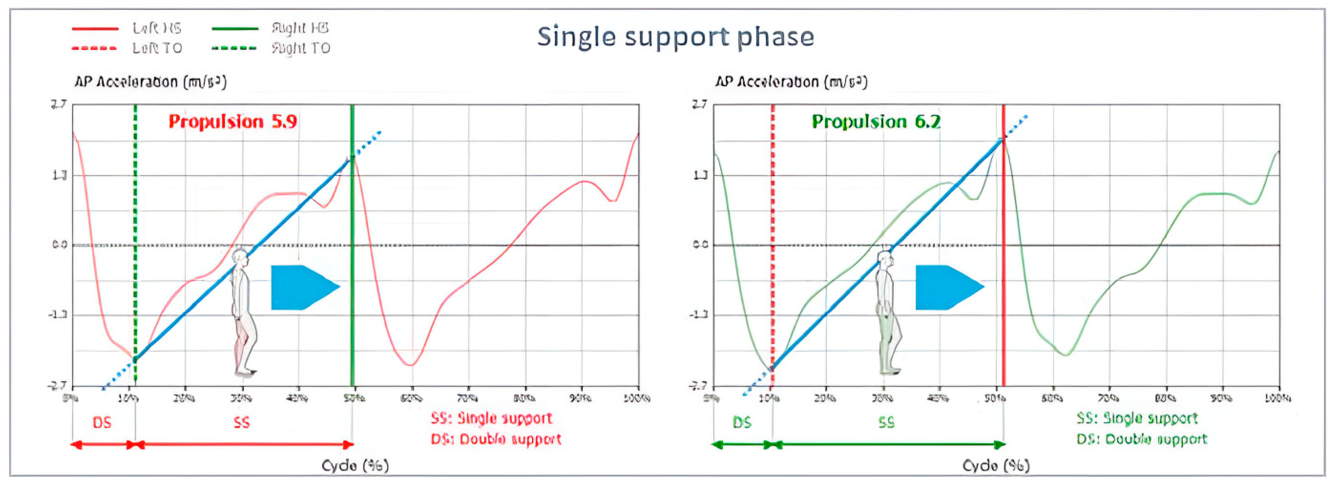
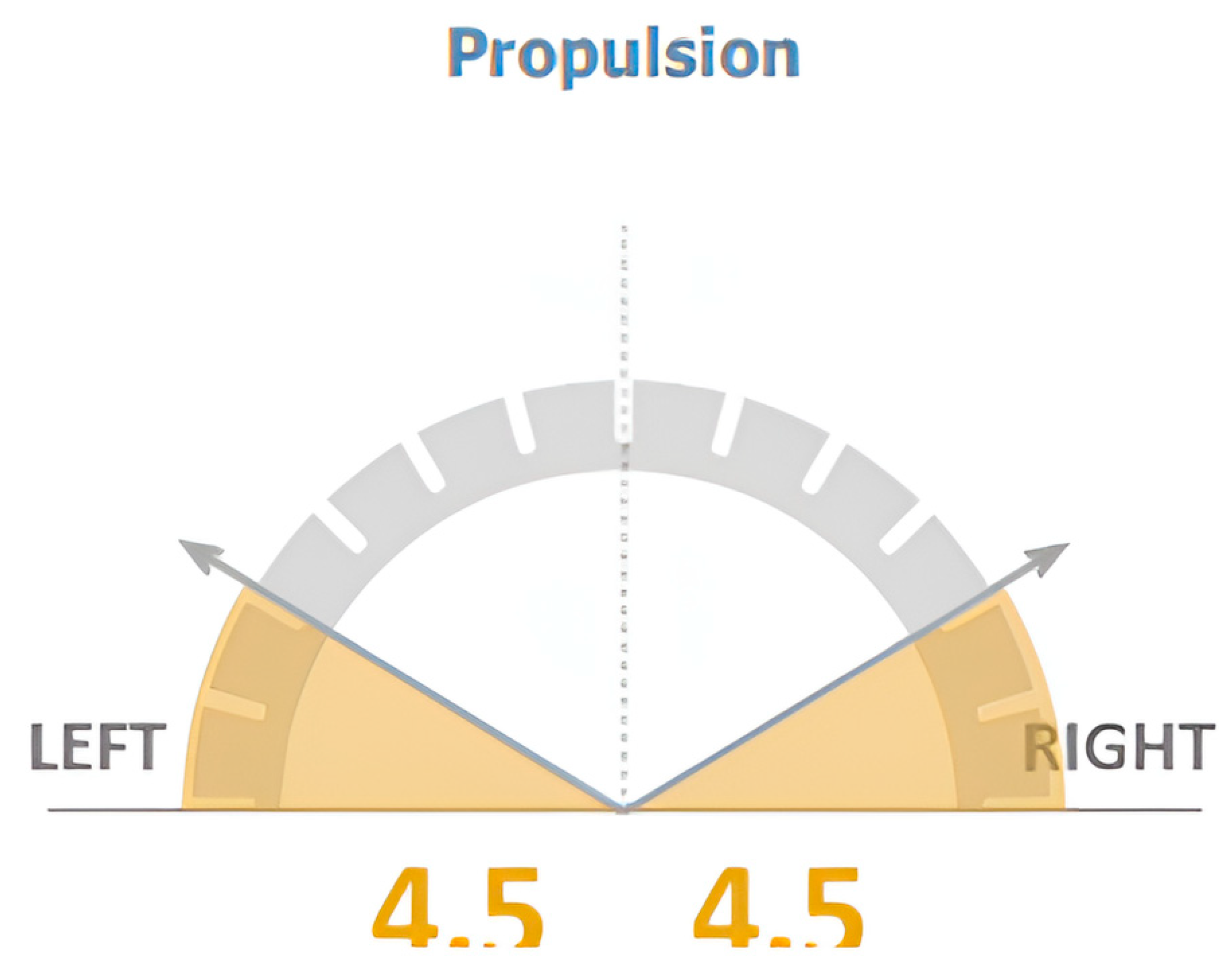
- Single Support Phases
- 5—
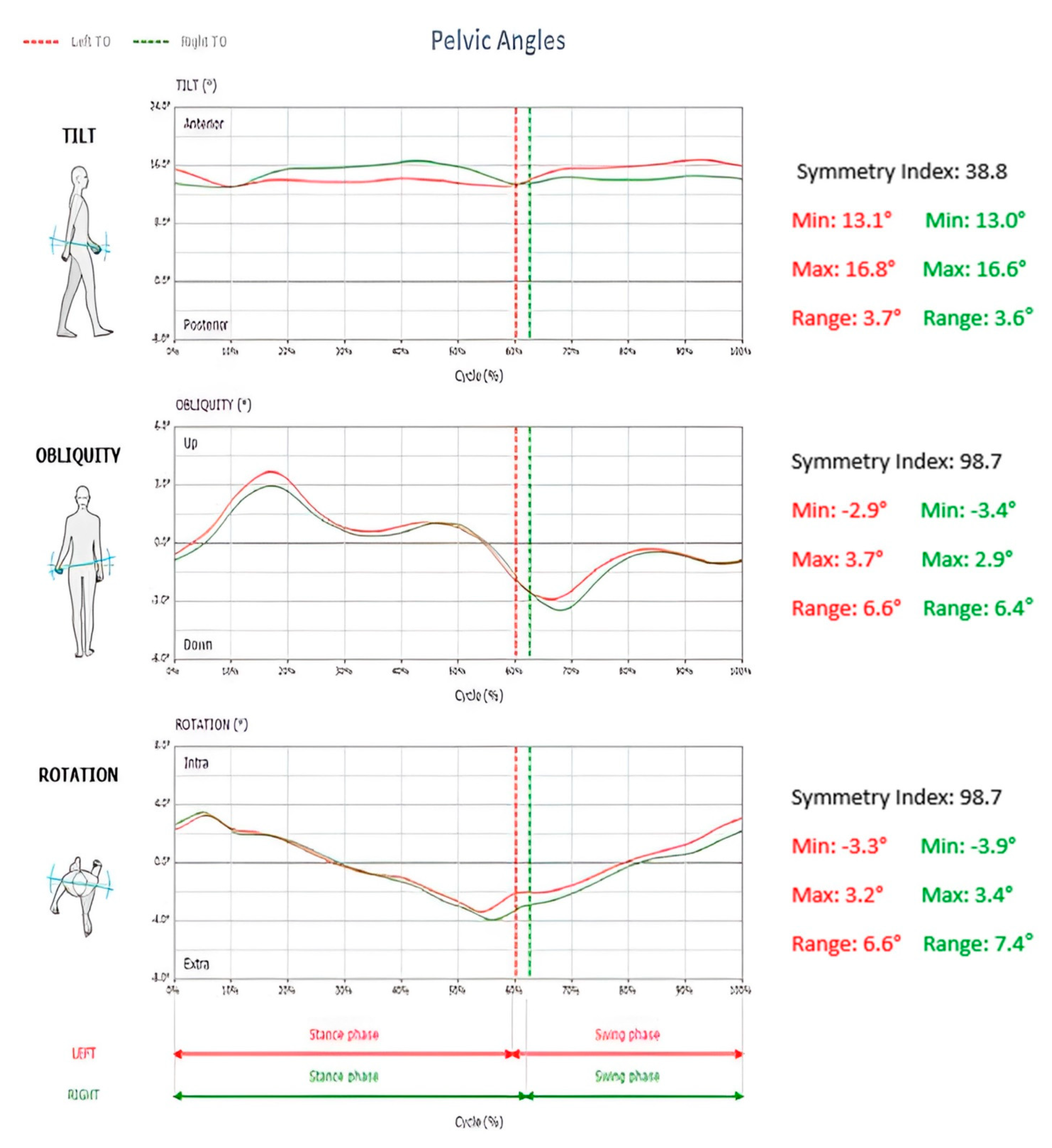
- Pelvic Angles
- Tilt: positive angular values indicate an anterior tilt of the pelvis, while negative angular values indicate a posterior tilt of the pelvis (sagittal plane movement);
- Obliquity: negative angular values indicate DOWN condition, while positive angular values indicate UP position for the considered side (frontal plane movement);
- Rotation: negative angular values indicate pelvis internally rotated, while positive angular values indicate a pelvis externally rotated (transverse plane movement);
- Relative symmetry index and minimum and maximum angles were shown for each of the 3 pelvic parameters mentioned above. All pelvic parameters are seen in Figure 4.
- 1-
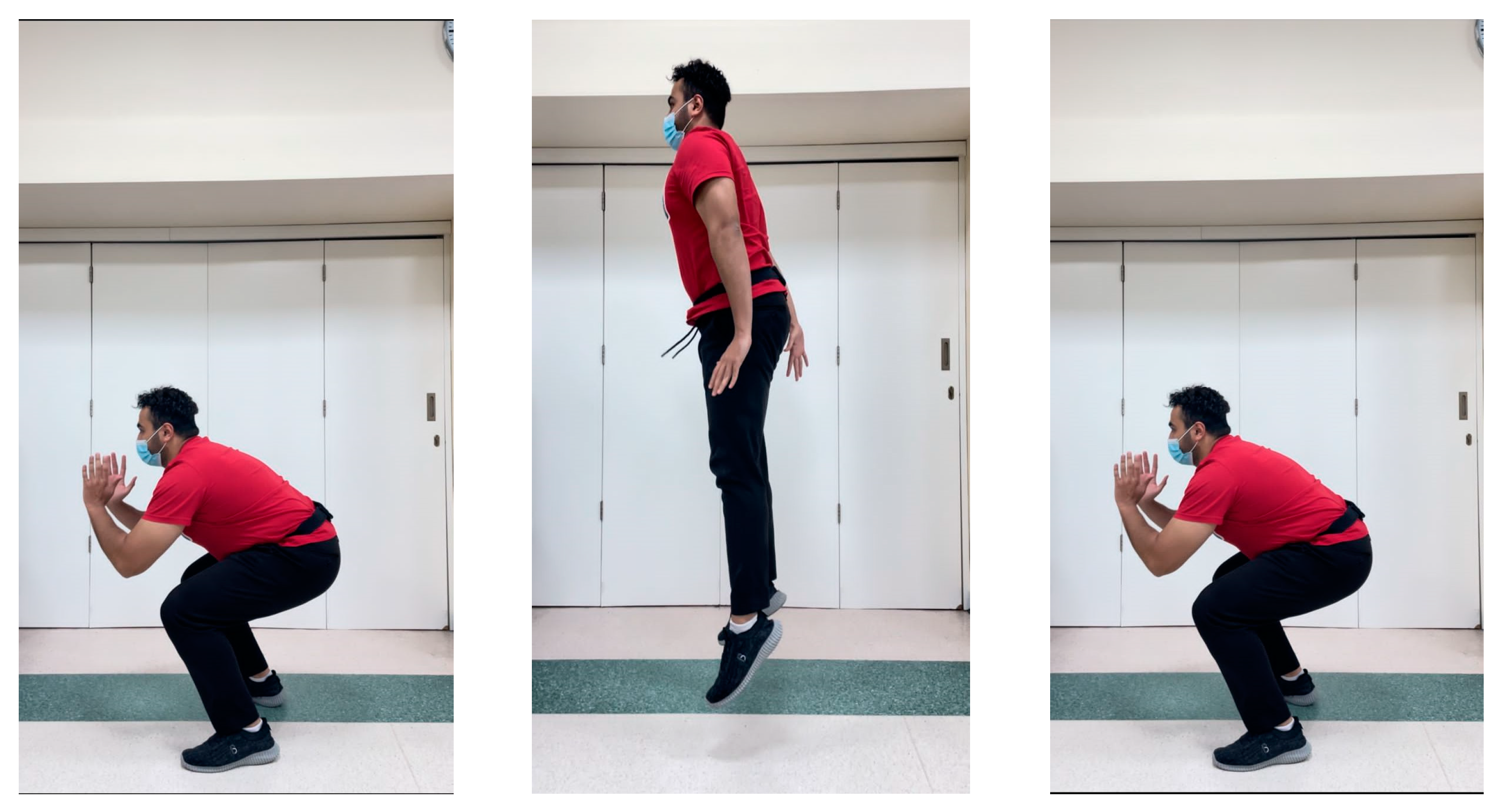
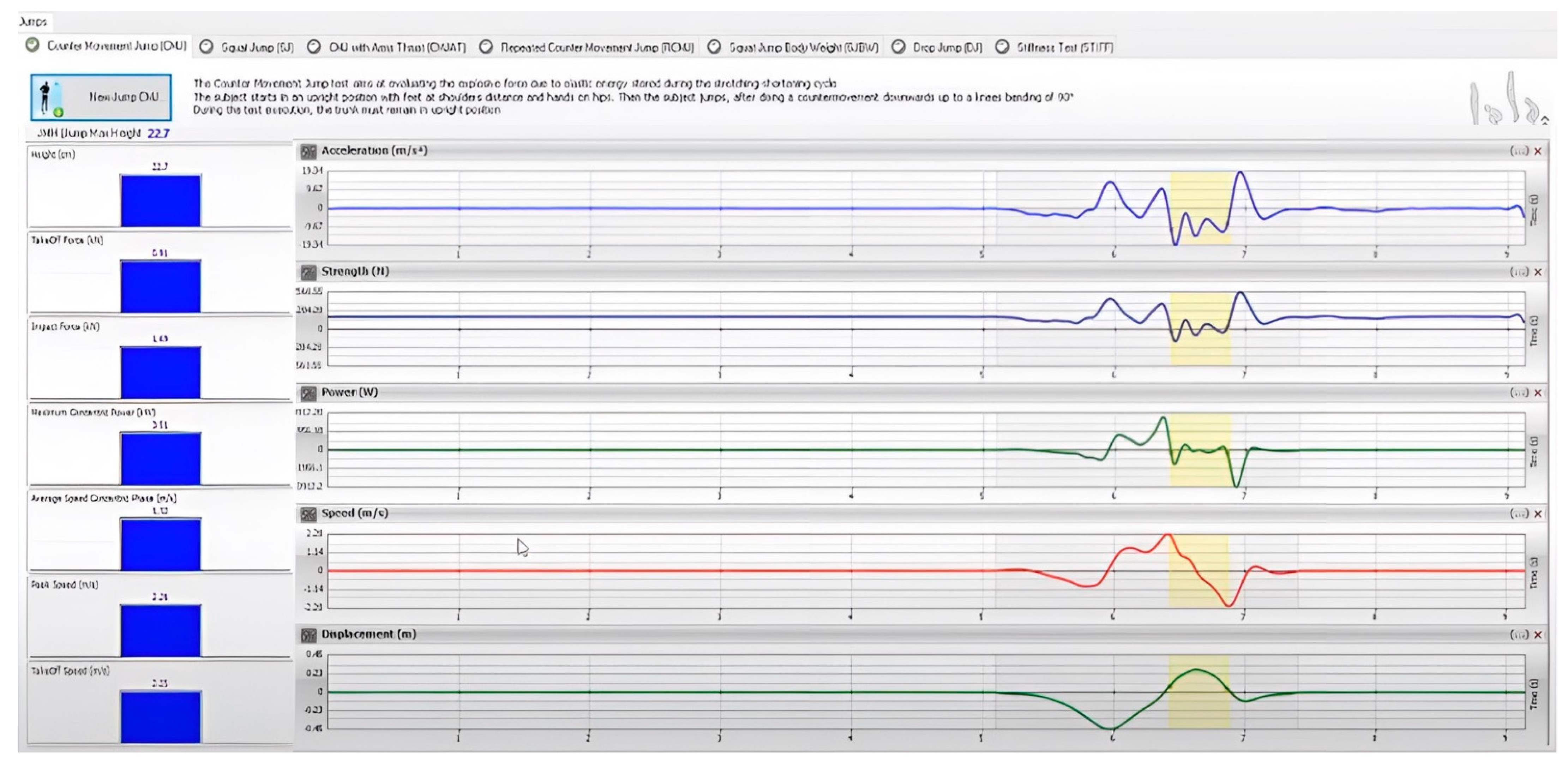
- Countermovement Jump (CMJ)
- The participant begins the test in upright position with their hands on the hips and their feet placed in line with the shoulders. They are then instructed to jump by performing a countermovement towards the downward direction and bending their knees by 90°. During the entire course of the test, the trunk should remain upright with hands near the hips (Figure 5).
- 2-
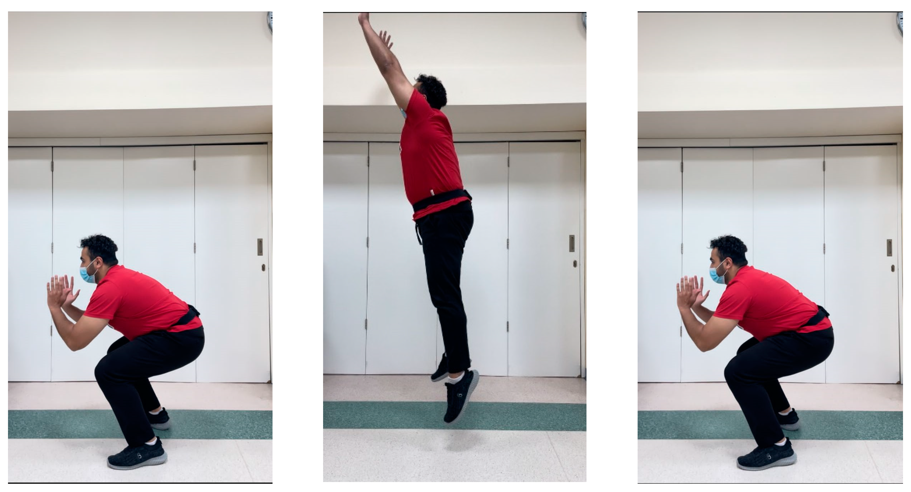
- CMJ with Arms Thrust (CMJAT)
- The participant begins the test in upright position with their hands on the sides and their feet placed in line with the shoulders. They are then instructed to jump by performing a countermovement towards the downward direction and bending their knees by 90°, with the help of using their arms as they extend them upwards. During the entire course of the test, the trunk should remain upright with arms and hands extending upwards in a thrust maneuver (Figure 6).
- Flight Height (cm);
- Take-Off Force (kN);
- Impact Force (kN);
- Take-Off Speed (m/s);
- Peak Speed (m/s);
- Average Speed Concentric Phase (m/s);
- Maximum Concentric Power (kW);
- Average Concentric Power (kW).

2.2. Data Analysis
2.2.1. Sample Size Determination
2.2.2. Statistical Analysis
3. Results
3.1. Participant Demographics and Characteristics
3.2. Correlations between Variables
4. Discussion
4.1. Posture and Athletic Skills
4.2. Posture and Gait Asymmetries
4.3. Study Limitations
5. Conclusions
Author Contributions
Funding
Institutional Review Board Statement
Informed Consent Statement
Data Availability Statement
Conflicts of Interest
References
- Menz, H.B.; Lord, S.R.; Fitzpatrick, R.C. Age-related differences in walking stability. Age Ageing 2003, 32, 137–142. [Google Scholar] [CrossRef] [PubMed]
- Promsri, A.; Cholamjiak, P.; Federolf, P. Walking Stability and Risk of Falls. Bioengineering 2023, 10, 471. [Google Scholar] [CrossRef] [PubMed]
- Biewener, A.A.; Farley, C.T.; Roberts, T.J.; Temaner, M.; Ludlow, L.W.; Weyand, P.G.; Beck, O.N.; Taboga, P.; Grabowski, A.M.; Giovanelli, N.; et al. Muscle mechanical advantage of human walking and running: Implications for energy cost. J. Appl. Physiol. 2004, 97, 2266–2274. [Google Scholar] [CrossRef] [PubMed]
- Kwak, S.T.; Chang, Y.H. Fascicle dynamics of the tibialis anterior muscle reflect whole-body walking economy. Sci. Rep. 2023, 13, 4660. [Google Scholar] [CrossRef] [PubMed]
- Graci, V.; Salsich, G.B. Trunk and lower extremity segment kinematics and their relationship to pain following movement instruction during a single-leg squat in females with dynamic knee valgus and patello-femoral pain. J. Sci. Med. Sport 2015, 18, 343–347. [Google Scholar] [CrossRef]
- Nunes, G.S.; de Moraes, W.S.L.A.; Sampaio, V.d.S.; Seda, N.R.; Mouta, G.d.S.; Dangui, A.J.M.; Petersen, R.d.S.; Nakagawa, T.H. Are Changes in Dynamic Knee Movement Control Related to Changes in Pain or Function in People with Knee Disorders? A Systematic Review and Meta-analysis. J. Orthop. Sports Phys. Ther. 2023, 53, 388–401. [Google Scholar] [CrossRef]
- Myer, G.D.; Ford, K.R.; Hewett, T.E. Tuck Jump Assessment for Reducing Anterior Cruciate Ligament Injury Risk. Athl. Ther. Today 2008, 13, 39–44. [Google Scholar] [CrossRef]
- Kang, J.-H.; Park, R.-Y.; Lee, S.-J.; Kim, J.-Y.; Yoon, S.-R.; Jung, K.-I. The Effect of The Forward Head Posture on Postural Balance in Long Time Computer Based Worker. Ann. Rehabil. Med. 2012, 36, 98–104. [Google Scholar] [CrossRef]
- Lee, J.-H. Effects of forward head posture on static and dynamic balance control. J. Phys. Ther. Sci. 2016, 28, 274–277. [Google Scholar] [CrossRef]
- Ruivo, R.M.; Pezarat-Correia, P.; Carita, A.I. Effects of a resistance and stretching training program on forward head and protracted shoulder posture in adolescents. J Manip. Physiol Ther 2017, 40, 1–10. [Google Scholar] [CrossRef]
- Muyor, J.M.; López-Miñarro, P.A.; Casimiro, A.J. Effect of stretching program in an industrial workplace on hamstring flexibility and sagittal spinal posture of adult women workers: A randomized controlled trial. J. Back. Musculoskelet. Rehabil. 2012, 25, 161–169. [Google Scholar] [CrossRef] [PubMed]
- Lin, G.; Zhao, X.; Wang, W.; Wilkinson, T. The relationship between forward head posture, postural control and gait: A systematic review. Gait Posture 2022, 98, 316–329. [Google Scholar] [CrossRef] [PubMed]
- Ahmadipoor, A.; Khademi-Kalantari, K.; Rezasoltani, A.; Naimi, S.-S.; Akbarzadeh-Baghban, A. Effect of Forward Head Posture on Dynamic Balance Based on the Biodex Balance System. J. Biomed. Phys. Eng. 2022, 12, 543–548. [Google Scholar] [CrossRef] [PubMed]
- Caneiro, J.P.; O’Sullivan, P.; Burnett, A.; Barach, A.; O’Neil, D.; Tveit, O.; Olafsdottir, K. The influence of different sitting postures on head/neck posture and muscle activity. Man Ther 2010, 15, 54–60. [Google Scholar] [CrossRef]
- Shaghayeghfard, B.; Ahmadi, A.; Maroufi, N.; Sarrafzadeh, J. Evaluation of forward head posture in sitting and standing positions. Eur. Spine J. 2016, 25, 3577–3582. [Google Scholar] [CrossRef]
- Moustafa, I.M.; Youssef, A.; Ahbouch, A.; Tamim, M.; Harrison, D.E. Is forward head posture relevant to autonomic nervous system function and cervical sensorimotor control? Cross sectional study. Gait Posture. 2020, 77, 29–35. [Google Scholar] [CrossRef]
- Jain, D. Effects of Forward Head Posture on Postural Balance in Young Adults. Int. J. Adv. Res. 2019, 7, 136–146. [Google Scholar] [CrossRef]
- Ha, S.-Y.; Sung, Y.-H. A temporary forward head posture decreases function of cervical proprioception. J. Exerc. Rehabil. 2020, 16, 168–174. [Google Scholar] [CrossRef]
- Lau, K.T.; Cheung, K.Y.; Chan, K.B.; Chan, M.H.; Lo, K.Y.; Chiu, T.T.W. Relationships between sagittal postures of thoracic and cervical spine, presence of neck pain, neck pain severity and disability. Man. Ther. 2010, 15, 457–462. [Google Scholar] [CrossRef]
- Lee, M.-Y.; Lee, H.-Y.; Yong, M.-S. Characteristics of Cervical Position Sense in Subjects with Forward Head Posture. J. Phys. Ther. Sci. 2014, 26, 1741–1743. [Google Scholar] [CrossRef]
- Harman, K.; Hubley-Kozey, C.L.; Butler, H. Effectiveness of an Exercise Program to Improve Forward Head Posture in Normal Adults: A Randomized, Controlled 10-Week Trial. J. Man. Manip. Ther. 2005, 13, 163–176. [Google Scholar] [CrossRef]
- Alowa, Z.; Elsayed, W. The impact of forward head posture on the electromyographic activity of the spinal muscles. J. Taibah Univ. Med. Sci. 2021, 16, 224–230. [Google Scholar] [CrossRef] [PubMed]
- Panjabi, M.M. The Stabilizing System of the Spine. Part, I.; Function, Dysfunction, Adaptation, and Enhancement. J. Spinal. Disord. 1992, 5, 383–389. [Google Scholar] [CrossRef] [PubMed]
- Cholewicki, J.; Vanvliet Iv, J.J. Relative contribution of trunk muscles to the stability of the lumbar spine during isometric exertions. Clin. Biomech. 2002, 17, 99–105. [Google Scholar] [CrossRef]
- Panjabi, M.; Abumi, K.; Duranceau, J.; Oxland, T.M. Spinal Stability and Intersegmental Muscle Forces. A biomechanical model. Spine 1989, 14, 194–200. [Google Scholar] [CrossRef]
- Boynton, A.M.; Carrier, D.R. The human neck is part of the musculoskeletal core: Cervical muscles help stabilize the pelvis during running and jumping. Integr. Org. Biol. 2022, 4, obac021. [Google Scholar] [CrossRef]
- Panjabi, M.M. The Stabilizing System of the Spine. Part II. Neutral Zone and Instability Hypothesis. J. Spinal. Disord. 1992, 5, 390–397. [Google Scholar] [CrossRef]
- Adams, M.; Dolan, P.; Hutton, W.C. The stages of disc degeneration as revealed by discograms. J. Bone Joint Surg. Br. 1986, 68, 36–41. [Google Scholar] [CrossRef]
- Harrison, D.E.; Oakley, P.A. An Introduction to Chiropractic BioPhysics® (CBP®) Technique: A Full Spine Rehabilitation Approach to Reducing Spine Deformities. In Complementary Therapies; Mary Ann Liebert, Inc.: New Rochelle, NY, USA, 2022. [Google Scholar] [CrossRef]
- Oakley, P.A.; Ehsani, N.N.; Moustafa, I.M.; Harrison, D.E. Restoring cervical lordosis by cervical extension traction methods in the treatment of cervical spine disorders: A systematic review of controlled trials. J. Phys. Ther. Sci. 2021, 33, 784–794. [Google Scholar] [CrossRef]
- Tao, W.; Liu, T.; Zheng, R.; Feng, H. Gait Analysis Using Wearable Sensors. Sensors 2012, 12, 2255–2283. [Google Scholar] [CrossRef]
- Wang, W.; Adamczyk, P.G. Analyzing gait in the real world using wearable movement sensors and frequently repeated movement paths. Sensors 2019, 19, 1925. [Google Scholar] [CrossRef]
- Muro-De-La-Herran, A.; Garcia-Zapirain, B.; Mendez-Zorrilla, A. Gait Analysis Methods: An Overview of Wearable and Non-Wearable Systems, Highlighting Clinical Applications. Sensors 2014, 14, 3362–3394. [Google Scholar] [CrossRef] [PubMed]
- Bisi, M.C.; Stagni, R. Development of gait motor control: What happens after a sudden increase in height during adolescence? Biomed Eng. Online 2016, 15, 47. [Google Scholar] [CrossRef] [PubMed]
- Szucs, K.A.; Brown, E.V.D. Rater reliability and construct validity of a mobile application for posture analysis. J. Phys. Ther. Sci. 2018, 30, 31–36. [Google Scholar] [CrossRef] [PubMed]
- Boland, D.M.; Neufeld, E.V.; Ruddell, J.; Dolezal, B.A.; Cooper, C.B. Inter- and intra-rater agreement of static posture analysis using a mobile application. J. Phys. Ther. Sci. 2016, 28, 3398–3402. [Google Scholar] [CrossRef]
- Volkan-Yazici, M.; Çobanoğlu, G.; Yazici, G. Test-retest reliability and minimal detectable change for measures of wearable gait analysis system (G-walk) in children with cerebral palsy. Turk J. Med. Sci. 2022, 52, 658–666. [Google Scholar] [CrossRef]
- De Ridder, R.; Lebleu, J.; Willems, T.; De Blaiser, C.; Detrembleur, C.; Roosen, P. Concurrent Validity of a Commercial Wireless Trunk Triaxial Accelerometer System for Gait Analysis. J. Sport. Rehabil. 2019, 28. [Google Scholar] [CrossRef]
- Randell, R.K.; Clifford, T.; Drust, B.; Moss, S.L.; Unnithan, V.B.; De Ste Croix, M.B.A.; Datson, N.; Martin, D.; Mayho, H.; Carter, J.M.; et al. Physiological characteristics of female soccer players and health and performance considerations: A narrative review. Sports Med. 2021, 51, 1377–1399. [Google Scholar] [CrossRef]
- Park, G.; Woo, Y. Comparison between a center of mass and a foot pressure sensor system for measuring gait parameters in healthy adults. J. Phys. Ther. Sci. 2015, 27, 3199–3202. [Google Scholar] [CrossRef]
- Yazici, G.; Yazici, M.V.; Çobanoğlu, G.; Küpeli, B.; Özkul, Ç.; Oskay, D.; Güzel, N.A. The reliability of a wearable movement analysis system (G-walk) on gait and jump assessment in healthy adults. J. Exerc. Ther. Rehabil. 2020, 7, 159–167. [Google Scholar]
- Pau, M.; Leban, B.; Collu, G.; Migliaccio, G.M. Effect of light and vigorous physical activity on balance and gait of older adults. Arch. Gerontol. Geriatr. 2014, 59, 568–573. [Google Scholar] [CrossRef] [PubMed]
- Balsalobre-Fernández, C.; Tejero-González, C.M.; del Campo-Vecino, J. Relationships between Training Load, Salivary Cortisol Responses and Performance during Season Training in Middle and Long Distance Runners. PLoS ONE 2014, 9, e106066. [Google Scholar] [CrossRef] [PubMed]
- Balsalobre-Fernandez, C.; Tejero-Gonzalez, C.M.; del Campo-Vecino, J. Hormonal and neuromuscular responses to high-level middle- and long-distance competition. Int. J. Sports Physiol. Perform 2014, 9, 839–844. [Google Scholar] [CrossRef] [PubMed]
- Gathercole, R.J.; Stellingwerff, T.; Sporer, B.C. Effect of acute fatigue and training adaptation on countermovement jump performance in elite snowboard cross athletes. J. Strength Cond. Res. 2015, 29, 37–46. [Google Scholar] [CrossRef] [PubMed]
- Jiménez-Reyes, P.; González-Badillo, J.J. Monitoring training load through the CMJ in sprints and jump events for optimizing performance in athletics. Cult. Cienc. Deporte 2011, 6, 207–217. [Google Scholar] [CrossRef]
- Loturco, I.; D’Angelo, R.A.; Fernandes, V.; Gil, S.; Kobal, R.; Abad, C.C.C.; Kitamura, K.; Nakamura, F.Y. Relationship between Sprint Ability and Loaded/Unloaded Jump Tests in Elite Sprinters. J. Strength. Cond. Res. 2015, 29, 758–764. [Google Scholar] [CrossRef]
- Sugiyono. Statistika untuk Penelitian; Alfabeta: Bandung, Indonesia, 2015. [Google Scholar]
- Khan, A.; Khan, Z.; Bhati, P.; Hussain, M.E. Influence of forward head posture on cervicocephalic kinesthesia and electromyographic activity of neck musculature in asymptomatic individuals. J. Chiropr. Med. 2020, 19, 230–240. [Google Scholar] [CrossRef]
- Mahmoud, N.F.; Hassan, K.A.; Abdelmajeed, S.F.; Moustafa, I.M.; Silva, A.G. The relationship between forward head posture and neck pain: A systematic review and meta-analysis. Curr. Rev. Musculoskelet. Med. 2019, 12, 562–577. [Google Scholar] [CrossRef]
- Pacheco, J.; Raimundo, J.; Santos, F.; Ferreira, M.; Lopes, T.; Ramos, L.; Silva, A.G. Forward head posture is associated with pressure pain threshold and neck pain duration in university students with subclinical neck pain. Somatosens. Mot. Res. 2018, 35, 103–108. [Google Scholar] [CrossRef]
- Naz, A.; Bashir, S.; Noor, R. Prevalence of forward head posture among university students. Rawal Med. J. 2018, 43, 260–262. [Google Scholar]
- Singh, S.; Kaushal, K.; Jasrotia, S. Prevalence of forward head posture and its impact on the activity of daily living among students of Adesh University—A cross-sectional study. Adesh Univ. J. Med. Sci. Res. 2020, 2, 99–102. [Google Scholar] [CrossRef]
- Hodges, P.W.; Smeets, R.J. Interaction between pain, movement, and physical activity: Short-term benefits, long-term consequences, and targets for treatment. Clin. J. Pain 2015, 31, 97–107. [Google Scholar] [CrossRef] [PubMed]
- Danna-Dos-Santos, A.; Degani, A.M.; Latash, M.L. Anticipatory control of head posture. Clin. Neurophysiol. 2007, 118, 1802–1814. [Google Scholar] [CrossRef] [PubMed]
- Massion, J. Movement, posture and equilibrium: Interaction and coordination. Prog. Neurobiol. 1992, 38, 35–56. [Google Scholar] [CrossRef] [PubMed]
- Frank, J.S.; Earl, M. Coordination of Posture and Movement. Phys. Ther. 1990, 70, 855–863. [Google Scholar] [CrossRef]
- Moustafa, I.; Kim, M.; Harrison, D.E. Comparison of sensorimotor integration and skill-related physical fitness components between college athletes with and without forward head posture. J. Sport Rehabil. 2022, 32, 53–62. [Google Scholar] [CrossRef]
- Abbruzzese, G.; Berardelli, A. Sensorimotor integration in movement disorders. Mov. Disord. 2002, 18, 231–240. [Google Scholar] [CrossRef]
- Haavik, H.; Murphy, B. The role of spinal manipulation in addressing disordered sensorimotor integration and altered motor control. J. Electromyogr. Kinesiol. 2012, 22, 768–776. [Google Scholar] [CrossRef]
- Haavik-Taylor, H.; Murphy, B. Cervical spine manipulation alters sensorimotor integration: A somatosensory evoked potential study. Clin. Neurophysiol. 2007, 118, 391–402. [Google Scholar] [CrossRef]
- Pickar, J.G. Neurophysiological effects of spinal manipulation. Spine J. 2002, 2, 357–371. [Google Scholar] [CrossRef]
- Taylor, H.H.; Murphy, B. Altered central integration of dual somatosensory input after cervical spine manipulation. J. Manip. Physiol. Ther. 2010, 33, 178–188. [Google Scholar] [CrossRef] [PubMed]
- Hishinuma, M.; Yamaguchi, T. Axonal projection of descending pathways responsible for eliciting forelimb stepping into the cat cervical spinal cord. Exp. Brain Res. 1990, 82, 597–605. [Google Scholar] [CrossRef] [PubMed]
- Desmurget, M.; Sirigu, A. A parietal-premotor network for movement intention and motor awareness. Trends Cogn. Sci. 2009, 13, 411–419. [Google Scholar] [CrossRef] [PubMed]
- Azevedo, R.d.A.; Cruz, R.; Couto, P.; Silva-Cavalcante, M.D.; Boari, D.; Lima-Silva, A.E.; Millet, G.Y.; Bertuzzi, R.; Drouin, P.J.; Kohoko, Z.I.N.; et al. Spinal and Supraspinal Factors in Human Muscle Fatigue. Physiol. Rev. 2001, 81, 1725–1789. [Google Scholar] [CrossRef]
- Krakauer, J.W.; Ghazanfar, A.A.; Gomez-Marin, A.; MacIver, M.A.; Poeppel, D. Neuroscience needs behavior: Correcting a reductionist bias. Neuron 2017, 93, 480–490. [Google Scholar] [CrossRef]
- Moustafa, I.; Youssef, M.A.S.; Ahbouch, M.A.; Harrison, D.D. Demonstration of Autonomic Nervous Function and Cervical Sensorimotor Control After Cervical Lordosis Rehabilitation: A Randomized Controlled Trial. J. Athl. Train. 2021, 56, 427–436. [Google Scholar] [CrossRef]
- Moustafa, I.M.; Diab, A.A.; Harrison, D.E. The effect of normalizing the sagittal cervical configuration on dizziness, neck pain, and cervicocephalic kinesthetic sensibility: A 1-year randomized controlled study. Eur. J. Phys. Rehabil. Med. 2017, 53, 57–71. [Google Scholar] [CrossRef]
- Al Suwaidi, A.S.; Moustafa, I.M.; Kim, M.; Oakley, P.A.; Harrison, D.E. A Comparison of Two Forward Head Posture Corrective Approaches in Elderly with Chronic Non-Specific Neck Pain: A Randomized Controlled Study. J. Clin. Med. 2023, 12, 542. [Google Scholar] [CrossRef]
- Oakley, P.A.; Moustafa, I.M.; Harrison, D.E. The Influence of Sagittal Plane Spine Alignment on Neurophysiology and Sensorimotor Control Measures: Optimization of Function through Structural Correction. In Therapy Approaches in Neurological Disorders; IntechOpen: London, UK, 2021. [Google Scholar] [CrossRef]
- Kawkabani, G.; Saliby, R.M.; Mekhael, M.; Rachkidi, R.; Massaad, A.; Ghanem, I.; Kharrat, K.; Kreichati, G.; Saad, E.; Lafage, V.; et al. Gait kinematic alterations in subjects with adult spinal deformity and their radiological determinants. Gait Posture 2021, 88, 203–209. [Google Scholar] [CrossRef]
- Mekhael, E.; El Rachkidi, R.; Saliby, R.M.; Nassim, N.; Semaan, K.; Massaad, A.; Karam, M.; Saade, M.; Ayoub, E.; Rteil, A.; et al. Functional assessment using 3D movement analysis can better predict health-related quality of life outcomes in patients with adult spinal deformity: A machine learning approach. Front. Surg. 2023, 10, 1166734. [Google Scholar] [CrossRef]
- Sato, K.; Tominaga, R.; Endo, T.; Miura, T.; Iwabuchi, M.; Ito, T.; Shirado, O. The association of dynamic spinal alignment on gait endurance of patients with adult spinal deformity: A cross-sectional study. Spine Deform. 2023, 11, 463–469. [Google Scholar] [CrossRef]
- Arima, H.; Yamato, Y.; Hasegawa, T.; Togawa, D.; Kobayashi, S.; Yasuda, T.; Banno, T.; Oe, S.; Matsuyama, Y. Discrepancy between Standing Posture and Sagittal Balance During Walking in Adult Spinal Deformity Patients. Spine 2017, 42, E25–E30. [Google Scholar] [CrossRef] [PubMed]
- Semaan, K.; Rachkidi, R.; Saad, E.; Massaad, A.; Kawkabani, G.; Saliby, R.M.; Mekhael, M.; Karam, K.A.; Fakhoury, M.; Jaber, E.; et al. Alterations of gait kinematics depend on the deformity type in the setting of adult spinal deformity. Eur. Spine J. 2022, 31, 3069–3080. [Google Scholar] [CrossRef] [PubMed]
- Khamis, S.; Carmeli, E. Relationship and significance of gait deviations associated with limb length discrepancy: A systematic review. Gait Posture 2017, 57, 115–123. [Google Scholar] [CrossRef] [PubMed]
- Shi, Y.; Pang, H.; Xu, H.; Li, X.; Cao, Y.; Merryweather, A.; Zheng, P.; Xiang, J. Effects of orthotic insole on gait patterns in children with mild leg length discrepancy. Gait Posture 2022, 93, 191–197. [Google Scholar] [CrossRef]
- Khamis, S.; Carmeli, E. The effect of simulated leg length discrepancy on lower limb biomechanics during gait. Gait Posture 2018, 61, 73–80. [Google Scholar] [CrossRef]


| Variable | N = 100 |
|---|---|
| Age (years) | 21.10 ± 1.70 |
| Weight (kg) | 67.77 ± 17.28 |
| Height (cm) | 166.68 ± 8.72 |
| BMI (kg/m2) | 24.27 ± 5.33 |
| Gender (%) | |
| Male | 27 |
| Female | 73 |
| Race/ethnicity (n) | |
| Arab–Non GCC | 51 |
| Arab–GCC | 31 |
| Non-Arab | 18 |
| Head Posture Parameters | N = 100 Median (IQR) |
|---|---|
| CVA (°) | 51.60 (46.60, 55.30) |
| Lateral translation head (cm) | 3.79 (2.68, 5.03) |
| AHT (cm) | 0.69 (0.18, 1.21) |
| Lateral angulation head (°) | 10.32 (6.67,14.3) |
| Gait Spatiotemporal Parameters | N = 100 Median (IQR) |
|---|---|
| Cadence | 109.70 (105.30, 116.30) |
| Speed (m/s) | 1.15 (0.98, 1.24) |
| Symmetry index | 92.20 (80.90, 95.60) |
| % Left stride length (% height) | 76 (66.70, 83.20) |
| % Right stride length (% height) | 75.40 (67.70, 82.60) |
| Left propulsion index | 5.50 (4.20, 7.50) |
| Right propulsion index | 4.80 (3.90, 7.60) |
| Tilt symmetry index | 47.10 (24.30, 77.90) |
| Tilt left range | 4.30 (3.30, 5.40) |
| Tilt right Range | 8.40 (3.70, 13.20) |
| Obliquity symmetry index | 95.80 (84.80, 98.10) |
| Obliquity left range | 7.50 (5.70, 10.40) |
| Obliquity right range | 80.00 (5.80, 10.50) |
| Rotation symmetry index | 96.70 (85.70, 98.10) |
| Rotation left range | 10.30 (6.90, 13.30) |
| Rotation right range | 10.50 (6.90, 13.70) |
| CMJ Parameters | N = 100 Median (IQR) |
|---|---|
| CMJ–flight height (cm) | 17.20 (14.20, 24.30) |
| CMJ–take–off force (kN) | 1.11 (0.92, 1.40) |
| CMJ–impact force (kN) | 1.35 (1.05, 1.69) |
| Take–off speed (m/s) | 2.12 (1.85, 2.48) |
| CMJ–peak speed (m/s) | 2.24 (1.99, 2.58) |
| CMJ–average speed concentric phase (m/s) | 1.23 (1.08, 1.41) |
| CMJ–maximum concentric power (kW) | 2.19 (1.67, 2.94) |
| CMJ–average concentric power (kW) | 1.02 (0.85, 1.41) |
| CMJAT Parameters | N = 100 Median (IQR) |
|---|---|
| CMJAT–flight height (cm) | 19.60 (15.90, 27.70) |
| CMJAT–take–off force (kN) | 1.15 (0.95, 1.55) |
| CMJAT–impact force (kN) | 1.45 (1.11, 1.95) |
| CMJAT–take–off speed (m/s) | 2.15 (1.94, 2.45) |
| CMJAT–peak speed (m/s) | 2.27 (2.09, 2.53) |
| CMJAT–average speed concentric phase (m/s) | 1.23 (1.12, 1.39) |
| CMJAT–maximum concentric power (kW) | 2.11 (1.76, 3.34) |
| CMJAT–average concentric power (kW) | 1.08 (0.89, 1.53) |
| Gait Parameters | CVA | Lateral Translation Head | AHT | Lateral Angulation Head | ||||
|---|---|---|---|---|---|---|---|---|
| r | p | r | p | r | p | r | p | |
| Cadence | 0.51 * | <0.001 | −0.74 * | <0.001 | −0.51 * | <0.001 | −0.27 * | 0.006 |
| Speed (m/s) | 0.52 * | <0.001 | −0.46 * | <0.001 | −0.44 * | <0.001 | −0.37 * | <0.001 |
| Symmetry index | 0.54 * | <0.001 | −0.70 * | <0.001 | 0.02 | 0.854 | −0.31 * | 0.003 |
| %Left stride length (% height) | 0.43 * | <0.001 | −0.42 * | <0.001 | −0.48 * | <0.001 | −0.31 * | 0.003 |
| %Right stride length (% height) | 0.51 * | <0.001 | −0.51 * | <0.001 | −0.46 * | <0.001 | −0.32 * | 0.01 |
| Left propulsion index | 0.54 * | <0.001 | −0.36 * | 0.001 | −0.47 * | <0.001 | −0.36 * | <0.001 |
| Right propulsion index | 0.55 * | <0.001 | −0.29 * | 0.015 | −0.39 * | 0.001 | −0.39 * | <0.001 |
| Tilt symmetry index | 0.57 * | <0.001 | −0.51 * | <0.001 | −0.62 * | <0.001 | −0.45 * | <0.001 |
| Tilt left range | 0.58 * | <0.001 | −0.21 * | 0.004 | −0.81 * | <0.001 | −0.37 * | <0.001 |
| Tilt right range | 0.54 * | <0.001 | −0.23 * | 0.004 | −0.47 * | <0.001 | −0.26 * | 0.011 |
| Obliquity symmetry index | 0.56 * | <0.001 | −0.06 * | <0.001 | −0.75 * | <0.001 | −0.40 * | <0.001 |
| Obliquity Left Range | 0.45 * | <0.001 | −0.49 * | <0.001 | −0.65 * | <0.001 | −0.26 * | 0.011 |
| Obliquity right Range | 0.54 * | <0.001 | −0.25 * | 0.005 | −0.64 * | <0.001 | −0.25 * | 0.011 |
| Rotation symmetry Index | 0.52 * | <0.001 | −0.69 * | <0.001 | −0.27 * | 0.010 | −0.43 * | <0.001 |
| Rotation left range | 0.46 * | <0.001 | −0.33 * | 0.001 | −0.42 * | <0.001 | −0.10 | 0.3 |
| Rotation right range | 0.46 * | <0.001 | −0.24 * | 0.005 | −0.41 * | <0.001 | −0.26 * | 0.011 |
| Physical Performance Skills | CVA | Lateral Translation Head | AHT | Lateral Angulation Head | ||||
|---|---|---|---|---|---|---|---|---|
| r | p | r | p | r | p | r | p | |
| CMJ–flight height (cm) | 0.41 * | <0.001 | −0.39 * | <0.001 | −0.44 * | <0.001 | −0.37 * | <0.001 |
| CMJ–take-off force (kN) | 0.39 * | <0.001 | −0.40 * | <0.001 | −0.65 * | <0.001 | −0.38 * | <0.001 |
| CMJ–impact force (kN) | 0.24 | 0.052 | −0.48 * | <0.001 | −0.55 * | <0.001 | −0.39 * | <0.001 |
| Take-off speed (m/s) | 0.39 * | <0.001 | −0.43 * | <0.001 | −0.49 * | <0.001 | −0.38 * | <0.001 |
| CMJ–peak speed (m/s) | 0.38 * | <0.001 | −0.42 * | <0.001 | −0.49 * | <0.001 | −0.40 * | <0.001 |
| CMJ–average speed concentric phase (m/s) | 0.41 * | <0.001 | −0.42 * | <0.001 | −0.57 * | <0.001 | −0.40 * | <0.001 |
| CMJ–maximum Concentric Power (kW) | 0.27 * | 0.03 | −0.31 * | 0.003 | −0.41 * | <0.001 | −0.40 * | <0.001 |
| CMJ–average concentric power (kW) | 0.52 * | <0.001 | −0.54 * | <0.001 | −0.48 * | <0.001 | −0.39 * | <0.001 |
| CMJAT | CVA | Lateral Translation Head | AHT | Lateral Angulation Head | ||||
|---|---|---|---|---|---|---|---|---|
| r | p | r | p | r | p | r | p | |
| CMJAT–flight height (cm) | 0.55 * | <0.001 | −0.39 * | <0.001 | −0.70 * | <0.001 | −0.39 * | <0.001 |
| CMJAT–take-off force (kN) | 0.43 * | <0.001 | −0.40 * | <0.001 | −0.65 * | <0.001 | −0.38 * | <0.001 |
| CMJAT–impact force (kN) | 0.39 * | <0.001 | −0.48 * | <0.001 | −0.55 * | <0.001 | −0.40 * | <0.001 |
| CMJAT–take-off Speed (m/s) | 0.44 * | <0.001 | −0.43 * | <0.001 | −0.50 * | <0.001 | −0.40 * | <0.001 |
| CMJAT–peak speed (m/s) | 0.48 * | <0.001 | −0.42 * | <0.001 | −0.55 * | <0.001 | −0.39 * | <0.001 |
| CMJAT–average Speed concentric phase (m/s) | 0.39 * | <0.001 | −0.42 * | <0.001 | −0.60 * | <0.001 | −0.40 * | <0.001 |
| CMJAT–maximum concentric power (kW) | 0.27 * | 0.03 | −0.31 * | 0.003 | −0.48 * | <0.001 | −0.31 * | 0.003 |
| CMJAT–average concentric power (kW) | 0.52 * | <0.001 | −0.54 * | <0.001 | −0.60 * | <0.001 | −0.31 * | 0.003 |
Disclaimer/Publisher’s Note: The statements, opinions and data contained in all publications are solely those of the individual author(s) and contributor(s) and not of MDPI and/or the editor(s). MDPI and/or the editor(s) disclaim responsibility for any injury to people or property resulting from any ideas, methods, instructions or products referred to in the content. |
© 2023 by the authors. Licensee MDPI, Basel, Switzerland. This article is an open access article distributed under the terms and conditions of the Creative Commons Attribution (CC BY) license (https://creativecommons.org/licenses/by/4.0/).
Share and Cite
Saad, N.; Moustafa, I.M.; Ahbouch, A.; Alsaafin, N.M.; Oakley, P.A.; Harrison, D.E. Are Rotations and Translations of Head Posture Related to Gait and Jump Parameters? J. Clin. Med. 2023, 12, 6211. https://doi.org/10.3390/jcm12196211
Saad N, Moustafa IM, Ahbouch A, Alsaafin NM, Oakley PA, Harrison DE. Are Rotations and Translations of Head Posture Related to Gait and Jump Parameters? Journal of Clinical Medicine. 2023; 12(19):6211. https://doi.org/10.3390/jcm12196211
Chicago/Turabian StyleSaad, Nabil, Ibrahim M. Moustafa, Amal Ahbouch, Nour Mustafa Alsaafin, Paul A. Oakley, and Deed E. Harrison. 2023. "Are Rotations and Translations of Head Posture Related to Gait and Jump Parameters?" Journal of Clinical Medicine 12, no. 19: 6211. https://doi.org/10.3390/jcm12196211
APA StyleSaad, N., Moustafa, I. M., Ahbouch, A., Alsaafin, N. M., Oakley, P. A., & Harrison, D. E. (2023). Are Rotations and Translations of Head Posture Related to Gait and Jump Parameters? Journal of Clinical Medicine, 12(19), 6211. https://doi.org/10.3390/jcm12196211