Article

Calcium, Phosphate, and Vitamin D Status in Patients with Sarcoidosis—Associations with Disease Activity and Symptoms

by
Łukasz Gwadera
*,
Adam J. Białas
,
Anna Kumor-Kisielewska
,
Joanna Miłkowska-Dymanowska
,
Sebastian Majewski
and
Wojciech J. Piotrowski
Department of Pneumology, Medical University of Lodz, 90-153 Lodz, Poland
*
Author to whom correspondence should be addressed.
J. Clin. Med. 2023, 12(14), 4745; https://doi.org/10.3390/jcm12144745
Submission received: 21 May 2023 / Revised: 13 July 2023 / Accepted: 15 July 2023 / Published: 18 July 2023
(This article belongs to the Special Issue Biomarkers and Diagnostics in Respiratory Diseases)

Abstract

:
Sarcoidosis is a systemic granulomatous disease with a variety of presentations. One of the known symptoms are altered vitamin D metabolism and hypercalcemia. In our study, we aimed to assess associations between disease activity, inflammatory parameters, and vitamin D and calcium status. The secondary aim was to find any dependencies between calcium and vitamin D metabolism and fatigue and quality of life in patients with sarcoidosis. We enrolled 58 patients with sarcoidosis (47 classified as active disease, 11 classified as non-active) and compared them with 25 healthy volunteers. Calcium concentration was significantly higher in the study group than in healthy controls. It correlated with some inflammatory markers but not with vitamin D status. Not calcium nor vitamin D, but phosphate concentration correlated with life quality was assessed with the use of the Sarcoidosis Health Questionnaire. An association between phosphate concentration and fatigue was also noted, but it did not reach statistical significance. Calcium concentration was higher in patients with sarcoidosis, but it was not an indicator of the disease activity, while phosphate concentration was significantly lower in patients with active sarcoidosis.

1. Introduction

Sarcoidosis is a granulomatous disease that can potentially affect every organ; thus, the presentation and natural history may vary strongly among the patients. Its etiology is yet to be fully explained and only a part of pathophysiological mechanisms is already known. In most of the cases, sarcoidosis is a self-limiting condition; nonetheless, it can significantly affect the quality of life. In sarcoidosis patients, alterations in vitamin D3 metabolism are frequently present. Activated macrophages, due to the increased activity of 1-alpha hydroxylase, transform 25(OH)D3 into 1,25(OH)2D3 (the more active form) without strict control and regardless of calcium concentration. This finally may lead to hypercalcemia as well as hypercalciuria [1,2,3]. One of the common complaints, even in cases of the disease remission, is chronic fatigue—not life-threatening, but a debilitating syndrome. The potential influence of vitamin D status on chronic fatigue seems probable as vitamin D has its role, for example, in the course of fibromyalgia—a condition sharing many similarities with chronic fatigue [4].
Our study aimed to find associations between the activity of sarcoidosis, evaluated with the use of clinical, biochemical, and lung function parameters, and vitamin D3 and calcium (Ca) metabolism. The secondary objective was to assess if vitamin D3 status would influence fatigue, as well as the quality of life of patients with sarcoidosis.

2. Materials and Methods

2.1. Participants

Participants were recruited from the inpatient pulmonary ward and outpatient clinic of the Barlicki Memorial University Teaching Hospital. The recruitment process took place between November 2016 and March 2020. A major criterion of inclusion was a diagnosis of sarcoidosis made in the past or during the present hospitalization, according to current guidelines [5,6]. Except for cases of Löfgren syndrome, histopathological confirmation of sarcoidosis was preferred; however, in some cases, clinical picture accompanied by typical findings in the radiological examination after the exclusion of other possible diagnoses were sufficient. The activity of the disease, as well as the treatment, were not discriminated at the recruitment stage. Only sarcoidosis of the heart with clinical signs of heart failure and neurosarcoidosis were excluded due to their potential to influence directly cognitive functions, effort tolerance, and fatigue. Other forms of extrapulmonary sarcoidosis did not disqualify patients. As exclusion criteria, we established comorbidities presenting with fatigue, exercise intolerance, cognitive function impairment (e.g., depression, dementia, heart failure more severe than NYHA II and renal failure), autoimmunological diseases, neoplasm in medical history, recent stressful life situations, vitamin D and Ca supplements intake. Patients were divided into two subgroups depending on the disease’s clinical activity. As clinically active sarcoidosis, we recognized a case meeting at least one of following criteria: Löfgren syndrome present at the time of enrollment or in the last 3 months, symptomatic pulmonary or extrapulmonary sarcoidosis, hypercalcemia or hypercalciuria (without other probable cause), radiological progression, worsening of lung function tests results or increased lymphocyte percentage in bronchoalveolar lavage fluid (BALf). The clinically inactive disease was diagnosed when there was radiological and clinical remission or, in the case of radiological stabilization, in a patient without any signs and symptoms of pulmonary and extrapulmonary sarcoidosis.
The control group was recruited from age- and sex-matched healthy volunteers.

2.2. Data Collection

Data concerning demography, comorbidities, presentation of sarcoidosis, radiological stage, basic biochemical blood parameters, and lung function tests (spirometry, TLCO) routinely performed during the diagnostic process or follow-up visit were collected in the study group. Additional blood samples had been taken and after preparation—they had been stored at −80 Celsius. They were then used for measurements of interferon-gamma (INF-gamma), soluble interleukin 2 receptors (sIL-2R), neopterin, angiotensin-converting enzyme (ACE), 25(OH)D3, 1,25(OH)2D3 with use of the ELISA and high sensitivity CRP (hsCRP) measurement with turbidimetric methods. The kits used to perform measurements are listed in the Supplementary Materials. Additionally, fatigue and life quality were assessed with the use of utility-proofed questionnaires, namely the Fatigue Assessment Scale (FAS) (https://www.wasog.org/dynamic/media/78/documents/Questionairres/679743_FAS_Polish.pdf, accessed on 1 December 2016) and Sarcoidosis Health Questionnaire (SHQ) [7,8,9].
In the control group, basic demographical data and medical history were noted and blood samples according to the same protocol as in the study group were taken and stored.

2.3. Statistical Analysis

Continuous data were presented as mean with standard deviation (SD) or quartiles and interquartile range (IQR), from LQ (25%) to UQ (75%), depending on the data normality. Variables were compared using the unpaired Student’s t-test, Welch t-test, or the Wilcoxon rank sum test with continuity correction, depending on data normality and homogeneity of variance. Differences among more than two groups were analyzed using the Kruskal–Wallis rank sum test or one-way ANOVA, with correction for multiple comparisons performed using the Bonferroni method. The correlation analysis was performed using Spearman’s rank correlation rho.
Categorical data were presented as absolute values and percentages. Such data were compared using Pearson’s Chi-square test (with Yates correction if appropriate) or Fisher’s exact test concordantly with test assumptions.
A comparison in biochemical parameters and FAS between the control and the study group and between the active and the non-active sarcoidosis group was made. In the subgroups of sarcoidosis patients (active vs. non-active), we also compared lung function parameters and quality of life.
For a more comprehensive analysis, a logistic regression model was generated. The sarcoidosis activity was used as an outcome and the clinical features (demographics, clinical characteristics of the disease, pulmonary function test results, blood count parameters, immunological markers, biochemical tests results, BALf, FAS, SHQ results and treatment) as predictors. First, the multivariate model that included all selected predictors was estimated, and then forward and backward stepwise selection approaches were used to restrict the model. The lowest Akaike information criterion (AIC) value was chosen as the determinant of the best final model. Receiver operating characteristics (ROC) and area under the curve (AUC) analysis were performed to determine the predictive power of the model.
Analysis was performed using the R software ver. 4.3.1 (R Core Team 2018). R: A language and environment for statistical computing. R Foundation for Statistical Computing, Vienna, Austria).

3. Results

3.1. Participant’s Characteristic

Of 103 patients enrolled, 58 were finally qualified for further analysis. Additional exclusions were made as patients treated with steroids and other immunosuppressive medications were finally disqualified. Furthermore, incomplete material banking resulted in a decrease in the final number of the study group participants. The control group consisted of 25 healthy volunteers. There were no significant between-group differences in median age (p = 0.06), sex (p = 0.08), or ethnicity—all participants were Caucasians. Details are shown in Table 1.

3.2. Control vs. Study Group

When comparing the control with the study group, statistically significant differences could be found in the concentration of the ACE median, 799.85 vs. 1588.02 ng/mL (p = 0.0005); sIL-2R, 3.01 vs. 8.70 pg/mL (p = 0.0002); INF-gamma, 9.40 vs. 11.93 pg/mL (p = 0.005); and hsCRP, 0.34 vs. 2.76 mg/L (p = 0.01). The relevant difference in Ca concentration was also noted with higher values in the study group: median, 2.14 vs. 2.42 mmol/L (p < 0.0001). The level of both forms of vitamin D3—25(OH)D3 and 1,25(OH)2D3—did not vary between healthy controls and sarcoidosis patients (p = 0.36 and 0.53, respectively). We also compared a 1,25(OH)2D3 to 25(OH)D3 ratio, but no between-group difference was noted. Similarly, parathormone (PTH) and phosphate (P) concentrations were not significantly different. The fatigue score measured with FAS was higher in the study than in the control group (only 22 FAS questionnaires from the study group were available). Details are shown in Table 2.

3.3. Active vs. Non-Active Group

A comparison between patients with clinically active and non-active sarcoidosis was also done. Age was not significantly different but mean body mass was higher in the active group, 95.5 vs. 85.5 kg (p = 0.01106). The groups did not differ according to reported comorbidities: congestive heart failure (p = 1.0), arterial hypertension (p = 0.052), nephrolithiasis (p = 0.32), and diabetes mellitus (p = 0.35), whereas we observed differences according to history of extrapulmonary manifestations (p = 0.04), including cardiac involvement (p = 0.047). Occurrence of Löfgren syndrome was comparable in patients with active and non-active sarcoidosis (p = 0.73). Both subgroups did not vary in lung function parameters (FEV1, FVC, TLCO, RV and TLC; RV and TLC were assessed during TLCO measurement). Significantly higher concentrations of CRP (but not hsCRP), 3.0 vs. 6.3 mg/L (p = 0.03327); INF-gamma, 9.94 vs. 12.74 pg/mL (p = 0.01606); neopterin, 5.20 vs. 8.82 nmol/L (p = 0.02139); and sIL-2R, 3.52 vs. 9.44 pg/mL (p = 0.0002342) were observed in patients with active sarcoidosis. The ACE concentration did not depend on the activity of sarcoidosis. Concentrations of Ca, both forms of vitamin D3, as well as its ratio were comparable in both groups. The PTH concentration tended to be higher in the non-active group, but it did not reach statistical significance, 34.18 vs. 27.35 pg/mL (p = 0.07414). What was interesting was that it was not Ca, but P which varied significantly in patients with active and non-active sarcoidosis—0.75 vs. 0.88 (p = 0.03). In addition, statistically significant findings were the differences in BALf lymphocytes percentage, 10.62 vs. 41.1% (p = 0.0004378); peripheral blood monocyte count, 0.55 vs. 0.7 103/μL (p < 0.007037); red blood cell count, 4.62 vs. 5.10 × 10⁶/μL (p = 0.002434); and creatinine level, 0.78 vs. 0.89 mg/dL (p = 0.03551), respectively, in the non-active and the active group. No relevant differences could be found in the severity of fatigue and SHQ score. However, only 22 (7 in the non-active and 15 in the active group) questionnaires were available for analysis. The results are summed up in Table 3.
The Ca concentration did not correlate with the vitamin D3 level. Weak correlations were noted between the Ca concentration and ACE, r = 0.39 (p = 001); sIL2-R, r = 0.36 (p = 0.003); hsCRP, r = 0.32 (p = 0.009), but not with the INF-gamma (p = 0.096) and neopterin (p = 0.23) concentrations. No associations could be found between the levels of Ca, vitamin D3, and quality of life and fatigue. A very interesting finding was the correlation between the P concentration and emotional—r = 0.554 (p = 0.00604)—and daily functioning—r = 0.469 (p = 0.0239)—aspects of life quality measured with SHQ. Some associations between the P concentration and FAS and other domains of SHQ were noted, but they did not reach statistical significance. The P concentration correlated also with the BALf lymphocyte percentage r = −0.45 (p = 0.008), and neopterin r = −0.33 (p = 0.006). Data shown in Table 4.

3.4. Control vs. Non-Active Group

When comparing the non-active and the control group, no significant differences could be found in the concentration of inflammatory biomarkers. What is interesting, the Ca concentration was significantly higher in the non-active than in the control group—2.41 vs. 2.14 mmol/L (p < 0.00001)—despite the lack of significant differences in concentrations of vitamin D, PTH, and P. A higher fatigue score in the non-active group was another relevant finding (32.29 vs. 21.18, p = 0.006). Details are presented in Table 5.

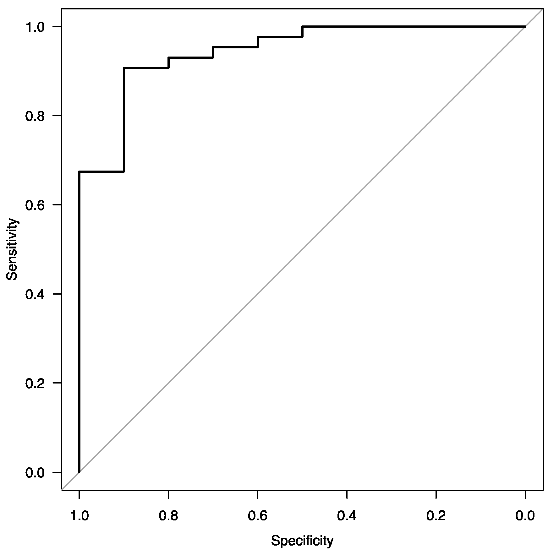
3.5. Logistic Regression and Sarcoidosis Activity

In the multivariate logistic regression analysis, the four analyzed parameters retained their significance. Namely, the final model (Table 6) consisted of RBC (p = 0.02), P (p = 0.05), extrapulmonary manifestation (p = 0.04), and monocytes (p = 0.04).
A ROC analysis for the model yielded an AUC of 0.94 (95% CI: 0.87–1.0). The ROC curve is shown in Figure 1.

4. Discussion

We aimed to find associations between sarcoidosis activity, its clinical presentation, and calcium and vitamin D3 status. The definition of sarcoidosis activity is still a matter of discussion, as there are no simple criteria for assessment. To make the evaluation more precise, we used not only clinical criteria and basic biochemical parameters, but also we measured ACE, sIL-2R, INF-gamma, hsCRP, and neopterin. ACE is one of the first biomarkers used in diagnosing sarcoidosis. Over time, its sensitivity and specificity have been questioned. Some studies find associations between ACE concentration and granuloma burden and radiological presentation of sarcoidosis. However, Popevic et al., in their study, questioned the value of ACE as a marker of activity when compared with the PET-CT assessment [10]. It was also shown that the serum ACE level does not correlate with BALf macrophages activation [11]. Moreover, other granulomatous diseases, genetic predispositions, and drugs can influence ACE concentrations [12]. Our results seem to be consistent with those findings, as there was a significant difference between the control and the study group. Differences between the active and the non-active, as well as the non-active and the control group, were not statistically significant. It seems that ACE may not be an accurate indicator of sarcoidosis activity. Other markers, such as CRP, INF-gamma, neopterin, and especially sIL-2R (with the most significant between-groups difference), seem to reflect disease activity as their concentrations were higher in the active than in the non-active group, and no differences could be found between the non-active and the control group. In the context of sIL-2R, our observation was similar to a study by Lawrence et al., which showed the dependence between sarcoidosis activity, treatment, and sIL-2R concentration [13]. sIL-2R is considered to be useful in diagnosing granulomatous diseases and it is highly specific for sarcoidosis-related uveitis [14,15]. Some data suggest that it can have a prognostic value [16]. It appears, however, that in the case of differential diagnosis in patients with low activity of the disease, usefulness of sIL-2R is questionable. An association between sarcoidosis activity and response to immunosuppressive treatment and INF-gamma inducible chemokines was found by Su et al. [17]. Additionally, Prior and Haslam showed elevated INF-gamma concentration in patients with active sarcoidosis, and its decrease after treatment with corticosteroids [18]. Our data were consistent with those findings, as INF-gamma concentration was elevated in patients with active sarcoidosis, while in non-active sarcoidosis it was comparable with healthy controls. Interestingly CRP, not hsCRP, varied significantly between the non-active and the active sarcoidosis group. As monocytes are key cells in forming granulomas, a higher monocyte count in peripheral blood in patients with active sarcoidosis seems to be consistent with disease pathobiology. A surprising finding was higher RBC in the active than in the non-active sarcoidosis group. The reason for such a phenomenon is unclear. Inflammation, especially chronic, is associated with anemia. Potentially, the activation of macrophages, which plays an essential role also in erythropoiesis [19], may have a certain meaning. Although none of the study group participants had a diagnosis of kidney disease or renal sarcoidosis, the mean creatinine concentration was slightly higher in patients with active sarcoidosis than in those with the non-active disease. This observation may have several explanations. One of the reasons is the higher mean body mass of the active group participants, while body mass correlates with serum creatinine concentration [20,21]. Another cause could be hypercalcemia, which may influence kidney functioning; however, no correlation between Ca and creatine concentration could be found. Also, inflammation itself could be a cause. CRP was proven to be associated with renal filtration impairment [22]. Obviously, an increased percentage of lymphocytes in BALf was observed in the active sarcoidosis group, which was an expected finding, because it is considered to be one of the markers of the disease activity.
Data concerning vitamin D status in sarcoidosis patients and its association with disease activity and prognosis remains incomplete. Some authors found associations between 25(OH)D3 deficiency and an increased activity of the disease and poorer prognosis [23,24]. Others found increased 1,25(OH)2D3 in patients with a more severe course of sarcoidosis [25,26]. Additionally, the matter of vitamin D supplementation in sarcoidosis patients is still questionable. Because increased conversion of 25(OH)D3 into its more active form—1,25(OH)2D3—is thought to be a pivotal mechanism of hypercalcemia, we measured not only both forms of vitamin D3, but we also compared the 25(OH)D3 to 1,25(OH)2D3 ratio. We suspected that this would be a more sensitive indicator of granuloma mass and activity, while vitamin D concentration is dependent on many factors and its normal range in the population is wide. Despite the statistically significant difference in concentration of Ca between the study and the control group, no differences could be found in levels of 25(OH)D3, 1,25(OH)2D3, nor its ratio. What is more, Ca concentration did not correlate with any form of vitamin D3 concentration. Our findings supported the opinion that patients with sarcoidosis have higher mean Ca concentrations (Ca even correlated with ACE level), but it is not a good indicator of disease activity except in cases of hypercalcemia. Interestingly, there was no correlation between INF-gamma and calcium. It is an unexpected finding, while INF-gamma is one of the major activators of macrophages, which are considered to be responsible for the overproduction of 1,25(OH)2D3 and hypercalcemia. Ca levels correlated with sIL-2R. sIL-2R probably also takes part in macrophage activation and is a marker of immunological response [15]. This is consistent with the study of Lawrence et al., who observed the highest concentration of sIL-2R in patients with hypercalcemia [27]. Vitamin D3 seems not to be a useful marker of disease activity. PTH and P were other parameters we measured, as they are inseparable elements of Ca metabolism. We suspected that change in PTH and P would be a sensitive feedback mechanism to disturbances in Ca concentration. What is more, the normal range for PTH is much better defined than for vitamin D3 in the general population. Interestingly, our data suggest that patients with active sarcoidosis have significantly lower P concentration than those with the inactive disease. As those patients tended to have lower PTH levels and no significant difference in vitamin D status was found, to explain this finding, we needed to refer to the complexity of the P metabolism and the role of FGF23 (tightly connected with 1,25(OH)2D3) and other phosphatonis [28]. First of all, inflammation itself may influence the FGF23 level. Another relevant issue may be the potential direct role of the lungs in P overturn. It is known that type IIb of the Na+/Pi co-transporter is expressed in the lungs and its clinical relevance is proved, as its inactivation may lead to pulmonary alveolar microlithiasis [29]. In our study, the P concentration correlated with neopterin and correlated negatively with BALf lymphocyte percentage, which would support the possible role of those mechanisms in alterations in P metabolism.
One of the most interesting findings of our study was that the P concentration was the only parameter, which correlated with scoring in certain domains of life quality measured with SHQ. Additionally, a tendency towards a lower P and more intensive fatigue was noted, but it did not reach statistical significance. The literature on this matter is scarce. The best-known association between P and fatigue is the accumulation of inorganic phosphate in muscles during effort and its role in mediating sensation of muscle fatigue [30]. Interestingly, the study by Harper et al. showed a significantly lower P level in the white matter of patients with major depressive disorder compared to healthy controls, and a significant negative correlation between the P level and the symptoms’ intensity, which is explained by altered bioenergetic metabolites turnover [31]. Kattenbach et al. found altered brain activation in patients with chronic fatigue [32]. Altogether, those findings may support the role of P in fatigue pathophysiology. What is more, fatigue is one of the complaints of patients with hypophosphatemia caused by other congenital and acquired conditions [33]. However, significance of this finding needs further study. Neither sarcoidosis activity nor inflammatory markers concentration were associated with fatigue level or life quality. As fatigue may be reported by some patients months, sometimes even years, after complete radiological remission of the sarcoidosis, the etiology of chronic fatigue seems far more complex than just chronic inflammation and direct cytokine action.
The number of participants was the main limitation of our study, especially in the context of fatigue and quality of life assessment. Furthermore, the single-point measurement of the parameters could be considered a disadvantage. Follow-up with reassessment would provide valuable information as a seasonal change in vitamin D status in Polish climate and latitude has material importance. Positron emission tomography (PET) is considered the most objective tool for the assessment of disease activity [34]. The lack of such an assessment is another limitation of the study. We acknowledge that our findings need confirmation in a more numerous study group.

5. Conclusions

Calcium concentration in patients with sarcoidosis correlated with several inflammatory markers and ACE, but not with vitamin D3 status and disease activity assessed with the use of typical clinical parameters. Fatigue did not depend on disease activity. No associations between vitamin D3 status and Ca concentration, fatigue and life quality could be found. The P concentration seemed to depend on sarcoidosis activity and was the only parameter correlating with some aspects of SHQ scoring.

Supplementary Materials

The following supporting information can be downloaded at: https://www.mdpi.com/article/10.3390/jcm12144745/s1, Table S1. Kits used for the measurement of the inflammatory parameters and vitamin D.

Author Contributions

Conceptualization: W.J.P., A.J.B. and Ł.G.; methodology: A.J.B., A.K.-K., W.J.P. and Ł.G.; validation: W.J.P., A.J.B. and Ł.G.; formal analysis: A.J.B., W.J.P. and Ł.G.; investigation: Ł.G., J.M.-D., S.M., W.J.P. and A.J.B.; resources: A.J.B., A.K.-K., W.J.P., S.M., J.M.-D. and Ł.G.; data curation: A.J.B. and A.K.-K.; writing—original draft preparation: Ł.G.; writing—review and editing: W.J.P., A.J.B., J.M.-D., S.M., Ł.G. and A.K.-K.; supervision, W.J.P.; project administration: W.J.P. and Ł.G.; funding acquisition: W.J.P. All authors have read and agreed to the published version of the manuscript.

Funding

The study was funded by the Medical University of Lodz. Study-related costs were defrayed from the research resources of the Department of Pneumology—account number 503/1-151-03/503-11-001-19-00. The APC was funded by the Department of Pneumology and Medical University of Lodz.

Institutional Review Board Statement

The study was conducted in accordance with the Declaration of Helsinki and approved by the Ethics Committee of Medical University of Lodz (protocol No. RNN/276/16/KE, 15 November 2016).

Informed Consent Statement

Informed consent was obtained from all subjects involved in the study.

Data Availability Statement

The data used and/or analyzed in this study are available from the corresponding author on reasonable request.

Acknowledgments

The authors would like to thank all study participants and personnel of the Pulmonology and Allergy ward and Laboratory of Pathobiology of Respiratory Diseases.

Conflicts of Interest

The authors declare no conflict of interest according to this study. The funders had no role in the design of the study; in the collection, analyses, or interpretation of data; in the writing of the manuscript; or in the decision to publish the results.

References

  1. Adams, J.S.; Sharma, O.P.; Gacad, M.A.; Singer, F.R. Metabolism of 25-Hydroxyvitamin D3 by Cultured Pulmonary Alveolar Macrophages in Sarcoidosis. J. Clin. Investig. 1983, 72, 1856–1860. [Google Scholar] [CrossRef] [PubMed]
  2. Reichel, H.; Koeffler, H.P.; Barbers, R.; Norman, A.W. Regulation of 1,25-Dihydroxyvitamin D3 Production by Cultured Alveolar Macrophages from Normal Human Donors and from Patients with Pulmonary Sarcoidosis. J. Clin. Endocrinol. Metab. 1987, 65, 1201–1209. [Google Scholar] [CrossRef]
  3. Dusso, A.S.; Kamimura, S.; Gallieni, M.; Zhong, M.; Negrea, L.; Shapiro, S.; Slatopolsky, E. Gamma-Interferon-Induced Resistance to 1,25-(OH)2 D3 in Human Monocytes and Macrophages: A Mechanism for the Hypercalcemia of Various Granulomatoses. J. Clin. Endocrinol. Metab. 1997, 82, 2222–2232. [Google Scholar] [CrossRef] [PubMed] [Green Version]
  4. Beserra, S.R.; Souza, F.I.S.; Sarni, R.O.S.; Pereira, M.M. de M. Association between Low Vitamin D Levels and the Greater Impact of Fibromyalgia. J. Clin. Med. Res. 2020, 12, 436–442. [Google Scholar] [CrossRef] [PubMed]
  5. Statement on Sarcoidosis. Joint Statement of the American Thoracic Society (ATS), the European Respiratory Society (ERS) and the World Association of Sarcoidosis and Other Granulomatous Disorders (WASOG) Adopted by the ATS Board of Directors and by the ERS Executive Committee, February 1999. Am. J. Respir. Crit. Care Med. 1999, 160, 736–755. [Google Scholar] [CrossRef]
  6. First Official ATS Practice Guidelines for Sarcoidosis Cover Diagnosis and Detection. Available online: https://www.thoracic.org/about/newsroom/press-releases/journal/2020/first-official-ats-practice-guidelines-for-sarcoidosis-cover-diagnosis-and-detection1.php (accessed on 15 January 2022).
  7. De Vries, J.; Michielsen, H.; Van Heck, G.L.; Drent, M. Measuring Fatigue in Sarcoidosis: The Fatigue Assessment Scale (FAS). Br. J. Health Psychol. 2004, 9, 279–291. [Google Scholar] [CrossRef] [Green Version]
  8. Cox, C.E.; Donohue, J.F.; Brown, C.D.; Kataria, Y.P.; Judson, M.A. The Sarcoidosis Health Questionnaire: A New Measure of Health-Related Quality of Life. Am. J. Respir. Crit. Care Med. 2003, 168, 323–329. [Google Scholar] [CrossRef]
  9. Górski, W.; Mokros, Ł.; Kumor-Kisielewska, A.; Pietras, T.; Piotrowski, W.J. The Utility of Selected Questionnaires in the Assessment of Fatigue, Depression and Health Quality in Post-Sarcoidosis Fatigue Syndrome. Adv. Respir. Med. 2017, 85, 313–321. [Google Scholar] [CrossRef] [Green Version]
  10. Popević, S.; Šumarac, Z.; Jovanović, D.; Babić, D.; Stjepanović, M.; Jovičić, S.; Šobić-Šaranović, D.; Filipović, S.; Gvozdenović, B.; Omčikus, M.; et al. Verifying Sarcoidosis Activity: Chitotriosidase versus ACE in Sarcoidosis—A Case-Control Study. J. Med. Biochem. 2016, 35, 390–400. [Google Scholar] [CrossRef]
  11. Sugimoto, M.; Nishi, R.; Ando, M.; Nakashima, H.; Araki, S. Activation of Alveolar Macrophages in Pulmonary Sarcoidosis: Lack of Correlation with Serum Angiotensin-Converting Enzyme Activity. Jpn. J. Med. 1986, 25, 135–143. [Google Scholar] [CrossRef] [Green Version]
  12. Kraaijvanger, R.; Janssen Bonás, M.; Vorselaars, A.D.M.; Veltkamp, M. Biomarkers in the Diagnosis and Prognosis of Sarcoidosis: Current Use and Future Prospects. Front. Immunol. 2020, 11, 1443. [Google Scholar] [CrossRef] [PubMed]
  13. Lawrence, E.C.; Brousseau, K.P.; Berger, M.B.; Kurman, C.C.; Marcon, L.; Nelson, D.L. Elevated Concentrations of Soluble Interleukin-2 Receptors in Serum Samples and Bronchoalveolar Lavage Fluids in Active Sarcoidosis. Am. Rev. Respir. Dis. 1988, 137, 759–764. [Google Scholar] [CrossRef] [PubMed]
  14. Ishihara, M.; Meguro, A.; Ishido, M.; Takeuchi, M.; Shibuya, E.; Mizuki, N. Usefulness of Combined Measurement of Serum Soluble IL-2R and Angiotensin-Converting Enzyme in the Detection of Uveitis Associated with Japanese Sarcoidosis. Clin. Ophthalmol. Auckl. NZ 2020, 14, 2311–2317. [Google Scholar] [CrossRef]
  15. Damoiseaux, J. The IL-2-IL-2 Receptor Pathway in Health and Disease: The Role of the Soluble IL-2 Receptor. Clin. Immunol. Orlando Fla 2020, 218, 108515. [Google Scholar] [CrossRef]
  16. Ogata-Suetsugu, S.; Hamada, N.; Takayama, K.; Tsubouchi, K.; Arimura-Omori, M.; Nakanishi, Y. The Clinical Value of Serum Soluble Interleukin-2 Receptor in Pulmonary Sarcoidosis. Sarcoidosis Vasc. Diffuse Lung Dis. Off. J. WASOG 2017, 34, 41–47. [Google Scholar] [CrossRef]
  17. Su, R.; Nguyen, M.-L.T.; Agarwal, M.R.; Kirby, C.; Nguyen, C.P.; Ramstein, J.; Darnell, E.P.; Gomez, A.D.; Ho, M.; Woodruff, P.G.; et al. Interferon-Inducible Chemokines Reflect Severity and Progression in Sarcoidosis. Respir. Res. 2013, 14, 121. [Google Scholar] [CrossRef] [Green Version]
  18. Prior, C.; Haslam, P.L. Increased Levels of Serum Interferon-Gamma in Pulmonary Sarcoidosis and Relationship with Response to Corticosteroid Therapy. Am. Rev. Respir. Dis. 1991, 143, 53–60. [Google Scholar] [CrossRef]
  19. Klei, T.R.L.; Meinderts, S.M.; van den Berg, T.K.; van Bruggen, R. From the Cradle to the Grave: The Role of Macrophages in Erythropoiesis and Erythrophagocytosis. Front. Immunol. 2017, 8, 73. [Google Scholar] [CrossRef] [Green Version]
  20. Banfi, G.; Del Fabbro, M.; Lippi, G. Relation between Serum Creatinine and Body Mass Index in Elite Athletes of Different Sport Disciplines. Br. J. Sports Med. 2006, 40, 675–678; discussion 678. [Google Scholar] [CrossRef] [Green Version]
  21. Asrin, M.; Nessa, A.; Hasan, M.I.; Das, R.K. Blood Pressure and Serum Creatinine in Obese Female. Mymensingh Med. J. MMJ 2015, 24, 34–39. [Google Scholar]
  22. Stuveling, E.M.; Hillege, H.L.; Bakker, S.J.L.; Gans, R.O.B.; de Jong, P.E.; de Zeeuw, D. C-Reactive Protein Is Associated with Renal Function Abnormalities in a Non-Diabetic Population. Kidney Int. 2003, 63, 654–661. [Google Scholar] [CrossRef] [PubMed] [Green Version]
  23. Barna, B.P.; Culver, D.A.; Kanchwala, A.; Singh, R.J.; Huizar, I.; Abraham, S.; Malur, A.; Marshall, I.; Kavuru, M.S.; Thomassen, M.J. Alveolar Macrophage Cathelicidin Deficiency in Severe Sarcoidosis. J. Innate Immun. 2012, 4, 569–578. [Google Scholar] [CrossRef]
  24. Kiani, A.; Abedini, A.; Adcock, I.M.; Mirenayat, M.S.; Taghavi, K.; Mortaz, E.; Kazempour-Dizaji, M. Association between Vitamin D Deficiencies in Sarcoidosis with Disease Activity, Course of Disease and Stages of Lung Involvements. J. Med. Biochem. 2018, 37, 103–109. [Google Scholar] [CrossRef] [PubMed]
  25. Bansal, A.S.; Bruce, J.; Hogan, P.G.; Allen, R.K. An Assessment of Peripheral Immunity in Patients with Sarcoidosis Using Measurements of Serum Vitamin D3, Cytokines and Soluble CD23. Clin. Exp. Immunol. 1997, 110, 92–97. [Google Scholar] [CrossRef] [PubMed]
  26. Kavathia, D.; Buckley, J.D.; Rao, D.; Rybicki, B.; Burke, R. Elevated 1, 25-Dihydroxyvitamin D Levels Are Associated with Protracted Treatment in Sarcoidosis. Respir. Med. 2010, 104, 564–570. [Google Scholar] [CrossRef] [Green Version]
  27. Lawrence, E.C.; Berger, M.B.; Brousseau, K.P.; Rodriguez, T.M.; Siegel, S.J.; Kurman, C.C.; Nelson, D.L. Elevated Serum Levels of Soluble Interleukin-2 Receptors in Active Pulmonary Sarcoidosis: Relative Specificity and Association with Hypercalcemia. Sarcoidosis 1987, 4, 87–93. [Google Scholar]
  28. Bergwitz, C.; Jüppner, H. Regulation of Phosphate Homeostasis by PTH, Vitamin D, and FGF23. Annu. Rev. Med. 2010, 61, 91–104. [Google Scholar] [CrossRef] [Green Version]
  29. Michigami, T. Advances in Understanding of Phosphate Homeostasis and Related Disorders. Endocr. J. 2022, 69, 881–896. [Google Scholar] [CrossRef]
  30. Ament, W.; Verkerke, G.J. Exercise and Fatigue. Sports Med. 2009, 39, 389–422. [Google Scholar] [CrossRef]
  31. Harper, D.G.; Jensen, J.E.; Ravichandran, C.; Perlis, R.H.; Fava, M.; Renshaw, P.F.; Iosifescu, D.V. Tissue Type-Specific Bioenergetic Abnormalities in Adults with Major Depression. Neuropsychopharmacology 2017, 42, 876–885. [Google Scholar] [CrossRef] [Green Version]
  32. Kettenbach, S.; Radke, S.; Müller, T.; Habel, U.; Dreher, M. Neuropsychobiological Fingerprints of Chronic Fatigue in Sarcoidosis. Front. Behav. Neurosci. 2021, 15, 633005. [Google Scholar] [CrossRef] [PubMed]
  33. Weber, T.J.; Quarles, L.D. Molecular Control of Phosphorus Homeostasis and Precision Treatment of Hypophosphatemic Disorders. Curr. Mol. Biol. Rep. 2019, 5, 75–85. [Google Scholar] [CrossRef] [PubMed]
  34. Maturu, V.N.; Rayamajhi, S.J.; Agarwal, R.; Aggarwal, A.N.; Gupta, D.; Mittal, B.R. Role of Serial F-18 FDG PET/CT Scans in Assessing Treatment Response and Predicting Relapses in Patients with Symptomatic Sarcoidosis. Sarcoidosis Vasc. Diffuse Lung Dis. Off. J. WASOG 2016, 33, 372–380. [Google Scholar]
Figure 1. ROC curve of the final model for assessing factors associated with sarcoidosis activity.
Jcm 12 04745 g001
Table 1. The basic characteristic of the control and the study group.
CharacteristicControl GroupStudy Groupp
Number2558
Age, median (IQR)32 (28–41)39 (34–43)0.06
SexMale13420.08
Female1216
Study group
Active sarcoidosisNon-active sarcoidosis
Number4711
SexMale366
Female115
BMI30.17 ± 4.4726.50 ± 3.950.0201
Extrapulmonary sarcoidosis in historytotal450.04
cardiac230.047
History of Löfgren syndrome2260.73
ComorbiditiesHypertension720.052
Heart failure101.0
Diabetes110.35
Nephrolithiasis420.32
Table 2. Comparison of ACE, inflammatory parameters, Ca and vitamin D status and FAS in the control and the study group.
ParameterControl GroupStudy Groupp
ACE [ng/mL]798.85 (142.80–1083.10)1588.02 (840.21–2353.40)0.00055
IFN-gamma [pg/mL]9.40 (8.54–12.74)11.93 (9.97–17.58)0.005
Neopterin [nmol/L]5.89 (2.57–8.46)7.55 (3.51–16.96)0.09
hsCRP [mg/L]0.34 (0–1.28)2.76 (0–13.36)0.01
sIL2-R [pg/mL]3.01 (2.43–4.29)8.70 (4.39–14.09)0.0002
P [mmol/L]0.79 ± 0.160.77 ± 0.210.68
Ca [mmol/L]2.14 (2.09–2.19)2.42 (2.377–2.49)<0.0001
25(OH)D3 [ng/mL]63.39 ± 10.3660.53 ± 13.280.36
1,25(OH)2D3 [ng/dL]10.69 (8.04–13.05)9.39 (7.94–14.47)0.53
1,25(OH)2D3/25(OH)D30.17 (0.13–0.22)0.17 (0.13–0.25)0.97
PTH [pg/mL]27.69 ± 10.5028.69 ± 11.390.7088
FAS21.18 ± 21.1830.36 30.360.0003
Data presented as median (IQR) for Wilcoxon rank sum test or as mean ± SD for the t-student test. In bold are statistically significant differences.
Table 3. Biochemical parameters, lung function tests, fatigue and quality of life comparison in active and non-active sarcoidosis patients.
ParameterNon-Active SarcoidosisActive Sarcoidosisp
Age [y]39.50 (35.75–42)39 (32.5–44.5)0.7885
Mass [kg]80.5 ± 13.6995.5 ± 16.820.01106
Activity biomarkers and basic biochemical parameters
ACE [ng/mL]1202.45 (1011.8–2078.9)1856.7 (728.15–2353.4)0.6346
CRP [mg/L]3.0 (1.5–3.7)6.3 (3.2–13.05)0.03327
hsCRP [mg/L]1.04 (0.93–2.64)4.40 (0–19.50)0.1722
IFN-gamma [pg/mL]9.94 (9.29–10.05)12.74 (10.80–19.30)0.01606
Neopterin [nmol/L]5.20 (0.72–6.87)8.82 (3.87–21.67)0.02139
sIL-2R [pg/mL]3.52 (2.36–4.29)9.44 (6.23–14.65)0.0002342
Creatinine [mg/dL]0.78 ± 0.080.89 ± 0.150.03551
Monocyte [103/μL]0.55 (0.42–0.6)0.7 (0.6–0.8)<0.007037
WBC [103/μL]6.25 (5.5–7.9)7.05 (6.07–8.02)0.6319
RBC [106/μL]4.62 ± 0.405.10 ± 0.440.002434
BALf limf. %10.62 ± 2.8241.10 ± 17.230.0004378
Calcium, phosphate, and vitamin D3
Ca [mmol/L]2.41 (2.36–2.44)2.43 (2.38–2.49)0.3802
P [mmol/L]0.88 ± 0.100.75 ± 0.220.03
25(OH)D3 [ng/mL]58.59 ± 9.0661.07 ± 14.280.63
1,25(OH)2D3 [ng/dL]8.20 (7.69–13.46)9.43 (8.15–14.56)0.7944
1,25(OH)2D3/25(OH)D30.14 (0.13–0.25)0.18 (0.13–0.25)0.833
PTH [pg/mL]34.18 ± 11.9327.35 ± 10.970.07414
Lung function tests
FEV1 [L]3.37 (3.09–4.40)3.90 (3.40–4.29)0.7468
FVCexpiratory [L]4.63 ± 1.114.86 ± 4.860.5455
FEV1/FVC ex z score−0.43 (−0.68–−0.17)−0.35 (−0.99–−0.17)0.904
RV [L]1.71 (1.65–2.04)1.86 (1.61–2.16)0.7427
TLC [L]6.26 ± 1.086.74 ± 1.080.2272
RV/TLC0.30 ± 0.030.29 ± 0.070.8994
TL,CO [mmol/min/kPa]9.46 ± 1.6410.88 ± 2.390.09733
Fatigue and life quality
#FAS32.29 ± 6.5229.47 ± 9.240.478
#SHQ emotional4.14 ± 0.514.14 ± 1.130.9829
#SHQ physical4.62 ± 1.003.81 ± 1.040.08476
#SHQ daily functioning3.86 ± 0.733.68 ± 1.250.7107
#SHQ total3.93 (3.64–4.22)4.14 (3.6–4.6)0.7779
Data presented as median (IQR) for Wilcoxon rank sum test or as mean ± SD for the t-student test. #—n = 7 in the non-active and n = 15 in the active sarcoidosis group. In bold are statistically significant results.
Table 4. Correlations between Ca, P, vitamin D and inflammation markers, life quality, and fatigue.
ParameterRhop
Ca and ACE0.390.001
Ca and hsCRP0.320.009
Ca and sIL2-R0.360.003
Ca and INF-gamma0.210.096
Ca and neopterin0.150.23
Ca and creatinie0.120.398
Ca and FAS0.160.47
Ca and SHQtotal0.200.36
P and BALf lymph. %−0.450.008
P and sIL2-R−0.240.05
P and neopterin−0.330.006
P and FAS−0.390.07
P and SHQtotal0.4060.0607
P and SHQdaily0.4690.0239
P and SHQemotional0.5540.00604
P and SHQphysical0.3770.0759
FAS and 25(OH)D3−0.020.97
FAS and 1,25(OH)2D3−0.0030.99
SHQtotal and 25(OH)D30.350.19
SHQtotal and 1,25(OH)2D30.020.94
Data presented as rho for the Spearman’s rank correlation. In bold are statistically significant correlations.
Table 5. Comparison of non-active sarcoidosis patients and healthy controls.
ParameterNon-Active GroupControl Groupp
ACE and inflammatory parameters
ACE [ng/mL]1202.45 (1011.8–2078.9)798.85 (142.8–1083.1)0.082
INF-gamma [pg/mL]9.94 (9.29–10.48)9.40 (8.54–12.74)1
Neopterin [nmol/L]5.20 (0.71–6.87)5.89 (2.57–8.46)1
hsCRP [mg/L]1.04 (0.93–2.64)0.34 (0–1.28)0.621
sIL2-R [pg/mL]3.52 (2.36–4.29)3.01 (2.43–4.29)1
Calcium, phosphate, and vitamin D
Ca [mmol/L]2.41 (2.36–2.44)2.14 (2.09–2.19)<0.00001
P [mmol/L]0.88 ± 0.100.79 ± 0.160.06526
Ca/P2.73 (2.56–2.93)2.53 (2.33–3.19)1
1,25(OH)2D3 [ng/dL]8.2 (7.69–13.46)10.69 (8.04–13.05)0.7928
25(OH)D3 [ng/mL]58.59 ± 9.0663.39 ± 10.360.572
1,25(OH)2D3/25(OH)D30.14 (0.13–0.25)0.17 (0.13–0.22)0.9743
PTH [pg/mL]34.18 ± 11.9327.69 ± 10.500.174
Fatigue
FAS32.29 ± 6.5221.18 ± 6.780.003
Data presented as median (IQR) for Kruskal–Wallis or as mean ± SD for the ANOVA. In bold are statistically significant differences.
Table 6. Final multivariate regression model for active sarcoidosis.
CoefficientEstimateStandard Errorz-Valuep
(Intercept)−33.815.33−2.210.03
RBC7.133.02.380.02
P−6.163.18−1.940.05
Monocytes 11.25.552.0180.04
Extrapulmonary
manifestation
−4.051.95−2.0820.04
Disclaimer/Publisher’s Note: The statements, opinions and data contained in all publications are solely those of the individual author(s) and contributor(s) and not of MDPI and/or the editor(s). MDPI and/or the editor(s) disclaim responsibility for any injury to people or property resulting from any ideas, methods, instructions or products referred to in the content.

