
Feasibility of CMR Imaging during Biventricular Pacing: Comparison with Invasive Measurement as a Pathway towards a Novel Optimization Strategy

 , , , ,
Abstract
:
1. Introduction
2. Materials and Methods
2.1. Study Population
2.2. Study Design
2.3. CMR Acquisition Protocol
2.4. Image Analysis
2.4.1. Image Quality
2.4.2. Volume Assessment
2.4.3. Deformation Assessment
2.5. Invasive Hemodynamic Measurements
2.6. Statistical Analysis
3. Results
3.1. CMR Scan Quality
3.2. CMR Characteristics at Baseline and Follow-up during CRT-off
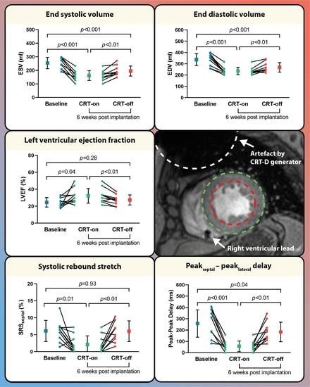
3.3. BIV Pacing Turned on vs. off during Follow-up
3.4. Invasive Volume Measurements
4. Discussion
4.1. CMR Imaging in CRT-D Patients
4.2. Acute Hemodynamic Changes and Reverse Remodeling during Follow-up
4.3. Device Optimization
5. Limitations
6. Conclusions
Supplementary Materials
Author Contributions
Funding
Institutional Review Board Statement
Informed Consent Statement
Data Availability Statement
Conflicts of Interest
Abbreviations
| AOO | atrial asynchronous mode |
| BIV pacing | biventricular pacing |
| CMR | cardiac magnetic resonance |
| CRT | cardiac resynchronization |
| EDV | end-diastolic volume |
| EF | ejection fraction |
| ESV | end-systolic volume |
| FU | follow-up |
| LGE | late gadolinium enhancement |
| LV | left ventricle |
| SRSseptal | systolic rebound stretch of the septum |
References
- Glikson, M.; Nielsen, J.C.; Kronborg, M.B.; Michowitz, Y.; Auricchio, A.; Barbash, I.M.; Barrabés, J.A.; Boriani, G.; Braunschweig, F.; Brignole, M.; et al. 2021 ESC Guidelines on cardiac pacing and cardiac resynchronization therapy: Developed by the Task Force on cardiac pacing and cardiac resynchronization therapy of the European Society of Cardiology (ESC) With the special contribution of the European Heart Rhythm Association (EHRA). Eur. Heart J. 2021, 42, 3427–3520. [Google Scholar] [CrossRef]
- Young, J.B.; Abraham, W.T.; Smith, A.L.; Leon, A.R.; Lieberman, R.; Wilkoff, B.; Canby, R.C.; Schroeder, J.S.; Liem, L.B.; Hall, S.; et al. Combined cardiac resynchronization and implantable cardioversion defibrillation in advanced chronic heart failure: The MIRACLE ICD Trial. Jama 2003, 289, 2685–2694. [Google Scholar] [CrossRef]
- Yu, C.M.; Bleeker, G.B.; Fung, J.W.; Schalij, M.J.; Zhang, Q.; van der Wall, E.E.; Chan, Y.-S.; Kong, S.-L.; Bax, J.J. Left ventricular reverse remodeling but not clinical improvement predicts long-term survival after cardiac resynchronization therapy. Circulation 2005, 112, 1580–1586. [Google Scholar] [CrossRef] [Green Version]
- Chung, E.S.; Leon, A.R.; Tavazzi, L.; Sun, J.P.; Nihoyannopoulos, P.; Merlino, J.; Abraham, W.T.; Ghio, S.; Leclercq, C.; Bax, J.J.; et al. Results of the Predictors of Response to CRT (PROSPECT) trial. Circulation 2008, 117, 2608–2616. [Google Scholar] [CrossRef] [Green Version]
- de Roest, G.J.; Wu, L.; de Cock, C.C.; Hendriks, M.L.; Delnoy, P.P.H.M.; van Rossum, A.C.; Allaart, C.P. Scar tissue–guided left ventricular lead placement for cardiac resynchronization therapy in patients with ischemic cardiomyopathy: An acute pressure-volume loop study. Am. Heart J. 2014, 167, 537–545. [Google Scholar] [CrossRef]
- Schwitter, J.; Kanal, E.; Schmitt, M.; Anselme, F.; Albert, T.; Hayes, D.L.; Bello, D.; Toth, A.; Chang, Y.; van Osch, D.; et al. Impact of the Advisa MRI pacing system on the diagnostic quality of cardiac MR images and contraction patterns of cardiac muscle during scans: Advisa MRI randomized clinical multicenter study results. Heart Rhythm 2013, 10, 864–872. [Google Scholar] [CrossRef] [Green Version]
- Vago, H.; Czimbalmos, C.; Papp, R.; Szabo, L.; Toth, A.; Dohy, Z.; Csecs, I.; Suhai, F.; Kosztin, A.; Molnar, L.; et al. Biventricular pacing during cardiac magnetic resonance imaging. EP Eur. 2019, 22, 117–124. [Google Scholar] [CrossRef]
- Brignole, M.; Auricchio, A.; Baron-Esquivias, G.; Bordachar, P.; Boriani, G.; Breithardt, O.A.; Cleland, J.; Deharo, J.C.; Delgado, V.; Elliott, P.M.; et al. 2013 ESC Guidelines on cardiac pacing and cardiac resynchronization therapy: The Task Force on cardiac pacing and resynchronization therapy of the European Society of Cardiology (ESC). Developed in collaboration with the European Heart Rhythm Association (EHRA). Eur. Heart J. 2013, 34, 2281–2329. [Google Scholar] [CrossRef] [Green Version]
- Steendijk, P.; Tulner, S.A.F.; Wiemer, M.; Bleasdale, R.A.; Bax, J.J.; van der Wall, E.E.; Vogt, J.; Schalij, M.J. Pressure–volume measurements by conductance catheter during cardiac resynchronization therapy. Eur. Heart J. Suppl. 2004, 6, D35–D42. [Google Scholar] [CrossRef] [Green Version]
- Burkhoff, D. Pressure-Volume Loops in Clinical Research∗: A Contemporary View. J. Am. Coll. Cardiol. 2013, 62, 1173–1176. [Google Scholar] [CrossRef] [Green Version]
- Zweerink, A.; van Everdingen, W.M.; Nijveldt, R.; Salden, O.A.E.; Meine, M.; Maass, A.H.; Vernooy, K.; de Lange, F.J.; Vos, M.A.; Croisille, P.; et al. Strain imaging to predict response to cardiac resynchronization therapy: A systematic comparison of strain parameters using multiple imaging techniques. ESC Heart Fail. 2018, 5, 1130–1140. [Google Scholar] [CrossRef]
- Mahrholdt, H.; Wagner, A.; Holly, T.A.; Elliott, M.D.; Bonow, R.O.; Kim, R.J.; Judd, R.M. Reproducibility of chronic infarct size measurement by contrast-enhanced magnetic resonance imaging. Circulation 2002, 106, 2322–2327. [Google Scholar] [CrossRef] [Green Version]
- Kawel-Boehm, N.; Maceira, A.; Valsangiacomo-Buechel, E.R.; Vogel-Claussen, J.; Turkbey, E.B.; Williams, R.; Plein, S.; Tee, M.; Eng, J.; Bluemke, D.A. Normal values for cardiovascular magnetic resonance in adults and children. J. Cardiovasc. Magn. Reson. 2015, 17, 29. [Google Scholar] [CrossRef] [Green Version]
- Zweerink, A.; de Roest, G.J.; Wu, L.; Nijveldt, R.; de Cock, C.C.; van Rossum, A.C.; Allaart, C.P. Prediction of Acute Response to Cardiac Resynchronization Therapy by Means of the Misbalance in Regional Left Ventricular Myocardial Work. J. Card Fail. 2016, 22, 133–142. [Google Scholar] [CrossRef]
- Zweerink, A.; Friedman, D.J.; Klem, I.; van de Ven, P.M.; Vink, C.; Biesbroek, P.S.; Hansen, S.M.; Kim, R.J.; van Rossum, A.C.; Atwater, B.D.; et al. Segment Length in Cine Strain Analysis Predicts Cardiac Resynchronization Therapy Outcome Beyond Current Guidelines. Circ. Cardiovasc. Imaging 2021, 14, e012350. [Google Scholar] [CrossRef]
- Salden, O.A.E.; van den Broek, H.T.; van Everdingen, W.M.; Mohamed Hoesein, F.A.A.; Velthuis, B.K.; Doevendans, P.A.; Cramer, M.J.; Tuinenburg, A.E.; Leufkens, P.; van Slochteren, F.J.; et al. Multimodality imaging for real-time image-guided left ventricular lead placement during cardiac resynchronization therapy implantations. Int. J. Cardiovasc. Imaging 2019, 35, 1327–1337. [Google Scholar] [CrossRef] [Green Version]
- Sasaki, T.; Hansford, R.; Zviman, M.M.; Kolandaivelu, A.; Bluemke, D.A.; Berger, R.D.; Calkins, H.; Halperin, H.R.; Nazarian, S. Quantitative Assessment of Artifacts on Cardiac Magnetic Resonance Imaging of Patients With Pacemakers and Implantable Cardioverter-Defibrillators. Circ. Cardiovasc. Imaging 2011, 4, 662–670. [Google Scholar] [CrossRef] [Green Version]
- Vuorinen, A.M.; Lehmonen, L.; Karvonen, J.; Holmström, M.; Kivistö, S.; Kaasalainen, T. Reducing cardiac implantable electronic device-induced artefacts in cardiac magnetic resonance imaging. Eur. Radiol. 2023, 33, 1229–1242. [Google Scholar] [CrossRef]
- Matsumoto, K.; Tanaka, H.; Okajima, K.; Hayashi, T.; Kajiya, T.; Sugiyama, D.; Kawai, H.; Hirata, K.-I. Reverse remodelling induces progressive ventricular resynchronization after cardiac resynchronization therapy ‘from vicious to virtuous cycle’. Eur. J. Echocardiogr. 2011, 12, 782–789. [Google Scholar] [CrossRef] [Green Version]
- Pouleur, A.C.; Knappe, D.; Shah, A.M.; Uno, H.; Bourgoun, M.; Foster, E.; McNitt, S.; Hall, W.J.; Zareba, W.; Goldenberg, I.; et al. Relationship between improvement in left ventricular dyssynchrony and contractile function and clinical outcome with cardiac resynchronization therapy: The MADIT-CRT trial. Eur. Heart J. 2011, 32, 1720–1729. [Google Scholar] [CrossRef] [Green Version]
- Gorcsan, J., 3rd; Oyenuga, O.; Habib, P.J.; Tanaka, H.; Adelstein, E.C.; Hara, H.; McNamara, D.M.; Saba, S. Relationship of echocardiographic dyssynchrony to long-term survival after cardiac resynchronization therapy. Circulation 2010, 122, 1910–1918. [Google Scholar] [CrossRef] [PubMed] [Green Version]
- Alvarez-Alvarez, B.; García-Seara, J.; Martínez-Sande, J.L.; Rodríguez-Mañero, M.; Fernández López, X.A.; González-Melchor, L.; Iglesias-Alvarez, D.; Gude, F.; Díaz-Louzao, C.; González-Juanatey, J.R. Long-term cardiac reverse remodeling after cardiac resynchronization therapy. J. Arrhythm. 2021, 37, 653–659. [Google Scholar] [CrossRef] [PubMed]
- Dauw, J.; Martens, P.; Mullens, W. CRT Optimization: What Is New? What Is Necessary? Curr. Treat Options Cardiovasc. Med. 2019, 21, 45. [Google Scholar] [CrossRef] [PubMed]
- Katbeh, A.; Van Camp, G.; Barbato, E.; Galderisi, M.; Trimarco, B.; Bartunek, J.; Vanderheyden, M.; Penicka, M. Cardiac Resynchronization Therapy Optimization: A Comprehensive Approach. Cardiology 2019, 142, 116–128. [Google Scholar] [CrossRef]
- Ellenbogen, K.A.; Gold, M.R.; Meyer, T.E.; Fernndez Lozano, I.; Mittal, S.; Waggoner, A.D.; Lemke, B.; Singh, J.P.; Spinale, F.G.; Van Eyk, J.E.; et al. Primary results from the SmartDelay determined AV optimization: A comparison to other AV delay methods used in cardiac resynchronization therapy (SMART-AV) trial: A randomized trial comparing empirical, echocardiography-guided, and algorithmic atrioventricular delay programming in cardiac resynchronization therapy. Circulation 2010, 122, 2660–2668. [Google Scholar] [CrossRef] [Green Version]
- Gao, X.; Abdi, M.; Auger, D.A.; Sun, C.; Hanson, C.A.; Robinson, A.A.; Schumann, C.; Oomen, P.J.; Ratcliffe, S.; Malhotra, R.; et al. Cardiac Magnetic Resonance Assessment of Response to Cardiac Resynchronization Therapy and Programming Strategies. JACC Cardiovasc. Imaging 2021, 14, 2369–2383. [Google Scholar] [CrossRef]
- Mafi Rad, M.; Blaauw, Y.; Prinzen, F.W.; Vernooy, K. The role of acute invasive haemodynamic measurements in cardiac resynchronization therapy: Looping towards prediction of long-term response and therapy optimization. Eur. J. Heart Fail. 2013, 15, 247–249. [Google Scholar] [CrossRef]
- Seemann, F.; Arvidsson, P.; Nordlund, D.; Kopic, S.; Carlsson, M.; Arheden, H.; Heiberg, E. Noninvasive Quantification of Pressure-Volume Loops From Brachial Pressure and Cardiovascular Magnetic Resonance. Circ. Cardiovasc. Imaging 2019, 12, e008493. [Google Scholar] [CrossRef] [Green Version]
- Yamamoto, K.; Masuyama, T.; Tanouchi, J.; Doi, Y.; Kondo, H.; Hori, M.; Kitabatake, A.; Kamada, T. Effects of heart rate on left ventricular filling dynamics: Assessment from simultaneous recordings of pulsed Doppler transmitral flow velocity pattern and haemodynamic variables. Cardiovasc. Res. 1993, 27, 935–941. [Google Scholar] [CrossRef]


| Parameter | n = 10 |
|---|---|
| Demographics | |
| Age in years | 70 ± 7 |
| Sex, male/female | 9/1 |
| BMI | 28.3 ± 6.9 |
| BSA | 2.1 ± 0.3 |
| Hypertension (%) | 4 (40%) |
| NYHA II/III | 1/9 |
| Etiology, ICMP/NICMP | 4/6 |
| Ischemic LV LGE pattern (%) | 3 (30%) |
| Non-ischemic LV LGE pattern (%) | 3 (30%) |
| QRS duration (ms) LBBB | 167 ± 15 10 (100%) |
| CRT-D (%) | 10 (100%) |
| Left sided pre-pectoral pocket (%) | 10 (100%) |
| Medications | |
| ACE inhibitors, ARBs or ARNi (%) | 10 (100%) |
| Beta-blockers (%) | 10 (100%) |
| Diuretics (%) | 9 (90%) |
| Baseline | FU–BIV Pacing | FU-Intrinsic Rhythm | Baseline vs. FU–BIV Pacing, p-Value | Baseline vs. FU–Intrinsic Rhythm, p-Value | FU–BIV Pacing vs. FU–Intrinsic Rhythm, p-Value | |
|---|---|---|---|---|---|---|
| Heart rate (bpm) LV volumetric function | 68 ± 9 | 81 ± 8 | 65 ± 11 | <0.01 | 0.33 | <0.001 |
| ESV (mL) | 253.8 ± 41.3 | 161.4 ± 36.3 | 194.9 ± 37.1 | <0.001 | <0.001 | <0.01 |
| EDV (mL) | 336.9 ± 52.8 | 236.2 ± 31.8 | 268.1 ± 42.3 | <0.001 | <0.001 | <0.01 |
| LVEF (%) | 24.6 ± 5.6 | 32.2 ± 8.7 | 27.4 ± 5.9 | 0.04 | 0.28 | <0.01 |
| LV strain and dyssynchrony | ||||||
| GLS (%) | −8.2 ± 2.1 | −6.2 ± 1.6 | −7.9 ± 2.2 | 0.06 | 0.74 | 0.02 |
| GCS (%) | −6.2 ± 3.9 | −7.8 ± 3.0 | −7.2 ± 2.4 | 0.35 | 0.53 | 0.18 |
| GRS (%) | 8.2 ± 4.6 | 10.3 ± 4.9 | 8.9 ± 3.5 | 0.37 | 0.74 | 0.08 |
| Peakseptal - peaklateral delay (ms) | 258 ± 121 | 57 ± 46 | 183 ± 86 | <0.001 | 0.04 | <0.01 |
| Septal systolic rebound stretch (%) | 6.1 ± 3.1 | 2.1 ± 2.6 | 6.0 ± 3.0 | 0.01 | 0.93 | <0.01 |
Disclaimer/Publisher’s Note: The statements, opinions and data contained in all publications are solely those of the individual author(s) and contributor(s) and not of MDPI and/or the editor(s). MDPI and/or the editor(s) disclaim responsibility for any injury to people or property resulting from any ideas, methods, instructions or products referred to in the content. |
© 2023 by the authors. Licensee MDPI, Basel, Switzerland. This article is an open access article distributed under the terms and conditions of the Creative Commons Attribution (CC BY) license (https://creativecommons.org/licenses/by/4.0/).
Share and Cite
Hopman, L.H.G.A.; Zweerink, A.; van der Lingen, A.-L.C.J.; Huntelaar, M.J.; Mulder, M.J.; Robbers, L.F.H.J.; van Rossum, A.C.; van Halm, V.P.; Götte, M.J.W.; Allaart, C.P. Feasibility of CMR Imaging during Biventricular Pacing: Comparison with Invasive Measurement as a Pathway towards a Novel Optimization Strategy. J. Clin. Med. 2023, 12, 3998. https://doi.org/10.3390/jcm12123998
Hopman LHGA, Zweerink A, van der Lingen A-LCJ, Huntelaar MJ, Mulder MJ, Robbers LFHJ, van Rossum AC, van Halm VP, Götte MJW, Allaart CP. Feasibility of CMR Imaging during Biventricular Pacing: Comparison with Invasive Measurement as a Pathway towards a Novel Optimization Strategy. Journal of Clinical Medicine. 2023; 12(12):3998. https://doi.org/10.3390/jcm12123998
Chicago/Turabian StyleHopman, Luuk H. G. A., Alwin Zweerink, Anne-Lotte C. J. van der Lingen, Marthe J. Huntelaar, Mark J. Mulder, Lourens F. H. J. Robbers, Albert C. van Rossum, Vokko P. van Halm, Marco J. W. Götte, and Cornelis P. Allaart. 2023. "Feasibility of CMR Imaging during Biventricular Pacing: Comparison with Invasive Measurement as a Pathway towards a Novel Optimization Strategy" Journal of Clinical Medicine 12, no. 12: 3998. https://doi.org/10.3390/jcm12123998
APA StyleHopman, L. H. G. A., Zweerink, A., van der Lingen, A.-L. C. J., Huntelaar, M. J., Mulder, M. J., Robbers, L. F. H. J., van Rossum, A. C., van Halm, V. P., Götte, M. J. W., & Allaart, C. P. (2023). Feasibility of CMR Imaging during Biventricular Pacing: Comparison with Invasive Measurement as a Pathway towards a Novel Optimization Strategy. Journal of Clinical Medicine, 12(12), 3998. https://doi.org/10.3390/jcm12123998