Effect of Vitamin D Supplementation on the Cerebral Placental Ratio in Pregnancy Complicated with Early Fetal Growth Restriction

Abstract
1. Introduction
2. Materials and Methods
Statistics
3. Results
4. Discussion
5. Conclusions
Author Contributions
Funding
Institutional Review Board Statement
Informed Consent Statement
Data Availability Statement
Conflicts of Interest
References
- Kamiński, K.; Fiegler-Rudol, P.; Węgrzyn, P. Nieprawidłowe wzrastanie płodu (hipotrofia i hipertrofia). In [W:] Położnictwo i Ginekologia; Bręborowicz, G., Ed.; PZWL: Warszawa, Poland, 2010. [Google Scholar]
- Bręborowicz, G. Położnictwo. Tom II; PZWL: Warszawa, Poland, 2012; pp. 105–119. [Google Scholar]
- Abdul-Karim, R.W.; Beydoun, S.N. Growth of the human fetus. Clin. Obs. Gynecol. 1974, 17, 37–52. [Google Scholar] [CrossRef] [Scilit]
- Royal College of Obstetricians and Gynaecologists (RCOG). The Investigation and Management of the Small–for–Gestational–Age Fetus; Green–Top Guideline No. 31; RCOG: London, UK, 2014; pp. 1–34. [Google Scholar]
- Bralewska, M.; Biesiada, L.; Grzesiak, M.; Rybak-Krzyszkowska, M.; Huras, H.; Gach, A.; Pietrucha, T.; Sakowicz, A. Chromogranin A demonstrates higher expression in preeclamptic placentas than in normal pregnancy. BMC Pregnancy Childbirth 2021, 21, 680. [Google Scholar] [CrossRef] [Scilit]
- Wysocka, U.; Sakowicz, A.; Jakubowski, L.; Pinkier, I.; Rybak-Krzyszkowska, M.; Alaszewski, W.; Dudarewicz, L.; Gach, A. Association between idiopathic recurrent pregnancy loss and genetic polymorphisms in cytokine and matrix metalloproteinase genes. Ginekol. Pol. 2021. Epub ahead of print. [Google Scholar] [CrossRef] [Scilit]
- Sakowicz, A.; Bralewska, M.; Pietrucha, T.; Figueras, F.; Habrowska-Górczyńska, D.E.; Piastowska-Ciesielska, A.W.; Gach, A.; Sakowicz, B.; Rybak-Krzyszkowska, M.; Huras, H.; et al. The Preeclamptic Environment Promotes the Activation of Transcription Factor Kappa B by P53/RSK1 Complex in a HTR8/SVneo Trophoblastic Cell Line. Int. J. Mol. Sci. 2021, 22, 10200. [Google Scholar] [CrossRef] [Scilit]
- Krawczyk, P.; Jastrzebska, A.; Lipka, D.; Huras, H. Pregnancy related and postpartum admissions to intensive care unit in the obstetric tertiary care center—An 8-year retrospective study. Ginekol. Pol. 2021, 92, 575–578. [Google Scholar] [CrossRef] [Scilit]
- Kowalska-jasiecka, J.; Szymański, P.; Ropacka, M.; Markwitz, W.; Bręborowicz, G.H. The effect of L-arginine treatment of Doppler measurments in pregnant woman with gestational hypertension and hypertrophy. Arch. Perinat. Med. 2008, 14, 12–17. [Google Scholar]
- Contini, C.; Rotondo, J.C.; Magagnoli, F.; Maritati, M.; Seraceni, S.; Graziano, A.; Poggi, A.; Capucci, R.; Vesce, F.; Tognon, M.; et al. Investigation on silent bacterial infections in specimens from pregnant women affected by spontaneous miscarriage. J. Cell Physiol. 2018, 234, 100–107. [Google Scholar] [CrossRef] [Scilit]
- Xiong, Y.Q.; Mo, Y.; Luo, Q.M.; Huo, S.T.; He, W.Q.; Chen, Q. The Risk of Human Papillomavirus Infection for Spontaneous Abortion, Spontaneous Preterm Birth, and Pregnancy Rate of Assisted Reproductive Technologies: A Systematic Review and Meta-Analysis. Gynecol. Obstet. Investig. 2018, 83, 417–427. [Google Scholar] [CrossRef] [Scilit]
- Royal College of Obstetricians and Gynaecologists (RCOG). The Investigation and Management of the Small-for-Gestational-Age Fetus; Green-Top Guideline, no. 31; Royal College of Obstetricians and Gynaecologists (RCOG): London, UK, 2013; p. 34. [Google Scholar]
- Lausman, A.; Kingdom, J.; Gagnon, R.; Basso, M.; Bos, H.; Crane, J.; Davies, G.; Delisle, M.F.; Hudon, L.; Menticoglou, S.; et al. Intrauterine growth restriction: Screening, diagnosis, and management. J. Obstet. Gynaecol. Can. 2013, 35, 741–748. [Google Scholar] [CrossRef] [Scilit]
- American College of Obstetricians and Gynecologists (ACOG). Fetal Growth Restriction; ACOG Practice Bulletin; no. 134; American College of Obstetricians and Gynecologists (ACOG): Washington, DC, USA, 2013; p. 12. [Google Scholar]
- Rytlewski, K.; Huras, H.; Kusmierska, K.; Jaworowski, A.; Gornisiewicz, T.; Ossowski, P.; Reron, A. Doppler velocimetry of the materno-fetal circulation in preterm delivered pregnancies complicated with hypertension. Neuro. Endocrinol. Lett. 2009, 30, 403–408. [Google Scholar]
- Mari, G.; Dater, R.L. Middle cerebral artery flow velocity veforms in normal and small-for-gestational-age fetuses. Am. J. Obstet. Gynecol. 1992, 166, 1262–1270. [Google Scholar] [CrossRef] [Scilit]
- Tarzamni, M.K. Nomograms of Iranian fetal middle cerebral artery Doppler waveforms and uniformity of their pattern with other populations nomograms. BMC Pregnancy Childbirth 2008, 12, 50. [Google Scholar] [CrossRef] [Scilit]
- Ebbing, C.; Rasmussen, S.; Kiserud, T. Middle cerebral artery blood flow velocities and pulsality index and the cerebroplacental pulsatility ratio: Longitudinal reference ranges and terms for serial measurments. Ultrasound Obstet. Gynecol. 2007, 30, 287–296. [Google Scholar] [CrossRef] [Scilit] [PubMed]
- Odibo, A.O.; Riddick, C.; Pare, E.; Stamilio, D.M.; Macones, G.A. Cerebroplacental Doppler ratio and adverse perinatal outcomes in intrauterine growth restriction: Evaluating the impact of using gestational agespecific reference values. J. Ultrasound Med. 2005, 24, 1223–1228. [Google Scholar] [CrossRef] [Scilit]
- Tesic, D.; Hawes, J.E.; Zosky, G.R.; Wyrwoll, C.S. Vitamin D Deficiency in BALB/c Mouse Pregnancy Increases Placental Transfer of Glucocorticoids. Endocrinology 2015, 156, 3673–3679. [Google Scholar] [CrossRef] [Scilit]
- Nancy, Q.L.; Yi, O.; Yasemin BVenu, L.; Shiao, Y.C.; Bruce, W.H.; Carol, W.; Ozlem, E.; Martin, H. Dietary vitamin D restriction in pregnant female mice is associated with maternal hypertension and altered placental and fetal development. Endocrinology 2013, 154, 2270–2280. [Google Scholar]
- Yates, N.; Crew, R.; Wyrwoll, C. Vitamin D deficiency and impaired placental function: Potential regulation by glucocorticoids? Reproduction 2017, 153, 163–171. [Google Scholar] [CrossRef] [Scilit] [PubMed]
- Grundmann, M.; von Versen-Höynck, F. Vitamin D—Roles in women’s reproductive health? Reprod. Biol. Endocrinol. 2011, 9, 146. [Google Scholar] [CrossRef] [Scilit] [PubMed]
- Nema, J.; Sundrani, D.; Joshi, S. Role of vitamin D in influencing angiogenesis in preeclampsia. Hypertens. Pregnancy 2019, 38, 201–207. [Google Scholar] [CrossRef] [Scilit]
- Li, N.; Wu, H.M.; Hang, F.; Zhang, Y.S.; Li, M.J. Women with recurrent spontaneous abortion have decreased 25(OH) vitamin D and VDR at the fetal-maternal interface. Braz. J. Med. Biol. Res. 2017, 50, e6527. [Google Scholar] [CrossRef] [Scilit]
- Schulz, E.V.; Cruze, L.; Wei, W.; Gehris, J.; Wagner, C.L. Maternal vitamin D sufficiency and reduced placental gene expression in angiogenic biomarkers related to comorbidities of pregnancy. J. Steroid. Biochem. Mol. Biol. 2017, 173, 273–279. [Google Scholar] [CrossRef] [Scilit] [PubMed]
- Jurisic, A.; Jurisic, Z.; Lefkou, E.; Pombo, J.; Girardi, G. Pravastatin and-L-arginine combination improves umbilical artery blood flow and neonatal outcomes in dichorionic twin pregnancies through an nitric oxide-dependent vasorelaxant effect. Vasc. Pharmacol. 2018, 110, 64–70. [Google Scholar] [CrossRef] [Scilit] [PubMed]
- Borowski, D.; Pietryga, M.; Basta, P.; Cnota, W.; Czuba, B.; Dubiel, M.; Fuchs, T.; Huras, H.; Iciek, R.; Jaczynska, R.; et al. Practice guidelines of the Polish Society of Gynecologists and Obstetricians—Ultrasound Section for ultrasound screening in uncomplicated pregnancy–2020. Ginekol. Pol. 2020, 91, 490–501. [Google Scholar] [CrossRef] [Scilit] [PubMed]
- ISUOG. Diagnosis and Management of Small-for-Gestational-Age Fetus and Fetal Growth Restriction; ISUOG: London, UK, 2020; Volume 56, pp. 298–312. [Google Scholar]
- Ou, F.S.; Le-Rademacher, J.G.; Ballman, K.V.; Adjei, A.A.; Mandrekar, S.J. Guidelines for Statistical Reporting in Medical Journals. J. Thorac. Oncol. 2020, 15, 1722–1726. [Google Scholar] [CrossRef] [Scilit] [PubMed]
- Alfirevic, Z.; Neilson, J.P. Doppler ultrasonography in high-risk pregnancies: Systematic review with meta-analysis. Am. J. Obstet. Gynecol. 1995, 172, 1379–1387. [Google Scholar] [CrossRef] [Scilit]
- Alfirevic, Z.; Neilson, J.P. WITHDRAWN. Doppler ultrasound for fetal assessment in high risk pregnancies. Cochrane Database Syst. Rev. 2010, 2010, CD000073. [Google Scholar] [CrossRef] [Scilit]
- Valcamonico, A.; Danti, L.; Frusca, T.; Soregaroli, M.; Zucca, S.; Abrami, F.; Tiberti, A. Absent end-diastolic velocity in umbilical artery: Risk of neonatal morbidity and brain damage. Am. J. Obstet. Gynecol. 1994, 170, 796–801. [Google Scholar] [CrossRef] [Scilit]
- Ferrazzi, E.; Bozzo, M.; Rigano, S.; Bellotti, M.; Morabito, A.; Pardi, G.; Galan, H.L. Temporal sequence of abnormal Doppler changes in the peripheral and central circulatory systems of the severely growth-restricted fetus. Ultrasound Obstet. Gynecol. 2002, 19, 140–146. [Google Scholar] [CrossRef] [Scilit]
- Cosmi, E.; Ambrosini, G.; D’Antona, D.; Saccardi, C.; Mari, G. Doppler, cardiotocography, and biophysical profile changes in growth-restricted fetuses. Obstet. Gynecol. 2005, 106, 1240–1245. [Google Scholar] [CrossRef] [Scilit]
- Shahinaj, R.; Manoku, N.; Kroi, E.; Tasha, I. The value of the middle cerebral to umbilical artery Doppler ratio in the prediction of neonatal outcome in patient with preeclampsia and gestational hypertension. J. Prenat. Med. 2010, 4, 17–21. [Google Scholar]
- Krzeszowski, W.; Janiak, K.; Kalinka, J.; Grzesiak, M.; Oszukowski, P.; Szaflik, T.; Wojtera, J.; Szaflik, K. Współczesne metody monitorowania dobrostanu płodu w ciąży powikłanej wewnątrzmacicznym zahamowaniem wzrastania. Ginekol. Pol. 2016, 87, 135–142. [Google Scholar] [CrossRef] [Scilit] [PubMed]
- Visser, G.H.A. Differences between the early and late IUGR. In Proceedings of the XXIV European Congress—Perinatal Medicine, Florence, Italy, 4–7 June 2014. [Google Scholar]
- Kalafatic, D. The relationship between fetal umbilical and middle cerebral artery Doppler indices, perinatal asphyxia and subsequent brain damage. Prenat. Neonatal Med. 1998, 3, 90. [Google Scholar]
- Nicolaides, K.; Rizzo, G.; Hecher, K.; Ximenes, R. Doppler in Obstetrics. Available online: https://fetalmedicine.org/var/uploads/Doppler-in-Obstetrics.pdf. (accessed on 9 March 2022).
- Lees, C.; Marlow, N.; Arabin, B.; Bilardo, C.M.; Brezinka, C.; Derks, J.B.; Duvekot, J.; Frusca, T.; Diemert, A.; TRUFFLE Group. Perinatal morbidity and mortality in early-onset fetal growth restriction: Cohort outcomes of the trial of randomized umbilical and fetal flow in Europe (TRUFFLE). Ultrasound Obs. Gynecol. 2013, 42, 400–408. [Google Scholar] [CrossRef] [Scilit] [PubMed]
- Bomba-Opoń, D.; Drews, K.; Huras, H.; Laudański, P.; Paszkowski, T.; Wielgoś, M. Rekomendacje Polskiego Towarzystwa Ginekologicznego dotyczące indukcji porodu. Ginekol. I Perinatol. Praktyczna. 2017, 2, 58–71. [Google Scholar]
- Figueras, F.; Gratacos, E. Update on the diagnosis and classification of fetal growth restriction and proposal of a stage-based management protocol. Fetal Diagn Ther. 2014, 36, 86–98. [Google Scholar] [CrossRef] [Scilit]




| Variable | Measure/Category | <500 | 2000 | Total | p |
|---|---|---|---|---|---|
| Abnormal CPR percentile baseline | n | 50 | 50 | 100 | 0.0050 |
| Yes | 18 (36.00%) | 6 (12.00%) | 24 (24.00%) | ||
| No | 32 (64.00%) | 44 (88.00%) | 76 (76.00%) | ||
| Abnormal CPR percentile 7 d | n | 50 | 50 | 100 | 0.0014 |
| Yes | 20 (40.00%) | 6 (12.00%) | 26 (26.00%) | ||
| No | 30 (60.00%) | 44 (88.00%) | 74 (74.00%) | ||
| Abnormal CPR percentile 14 d | n | 42 | 50 | 92 | <0.0001 |
| Yes | 20 (47.62%) | 4 (8.00%) | 24 (26.09%) | ||
| No | 22 (52.38%) | 46 (92.00%) | 68 (73.91%) |
| Variable | Measure/Category | <500 | 2000 | Total | p |
|---|---|---|---|---|---|
| CPR baseline | n | 50 | 50 | 100 | 0.0032 |
| Mean (±SD) | 1.69 (±0.79) | 2.01 (±0.69) | 1.85 (±0.76) | ||
| Mean 95% CI | (1.47; 1.91) | (1.81; 2.21) | (1.70; 2.00) | ||
| Me (Q1; Q3) | 1.47 (1.16; 2.02) | 1.90 (1.54;2.32) | 1.66 (1.33; 2.31) | ||
| Min/Max | 0.70/3.50 | 0.60/3.75 | 0.60/3.75 | ||
| CPR 7d | n | 50 | 50 | 100 | 0.0455 |
| Mean (±SD) | 1.58 (±0.65) | 1.80 (±0.59) | 1.69 (±0.63) | ||
| Mean 95% CI | (1.39; 1.76) | (1.63; 1.97) | (1.57; 1.81) | ||
| Me (Q1; Q3) | 1.55 (1.04; 1.92) | 1.75 (1.47; 2.06) | 1.64 (1.20; 2.01) | ||
| Min/Max | 0.69/2.84 | 0.70/3.23 | 0.69/3.23 | ||
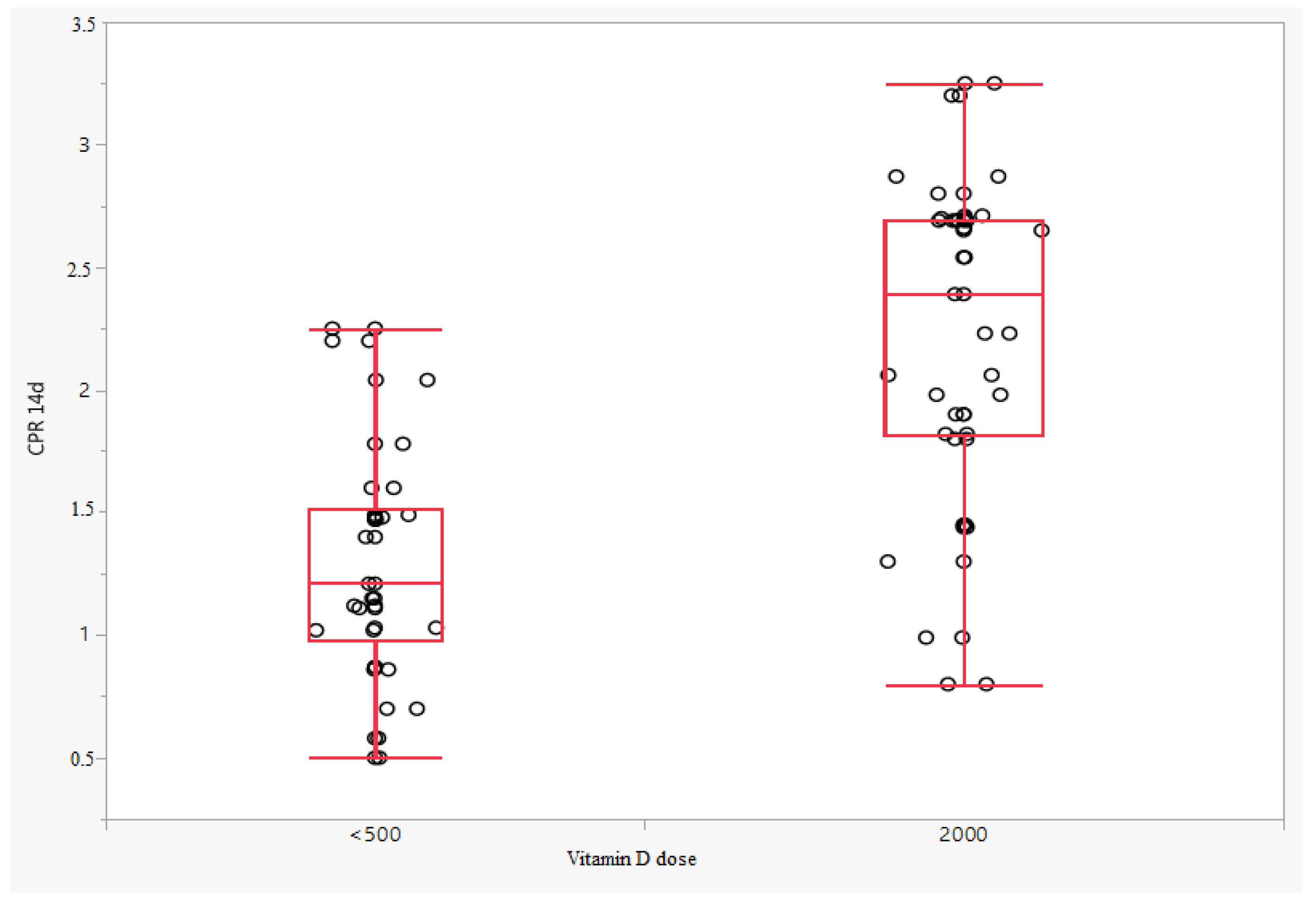
| CPR 14 d | n | 42 | 50 | 92 | <0.0001 |
| Mean (±SD) | 1.30 (±0.49) | 2.22 (±0.66) | 1.80 (±0.74) | ||
| Mean 95% CI | (1.15; 1.45) | (2.03; 2.41) | (1.65; 1.95) | ||
| Me (Q1; Q3) | 1.21 (0.98; 1.52) | 2.39 (1.82; 2.69) | 1.79 (1.15; 2.54) | ||
| Min/Max | 0.50/2.25 | 0.80/3.25 | 0.50/3.25 | ||
| Odds CPR (7-d-baseline) | n | 50 | 50 | 100 | 0.7933 |
| Mean (±SD) | −0.11 (±0.44) | −0.21 (±0.65) | −0.16 (±0.55) | ||
| Mean 95% CI | (−0.23; 0.01) | (−0.39; −0.02) | (−0.27; −0.05) | ||
| Me (Q1; Q3) | −0.01 (−0.40; 0.12) | −0.23 (−0.55; 0.27) | −0.01 (−0.46; 0.17) | ||
| Min/Max | −0.99/0.93 | −1.70/1.11 | −1.70/1.11 | ||
| Odds CPR (14-d-baseline) | n | 42 | 50 | 92 | <0.0001 |
| Mean (±SD) | −0.47 (±0.56) | 0.21 (±0.56) | −0.10 (±0.65) | ||
| Mean 95% CI | (−0.65; −0.30) | (0.05; 0.37) | (−0.24; 0.04) | ||
| Me (Q1; Q3) | −0.33 (−0.64; −0.11) | 0.23 (−0.18; 0.64) | −0.10 (−0.42; 0.35) | ||
| Min/Max | −1.90/0.42 | −1.10/1.30 | −1.90/1.30 | ||
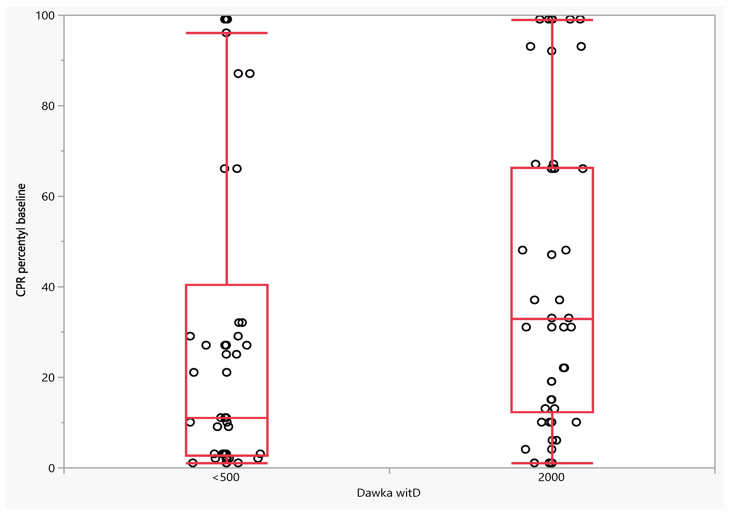
| CPR percentile baseline | n | 50 | 50 | 100 | 0.0181 |
| Mean (±SD) | 30.64 (±36.10) | 43.00 (±33.95) | 36.82 (±35.41) | ||
| Mean 95% CI | (20.38; 40.90) | (33.35; 52.65) | (29.79; 43.85) | ||
| Me (Q1; Q3) | 11.00 (2.75; 40.50) | 33.00 (12.25; 66.25) | 26.00 (6.00; 66.00) | ||
| Min/Max | 1.00/99.00 | 1.00/99.00 | 1.00/99.00 | ||
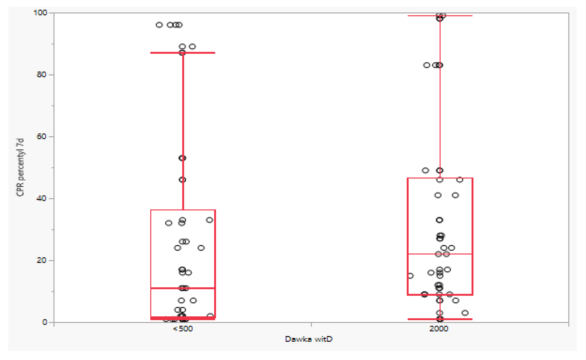
| CPR percentile 7 d | n | 50 | 50 | 100 | 0.0723 |
| Mean (±SD) | 26.40 (±32.36) | 31.60 (±29.82) | 29.00 (±31.07) | ||
| Mean 95% CI | (17.20; 35.60) | (23.13; 40.07) | (22.84; 35.16) | ||
| Me (Q1; Q3) | 11.00 (1.75; 36.25) | 22.00 (9.00; 46.75) | 16.50 (4.00;46.00) | ||
| Min/Max | 1.00/96.00 | 1.00/99.00 | 1.00/99.00 | ||
| CPR percentile 14 d | n | 42 | 50 | 92 | <0.0001 |
| Mean (±SD) | 13.19 (±18.67) | 58.28 (±36.02) | 37.70 (±36.95) | ||
| Mean 95% CI | (7.37; 19.01) | (48.04; 68.52) | (30.04; 45.35) | ||
| Me (Q1; Q3) | 5.00 (1.00; 13.50) | 69.00 (25.75; 92.25) | 25.50 (4.00; 81.00) | ||
| Min/Max | 1.00/60.00 | 1.00/99.00 | 1.00/99.00 | ||
| Odds CPR percentile (7-d-baseline) | n | 50 | 50 | 100 | 0.8683 |
| Mean (±SD) | −4.24 (±18.74) | −11.40 (±36.21) | −7.82 (±28.91) | ||
| Mean 95% CI | (−9.56; 1.08) | (−21.69; −1.11) | (−13.56; −2.08) | ||
| Me (Q1; Q3) | −1.00 (−7.50; 0.00) | −1.00 (−33.75;13.00) | −1.00 (−19.00;6.00) | ||
| Min/Max | −55.00/44.00 | −88.00/51.00 | −88.00/51.00 | ||
| Odds CPR percentile (14-d-baseline) | n | 42 | 50 | 92 | <0.0001 |
| Mean (±SD) | −20.43 (±25.29) | 15.28 (±29.24) | −1.02 (±32.69) | ||
| Mean 95% CI | (−28.31; −12.55) | (6.97; 23.59) | (−7.79; 5.75) | ||
| Me (Q1; Q3) | −9.00 (−30.75; −1.00) | 3.00 (−6.75; 30.25) | −1.00 (−20.00; 20.00) | ||
| Min/Max | −75.00/3.00 | −23.00/83.00 | −75.00/83.00 |
| Model/Responsible Variable | Parameter | (95% CI) | p-Value | Statistics | Value |
|---|---|---|---|---|---|
| Model: CPR baseline | 0.0100 | R˛ | 12.92% | ||
| Intercept | 2.9624 | (1.3863; 4.5385) | 0.0003 | Adjusted R˛ | 9.26% |
| Mother age (years) | −0.0471 | (−0.0823; −0.0119) | 0.0092 | Observations | 100 |
| Gestational age (weeks) | 0.0093 | (−0.0394; 0.0579) | 0.7057 | ||
| Pregnancy | 0.0151 | (−0.2017; 0.2318) | 0.8906 | ||
| Dose of vitD (<500) | −0.1997 | (−0.3609; −0.0385) | 0.0157 | ||
| Model: CPR 7 d | 0.1079 | R˛ | 7.60% | ||
| Intercept | 2.0236 | (0.6784; 3.3687) | 0.0036 | Adjusted R˛ | 3.71% |
| Mother age (years) | −0.0132 | (−0.0432; 0.0168) | 0.3840 | Observations | 100 |
| Gestational age (weeks) | 0.0077 | (−0.0338; 0.0492) | 0.7145 | ||
| Pregnancy | −0.1133 | (−0.2983; 0.0717) | 0.2271 | ||
| Dose of vitD (<500) | −0.1068 | (−0.2444; 0.0307) | 0.1265 | ||
| Model: CPR 14 d | <0.0001 | R˛ | 41.83% | ||
| Intercept | 2.2803 | (0.9709; 3.5896) | 0.0008 | Adjusted R˛ | 39.15% |
| Mother age (years) | 0.0062 | (−0.0248; 0.0373) | 0.6899 | Observations | 92 |
| Gestational age (weeks) | −0.0145 | (−0.0556; 0.0266) | 0.4849 | ||
| Pregnancy | −0.1792 | (−0.3610; 0.0025) | 0.0532 | ||
| Dose of vitD (<500) | −0.4521 | (−0.5874; −0.3168) | <0.0001 | ||
| Model: Róznica CPR (7-d-baseline) | 0.1227 | R˛ | 7.29% | ||
| Intercept | −0.9388 | (−2.1223; 0.2446) | 0.1186 | Adjusted R˛ | 3.38% |
| Mother age (years) | 0.0339 | (0.0075; 0.0603) | 0.0124 | Observations | 100 |
| Gestational age (weeks) | −0.0016 | (−0.0381; 0.0349) | 0.9305 | ||
| Pregnancy | −0.1284 | (−0.2911; 0.0344) | 0.1208 | ||
| Dose of vitD (<500) | 0.0928 | (−0.0282; 0.2139) | 0.1312 | ||
| Model: Róznica CPR (14-d-baseline) | <0.0001 | R˛ | 36.90% | ||
| Intercept | −0.6243 | (−1.8224; 0.5737) | 0.3032 | Adjusted R˛ | 34.00% |
| Mother age (years) | 0.0503 | (0.0219; 0.0787) | 0.0007 | Observations | 92 |
| Gestational age (weeks) | −0.0234 | (−0.0610; 0.0143) | 0.2203 | ||
| Pregnancy | −0.2011 | (−0.3674; −0.0348) | 0.0183 | ||
| Dose of vitD (<500) | −0.2854 | (−0.4093; −0.1616) | <0.0001 |
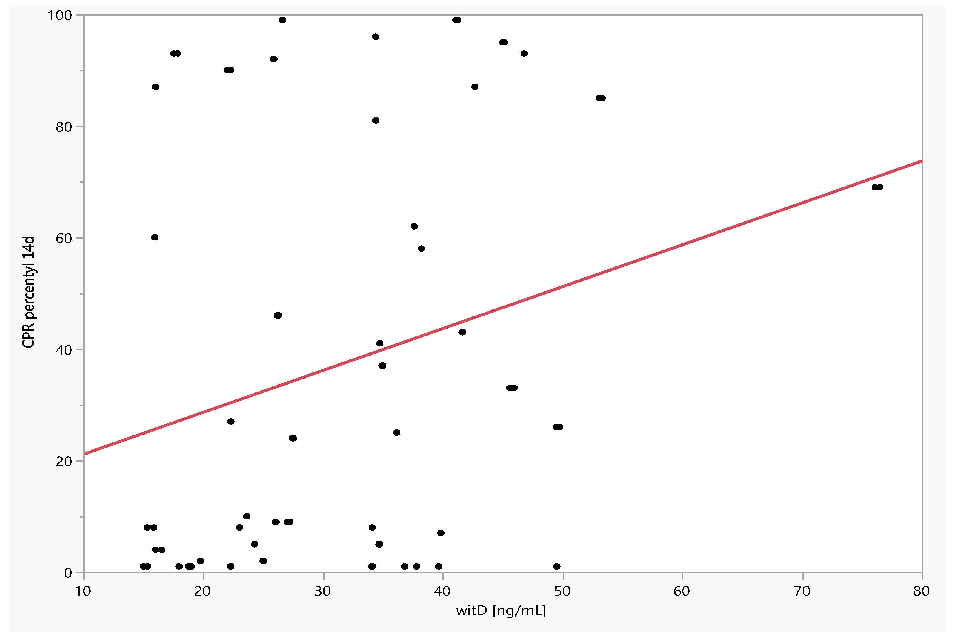
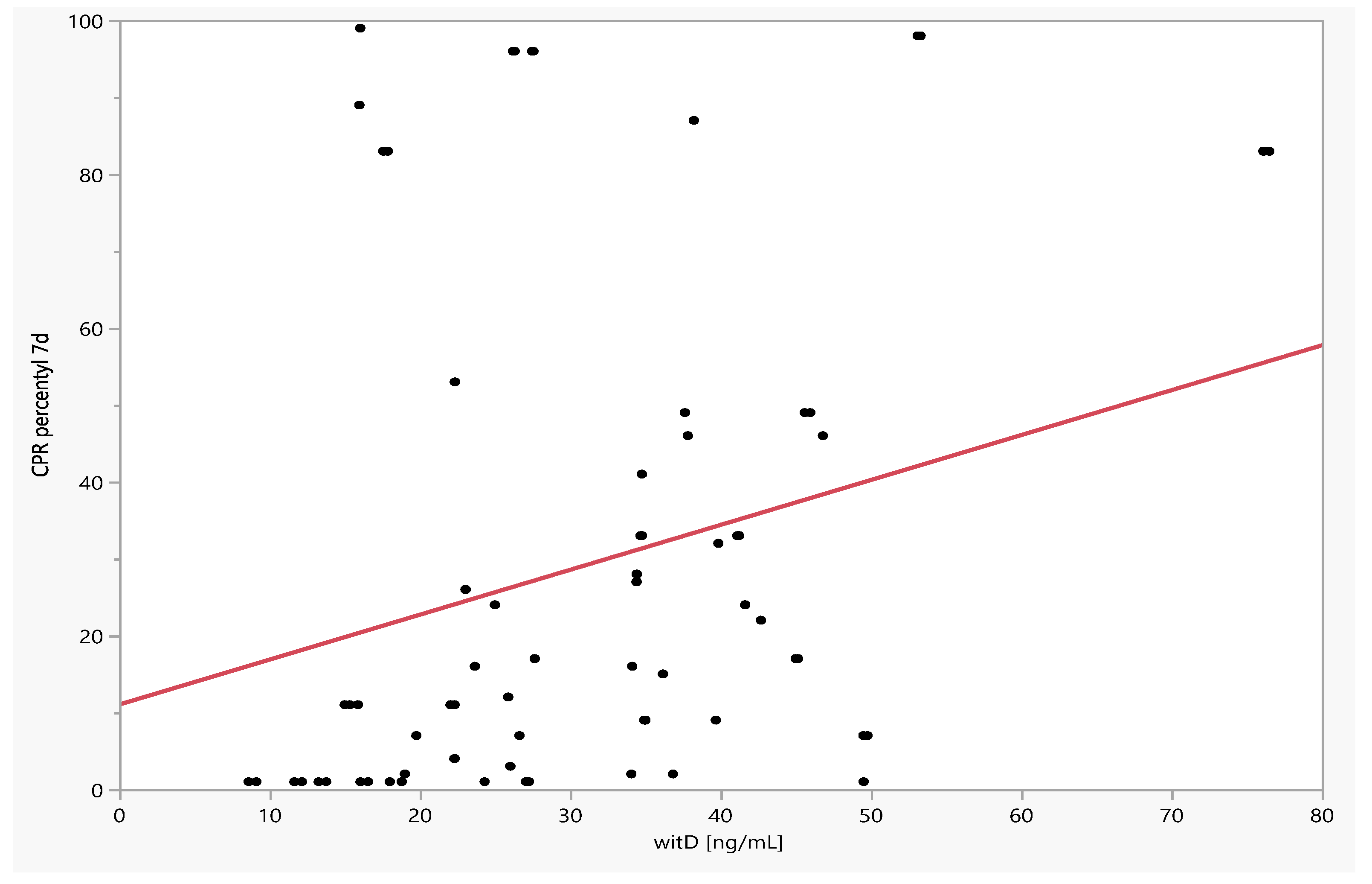
| By Variable | Spearman’s Correlation | Spearman’s Correlation p-Value |
|---|---|---|
| CPR percentile 7 d | 0.3310 | 0.0008 |
| CPR 7 d | 0.3210 | 0.0011 |
| CPR 14 d | 0.2569 | 0.0134 |
| CPR percentile 14 d | 0.2380 | 0.0223 |
| Odds CPR (14-d-baseline) | 0.1863 | 0.0754 |
| CPR baseline | 0.1649 | 0.1011 |
| Odds CPR percentile (14-d-baseline) | 0.1587 | 0.1308 |
| CPR percentile baseline | 0.1463 | 0.1463 |
| Odds CPR percentile (7-d-baseline) | 0.1304 | 0.1960 |
| Odds CPR (7-d-baseline) | 0.0529 | 0.6009 |
Publisher’s Note: MDPI stays neutral with regard to jurisdictional claims in published maps and institutional affiliations. |
© 2022 by the authors. Licensee MDPI, Basel, Switzerland. This article is an open access article distributed under the terms and conditions of the Creative Commons Attribution (CC BY) license (https://creativecommons.org/licenses/by/4.0/).
Share and Cite
Jakubiec-Wisniewska, K.; Huras, H.; Kolak, M. Effect of Vitamin D Supplementation on the Cerebral Placental Ratio in Pregnancy Complicated with Early Fetal Growth Restriction. J. Clin. Med. 2022, 11, 2627. https://doi.org/10.3390/jcm11092627
Jakubiec-Wisniewska K, Huras H, Kolak M. Effect of Vitamin D Supplementation on the Cerebral Placental Ratio in Pregnancy Complicated with Early Fetal Growth Restriction. Journal of Clinical Medicine. 2022; 11(9):2627. https://doi.org/10.3390/jcm11092627
Chicago/Turabian StyleJakubiec-Wisniewska, Karolina, Hubert Huras, and Magdalena Kolak. 2022. "Effect of Vitamin D Supplementation on the Cerebral Placental Ratio in Pregnancy Complicated with Early Fetal Growth Restriction" Journal of Clinical Medicine 11, no. 9: 2627. https://doi.org/10.3390/jcm11092627
APA StyleJakubiec-Wisniewska, K., Huras, H., & Kolak, M. (2022). Effect of Vitamin D Supplementation on the Cerebral Placental Ratio in Pregnancy Complicated with Early Fetal Growth Restriction. Journal of Clinical Medicine, 11(9), 2627. https://doi.org/10.3390/jcm11092627