Review

Risk Stratification in Patients with Acute Pulmonary Embolism: Current Evidence and Perspectives

1
Division of General Internal Medicine, Department of Medicine, Geneva University Hospitals, 1205 Geneva, Switzerland
2
Division of Angiology and Haemostasis, Department of Medicine, Geneva University Hospitals, 1205 Geneva, Switzerland
*
Author to whom correspondence should be addressed.
These authors contributed equally to the manuscript.
J. Clin. Med. 2022, 11(9), 2533; https://doi.org/10.3390/jcm11092533
Submission received: 22 March 2022 / Revised: 26 April 2022 / Accepted: 27 April 2022 / Published: 30 April 2022
(This article belongs to the Special Issue Management of Venous and Arterial Thrombosis)

Abstract

:
Risk stratification is one of the cornerstones of the management of acute pulmonary embolism (PE) and determines the choice of both diagnostic and therapeutic strategies. The first step is the identification of patent circulatory failure, as it is associated with a high risk of immediate mortality and requires a rapid diagnosis and prompt reperfusion. The second step is the estimation of 30-day mortality based on clinical parameters (e.g., original and simplified version of the pulmonary embolism severity index): low-risk patients without right ventricular dysfunction are safely managed with ambulatory anticoagulation. The remaining group of hemodynamically stable patients, labeled intermediate-risk PE, requires hospital admission, even if most of them will heal without complications. In recent decades, efforts have been made to identify a subgroup of patients at an increased risk of adverse outcomes (intermediate-high-risk PE), who might benefit from a more aggressive approach, including reperfusion therapies and admission to a monitored unit. The cur-rent approach, combining markers of right ventricular dysfunction and myocardial injury, has an insufficient positive predictive value to guide primary thrombolysis. Sensitive markers of circulatory failure, such as plasma lactate, have shown interesting prognostic accuracy and may play a central role in the future. Furthermore, the improved security of reduced-dose thrombolysis may enlarge the indication of this treatment to selected intermediate–high-risk PE.

1. Introduction

Pulmonary embolism (PE) is the third most frequent cardiovascular disease and is associated with a high mortality burden, accounting for approximately 300,000 deaths in Europe every year [1,2]. PE is defined as the obstruction of a pulmonary artery, mostly resulting from the dislodgement of thrombotic material from the lower limbs. It has a wide variety of presentations, ranging from an asymptomatic incidental finding to circulatory collapse and sudden death. Diagnosis of PE often requires a sequential strategy combining a pre-test probability assessment, D-dimers measurement when indicated and thoracic imaging. Risk stratification immediately guides the management of acute PE, as it determines the need for urgent reperfusion therapy (high-risk PE) and identifies patients who can be safely treated as outpatients (low-risk PE). The remaining group of patients, called intermediate-risk PE, is highly heterogeneous with most of the patients recovering, but a significant proportion being at risk of complications. The present article will review the evidence supporting risk stratification and reperfusion strategies in the management of PE, with a particular focus on intermediate-risk pulmonary embolism.

2. Risk Stratification in Acute Pulmonary Embolism

Risk stratification is applied in various medical conditions to stratify patients’ severity in therapeutic trials or guide specific diagnostic or therapeutic interventions. Risk stratification often relies on prognostic scores built on clinical or biological parameters. Traditional steps in the development of a prognostic tool include derivation, internal and external validation and impact studies (i.e., studies evaluating the benefit of a risk stratification strategy). While derivation and validation studies are plentiful, impact studies are scarce [3]. Risk stratification in the setting of acute PE includes three main steps: identification of patients at a high risk of early mortality, hence, requiring immediate reperfusion treatment; identification of patients at a low risk of complications who can be safely treated as outpatients; identification of patients with an increased risk of complications requiring hospitalization for close monitoring and potential primary or rescue reperfusion therapy (Figure 1) [4].
Nomenclature and definitions slightly differ between the European Society of Cardiology (ESC) and American Heart Association (AHA) guidelines; differences are highlighted in Table 1 [4,5]. This review mainly relies on the principles outlined in the 2019 ESC guidelines [4].

2.1. Step 1: Identification of High-Risk Patients

The first step in risk stratification of acute PE is the identification of patients at a high risk of early mortality. The most feared complication of acute PE is right ventricular overload and dysfunction which may lead to circulatory collapse and death. Therefore, patients with patent hemodynamic instability are considered as high-risk, according to the ESC criteria. Hemodynamic instability is defined by a systolic blood pressure (SBP) inferior to 90 mmHg for more than 15 min in the absence of hypovolemia, sepsis or arrhythmia; and/or the need of vasopressors in combination with end-organ hypoperfusion. In a recent systematic review including forty thousand patients with PE, 3.9% had high-risk PE. Short-term mortality was 19% among patients presenting with unstable PE versus 5.7% among patients with stable PE (OR 5.9; 95% CI 2.7 to 13.0) [6]. In a recent cohort of 7438 Chinese patients, the prevalence of unstable PE was 4.2% and mortality was 15.8% [7]. In high-risk acute PE, management relies on organ support and prompt reperfusion with thrombolytic therapies or percutaneous/surgical thrombectomy. The benefit of systemic thrombolysis has been demonstrated in small randomized controlled trials (RCTs) and large observational databases [6,8]. In the landmark study by Jerjes-Sanchez et al., eight patients with high-risk PE were randomized to anticoagulation (AC) alone or in combination with streptokinase [8]. The four patients randomized to AC died and the four patients randomized to thrombolysis survived. However, this small size, open-label trial was limited by an imbalance between groups. Recent RCTs evaluating thrombolytic therapy usually excluded high-risk PE [9,10,11], while older studies did not separately report outcomes for high-risk PE [12,13]. In a large North American database including more than two million patients with PE, in-hospital mortality was 15% among patients with unstable PE receiving TT and 47% among patients with unstable PE treated with AC alone. However, 70% of unstable patients did not receive TT [14,15]. This underutilization may be explained by the reluctance of physicians to administer TT because of its bleeding complications. Moreover, the definition of high-risk PE in previous ESC criteria, based exclusively on the presence of hypotension, was probably too simplistic as systolic blood pressure should probably be considered as a continuous risk marker rather than a dichotomized variable [16]. In the 2019 recommendations, they have been enriched by evidence of end-organ dysfunction and the exclusion of alternative contributors of shock [4,17].
In summary, identification of high-risk patients based on the presence of hemodynamic instability is recommended for rapid diagnosis and prompt reperfusion therapy.

2.2. Step 2: Outpatient Management of Low-Risk Pulmonary Embolism

Historically, all patients with acute PE were admitted to the hospital. Short-term mortality prediction rules have, therefore, been developed to identify patients at a low risk of mortality who could be treated as outpatients. The pulmonary embolism severity index (PESI) was derived and internally validated in 2005 in a cohort of 15,531 patients. The PESI score comprises 11 clinical variables and stratifies patients into five severity classes [18]. A simplified version of the PESI (sPESI) was derived including six clinical variables, each scoring one point (Supplementary Table S1) [19]. According to a 2012 meta-analysis including 50,021 patients, the area under the curve (AUC) of sPESI was 0.79 for all-cause mortality with a pooled sensitivity of 0.92 and a pooled specificity of 0.38, which is similar to the original PESI score. The pooled mortality was 2% among patients with PESI class I or II and 1.8% among patients with 0 points in sPESI (Table 2) [20]. A non-inferiority interventional study compared PESI-based outpatient management with hospital admission [21]. Three hundred and forty-four patients with low-risk PE (PESI class I-II) were randomly allocated to outpatient versus inpatient management. Ninety-day mortality was non-inferior in outpatients compared to patients admitted in-hospital (0.6% in both groups, upper confidence limit (UCL) for difference 2.1%) as well as PE recurrence (0.6% in outpatients versus 0% for inpatients, UCL 2.7%). At three months, three outpatients (1.8%), but no inpatients, developed major bleeding (UCL 4.5%).
Interestingly, when a low-risk PESI is combined with the absence of right ventricular dysfunction (RVD), 30-day mortality decreases even further (0.2–0.3%) [23,24]. In a single arm study evaluating the early discharge of low-risk PE (normotensive, absence of RVD and absence of serious comorbidities) treated with rivaroxaban, the rate of major bleeding at 3 months was low (1.2%) [25]. However, this increased sensitivity is obtained at the expense of a significantly lower proportion of patients identified as low risk [26]. An alternative approach to identify low-risk patients is the use of the Hestia criteria (Supplementary Table S2), consisting of a checklist of eleven criteria requiring hospital admission. Home treatment of patients without these criteria has been shown to be safe and non-inferior to sPESI-based home treatment [27,28,29].
In summary, outpatient treatment appears to be safe for low-risk PE patients identified by PESI, sPESI or Hestia criteria and absence of RVD.

3. Step 3: Further Classification of Intermediate-Risk Pulmonary Embolism

About 4% and 40% of acute pulmonary embolisms are categorized as high risk and low risk, respectively [30]. The remaining patients (i.e., normotensive patients with PESI III-V or sPESI ≥ 1) are classified as intermediate-risk PE with an overall 30-day mortality between 5% and 15% [18,19,31]. This group of patients is highly heterogeneous, with the vast majority experiencing a favorable outcome with AC alone, and a small, albeit significant, proportion requiring rescue reperfusion and cardiopulmonary resuscitation. For this reason, efforts have been made in recent decades to identify a subgroup of patients at risk of deterioration who might benefit from initial aggressive therapy and/or admission to a monitored unit. Clinical scores, biological and radiological markers of right ventricular overload and circulatory failure, alone or in combination, have been proposed to further stratify intermediate-risk acute PE.

3.1. Clinical Scores

Despite a high sensitivity and negative predictive value, PESI and sPESI lack specificity to predict early mortality (Table 2). Moreover, these scores rely heavily on demographic and co-morbid conditions rather than the severity of the acute PE event. PE-attributable mortality represents less than half of the overall 3-month mortality among patients with an acute PE [32,33]. Clinical scores alone are, therefore, inadequate to guide admission to monitored units or to initiate reperfusion therapies. Right ventricular dysfunction with circulatory collapse is the most common mechanism leading to fatal PE. Various markers of RVD and circulatory failure have been investigated as potential tools to further stratify normotensive patients; they are detailed in the following sections and summarized in Table 3.

3.2. Markers of Right Ventricular Dysfunction

  • Cardiac troponin
The prognostic value of Troponin I and T has been evaluated in a meta-analysis including 1985 patients [36]. Elevated troponin was associated with increased short-term mortality in the whole cohort and in the subgroup of normotensive patients (OR 5.90, 95% CI 2.68 to 12.95, overall short-term mortality 17.9%). Subsequent meta-analyses questioned the prognostic value of elevated troponin in normotensive patients (positive likelihood ratio 2.13, negative likelihood ratio 0.51) and it has been suggested to combine it with other prognostic factors [34,37].
  • Brain natriuretic peptides
Brain natriuretic peptide (BNP) and its N-terminal portion (NT-proBNP) are secreted by cardiomyocytes in response to ventricular stretching due to volume or pressure overload. The prognostic value of natriuretic peptides has been evaluated in at least eight studies [35,38]. In a meta-analysis, the pooled relative risk for 30-day mortality was 9.5 (95% CI 3.1 to 28.6) for BNP and 8.3 (95% CI 3.6 to 19.3) for NT-proBNP [35]. A meta-analysis of five more recent studies reported a relative risk for the complicated clinical course of 5.63 (95% CI 2.77 to 11.43) when the NT-proBNP value was over 1000 pg/mL [39]. Interestingly, troponin and brain natriuretic peptides seem to have an additive prognostic value [38,40,41].
Other biomarkers, such as serum creatinine, heart fatty acid-binding protein and copeptin, have been studied but are less extensively validated and/or not available in clinical practice [42,43,44,45].
  • Computer tomography pulmonary angiography
CTPA signs of RVD include an elevated RV/left ventricular (LV) end diastolic diameter ratio (cut-off of 0.9 or 1.0), interventricular septum bowing, pulmonary artery enlargement, and retrograde reflux of contrast into the vena cava [46]. The right-to-left ventricular ratio can be easily estimated using the largest transverse diameters which may be measured on different CTPA slices. Septum bowing has an excellent specificity (98%) but poor sensitivity (31%), and inter-observer reproducibility limits its clinical utilization [46,47]. CT obstruction indexes have also been proposed by Qanadli et al. and [48] have shown to be associated with increased mortality, mostly among patients without comorbidities [49].
  • Bedside echocardiography
Increased right-to-left ventricular ratio, hypokinesis of the free RV wall and the presence of pulmonary hypertension estimated from tricuspid regurgitation velocity have been reported to be associated with an increased risk of early complications. More advanced measures, such as the ratio of tricuspid annular plane systolic excursion to pulmonary arterial systolic pressure (TAPSE/PASP) are being investigated to stratify the risk among PE patients, but they are impractical for daily use and bedside stratification [50]. More recently, additional echocardiographic markers such as left or right ventricular outflow tract velocity time integral (LVOT and RVOT VTI) and stroke volume index have been reported to be associated with death or clinical deterioration, with interesting discriminative performance among intermediate-risk patients [51,52,53,54].
Despite a consistent association with short-term mortality, markers of RVD have poor diagnostic performances when they are used as a stand-alone test (Table 3) [17,35]. They have, therefore, been combined in current risk stratification guidelines.

3.3. Current Stratification of Intermediate-Risk Pulmonary Embolism

The ESC 2019 risk stratification of patients with acute PE relies on the three-step process described above, based on the presence of hemodynamic instability, clinical scores (PESI or sPESI) and the combined presence of two markers of RVD (Figure 1 and Table 1). This revision of the previously published 2014 criteria allows us to identify a subgroup of intermediate-risk PE patients at risk of short-term circulatory collapse or mortality, labelled as intermediate–high risk. These criteria were conceived following expert opinions, and no impact study has been published to date. In a prospective cohort of 1015 patients with normotensive acute PE, the ESC 2019 criteria classified 347 (34%), 571 (56%) and 97 (9.6%) of patients as low, intermediate–low and intermediate–high-risk PE, respectively. All cause 30-day mortality was significantly higher in intermediate–high-risk patients (10%) than in those with low risk or intermediate–low risk (4%) [55].

4. Reperfusion Therapy for Intermediate–High-Risk Pulmonary Embolism

Systemic thrombolysis using plasminogen activators is the most widely studied reperfusion strategy. Tissue plasminogen activators (tPA), such as urokinase, alteplase or tenecteplase, have a fibrinolytic effect, allowing clot dissolution and improvement of hemodynamic parameters in patients with high-risk PE [8,13]. Several randomized controlled trials aimed to evaluate their potential benefit among normotensive patients with elevated markers of RVD. The most informative evidence is provided by the European Pulmonary Embolism Trombolysis (PEITHO) trial [10]. This large RCT included 1005 patients with acute PE and RVD on imaging (CTPA or echocardiography) and myocardial injury (elevated troponin T or I), corresponding to the current intermediate–high-risk category. Patients were randomly assigned to unfractionated heparin (UFH) plus tenecteplase or UFH alone. The incidence of the primary outcome (death or hemodynamic collapse within 1 week) was significantly lower among patients allocated to tenecteplase than to UFH alone (2.6% vs. 5.6%, p = 0.02). This difference was mainly driven by an increased risk in hemodynamic decompensation among patients allocated to UFH (5.0% vs. 1.6%) while mortality was low and did not significantly differ (1.2% vs. 1.8%). This was counterbalanced by a significant increase in the risk of both major (11.5% vs. 2.4%) and intracranial bleeding (2.0% vs. 0.2%). When thrombolysis studies exclusively including acute PE with RVD are pooled, the uncertain benefit in overall mortality is mitigated by the significant increase in both major and intracranial bleeding [11]. Of note, the increased risk of major bleeding was more pronounced in studies using tenecteplase than in those using alteplase, but direct comparison studies are lacking to confirm this observation [56].

5. Toward a Better Identification of Thrombolysis Candidates among Intermediate-Risk Pulmonary Embolism

The current ESC classification seems to have an insufficient positive predictive value to identify a subgroup of intermediate-risk patients warranting more aggressive therapy. The benefits of a full-dose systemic thrombolysis are outweighed by bleeding risks, particularly when using tenecteplase. Two strategies have been identified by researchers to enhance the benefit-risk ratio: identifying patients at a higher basal risk and improving the safety of TT.

Identifying Patients at Higher Basal Risk: Markers of Circulatory Failure and Alternative Scores

Various alternative prediction rules, including early markers of circulatory failure, have been studied to identify normotensive patients with a higher risk of hemodynamic collapse.
  • Plasma lactate
Plasma lactate is an important prognostic marker of organ dysfunction and is widely used in patients with sepsis or trauma [57]. Several studies evaluated the prognostic value of plasma lactate among patients with acute PE [58,59]. A retrospective study including 287 patients with acute PE reported a significant association between plasma lactate levels above 2 mmol/L and in-hospital mortality (OR 4.6; 95% CI 1.57 to 13.53) [58]. An association with 30-day mortality was subsequently observed in a prospective study (HR 11.67; 95% CI 3.32 to 41.03) [59]. Interestingly, this association was independent of shock state, hypotension, RVD or elevated troponin. A single center registry of 419 consecutive PE patients confirmed the association of elevated venous lactate with adverse outcomes, and found that levels above 3.3 mmol/L had the best predictive performance for in-hospital adverse events (PPV 0.27 and NPV 0.97) [60]. Moreover, adding venous lactate levels to the ESC 2019 risk criteria allowed us to further fine-tune stratification. Intermediate–high-risk patients with venous lactate ≥3.3 mmol/L had a 27.5% prevalence of adverse events, versus 6.8% if lactate was <3.3 mmol/L. Intermediate low-risk patients with lactate levels <2.3 mmol/L were at a low risk of adverse events (0.6%, versus 12.2% if ≥2.3 mmol/L) [60].
  • BOVA score
The Bova score was derived from pooled results of six prospective studies, including 2874 patients with hemodynamically stable acute PE [61]. Model predictors included heart rate, SBP, biomarkers (cardiac troponin or BNP) and echocardiography (Table 4 and Supplementary Table S3) [61]. RVD was defined by the presence of RV/LV >0.9 or 1, RV free wall hypokinesis, RV end-diastolic diameter >30 mm or estimated systolic pulmonary artery pressure > 30 mmH. The primary composite outcome was PE-related death, hemodynamic collapse or recurrent PE at 30 days. Thirty-day complications differed significantly across categories of the model (0–2 points 4.2%; 3–4 points 10.8%; >4 points 29.2%). The area under the ROC curve was 0.73 (95% CI 0.68–0.77) and 5.8% of patients were classified in the stage III category. Recently, a meta-analysis including the derivation study and eight prospective and retrospective external validation cohort studies were conducted [62]. The pooled cumulative incidence of PE-related complications (PE-related death, hemodynamic collapse or recurrent PE) at 30 days was 3.8% for stage I, 10.8% for stage II and 19.9% for stage III (1.9, 5.5 and 12.1 for 30-day PE mortality) with an AUC of 0.73 (Table 4) [63]. In another retrospective cohort including 994 normotensive patients, 5.9% of patients were classified in the stage III category. Death or hemodynamic collapse at 7 days occurred in 18.6% of patients in the stage III category. When lactate elevation was incorporated into an extended Bova score, the proportion of patients in the stage III category increased to 11.2 %, with a primary outcome rate of 25.9 %. Hemodynamic collapse by day 7 occurred in 15.3% of patients in the class III category according to the standard BOVA score, compared to 24.1% in the model including lactate elevation [64].
  • TELOS score
The TELOS score was derived in a prospective cohort of 496 normotensive PE patients. The primary outcome was PE-related death or hemodynamic collapse within 7 days. A model including RVD, troponin and plasma lactate elevation resulted in a 17.9% PPV [65]. The TELOS rule was further validated by the same group in a prospective cohort of 994 normotensive patients. A total of 5.9% of patients were allocated to the intermediate–high-risk category according to the TELOS criteria, with a cumulative incidence of the primary outcome (death or hemodynamic collapse at 7 days) of 21.1% (Table 5) [64].
  • SHIELD score
The SHIELD score was derived from a retrospective monocentric cohort of 554 normotensive patients and was externally validated. Predictors of the model included shock index ≥1, hypoxemia, lactate elevation and signs of RVD (i.e., elevated troponin, NT-pro BNP and RV/LV ratio >1 using CTPA) [68]. The risk of 30-day mortality or rescue thrombolysis for each tercile was 0.6%, 1.8% and 16.4% (AUC 0.90, 95% CI 0.85 to 0.94) in the derivation cohort and 0.6%, 1.9% and 15.3% (AUC 0.82; 95% CI 0.75 to 0.87) in the external validation cohort.
  • Other scores
Lankeit et al. derived a clinical prediction rule including heart fatty acid-binding protein (H-FABP), syncope and heart rate (FAST score). The positive predictive value was 20.5% and the AUC was 0.85 (95% CI 0.75 to 0.95) [69]. This score was validated in another cohort of the same center with a positive predictive value of 18.9% and an AUC of 0.82 (95% CI 0.75 to 0.89) [66].
A prospective study including 268 normotensive PE patients showed an association between copeptin level >24 pmol/L and 30-day mortality or adverse outcome. The positive predictive value was 11% (95% CI 7 to 19%) and the negative predictive value was 98% (95% CI 95 to 99%), suggesting that this biomarker could be combined with markers of RVD such as NT-proBNP or highly sensitive troponin T [42].
Other scores combining clinical variables, imaging and biomarkers have been studied with less extensive validation [70,71].
  • Between score comparison
Vanni et al. compared the prognostic accuracy of the ESC 2014 criteria, TELOS and Bova score in a cohort of 994 normotensive patients with PE. The Bova and TELOS scores classified the same proportion of patients in the intermediate–high-risk category (5.9 and 5.7%) with a similar rate of early adverse events (18.6 and 21.1% 7-day death or hemodynamic collapse), while the ESC criteria classified a higher proportion of patients in the intermediate–high-risk category (12.5%, p < 0.001) with a lower rate of events (13% p = 0.18) [54]. Diagnostic performances of several existing scores are summarized in Table 5. While the risk of adverse events was comprised between 7% and 15% in the intermediate–high-risk group according to the ESC criteria, recent prediction rules, including markers of circulatory dysfunction (plasma lactates) or adjunction of plasma lactates to the Bova or ESC 2019 criteria, appeared to have a promising positive predictive value with event rates of around 25%. However, some variation in their PPVs was observed across studies, which is partly explained by variations in the outcome definitions. Moreover, the plasma lactate cut-off varied across studies and the optimal cut-off remains to be determined.

6. Expected Benefits from a Better Identification of Intermediate–High-Risk Patients

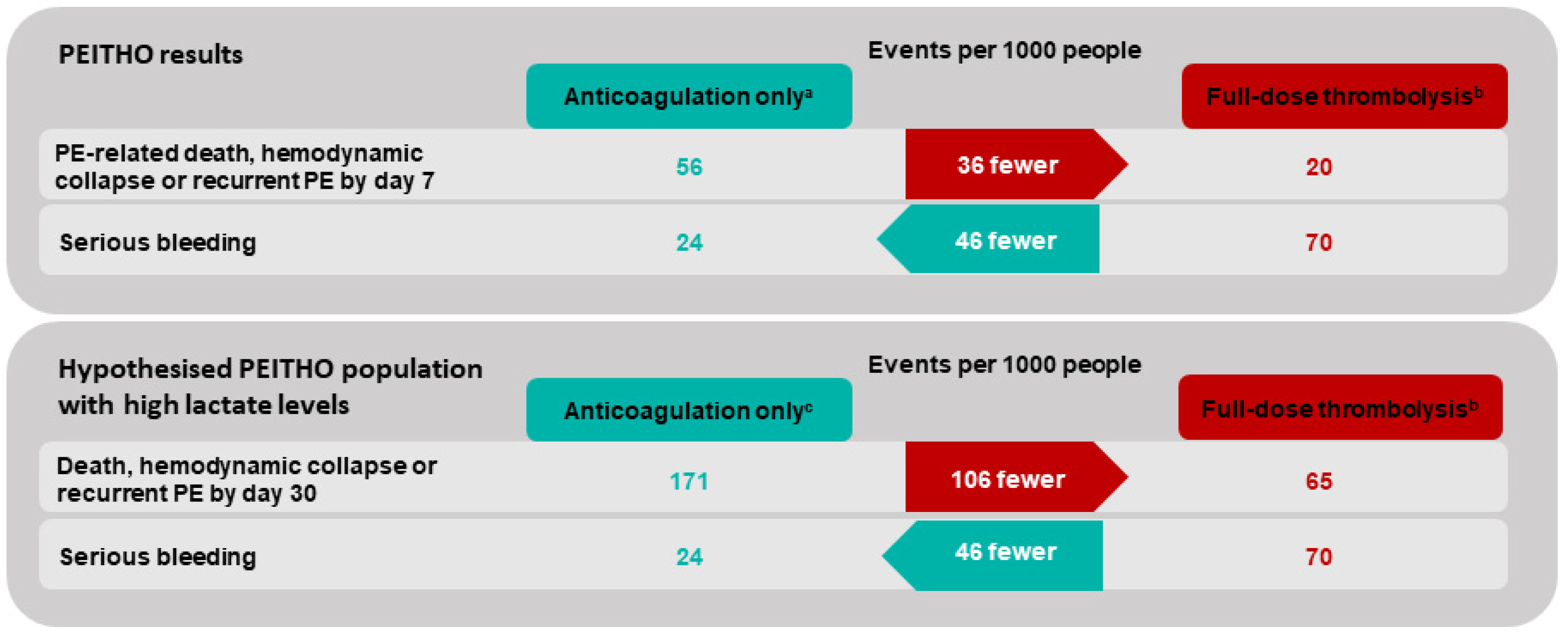
As discussed above, recent prediction rules, including markers of circulatory dysfunction, might allow us to identify a subgroup of patients with a significantly increased (>25%) risk in adverse events who might benefit from a more aggressive therapy. Figure 2 illustrates the extrapolated benefits of thrombolytic therapy in a theoretical cohort of patients with a 17.1% basal risk of adverse outcomes. This basal risk was obtained by combining the basal risk observed in the PEITHO trial and the positive likelihood ratio of elevated blood lactates [9]. Net benefits and harms were computed based on the relative risks reported in our previous meta-analysis on thrombolytic therapy in PE [11]. These extrapolations are mainly illustrative and should be evaluated in interventional studies, as bleeding complication risks might also increase among patients with a higher basal risk of a PE-related adverse event.

7. Improving the Safety Profile: Reduced-Dose Thrombolytic Therapy

Another strategy to optimize the risk-benefit ratio of TT among intermediate–high-risk PE is the reduction in TT bleeding complications. In this perspective, several trials investigated reduced-dose systemic thrombolysis regimens. In the Moderate Pulmonary Embolism Treated with Thrombolysis (MOPETT) trial, 121 patients with acute PE and a high thrombotic burden were randomized to half-dose tPA and heparin versus heparin alone. Half-dose thrombolysis was associated with a lower rate of pulmonary hypertension or recurrent PE at 28 months (16% versus 63% in the AC group) and no significantly increased risk of bleeding [72]. Similarly, a systematic review including 5 small-size randomized trials suggested a similar efficacy and reduced bleeding complications when half-dose was compared to standard dose thrombolysis [73]. These preliminary results based on a limited sample of patients need to be further confirmed.
Catheter-based thrombolytic therapies, including catheter-released thrombolysis (CRT), ultrasound-assisted thrombolysis and mechanical fragmentation or aspiration, alone or in combination, are alternative strategies to reduce bleeding risks associated with systemic TT [74,75]. Catheter-based therapies are an evolving technology and several devices have been shown to improve echocardiographic signs of RVD in single-arm studies [75,76]. Their impact regarding clinically relevant outcomes should be further evaluated in randomized controlled studies [77]. These alternatives may be proposed to high-risk patients at an increased risk of bleeding. More recently, circulatory support using extracorporeal membrane oxygenation (ECMO) in combination with surgical embolectomy has been proposed for high-risk patients with high in-hospital survival rates [78].

8. Evidence to Come: The PEITHO-3 Study

The ongoing PEITHO-3 study is expected to add further evidence for the management of intermediate–high-risk PE. This ongoing, multicentric, randomized controlled trial aims to combine the two previously discussed strategies. Identification of patients with a higher basal risk of adverse events will be obtained based on a retrospective analysis of the PEITHO population by adding clinical markers of severity (SBP ≤ 110 mmHg, respiratory rate >20/min, history of chronic heart failure) to the ESC intermediate–high-risk criteria with an expected event rate of 11.2% [79,80]. At the same time, a reduced dose of alteplase (0.6 mg/kg) will be used. The PEITHO-3 trial plans to include 650 patients with, expected to be completed in September 2025 [80]. The expected benefits in the hypothesized PEITHO-3 population are illustrated in Figure 3, assuming a constant relative effect of treatment and a constant basal bleeding risk with important uncertainty regarding the increase in bleeding complications with reduced-dose TT.

9. Conclusions

The management of patients with acute PE requires an accurate step-by-step risk stratification. Hemodynamic instability allows to quickly detect high-risk patients who will benefit from TT; clinical prediction rules, such as PESI or sPESI, allow us to identify low-risk patients who can be safely treated as outpatients. The intermediate-risk group is composed of a highly heterogeneous group of patients, most of whom will experience favorable short-term outcomes. Individual evaluations by dedicated multidisciplinary PE response teams may be useful to decide the optimal treatment strategy [81,82]. Several prediction models combining clinical variables, biomarkers and medical imaging have been developed to identify the subgroup of patients with a significant risk of early adverse events who might benefit from early aggressive management. The inclusion of plasma lactate may increase the performance of risk stratification models, but it needs to be further validated in future studies. Optimization of reperfusion strategies by using reduced regimens of thrombolysis or catheter-directed reperfusion techniques might contribute to improving the prognosis of patients by limiting the side effects. Results of ongoing studies (NCT04430569) will clarify the benefits of initial reperfusion strategies in this subgroup of patients. While waiting for these results, current guidelines recommend admitting patients of the intermediate–high-risk category to a monitored unit for early detection of clinical deterioration and the potential need for rescue reperfusion.

Supplementary Materials

The following supporting information can be downloaded at: https://www.mdpi.com/article/10.3390/jcm11092533/s1, Supplementary Table S1. Original and simplified pulmonary embolism severity index (PESI and sPESI); Supplementary Table S2. The Hestia rule; Supplementary Table S3. Components of several prediction scores.

Author Contributions

Conceptualization, A.L., S.B., C.M.; methodology, A.L., S.B., C.M.; validation, A.L., S.B., C.M., A.B., O.G., M.R.; resources, A.L., C.M., S.B.; writing—original draft preparation, A.L., S.B., C.M.; writing—review and editing, A.L., S.B., C.M., M.R., O.G., A.B.; supervision, C.M., M.R.; Y. All authors have read and agreed to the published version of the manuscript.

Funding

This research received no external funding.

Institutional Review Board Statement

Not applicable.

Informed Consent Statement

Not applicable.

Conflicts of Interest

The authors declare no conflict of interest.

References

  1. ISTH Steering Committee for World Thrombosis Day. Thrombosis: A major contributor to the global disease burden. J. Thromb. Haemost. 2014, 12, 1580–1590. [Google Scholar] [CrossRef] [PubMed]
  2. Cohen, A.T.; Agnelli, G.; Anderson, F.A.; Arcelus, J.I.; Bergqvist, D.; Brecht, J.G.; Greer, I.A.; Heit, J.A.; Hutchinson, J.L.; Kakkar, A.K. Venous thromboembolism (VTE) in Europe. The number of VTE events and associated morbidity and mortality. Thromb. Haemost. 2007, 98, 756–764. [Google Scholar] [PubMed]
  3. Reilly, B.M.; Evans, A.T. Translating clinical research into clinical practice: Impact of using prediction rules to make decisions. Ann. Intern. Med. 2006, 144, 201–209. [Google Scholar] [CrossRef]
  4. Konstantinides, S.V.; Meyer, G. The 2019 ESC Guidelines on the Diagnosis and Management of Acute Pulmonary Embolism. Eur. Heart J. 2019, 40, 3453–3455. [Google Scholar] [CrossRef] [PubMed] [Green Version]
  5. Jaff, M.R.; McMurtry, M.S.; Archer, S.L.; Cushman, M.; Goldenberg, N.; Goldhaber, S.Z.; Jenkins, J.S.; Kline, J.A.; Michaels, A.D.; Thistlethwaite, P.; et al. Management of massive and submassive pulmonary embolism, iliofemoral deep vein thrombosis, and chronic thromboembolic pulmonary hypertension: A scientific statement from the American Heart Association. Circulation 2011, 123, 1788–1830. [Google Scholar] [CrossRef] [Green Version]
  6. Quezada, C.A.; Bikdeli, B.; Barrios, D.; Barbero, E.; Chiluiza, D.; Muriel, A.; Casazza, F.; Monreal, M.; Yusen, R.D.; Jiménez, D. Meta-Analysis of Prevalence and Short-Term Prognosis of Hemodynamically Unstable Patients with Symptomatic Acute Pulmonary Embolism. Am. J. Cardiol. 2019, 123, 684–689. [Google Scholar] [CrossRef]
  7. Zhai, Z.; Wang, D.; Lei, J.; Yang, Y.; Xu, X.; Ji, Y.; Yi, Q.; Chen, H.; Hu, X.; Liu, Z.; et al. Trends in risk stratification, in-hospital management and mortality of patients with acute pulmonary embolism: An analysis from the China pUlmonary thromboembolism REgistry Study (CURES). Eur. Respir. J. 2021, 58, 2002963. [Google Scholar] [CrossRef]
  8. Jerjes-Sanchez, C.; Ramirez-Rivera, A.; de Lourdes Garcia, M.; Arriaga-Nava, R.; Valencia, S.; Rosado-Buzzo, A.; Pierzo, J.A.; Rosas, E. Streptokinase and heparin versus heparin alone in massive pulmonary embolism: A randomized controlled trial. J. Thromb. Thrombolysis 1995, 2, 227–229. [Google Scholar] [CrossRef]
  9. Konstantinides, S.V.; Geibel, A.; Heusel, G.; Heinrich, F.; Kasper, W. Heparin plus Alteplase Compared with Heparin Alone in Patients with Submassive Pulmonary Embolism. N. Engl. J. Med. 2002, 347, 1143–1150. [Google Scholar] [CrossRef] [Green Version]
  10. Meyer, G.; Vicaut, E.; Danays, T.; Agnelli, G.; Becattini, C.; Beyer-Westendorf, J.; Bluhmki, E.; Bouvaist, H.; Brenner, B.; Couturaud, F.; et al. Fibrinolysis for patients with intermediate-risk pulmonary embolism. N. Engl. J. Med. 2014, 370, 1402–1411. [Google Scholar] [CrossRef]
  11. Marti, C.; John, G.; Konstantinides, S.; Combescure, C.; Sanchez, O.; Lankeit, M.; Meyer, G.; Perrier, A. Systemic thrombolytic therapy for acute pulmonary embolism: A systematic review and meta-analysis. Eur. Heart J. 2014, 36, 605–614. [Google Scholar] [CrossRef] [PubMed] [Green Version]
  12. Dotter, C.T.; Seaman, A.J.; Rösch, J.; Porter, J.M. Streptokinase and Heparin in the Treatment of Pulmonary Embolism: A Randomized Comparison. Vasc. Surg. 1979, 13, 42–52. [Google Scholar] [CrossRef]
  13. Urokinase Pulmonary Embolism Trial Study Group. Urokinase pulmonary embolism trial. Phase 1 results: A cooperative study. JAMA 1970, 214, 2163–2172. [Google Scholar] [CrossRef]
  14. Stein, P.D.; Matta, F. Thrombolytic therapy in unstable patients with acute pulmonary embolism: Saves lives but underused. Am. J. Med. 2012, 125, 465–470. [Google Scholar] [CrossRef] [PubMed]
  15. Stein, P.D.; Matta, F.; Hughes, P.G.; Hughes, M.J. Adjunctive Therapy and Mortality in Patients With Unstable Pulmonary Embolism. Am. J. Cardiol. 2020, 125, 1913–1919. [Google Scholar] [CrossRef]
  16. Quezada, A.; Jiménez, D.; Bikdeli, B.; Moores, L.; Porres-Aguilar, M.; Aramberri, M.; Lima, J.; Ballaz, A.; Yusen, R.D.; Monreal, M. Systolic blood pressure and mortality in acute symptomatic pulmonary embolism. Int. J. Cardiol. 2019, 302, 157–163. [Google Scholar] [CrossRef]
  17. Konstantinides, S.V. 2014 ESC Guidelines on the diagnosis and management of acute pulmonary embolism. Eur. Heart J. 2014, 35, 3145–3146. [Google Scholar] [CrossRef] [Green Version]
  18. Aujesky, D.; Obrosky, D.S.; Stone, R.A.; Auble, T.E.; Perrier, A.; Cornuz, J.; Roy, P.-M.; Fine, M.J. Derivation and Validation of a Prognostic Model for Pulmonary Embolism. Am. J. Respir. Crit. Care Med. 2005, 172, 1041–1046. [Google Scholar] [CrossRef]
  19. Jiménez, D.; Aujesky, D.; Moores, L.; Gómez, V.; Lobo, J.L.; Uresandi, F.; Otero, R.; Monreal, M.; Muriel, A.; Yusen, R.D. Simplification of the Pulmonary Embolism Severity Index for Prognostication in Patients With Acute Symptomatic Pulmonary Embolism. Arch. Intern. Med. 2010, 170, 1383–1389. [Google Scholar] [CrossRef] [Green Version]
  20. Zhou, X.-Y.; Ben, S.-Q.; Chen, H.-L.; Ni, S.-S. The prognostic value of pulmonary embolism severity index in acute pulmonary embolism: A meta-analysis. Respir. Res. 2012, 13, 1–12. [Google Scholar] [CrossRef] [Green Version]
  21. Aujesky, D.; Roy, P.-M.; Verschuren, F.; Righini, M.; Osterwalder, J.; Egloff, M.; Renaud, B.; Verhamme, P.; Stone, R.A.; Legall, C.; et al. Outpatient versus inpatient treatment for patients with acute pulmonary embolism: An international, open-label, randomised, non-inferiority trial. Lancet 2011, 378, 41–48. [Google Scholar] [CrossRef]
  22. Kohn, C.G.; Mearns, E.S.; Parker, M.W.; Hernandez, A.V.; Coleman, C.I. Prognostic accuracy of clinical prediction rules for early post-pulmonary embolism all-cause mortality: A bivariate meta-analysis. Chest 2015, 147, 1043–1062. [Google Scholar] [CrossRef] [PubMed]
  23. Barco, S.; Mahmoudpour, S.H.; Planquette, B.; Sanchez, O.; Konstantinides, S.V.; Meyer, G. Prognostic value of right ventricular dysfunction or elevated cardiac biomarkers in patients with low-risk pulmonary embolism: A systematic review and meta-analysis. Eur. Heart J. 2019, 40, 902–910. [Google Scholar] [CrossRef] [PubMed] [Green Version]
  24. Becattini, C.; Maraziti, G.; Vinson, D.R.; Ng, A.C.C.; Exter, P.L.D.; Côté, B.; Vanni, S.; Doukky, R.; Khemasuwan, D.; Weekes, A.J.; et al. Right ventricle assessment in patients with pulmonary embolism at low risk for death based on clinical models: An individual patient data meta-analysis. Eur. Heart J. 2021, 42, 3190–3199. [Google Scholar] [CrossRef] [PubMed]
  25. Barco, S.; Schmidtmann, I.; Ageno, W.; Bauersachs, R.M.; Becattini, C.; Bernardi, E.; Beyer-Westendorf, J.; Bonacchini, L.; Brachmann, J.; Christ, M.; et al. Early discharge and home treatment of patients with low-risk pulmonary embolism with the oral factor Xa inhibitor rivaroxaban: An international multicentre single-arm clinical trial. Eur. Heart J. 2019, 41, 509–518. [Google Scholar] [CrossRef] [PubMed] [Green Version]
  26. Myc, L.A.; Richardson, E.D.; Barros, A.J.; Watson, J.T.; Sharma, A.M.; Kadl, A. Risk stratification in acute pulmonary embolism: Half of the way there? Ann. Am. Thorac. Soc. 2021, 18, 1066–1068. [Google Scholar] [CrossRef] [PubMed]
  27. Roy, P.-M.; Penaloza, A.; Hugli, O.; Klok, F.A.; Arnoux, A.; Elias, A.; Couturaud, F.; Joly, L.-M.; Lopez, R.; Faber, L.M.; et al. Triaging acute pulmonary embolism for home treatment by Hestia or simplified PESI criteria: The HOME-PE randomized trial. Eur. Heart J. 2021, 42, 3146–3157. [Google Scholar] [CrossRef]
  28. den Exter, P.L.; Zondag, W.; Klok, F.A.; Brouwer, R.E.; Dolsma, J.; Eijsvogel, M.; Faber, L.M.; van Gerwen, M.; Grootenboers, M.J.; Heller-Baan, R.; et al. Efficacy and Safety of Outpatient Treatment Based on the Hestia Clinical Decision Rule with or without N-Terminal Pro-Brain Natriuretic Peptide Testing in Patients with Acute Pulmonary Embolism. A Randomized Clinical Trial. Am. J. Respir. Crit. Care Med. 2016, 194, 998–1006. [Google Scholar] [CrossRef]
  29. Zondag, W.; Hiddinga, B.I.; Crobach, M.J.; Labots, G.; Dolsma, A.; Durian, M.; Faber, L.M.; Hofstee, H.M.; Melissant, C.F.; Ullmann, E.F.; et al. Hestia criteria can discriminate high- from low-risk patients with pulmonary embolism. Eur. Respir. J. 2013, 41, 588–592. [Google Scholar] [CrossRef] [Green Version]
  30. Elias, A.; Mallett, S.; Daoud-Elias, M.; Poggi, J.-N.; Clarke, M. Prognostic models in acute pulmonary embolism: A systematic review and meta-analysis. BMJ Open 2016, 6, e010324. [Google Scholar] [CrossRef]
  31. Sanchez, D.; De Miguel, J.; Sam, A.; Wagner, C.; Zamarro, C.; Nieto, R.; Garcia, L.; Aujesky, D.; Yusen, R.D.; Jiménez, D. The effects of cause of death classification on prognostic assessment of patients with pulmonary embolism. J. Thromb. Haemost. 2011, 9, 2201–2207. [Google Scholar] [CrossRef] [PubMed]
  32. Goldhaber, S.Z.; Visani, L.; De Rosa, M. Acute pulmonary embolism: Clinical outcomes in the International Cooperative Pulmonary Embolism Registry (ICOPER). Lancet 1999, 353, 1386–1389. [Google Scholar] [CrossRef]
  33. Laporte, S.; Mismetti, P.; Décousus, H.; Uresandi, F.; Otero, R.; Lobo, J.L.; Monreal, M.; The RIETE Investigators. Clinical predictors for fatal pulmonary embolism in 15,520 patients with venous thromboembolism: Findings from the Registro Informatizado de la Enfermedad TromboEmbolica venosa (RIETE) Registry. Circulation 2008, 117, 1711–1716. [Google Scholar] [CrossRef] [PubMed]
  34. Bajaj, A.; Saleeb, M.; Rathor, P.; Sehgal, V.; Kabak, B.; Hosur, S. Prognostic value of troponins in acute nonmassive pulmonary embolism: A meta-analysis. Heart Lung 2015, 44, 327–334. [Google Scholar] [CrossRef] [PubMed]
  35. Sanchez, O.; Trinquart, L.; Colombet, I.; Durieux, P.; Huisman, M.V.; Chatellier, G.; Meyer, G. Prognostic value of right ventricular dysfunction in patients with haemodynamically stable pulmonary embolism: A systematic review. Eur. Heart J. 2008, 29, 1569–1577. [Google Scholar] [CrossRef] [PubMed] [Green Version]
  36. Becattini, C.; Vedovati, M.C.; Agnelli, G. Prognostic value of troponins in acute pulmonary embolism: A meta-analysis. Circulation 2007, 116, 427–433. [Google Scholar] [CrossRef] [Green Version]
  37. Jiménez, D.; Uresandi, F.; Otero, R.; Lobo, J.L.; Monreal, M.; Martí, D.; Zamora, J.; Muriel, A.; Aujesky, D.; Yusen, R.D. Troponin-based risk stratification of patients with acute nonmassive pulmonary embolism: Systematic review and metaanalysis. Chest 2009, 136, 974–982. [Google Scholar] [CrossRef]
  38. Lankeit, M.; Jiménez, D.; Kostrubiec, M.; Dellas, C.; Kuhnert, K.; Hasenfuß, G.; Pruszczyk, P.; Konstantinides, S. Validation of N-terminal pro-brain natriuretic peptide cut-off values for risk stratification of pulmonary embolism. Eur. Respir. J. 2014, 43, 1669–1677. [Google Scholar] [CrossRef] [Green Version]
  39. Nithianandan, H.; Reilly, A.; Tritschler, T.; Wells, P. Applying rigorous eligibility criteria to studies evaluating prognostic utility of serum biomarkers in pulmonary embolism: A systematic review and meta-analysis. Thromb. Res. 2020, 195, 195–208. [Google Scholar] [CrossRef]
  40. Jiménez, D.; Kopecna, D.; Tapson, V.; Briese, B.; Schreiber, D.; Lobo, J.L.; Monreal, M.; Aujesky, D.; Sanchez, O.; Meyer, G.; et al. Derivation and validation of multimarker prognostication for normotensive patients with acute symptomatic pulmonary embolism. Am. J. Respir. Crit. Care Med. 2014, 189, 718–726. [Google Scholar] [CrossRef]
  41. Santos, A.R.; Freitas, P.; Ferreira, J.; Félix-Oliveira, A.; Gonçalves, M.; Faria, D.; Augusto, J.; Simões, J.; Gago, M.; Oliveira, J.; et al. Risk stratification in normotensive acute pulmonary embolism patients: Focus on the intermediate–high risk subgroup. Eur. Heart J. Acute Cardiovasc. Care 2020, 9, 279–285. [Google Scholar] [CrossRef] [PubMed]
  42. Hellenkamp, K.; Schwung, J.; Rossmann, H.; Kaeberich, A.; Wachter, R.; Hasenfuß, G.; Konstantinides, S.; Lankeit, M. Risk stratification of normotensive pulmonary embolism: Prognostic impact of copeptin. Eur. Respir. J. 2015, 46, 1701–1710. [Google Scholar] [CrossRef] [PubMed] [Green Version]
  43. Kostrubiec, M.; Pływaczewska, M.; Jiménez, D.; Lankeit, M.; Ciurzynski, M.; Konstantinides, S.; Pruszczyk, P. The Prognostic Value of Renal Function in Acute Pulmonary Embolism—A Multi-Centre Cohort Study. Thromb. Haemost. 2019, 119, 140–148. [Google Scholar] [CrossRef] [PubMed]
  44. Chopard, R.; Jimenez, D.; Serzian, G.; Ecarnot, F.; Falvo, N.; Kalbacher, E.; Bonnet, B.; Capellier, G.; Schiele, F.; Bertoletti, L.; et al. Renal dysfunction improves risk stratification and may call for a change in the management of intermediate- and high-risk acute pulmonary embolism: Results from a multicenter cohort study with external validation. Crit. Care 2021, 25, 1–11. [Google Scholar] [CrossRef]
  45. Dellas, C.; Puls, M.; Lankeit, M.; Schäfer, K.; Cuny, M.; Berner, M.; Hasenfuss, G.; Konstantinides, S. Elevated heart-type fatty acid-binding protein levels on admission predict an adverse outcome in normotensive patients with acute pulmonary embolism. J. Am. Coll. Cardiol. 2010, 55, 2150–2157. [Google Scholar] [CrossRef] [Green Version]
  46. John, G.; Marti, C.; Poletti, P.-A.; Perrier, A. Hemodynamic Indexes Derived from Computed Tomography Angiography to Predict Pulmonary Embolism Related Mortality. BioMed Res. Int. 2014, 2014, 1–8. [Google Scholar] [CrossRef] [Green Version]
  47. Chornenki, N.L.J.; Poorzargar, K.; Shanjer, M.; Mbuagbaw, L.; Delluc, A.; Crowther, M.; Siegal, D.M. Detection of right ventricular dysfunction in acute pulmonary embolism by computed tomography or echocardiography: A systematic review and meta-analysis. J. Thromb. Haemost. 2021, 19, 2504–2513. [Google Scholar] [CrossRef]
  48. Qanadli, S.D.; El Hajjam, M.; Vieillard-Baron, A.; Joseph, T.; Mesurolle, B.; Oliva, V.L.; Barré, O.; Bruckert, F.; Dubourg, O.; Lacombe, P. New CT index to quantify arterial obstruction in pulmonary embolism: Comparison with angiographic index and echocardiography. AJR Am. J. Roentgenol. 2001, 176, 1415–1420. [Google Scholar] [CrossRef]
  49. Kay, F.U.; Abbara, S. Refining Risk Stratification in Nonmassive Acute Pulmonary Embolism. Radiol. Cardiothorac. Imaging 2020, 2, e200458. [Google Scholar] [CrossRef]
  50. Lyhne, M.D.; Kabrhel, C.; Giordano, N.; Andersen, A.; Nielsen-Kudsk, J.E.; Zheng, H.; Dudzinski, D.M. The echocardiographic ratio tricuspid annular plane systolic excursion/pulmonary arterial systolic pressure predicts short-term adverse outcomes in acute pulmonary embolism. Eur. Heart J. Cardiovasc. Imaging 2020, 22, 285–294. [Google Scholar] [CrossRef]
  51. Brailovsky, Y.; Lakhter, V.; Weinberg, I.; Porcaro, K.; Haines, J.; Morris, S.; Masic, D.; Mancl, E.; Bashir, R.; Alkhouli, M.; et al. Right Ventricular Outflow Doppler Predicts Low Cardiac Index in Intermediate Risk Pulmonary Embolism. Clin. Appl. Thromb. 2019, 25, 1076029619886062. [Google Scholar] [CrossRef] [PubMed] [Green Version]
  52. Yuriditsky, E.; Mitchell, O.J.; Sibley, R.A.; Xia, Y.; Sista, A.; Zhong, J.; Moore, W.H.; Amoroso, N.E.; Goldenberg, R.; Smith, D.E.; et al. Low left ventricular outflow tract velocity time integral is associated with poor outcomes in acute pulmonary embolism. Vasc. Med. 2020, 25, 133–140. [Google Scholar] [CrossRef] [PubMed]
  53. Yuriditsky, E.; Mitchell, O.J.; Sista, A.; Xia, Y.; Sibley, R.A.; Zhong, J.; Moore, W.H.; Amoroso, N.E.; Goldenberg, R.; Smith, D.E.; et al. Right ventricular stroke distance predicts death and clinical deterioration in patients with pulmonary embolism. Thromb. Res. 2020, 195, 29–34. [Google Scholar] [CrossRef] [PubMed]
  54. Prosperi-Porta, G.; Solverson, K.; Fine, N.; Humphreys, C.J.; Ferland, A.; Weatherald, J. Echocardiography-Derived Stroke Volume Index Is Associated with Adverse In-Hospital Outcomes in Intermediate-Risk Acute Pulmonary Embolism: A Retrospective Cohort Study. Chest 2020, 158, 1132–1142. [Google Scholar] [CrossRef]
  55. Mirambeaux, R.; León, F.; Bikdeli, B.; Morillo, R.; Barrios, D.; Mercedes, E.; Moores, L.; Tapson, V.; Yusen, R.D.; Jiménez, D. Intermediate-High Risk Pulmonary Embolism. TH Open 2019, 03, e356–e363. [Google Scholar] [CrossRef] [Green Version]
  56. Guillermin, A.; Yan, D.J.; Perrier, A.; Marti, C. Safety and efficacy of tenecteplase versus alteplase in acute coronary syndrome: A systematic review and meta-analysis of randomized trials. Arch. Med Sci. 2016, 12, 1181–1187. [Google Scholar] [CrossRef] [Green Version]
  57. Jones, A.E.; Shapiro, N.I.; Trzeciak, S.; Arnold, R.C.; Claremont, H.A.; Kline, J.A.; Emergency Medicine Shock Research Network (EMShockNet) Investigators. Lactate clearance vs central venous oxygen saturation as goals of early sepsis therapy: A randomized clinical trial. JAMA 2010, 303, 739–746. [Google Scholar] [CrossRef] [Green Version]
  58. Vanni, S.; Socci, F.; Pepe, G.; Nazerian, P.; Viviani, G.; Baioni, M.; Conti, A.; Grifoni, S. High Plasma Lactate Levels Are Associated with Increased Risk of In-hospital Mortality in Patients With Pulmonary Embolism. Acad. Emerg. Med. 2011, 18, 830–835. [Google Scholar] [CrossRef]
  59. Vanni, S.; Viviani, G.; Baioni, M.; Pepe, G.; Nazerian, P.; Socci, F.; Bartolucci, M.; Bartolini, M.; Grifoni, S. Prognostic Value of Plasma Lactate Levels Among Patients with Acute Pulmonary Embolism: The Thrombo-Embolism Lactate Outcome Study. Ann. Emerg. Med. 2013, 61, 330–338. [Google Scholar] [CrossRef]
  60. Ebner, M.; Pagel, C.F.; Sentler, C.; Harjola, V.-P.; Bueno, H.; Lerchbaumer, M.H.; Stangl, K.; Pieske, B.; Hasenfuß, G.; Konstantinides, S.V.; et al. Venous lactate improves the prediction of in-hospital adverse outcomes in normotensive pulmonary embolism. Eur. J. Intern. Med. 2021, 86, 25–31. [Google Scholar] [CrossRef]
  61. Bova, C.; Sanchez, O.; Prandoni, P.; Lankeit, M.; Konstantinides, S.; Vanni, S.; Jiménez, D. Identification of intermediate-risk patients with acute symptomatic pulmonary embolism. Eur. Respir. J. 2014, 44, 694–703. [Google Scholar] [CrossRef] [PubMed] [Green Version]
  62. Chen, X.; Shao, X.; Zhang, Y.; Zhang, Z.; Tao, X.; Zhai, Z.; Wang, C. Assessment of the Bova score for risk stratification of acute normotensive pulmonary embolism: A systematic review and meta-analysis. Thromb. Res. 2020, 193, 99–106. [Google Scholar] [CrossRef] [PubMed]
  63. Fernández, C.; Bova, C.; Sanchez, O.; Prandoni, P.; Lankeit, M.; Konstantinides, S.; Vanni, S.; Fernández-Golfín, C.; Yusen, R.D.; Jiménez, D. Validation of a Model for Identification of Patients at Intermediate to High Risk for Complications Associated With Acute Symptomatic Pulmonary Embolism. Chest 2015, 148, 211–218. [Google Scholar] [CrossRef]
  64. Vanni, S.; Nazerian, P.; Bova, C.; Bondi, E.; Morello, F.; Pepe, G.; Paladini, B.; Liedl, G.; Cangioli, E.; Grifoni, S.; et al. Comparison of clinical scores for identification of patients with pulmonary embolism at intermediate–high risk of adverse clinical outcome: The prognostic role of plasma lactate. Intern. Emerg. Med. 2016, 12, 657–665. [Google Scholar] [CrossRef] [PubMed]
  65. Vanni, S.; Jiménez, D.; Nazerian, P.; Morello, F.; Parisi, M.; Daghini, E.; Pratesi, M.; López, R.; Bedate, P.; Lobo, J.L.; et al. Short-term clinical outcome of normotensive patients with acute PE and high plasma lactate. Thorax 2015, 70, 333–338. [Google Scholar] [CrossRef] [PubMed] [Green Version]
  66. Hobohm, L.; Hellenkamp, K.; Hasenfuß, G.; Münzel, T.; Konstantinides, S.; Lankeit, M. Comparison of risk assessment strategies for not-high-risk pulmonary embolism. Eur. Respir. J. 2016, 47, 1170–1178. [Google Scholar] [CrossRef]
  67. Hobohm, L.; Becattini, C.; Konstantinides, S.V.; Casazza, F.; Lankeit, M. Validation of a fast prognostic score for risk stratification of normotensive patients with acute pulmonary embolism. Clin. Res. Cardiol. 2020, 109, 1008–1017. [Google Scholar] [CrossRef] [Green Version]
  68. Freitas, P.; Santos, A.R.; Ferreira, A.M.; Oliveira, A.; Gonçalves, M.; Corte-Real, A.; Lameiras, C.; Maurício, J.; Ornelas, E.; Matos, C.; et al. Derivation and external validation of the SHIeLD score for predicting outcome in normotensive pulmonary embolism. Int. J. Cardiol. 2019, 281, 119–124. [Google Scholar] [CrossRef] [PubMed]
  69. Lankeit, M.; Friesen, D.; Schäfer, K.; Hasenfuß, G.; Konstantinides, S.; Dellas, C. A simple score for rapid risk assessment of non-high-risk pulmonary embolism. Clin. Res. Cardiol. 2013, 102, 73–80. [Google Scholar] [CrossRef] [Green Version]
  70. Skowrońska, M.; Skrzyńska, M.; Machowski, M.; Bartoszewicz, Z.; Paczyńska, M.; Ou-Pokrzewińska, A.; Kurnicka, K.; Ciurzyński, M.; Roik, M.; Wiśniewska, M.; et al. Plasma growth differentiation factor 15 levels for predicting serious adverse events and bleeding in acute pulmonary embolism: A prospective observational study. Pol. Arch. Intern. Med. 2020, 130, 757–765. [Google Scholar]
  71. Kaeberich, A.; Seeber, V.; Jiménez, D.; Kostrubiec, M.; Dellas, C.; Hasenfuß, G.; Giannitsis, A.; Pruszczyk, P.; Konstantinides, S.; Lankeit, M. Age-adjusted high-sensitivity troponin T cut-off value for risk stratification of pulmonary embolism. Eur. Respir. J. 2015, 45, 1323–1331. [Google Scholar] [CrossRef] [PubMed] [Green Version]
  72. Sharifi, M.; Bay, C.; Skrocki, L.; Rahimi, F.; Mehdipour, M. Moderate pulmonary embolism treated with thrombolysis (from the “MOPETT” Trial). Am. J. Cardiol. 2013, 111, 273–277. [Google Scholar] [CrossRef] [PubMed]
  73. Zhang, Z.; Zhai, Z.-G.; Liang, L.-R.; Liu, F.-F.; Yang, Y.-H.; Wang, C. Lower dosage of recombinant tissue-type plasminogen activator (rt-PA) in the treatment of acute pulmonary embolism: A systematic review and meta-analysis. Thromb. Res. 2014, 133, 357–363. [Google Scholar] [CrossRef] [PubMed]
  74. Avgerinos, E.D.; Jaber, W.; Lacomis, J.; Markel, K.; McDaniel, M.; Rivera-Lebron, B.N.; Ross, C.B.; Sechrist, J.; Toma, C.; Chaer, R. Randomized Trial Comparing Standard Versus Ultrasound-Assisted Thrombolysis for Submassive Pulmonary Embolism: The SUNSET sPE Trial. JACC Cardiovasc. Interv. 2021, 14, 1364–1373. [Google Scholar] [CrossRef] [PubMed]
  75. Sista, A.K.; Horowitz, J.M.; Tapson, V.F.; Rosenberg, M.; Elder, M.D.; Schiro, B.J.; Dohad, S.; Amoroso, N.E.; Dexter, D.J.; Loh, C.T.; et al. Indigo Aspiration System for Treatment of Pulmonary Embolism: Results of the EXTRACT-PE Trial. JACC Cardiovasc. Interv. 2021, 14, 319–329. [Google Scholar] [CrossRef]
  76. Tu, T.; Toma, C.; Tapson, V.F.; Adams, C.; Jaber, W.A.; Silver, M.; Khandhar, S.; Amin, R.; Weinberg, M.; Engelhardt, T.; et al. A Prospective, Single-Arm, Multicenter Trial of Catheter-Directed Mechanical Thrombectomy for Intermediate-Risk Acute Pulmonary Embolism: The FLARE Study. JACC Cardiovasc. Interv. 2019, 12, 859–869. [Google Scholar] [CrossRef]
  77. Pei, D.T.; Liu, J.; Yaqoob, M.; Ahmad, W.; Bandeali, S.S.; Hamzeh, I.R.; Virani, S.S.; Hira, R.S.; Lakkis, N.M.; Alam, M. Meta-Analysis of Catheter Directed Ultrasound-Assisted Thrombolysis in Pulmonary Embolism. Am. J. Cardiol. 2019, 124, 1470–1477. [Google Scholar] [CrossRef]
  78. Pasrija, C.; Kronfli, A.; Rouse, M.; Raithel, M.; Bittle, G.J.; Pousatis, S.; Ghoreishi, M.; Gammie, J.S.; Griffith, B.P.; Sanchez, P.G.; et al. Outcomes after surgical pulmonary embolectomy for acute submassive and massive pulmonary embolism: A single-center experience. J. Thorac. Cardiovasc. Surg. 2018, 155, 1095–1106.e2. [Google Scholar] [CrossRef] [Green Version]
  79. Barco, S.; Vicaut, E.; Klok, F.A.; Lankeit, M.; Meyer, G.; Konstantinides, S.V. Improved identification of thrombolysis candidates amongst intermediate-risk pulmonary embolism patients: Implications for future trials. Eur. Respir. J. 2018, 51, 1701775. [Google Scholar] [CrossRef] [Green Version]
  80. Sanchez, O.; Charles-Nelson, A.; Ageno, W.; Barco, S.; Binder, H.; Chatellier, G.; Duerschmied, D.; Empen, K.; Ferreira, M.; Girard, P.; et al. Reduced-Dose Intravenous Thrombolysis for Acute Intermediate-High-risk Pulmonary Embolism: Rationale and Design of the Pulmonary Embolism International THrOmbolysis (PEITHO)-3 trial. Thromb. Haemost. 2021. [Google Scholar] [CrossRef]
  81. Araszkiewicz, A.; Kurzyna, M.; Kopeć, G.; Sławek-Szmyt, S.; Wrona, K.; Stępniewski, J.; Jankiewicz, S.; Pietrasik, A.; Machowski, M.; Darocha, S.; et al. Pulmonary embolism response team: A multidisciplinary approach to pulmonary embolism treatment. Polish PERT Initiative Report. Kardiologia Polska 2021, 79, 1311–1319. [Google Scholar] [CrossRef] [PubMed]
  82. Dudzinski, D.M.; Piazza, G. Jd Multidisciplinary Pulmonary Embolism Response Teams. Circulation 2016, 133, 98–103. [Google Scholar] [CrossRef] [PubMed] [Green Version]
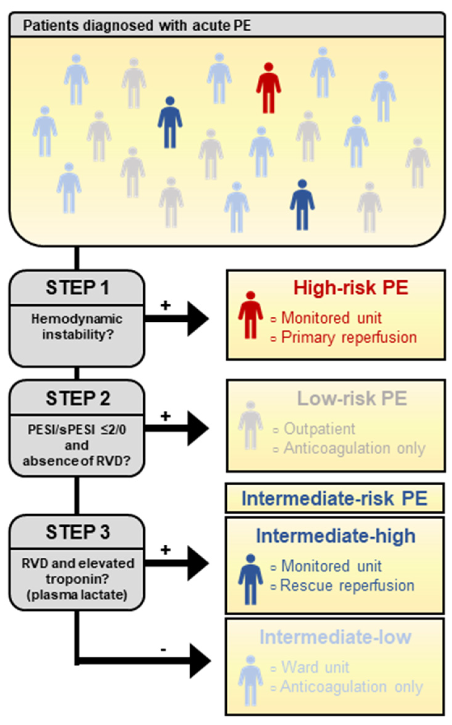
Figure 1. Step-by-step risk stratification in acute pulmonary embolism.
Jcm 11 02533 g001
Figure 2. Extrapolated effect of full-dose thrombolysis in cohorts of patients with a different basal risk of an early adverse event. a Basal risk according to PEITHO trial [10], b relative effect according to Marti et al. [11]. c Basal risk obtained by combining the risk of the PEITHO population and the prognostic modulation obtained by adding lactate [60].
Jcm 11 02533 g002
Figure 3. Extrapolated effect of half-dose thrombolysis in the hypothesized PEITHO-3 population. c Basal risk derived from applying the PEITHO-3 inclusion criteria and outcome to the PEITHO population [79]. d Relative treatment effect of full-dose thrombolysis is applied, and no increased risk of serious bleeding is assumed, according to a meta-analysis on half-dose thrombolysis [73].
Jcm 11 02533 g003
Table 1. Nomenclature in current European and American guidelines.
NomenclatureHemodynamic InstabilityRVDElevated
Troponin
PESI > Class II
or sPESI > 0
European Society of Cardiology (ESC) 2019
High risk+(+)(+)+
Intermediate–high risk+++ *
Intermediate–low riskOne or none+ *
Low risk(−)
American Heart Association (AHA) 2011
Massive+(+)(+)NA
SubmassiveOne or bothNA
Low riskNA
RVD: right ventricular dysfunction; PESI: pulmonary embolism severity index; sPESI: simplified pulmonary embolism severity index; NA: not assessed. * Presence of RVD despite PESI ≤ 2 or sPESI 0 classifies patients in intermediate-risk category.
Table 2. Operative characteristics of original and simplified pulmonary embolism severity index for early all-cause mortality [22].
Prediction
Index
Validation Cohorts
(Patients)
Sensitivity
(95% CI)
Specificity
(95% CI)
PLR (95% CI)NLR (95% CI)
PESI19 (23,997)0.89
(0.87–0.90)
0.49
(0.44–0.53)
1.72
(1.57–1.89)
0.22
(0.18–0.25)
sPESI9 (26,610)0.92
(0.89–0.94)
0.38
(0.32–0.44)
1.47
(1.28–1.68)
0.20
(0.13–0.31)
PESI: pulmonary embolism severity index; sPESI: simplified pulmonary embolism severity index; CI: confidence interval; PLR: positive likelihood ratio; NLR: negative likelihood ratio.
Table 3. Prognostic value of markers of right ventricular dysfunction for short-term mortality.
MarkerSensitivity (95% CI)Specificity
(95% CI)
PLR
(95% CI)
NLR
(95% CI)
Troponin [34]0.66
(0.61 to 0.70)
0.66
(0.65 to 0.67)
2.13
(1.84 to 2.47)
0.51
(0.40 to 0.60)
BNP [35]0.88
(0.65 to 0.96)
0.70
(0.64 to 0.75)
2.13
(1.84 to 2.47)
0.51
(0.40 to 0.60)
NT-proBNP [35]0.93
(0.14 to 1.00)
0.58
(0.14 to 0.92)
2.93
(2.28 to 3.77)
0.17
(0.05 to 0.58)
RVD US [35]0.70
(0.46 to 0.86)
0.57
(0.47 to 0.66)
1.48
(1.05 to 2.08)
0.82
(0.65 to 1.03)
RVD CT [35]0.65
(0.35 to 0.85)
0.56
(0.39 to 0.71)
1.63
(1.27 to 2.08)
0.53
(0.31 to 0.89)
CI: confidence interval; PLR: positive likelihood ratio; NLR: negative likelihood ratio; BNP: brain natriuretic peptide; NT-proBNP: N-terminal brain natriuretic peptide; RVD: right ventricular dysfunction; US: ultrasonography; CT: computer tomography.
Table 4. Components of the Bova score.
PredictorPoints
SBP 90–100 mmHg2
Elevated troponin2
RV dysfunction2
Heart rate > 100/min1
Table 5. Prognostic value of stratification scores dichotomized at an intermediate–high-risk level.
ScoreSensitivitySpecificityPLRPPVOutcome
Scores including Plasma Lactate
ESC 2019 + lactate [60]0.33
(0.16 to 0.55)
0.95
(0.92 to 0.97)
6.27
(3.11 to 12.66)
0.27In-hospital adverse outcome
Bova + lactate [64]0.46
(0.34 to 0.58)
0.91
(0.90 to 0.92)
5.16
(3.55 to 7.13)
0.26Adverse 7-day outcome
TELOS [64]0.19
(0.11 to 0.30)
0.95
(0.95 to 0.96)
3.94
(2.04 to 7.15)
0.21Adverse 7-day outcome
Scores without plasma lactate
Bova [66]0.48
(0.30 to 0.67)
0.86
(0.82 to 0.90)
3.41
(2.11 to 5.52)
0.19Adverse 30-day outcome
ESC 2014 [66]0.80
(0.61 to 0.91)
0.69
(0.64 to 0.73)
2.60
(2.00 to 3.30)
0.15Adverse 30-day outcome
ESC 2019 [67]0.52
(0.34 to 0.70)
0.79
(0.77 to 0.82)
2.5
(1.7 to 3.7)
0.07In-hospital adverse outcome
ESC: European Society of Cardiology; PLR: positive likelihood ratio; PPV: positive predictive value.
Publisher’s Note: MDPI stays neutral with regard to jurisdictional claims in published maps and institutional affiliations.