
Real-Time Spiral CMR Is Superior to Conventional Segmented Cine-Imaging for Left-Ventricular Functional Assessment in Patients with Arrhythmia

 , , and
Abstract
:1. Introduction
2. Materials and Methods
2.1. Study Population
2.2. CMR Protocol
2.3. Image Quality Assessment and CMR Data Analysis
2.4. Echocardiographic Assessment
2.5. Statistical Analysis
3. Results
3.1. Patients’ Characteristics
3.2. Volumetry Assessment in Patients with Sinus Rhythms
3.3. Volumetry Assessment in Patients with Arrhythmia
4. Discussion
Clinical Utility in Arrhythmic Patients
5. Limitations
6. Conclusions
Author Contributions
Funding
Institutional Review Board Statement
Informed Consent Statement
Data Availability Statement
Conflicts of Interest
References
- Karamitsos, T.D.; Francis, J.M.; Myerson, S.; Selvanayagam, J.B.; Neubauer, S. The Role of Cardiovascular Magnetic Resonance Imaging in Heart Failure. J. Am. Coll. Cardiol. 2009, 54, 1407–1424. [Google Scholar] [CrossRef] [PubMed] [Green Version]
- Knauth, A.L.; Gauvreau, K.; Powell, A.J.; Landzberg, M.J.; Walsh, E.P.; Lock, J.E.; Nido, P.J.d.; Geva, T. Ventricular Size and Function Assessed by Cardiac MRI Predict Major Adverse Clinical Outcomes Late after Tetralogy of Fallot Repair. Heart 2008, 94, 211–216. [Google Scholar] [CrossRef] [PubMed]
- Feng, L.; Srichai, M.B.; Lim, R.P.; Harrison, A.; King, W.; Adluru, G.; Dibella, E.V.R.; Sodickson, D.K.; Otazo, R.; Kim, D. Highly Accelerated Real-Time Cardiac Cine MRI Using k-t SPARSE-SENSE: Accelerated Real-Time Cardiac Cine MRI with k-t SPARSE-SENSE. Magn. Reson. Med. 2013, 70, 64–74. [Google Scholar] [CrossRef] [PubMed] [Green Version]
- Saybasili, H.; Herzka, D.A.; Seiberlich, N.; Griswold, M.A. Real-Time Imaging with Radial GRAPPA: Implementation on a Heterogeneous Architecture for Low-Latency Reconstructions. Magn. Reson. Imaging 2014, 32, 747–758. [Google Scholar] [CrossRef] [Green Version]
- Voit, D.; Zhang, S.; Unterberg-Buchwald, C.; Sohns, J.M.; Lotz, J.; Frahm, J. Real-Time Cardiovascular Magnetic Resonance at 1.5 T Using Balanced SSFP and 40 Ms Resolution. J. Cardiovasc. Magn. Reson. 2013, 15, 79. [Google Scholar] [CrossRef] [Green Version]
- Xue, H.; Kellman, P.; LaRocca, G.; Arai, A.E.; Hansen, M.S. High Spatial and Temporal Resolution Retrospective Cine Cardiovascular Magnetic Resonance from Shortened Free Breathing Real-Time Acquisitions. J. Cardiovasc. Magn. Reson. 2013, 15, 102. [Google Scholar] [CrossRef] [Green Version]
- Kido, T.; Kido, T.; Nakamura, M.; Watanabe, K.; Schmidt, M.; Forman, C.; Mochizuki, T. Compressed Sensing Real-Time Cine Cardiovascular Magnetic Resonance: Accurate Assessment of Left Ventricular Function in a Single-Breath-Hold. J. Cardiovasc. Magn. Reson. 2016, 18, 50. [Google Scholar] [CrossRef] [Green Version]
- Sudarski, S.; Henzler, T.; Haubenreisser, H.; Dösch, C.; Zenge, M.O.; Schmidt, M.; Nadar, M.S.; Borggrefe, M.; Schoenberg, S.O.; Papavassiliu, T. Free-Breathing Sparse Sampling Cine MR Imaging with Iterative Reconstruction for the Assessment of Left Ventricular Function and Mass at 3.0 T. Radiology 2017, 282, 74–83. [Google Scholar] [CrossRef] [Green Version]
- Kellman, P.; Epstein, F.H.; McVeigh, E.R. Adaptive Sensitivity Encoding Incorporating Temporal Filtering (TSENSE). Magn. Reson. Med. 2001, 45, 846–852. [Google Scholar] [CrossRef] [Green Version]
- Breuer, F.A.; Kellman, P.; Griswold, M.A.; Jakob, P.M. Dynamic Autocalibrated Parallel Imaging Using Temporal GRAPPA (TGRAPPA). Magn. Reson. Med. 2005, 53, 981–985. [Google Scholar] [CrossRef]
- Hori, Y.; Yamada, N.; Higashi, M.; Hirai, N.; Nakatani, S. Rapid Evaluation Of Right and Left Ventricular Function and Mass Using Real-Time True-Fisp Cine Mr Imaging Without Breath-Hold: Comparison with Segmented True-Fisp Cine Mr Imaging with Breath-Hold. J. Cardiovasc. Magn. Reson. 2003, 5, 439–450. [Google Scholar] [CrossRef]
- Kaji, S.; Yang, P.C.; Kerr, A.B.; Tang, W.H.W.; Meyer, C.H.; Macovski, A.; Pauly, J.M.; Nishimura, D.G.; Hu, B.S. Rapid Evaluation of Left Ventricular Volume and Mass without Breath-Holding Using Real-Time Interactive Cardiac Magnetic Resonance Imaging System. J. Am. Coll. Cardiol. 2001, 38, 527–533. [Google Scholar] [CrossRef] [Green Version]
- Schwab, F.; Schwarz, F.; Dietrich, O.; Lanz, T.; Resmer, F.; Wichmann, T.; Wintersperger, B.J.; Bamberg, F.; Nikolaou, K.; Reiser, M.; et al. Free Breathing Real-Time Cardiac Cine Imaging with Improved Spatial Resolution at 3 T. Investig. Radiol. 2013, 48, 158–166. [Google Scholar] [CrossRef] [Green Version]
- Vincenti, G.; Monney, P.; Chaptinel, J.; Rutz, T.; Coppo, S.; Zenge, M.O.; Schmidt, M.; Nadar, M.S.; Piccini, D.; Chèvre, P.; et al. Compressed Sensing Single–Breath-Hold CMR for Fast Quantification of LV Function, Volumes, and Mass. JACC Cardiovasc. Imaging 2014, 7, 882–892. [Google Scholar] [CrossRef] [Green Version]
- Wundrak, S.; Paul, J.; Ulrici, J.; Hell, E.; Rasche, V. A Small Surrogate for the Golden Angle in Time-Resolved Radial MRI Based on Generalized Fibonacci Sequences. IEEE Trans. Med. Imaging 2015, 34, 1262–1269. [Google Scholar] [CrossRef]
- Wundrak, S.; Paul, J.; Ulrici, J.; Hell, E.; Geibel, M.-A.; Bernhardt, P.; Rottbauer, W.; Rasche, V. Golden Ratio Sparse MRI Using Tiny Golden Angles: Golden Ratio Sparse MRI Using Tiny Golden Angles. Magn. Reson. Med. 2016, 75, 2372–2378. [Google Scholar] [CrossRef]
- Klinke, V.; Muzzarelli, S.; Lauriers, N.; Locca, D.; Vincenti, G.; Monney, P.; Lu, C.; Nothnagel, D.; Pilz, G.; Lombardi, M.; et al. Quality Assessment of Cardiovascular Magnetic Resonance in the Setting of the European CMR Registry: Description and Validation of Standardized Criteria. J. Cardiovasc. Magn. Reson. 2013, 15, 55. [Google Scholar] [CrossRef] [Green Version]
- Zhong, X.; Spottiswoode, B.S.; Meyer, C.H.; Kramer, C.M.; Epstein, F.H. Imaging Three-Dimensional Myocardial Mechanics Using Navigator-Gated Volumetric Spiral Cine DENSE MRI. Magn. Reson. Med. 2010, 64, 1089–1097. [Google Scholar] [CrossRef] [Green Version]
- Nayak, K.S.; Hargreaves, B.A.; Hu, B.S.; Nishimura, D.G.; Pauly, J.M.; Meyer, C.H. Spiral Balanced Steady-State Free Precession Cardiac Imaging. Magn. Reson. Med. 2005, 53, 1468–1473. [Google Scholar] [CrossRef]
- Delattre, B.M.A.; Heidemann, R.M.; Crowe, L.A.; Vallée, J.-P.; Hyacinthe, J.-N. Spiral Demystified. Magn. Reson. Imaging 2010, 28, 862–881. [Google Scholar] [CrossRef]
- Eirich, P.; Wech, T.; Heidenreich, J.F.; Stich, M.; Petri, N.; Nordbeck, P.; Bley, T.A.; Köstler, H. Cardiac Real-time MRI Using a Pre-emphasized Spiral Acquisition Based on the Gradient System Transfer Function. Magn. Reson. Med. 2021, 85, 2747–2760. [Google Scholar] [CrossRef]
- Steeden, J.A.; Kowalik, G.T.; Tann, O.; Hughes, M.; Mortensen, K.H.; Muthurangu, V. Real-Time Assessment of Right and Left Ventricular Volumes and Function in Children Using High Spatiotemporal Resolution Spiral BSSFP with Compressed Sensing. J. Cardiovasc. Magn. Reson. 2018, 20, 79. [Google Scholar] [CrossRef]
- Narayan, G.; Nayak, K.; Pauly, J.; Hu, B. Single-Breathhold, Four-Dimensional, Quantitative Assessment of LV and RV Function Using Triggered, Real-Time, Steady-State Free Precession MRI in Heart Failure Patients. J. Magn. Reson. Imaging 2005, 22, 59–66. [Google Scholar] [CrossRef]
- Barkauskas, K.J.; Rajiah, P.; Ashwath, R.; Hamilton, J.I.; Chen, Y.; Ma, D.; Wright, K.L.; Gulani, V.; Griswold, M.A.; Seiberlich, N. Quantification of Left Ventricular Functional Parameter Values Using 3D Spiral BSSFP and Through-Time Non-Cartesian GRAPPA. J. Cardiovasc. Magn. Reson. 2014, 16, 65. [Google Scholar] [CrossRef] [Green Version]
- Yamamuro, M.; Tadamura, E.; Kanao, S.; Okayama, S.; Okamoto, J.; Urayama, S.; Kimura, T.; Komeda, M.; Kita, T.; Togashi, K. Cardiac Functional Analysis by Free-Breath Real-Time Cine CMR with a Spatiotemporal Filtering Method, TSENSE: Comparison with Breath-Hold Cine CMR. J. Cardiovasc. Magn. Reson. 2006, 8, 801–807. [Google Scholar] [CrossRef]
- Cui, C.; Yin, G.; Lu, M.; Chen, X.; Cheng, S.; Li, L.; Yan, W.; Song, Y.; Prasad, S.; Zhang, Y.; et al. Retrospective Electrocardiography-Gated Real-Time Cardiac Cine MRI at 3T: Comparison with Conventional Segmented Cine MRI. Korean J. Radiol. 2019, 20, 114. [Google Scholar] [CrossRef] [PubMed]
- Allen, B.D.; Carr, M.L.; Markl, M.; Zenge, M.O.; Schmidt, M.; Nadar, M.S.; Spottiswoode, B.; Collins, J.D.; Carr, J.C. Accelerated Real-Time Cardiac MRI Using Iterative Sparse SENSE Reconstruction: Comparing Performance in Patients with Sinus Rhythm and Atrial Fibrillation. Eur. Radiol. 2018, 28, 3088–3096. [Google Scholar] [CrossRef] [PubMed]
- Yilmaz Cankaya, B.; Kantarci, M.; Gundogdu, F. Evaluation and Comparison of Left Ventricular Functions by Cardiac MRI and 2D Transthoracic Echocardiography. Eurasian J. Med. 2021, 53, 28–33. [Google Scholar] [CrossRef] [PubMed]
- Zhao, D.; Quill, G.M.; Gilbert, K.; Wang, V.Y.; Houle, H.C.; Legget, M.E.; Ruygrok, P.N.; Doughty, R.N.; Pedrosa, J.; D’hooge, J.; et al. Systematic Comparison of Left Ventricular Geometry Between 3D-Echocardiography and Cardiac Magnetic Resonance Imaging. Front. Cardiovasc. Med. 2021, 8, 728205. [Google Scholar] [CrossRef] [PubMed]
- Lee, V.S.; Resnick, D.; Bundy, J.M.; Simonetti, O.P.; Lee, P.; Weinreb, J.C. Cardiac Function: MR Evaluation in One Breath Hold with Real-Time True Fast Imaging with Steady-State Precession. Radiology 2002, 222, 835–842. [Google Scholar] [CrossRef]
- Unterberg-Buchwald, C.; Fasshauer, M.; Sohns, J.M.; Staab, W.; Schuster, A.; Voit, D.; Kowallick, J.T.; Steinmetz, M.; Frahm, J.; Lotz, J. Real Time Cardiac MRI and Its Clinical Usefulness in Arrhythmias and Wall Motion Abnormalities. J. Cardiovasc. Magn. Reson. 2014, 16, P34. [Google Scholar] [CrossRef] [Green Version]


| Parameters | Cartesian Segmented CMR | Real-Time Spiral CMR |
|---|---|---|
| Sequence type | 2D cine b-SSFP | 2D cine b-SSFP |
| TR (ms) | 2.42 | 5.73 |
| TE (ms) | 1.2 | 1.11 |
| Flip angle (°) | 60 | 70 |
| FOV (mm2) | 380 × 380 | 340 × 340 |
| Thickness (mm) | 8 | 8 |
| Number of slices | 11–17 | 11–17 |
| In plane resolution (mm2) | 1.4 × 1.4 | 2 × 2 |
| Temporal resolution (ms) | 32 Phases * | 39.9 ms |
| Time per slice (s) | 4 (3 Slices/BH) | 3 |
| ECG Mode | Retrospective | Retrospective |
| Acceleration factor | 1.7 (SENSE) | 5 (CS with TV regularization) |
| All (n = 32) | Sinus Rhythm (n = 21) | Arrythmia (n = 11) | |
|---|---|---|---|
| Age, years | 57 ± 16 | 55 ± 18 | 61 ± 12 |
| Male (%) | 19 (59.4) | 12 (57.1) | 7 (63.6) |
| BMI (kg/m2) | 26.34 ± 2.06 | 26.11 ± 1.94 | 26.78 ± 1.71 |
| BSA (m2) | 1.99 ± 0.18 | 1.99 ± 0.18 | 1.99 ± 0.17 |
| HR (beats/minute) * | 72 ± 11 | 70 ± 11 | 77 ± 8 |
| Cardiovascular risk factors | |||
| Hypertension | 16 (50) | 11 (52.4) | 5 (45.5) |
| Hypercholesterolemia | 20 (62.5) | 13 (61.9) | 7 (63.6) |
| Diabetes | 6 (18.8) | 5 (23.8) | 1 (9.1) |
| Smoking | 14 (43.8) | 9 (42.9) | 5 (45.5) |
| CMR-Diagnosis | |||
| Coronary artery disease | 11 (34.4) | 7 (33.3) | 4 (36.4) |
| Dilated Cardiomyopathy | 1 (3.1) | 0 (0) | 1 (9.1) |
| Hypertrophic Cardiomyopathy | 1 (3.1) | 1 (4.8) | 0 (0) |
| Valvular Disease | 2 (6.3) | 1 (4.8) | 1(9.1) |
| Myocarditis | 4 (12.5) | 3 (14.3) | 1 (9.1) |
| Other | 8 (25) | 5 (23.8) | 3 (27.2) |
| Normal finding | 5 (15.6) | 4 (19) | 1 (9.1) |
| Arrythmia (%) | 11 (34.4) | 0 | 11 (100) |
| Atrial Fibrillation | 9 (28.2) | 0 | 9 (81.8) |
| Ventricular premature beats | 2 (6.2) | 0 | 2 (18.2) |
| Segmented Cine | Spiral Real Time | p-Value | |
|---|---|---|---|
| Wrap-around | 0 ± 0 | 0 ± 0 | |
| Respiratory ghost | 0 ± 0 | 0 ± 0 | |
| Cardiac ghost | 0 ± 0 | 0.1 ± 0.3 | 0.15 |
| Blurring/mis-triggering | 0 ± 0 | 0 ± 0 | |
| Metallic artifact | 0 ± 0 | 0 ± 0 | |
| Shimming artifact | 0.2 ± 0.4 | 0 ± 0 | 0.08 |
| Total scoring | 0.14 ± 0.35 | 0.09 ± 0.3 | 0.64 |
| Segmented Cartesian CMR | Real-Time Spiral CMR | p-Value | |
|---|---|---|---|
| Left ventricle | |||
| LV-EDV (mL) | 137.15 ± 31 | 135.95 ± 30 | 0.164 |
| LV-ESV (mL) | 63.90 ± 20 | 63.76 ± 19 | 0.883 |
| LV-SV (mL) | 72.42 ± 16 | 72.19 ± 15 | 0.818 |
| LV-EF | 54.6 ± 6.4 | 54.00 ± 6.4 | 0.412 |
| Right ventricle | |||
| RV-EDV (mL) | 140.19 ± 31 | 140.95 ± 31 | 0.350 |
| RV-ESV (mL) | 64.33 ± 20 | 65.66 ± 19 | 0.122 |
| RV-SV (mL) | 75.80 ± 16 | 75.19 ± 17 | 0.522 |
| RV-EF | 54.67 ± 6.3 | 54.04 ± 7.0 | 0.399 |
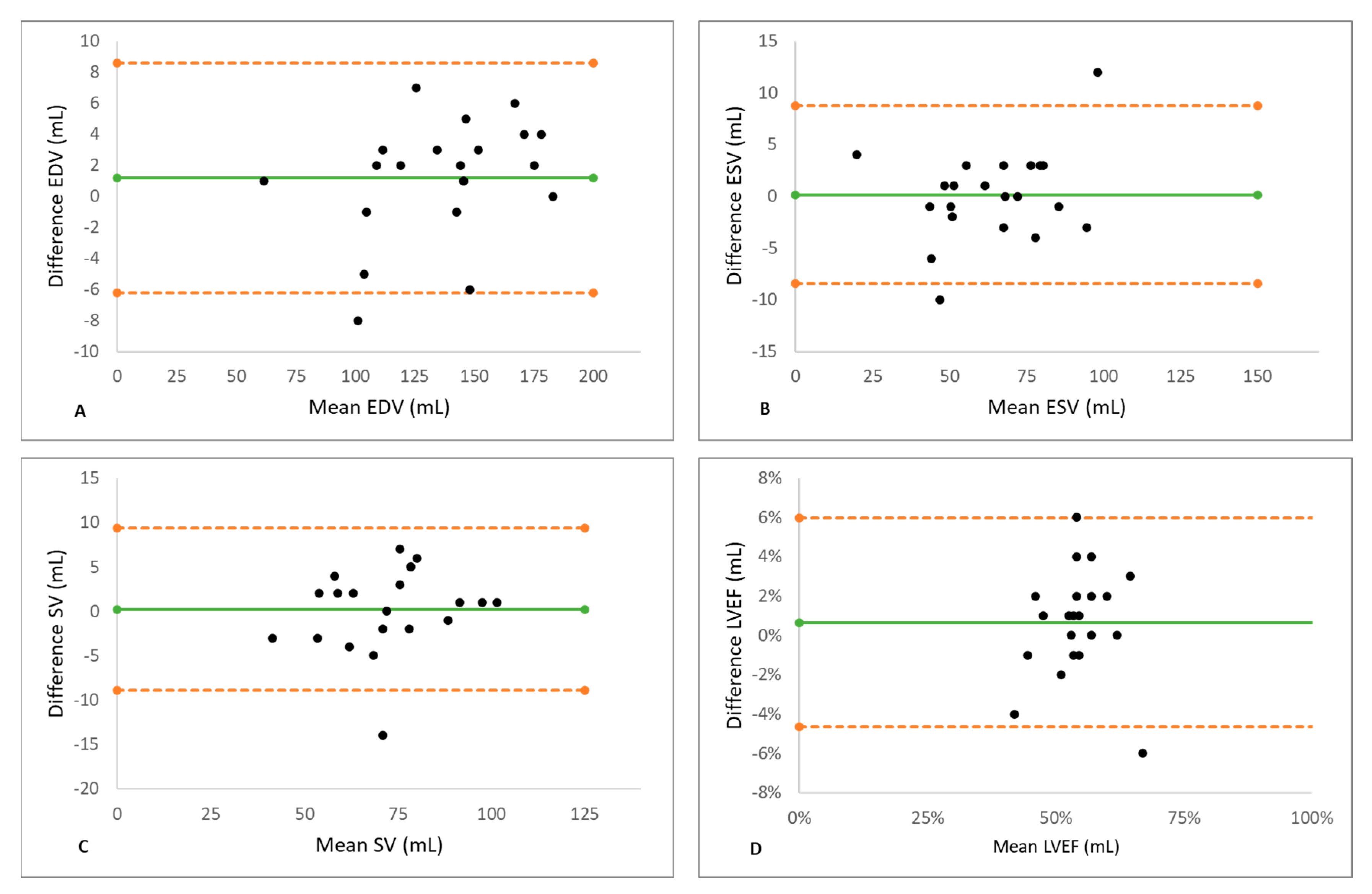
| LV-EDV | LV-ESV | LV-SV | LV-EF | |
|---|---|---|---|---|
| Mean difference ± SD | 1.19 ± 3.77 | 0.14 ± 4.38 | 0.23 ± 4.67 | 0.67 ± 2.71 |
| Limits of agreement | −6.21–8.59 | −8.45–8.73 | −8.93–9.40 | −4.64–5.97 |
| 95% Confidence interval | −0.52–2.90 | −1.85–2.13 | −1.89–2.36 | −0.57–1.90 |
| p-value | 0.164 | 0.883 | 0.818 | 0.273 |
| SE of the mean difference | 0.82 | 0.95 | 1.02 | 0.59 |
| Regression line | y = 0.041x − 4.36 | y = 0.066x − 4.099 | y = 0.061x − 4.176 | y = 0.026x − 0.739 |
| p-value, mean coefficient | 0.141 | 0.200 | 0.393 | 0.800 |
| RV-EDV | RV-ESV | RV-SV | RV-EF | |
| Mean difference ± SD | −0.76 ± 3.64 | −1.33 ± 3.78 | 0.61 ± 4.35 | 0.38 ± 3.79 |
| Limits of agreement | −7.90–6.38 | −8.75–6.08 | −7.91–9.15 | −0.07–0.07 |
| 95% Confidence interval | −2.42–0.89 | −3.05–0.39 | −1.36–2.60 | −1.34–2.10 |
| p-value | 0.350 | 0.122 | 0.522 | 0.650 |
| SE of the mean difference | 0.79 | 0.82 | 0.95 | 0.83 |
| Regression line | y = −0.01x + 0.576 | y = −0.019x − 0.130 | y = −0.014x + 1.645 | y = 0.036x + 2.052 |
| p-value, mean coefficient | 0.723 | 0.680 | 0.827 | 0.30 |
| Segmented Cine | Spiral Real Time | p-Value | |
|---|---|---|---|
| Wrap-around | 0 ± 0 | 0 ± 0 | |
| Respiratory ghost | 1.18 ± 1.16 | 0 ± 0 | 0.003 |
| Cardiac ghost | 1.18 ± 1.16 | 0.18 ± 0.4 | 0.014 |
| Blurring/mis-triggering | 2.09 ± 0.94 | 0.09 ± 0.30 | <0.001 |
| Metallic artifact | 0 ± 0 | 0 ± 0 | |
| Shimming artifact | 0.27 ± 0.64 | 0 ± 0 | 0.19 |
| Total scoring | 4.72 ± 2.93 | 0.27 ± 0.64 | <0.001 |
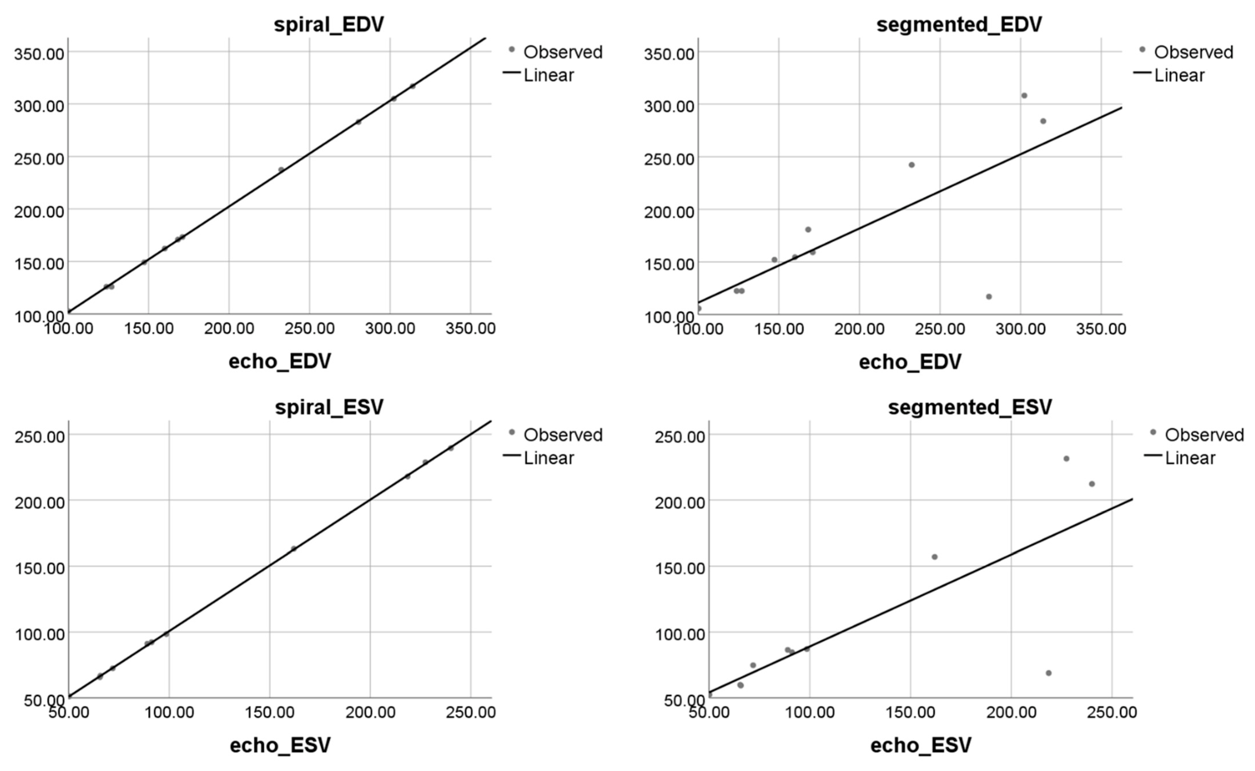
| Segmented Cartesian CMR | Reference Echo | p-Value | |
|---|---|---|---|
| LVEDV (mL) | 177.11 ± 69.98 | 194.85 ± 75.47 | 0.264 |
| LVESV (mL) | 106.72 ± 63.51 | 125.47 ± 72.41 | 0.026 |
| LVSV (mL) | 70.40 ± 16.03 | 69.95 ± 12.90 | 0.849 |
| LVEF (%) | 42.96 ± 10.81 | 39.02 ± 11.72 | 0.039 |
| Real-time spiral CMR | Reference Echo | p-value | |
| LVEDV (mL) | 195.74 ± 76.66 | 194.85 ± 75.47 | 0.174 |
| LVESV (mL) | 125.64 ± 72.10 | 125.47 ± 72.41 | 0.328 |
| LVSV (mL) | 69.11 ± 12.70 | 69.95 ± 12.90 | 0.976 |
| LVEF (%) | 39.44 ± 11.80 | 39.02 ± 11.72 | 0.139 |
Publisher’s Note: MDPI stays neutral with regard to jurisdictional claims in published maps and institutional affiliations. |
© 2022 by the authors. Licensee MDPI, Basel, Switzerland. This article is an open access article distributed under the terms and conditions of the Creative Commons Attribution (CC BY) license (https://creativecommons.org/licenses/by/4.0/).
Share and Cite
Nita, N.; Kersten, J.; Pott, A.; Weber, F.; Tesfay, T.; Benea, M.-T.; Metze, P.; Li, H.; Rottbauer, W.; Rasche, V.; et al. Real-Time Spiral CMR Is Superior to Conventional Segmented Cine-Imaging for Left-Ventricular Functional Assessment in Patients with Arrhythmia. J. Clin. Med. 2022, 11, 2088. https://doi.org/10.3390/jcm11082088
Nita N, Kersten J, Pott A, Weber F, Tesfay T, Benea M-T, Metze P, Li H, Rottbauer W, Rasche V, et al. Real-Time Spiral CMR Is Superior to Conventional Segmented Cine-Imaging for Left-Ventricular Functional Assessment in Patients with Arrhythmia. Journal of Clinical Medicine. 2022; 11(8):2088. https://doi.org/10.3390/jcm11082088
Chicago/Turabian StyleNita, Nicoleta, Johannes Kersten, Alexander Pott, Fabian Weber, Temsgen Tesfay, Marius-Tudor Benea, Patrick Metze, Hao Li, Wolfgang Rottbauer, Volker Rasche, and et al. 2022. "Real-Time Spiral CMR Is Superior to Conventional Segmented Cine-Imaging for Left-Ventricular Functional Assessment in Patients with Arrhythmia" Journal of Clinical Medicine 11, no. 8: 2088. https://doi.org/10.3390/jcm11082088
APA StyleNita, N., Kersten, J., Pott, A., Weber, F., Tesfay, T., Benea, M.-T., Metze, P., Li, H., Rottbauer, W., Rasche, V., & Buckert, D. (2022). Real-Time Spiral CMR Is Superior to Conventional Segmented Cine-Imaging for Left-Ventricular Functional Assessment in Patients with Arrhythmia. Journal of Clinical Medicine, 11(8), 2088. https://doi.org/10.3390/jcm11082088