
Clinical, Radiological, and Histopathological Characteristics of Periosteal Chondrosarcoma with a Focus on the Frequency of Medullary Invasion

 ,
,  , , , ,
Abstract
:1. Introduction
2. Materials and Methods
Case Selection
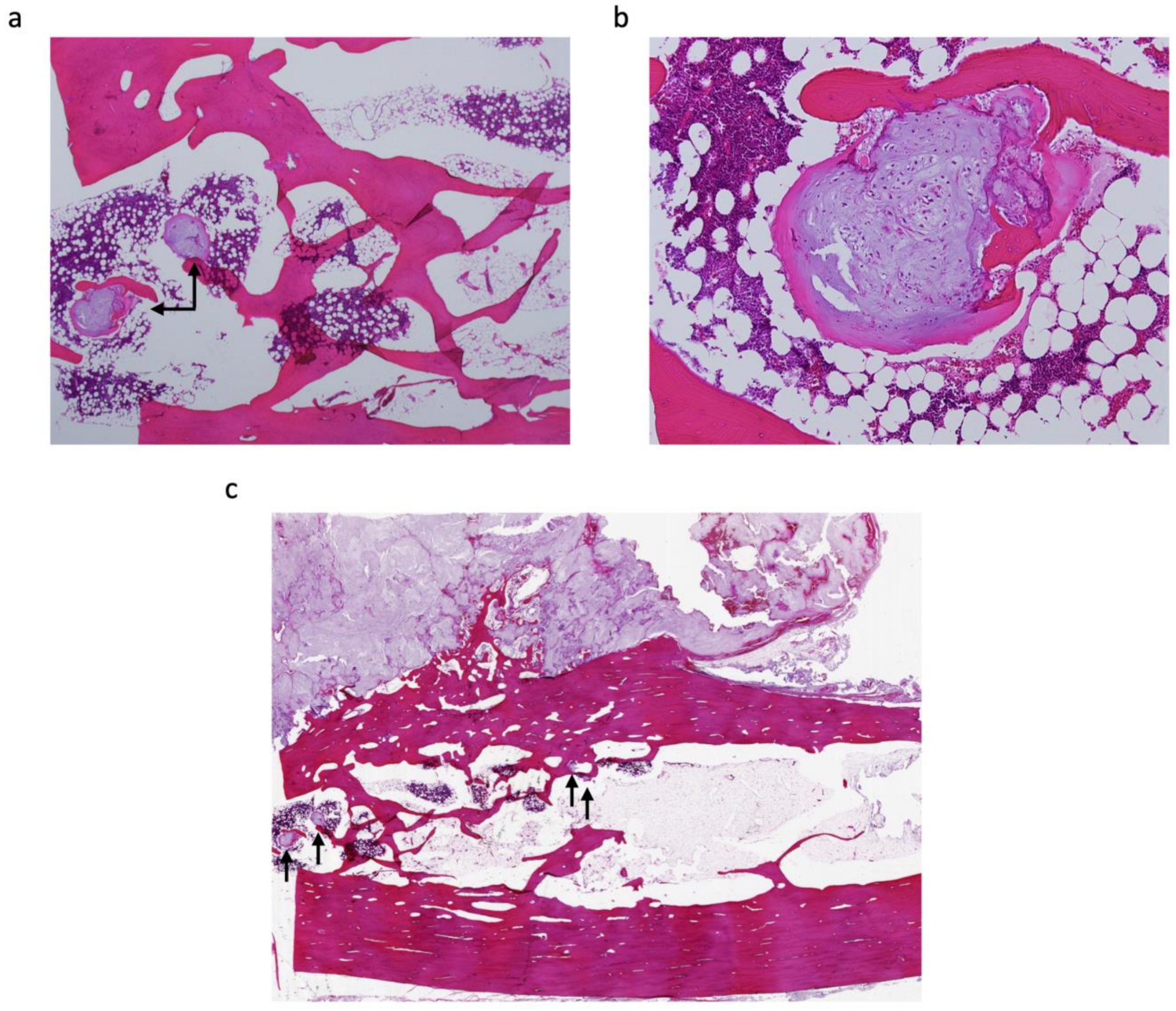
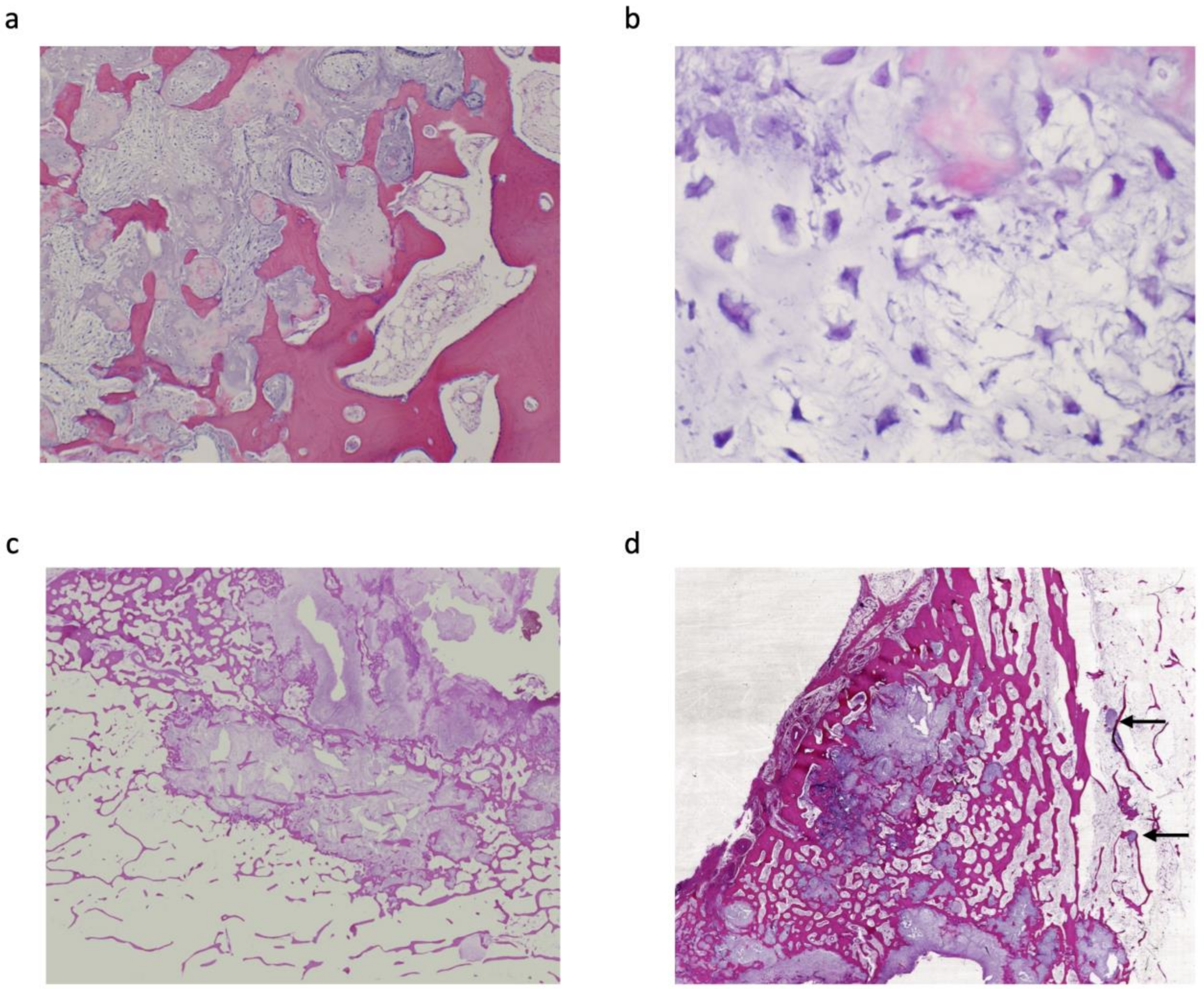
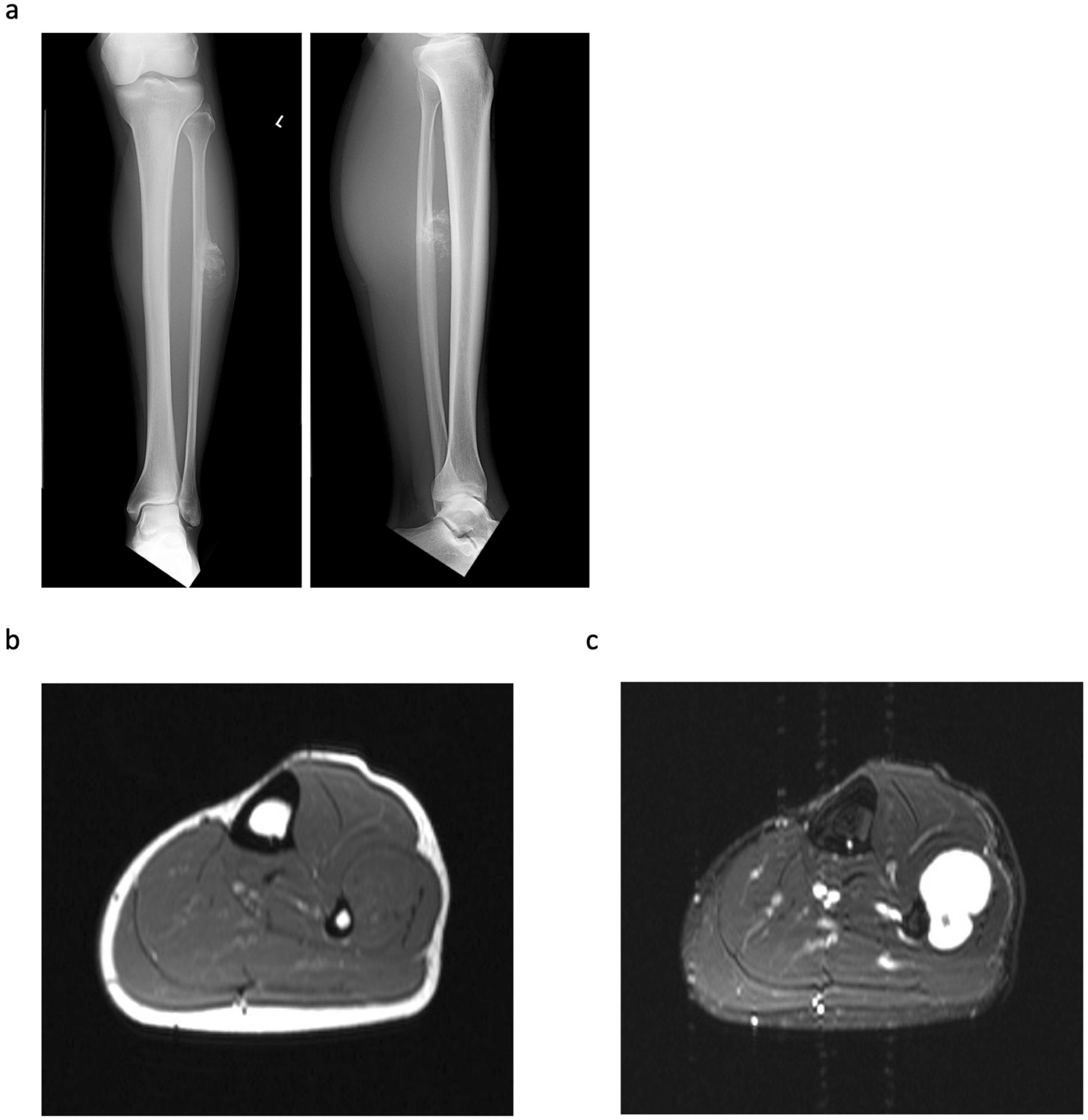
3. Results
4. Discussion
5. Conclusions
Author Contributions
Funding
Institutional Review Board Statement
Informed Consent Statement
Data Availability Statement
Conflicts of Interest
References
- WHO Classification of Tumours Editorial Board. Soft tissue and bone tumours. In World Health Organization Classification of Tumours, 5th ed.; World Health Organization International Agency for Research on Cancer: Lyon, France, 2020. [Google Scholar]
- Goedhart, L.M.; Ploegmakers, J.J.; Kroon, H.M.; Zwartkruis, E.C.; Jutte, P.C. The presentation, treatment and outcome of periosteal chondrosarcoma in the Netherlands. Bone Jt. J. 2014, 96, 823–828. [Google Scholar] [CrossRef] [PubMed]
- Papagelopoulos, P.J.; Galanis, E.C.; Mavrogenis, A.F.; Savvidou, O.D.; Bond, J.R.; Unni, K.K.; Sim, F.H. Survivorship analysis in patients with periosteal chondrosarcoma. Clin. Orthop. Relat. Res. 2006, 448, 199–207. [Google Scholar] [CrossRef] [PubMed]
- Liu, X.; Min, L.; Chen, G.; Hong, S.; Tu, C. Bony metastases following complete resection of periosteal chondrosarcoma. World J. Surg. Oncol. 2015, 13, 121. [Google Scholar] [CrossRef] [PubMed] [Green Version]
- Robinson, P.; White, L.M.; Sundaram, M.; Kandel, R.; Wunder, J.; McDonald, D.J.; Janney, C.; Bell, R.S. Periosteal chondroid tumors: Radiologic evaluation with pathologic correlation. Am. J. Roentgenol. 2001, 177, 1183–1188. [Google Scholar] [CrossRef] [PubMed]
- Cleven, A.H.; Zwartkruis, E.; Hogendoorn, P.C.; Kroon, H.M.; Briaire-de Bruijn, I.; Bovee, J.V. Periosteal chondrosarcoma: A histopathological and molecular analysis of a rare chondrosarcoma subtype. Histopathology 2015, 67, 483–490. [Google Scholar] [CrossRef] [PubMed]
- Hatano, H.; Ogose, A.; Hotta, T.; Otsuka, H.; Takahashi, H.E. Periosteal chondrosarcoma invading the medullary cavity. Skelet. Radiol. 1997, 26, 375–378. [Google Scholar] [CrossRef] [PubMed]
- Vanel, D.; De Paolis, M.; Monti, C.; Mercuri, M.; Picci, P. Radiological features of 24 periosteal chondrosarcomas. Skelet. Radiol. 2001, 30, 208–212. [Google Scholar] [CrossRef] [PubMed]
- Harper, K.; Sathiadoss, P.; Saifuddin, A.; Sheikh, A. A review of imaging of surface sarcomas of bone. Skelet. Radiol. 2021, 50, 9–28. [Google Scholar] [CrossRef] [PubMed]
- Murphey, M.D.; Jelinek, J.S.; Temple, H.T.; Flemming, D.J.; Gannon, F.H. Imaging of Periosteal Osteosarcoma: Radiologic-Pathologic Comparison. Radiology 2004, 233, 129–138. [Google Scholar] [CrossRef] [PubMed]
- Amary, M.F.; Bacsi, K.; Maggiani, F.; Damato, S.; Halai, D.; Berisha, F.; Pollock, R.; O’Donnell, P.; Grigoriadis, A.; Diss, T.; et al. IDH1 and IDH2 mutations are frequent events in central chondrosarcoma and central and periosteal chondromas but not in other mesenchymal tumours. J. Pathol. 2011, 224, 334–343. [Google Scholar] [CrossRef] [PubMed]
- Pansuriya, T.C.; van Eijk, R.; d’Adamo, P.; van Ruler, M.A.; Kuijjer, M.L.; Oosting, J.; Cleton-Jansen, A.M.; van Oosterwijk, J.G.; Verbeke, S.L.; Meijer, D.; et al. Somatic mosaic IDH1 and IDH2 mutations are associated with enchondroma and spindle cell hemangioma in Ollier disease and Maffucci syndrome. Nat. Genet. 2011, 43, 1256–1261. [Google Scholar] [CrossRef] [PubMed]


| Case | Sex | Age | Symptoms | Duration of Symptom (Months) | Site | Size (cm) | Cortex | Surgical Margin | Reconstruction | Histological Grade | Follow-Up Period (Months) | Recurrence | Metastasis |
|---|---|---|---|---|---|---|---|---|---|---|---|---|---|
| 1 | M | 22 | pain swelling | 1 1 | distal femur metaphysis | 2 × 1 × 2.5 | erosion | wide | β-TCP | I | 1 | − | − |
| 2 | M | 36 | swelling | 2 | distal femur metaphysis | 2 × 1.5 × 2 | no change | wide | bone autograft + plate | II | 72 | − | − |
| 3 | M | 23 | pain swelling | 6 3 | distal femur metaphysis | 6.0 × 4.0 × 3.0 | thickening | wide | DFR | II | 134 | − | − |
| 4 | M | 37 | swelling | 12 | fibula diaphysis | 5.5 × 2.5 × 3.5 | erosion | marginal | − | I | 59 | − | − |
| 5 | M | 41 | pain | 2 | proximal humerus metaphysis | 5.2 × 4.8 × 4.4 | no change | wide | PHR | II | 77 | − | − |
| 6 | M | 49 | swelling | 2 | pubis | 8.8 × 5.4 × 5.4 | thickening | wide | − | I | 36 | − | − |
| 7 | M | 35 | swelling | 1 | proximal humerus metaphysis | 7.5 × 5.5 × 5.5 | thickening | wide | bone autograft + plate | II | 32 | − | − |
| Case | Medullary Invasion (MRI) | Medullary Invasion (Pathological) | Distance from the Cortex to the Deepest Lesion of the Intramedullary Tumour (mm) | Skip or Continuous from Periosteal Tumour |
|---|---|---|---|---|
| 1 | − | + | 15 | continuous |
| 2 | + | + | 15 | continuous |
| 3 | + | + | 25 | skip |
| 4 | − | + | 3 | skip |
| 5 | + | + | 18 | skip |
| 6 | − | + | 1 | continuous |
| 7 | − | − |
Publisher’s Note: MDPI stays neutral with regard to jurisdictional claims in published maps and institutional affiliations. |
© 2022 by the authors. Licensee MDPI, Basel, Switzerland. This article is an open access article distributed under the terms and conditions of the Creative Commons Attribution (CC BY) license (https://creativecommons.org/licenses/by/4.0/).
Share and Cite
Nakagawa, M.; Endo, M.; Susuki, Y.; Yokoyama, N.; Maekawa, A.; Nabeshima, A.; Iida, K.; Fujiwara, T.; Setsu, N.; Matsunobu, T.; et al. Clinical, Radiological, and Histopathological Characteristics of Periosteal Chondrosarcoma with a Focus on the Frequency of Medullary Invasion. J. Clin. Med. 2022, 11, 2062. https://doi.org/10.3390/jcm11072062
Nakagawa M, Endo M, Susuki Y, Yokoyama N, Maekawa A, Nabeshima A, Iida K, Fujiwara T, Setsu N, Matsunobu T, et al. Clinical, Radiological, and Histopathological Characteristics of Periosteal Chondrosarcoma with a Focus on the Frequency of Medullary Invasion. Journal of Clinical Medicine. 2022; 11(7):2062. https://doi.org/10.3390/jcm11072062
Chicago/Turabian StyleNakagawa, Makoto, Makoto Endo, Yosuke Susuki, Nobuhiko Yokoyama, Akira Maekawa, Akira Nabeshima, Keiichiro Iida, Toshifumi Fujiwara, Nokitaka Setsu, Tomoya Matsunobu, and et al. 2022. "Clinical, Radiological, and Histopathological Characteristics of Periosteal Chondrosarcoma with a Focus on the Frequency of Medullary Invasion" Journal of Clinical Medicine 11, no. 7: 2062. https://doi.org/10.3390/jcm11072062
APA StyleNakagawa, M., Endo, M., Susuki, Y., Yokoyama, N., Maekawa, A., Nabeshima, A., Iida, K., Fujiwara, T., Setsu, N., Matsunobu, T., Matsumoto, Y., Yokoyama, R., Yamada, Y., Kohashi, K., Yamamoto, H., Oda, Y., Iwamoto, Y., & Nakashima, Y. (2022). Clinical, Radiological, and Histopathological Characteristics of Periosteal Chondrosarcoma with a Focus on the Frequency of Medullary Invasion. Journal of Clinical Medicine, 11(7), 2062. https://doi.org/10.3390/jcm11072062