Article

The 10 Year Outcomes of Implants Inserted with Dehiscence or Fenestrations in the Rehabilitation of Completely Edentulous Jaws with the All-on-4 Concept

by
Miguel de Araújo Nobre
1,*,
Armando Lopes
2 and
Elsa Antunes
3
1
Research, Development and Education Department, Maló Clinic, 1600-042 Lisboa, Portugal
2
Oral Surgery Department, Maló Clinic, 1600-042 Lisboa, Portugal
3
Dental Hygiene Department, Maló Clinic, 1600-042 Lisboa, Portugal
*
Author to whom correspondence should be addressed.
J. Clin. Med. 2022, 11(7), 1939; https://doi.org/10.3390/jcm11071939
Submission received: 10 February 2022 / Revised: 25 March 2022 / Accepted: 27 March 2022 / Published: 31 March 2022
(This article belongs to the Special Issue Implant Dentistry—Trends, Challenges and Innovations)

Abstract

:
Background: There is a need for a long-term evidence of implants placed in challenging conditions. The aim of this study was to investigate the outcome of full-arch rehabilitations with the All-on-4 concept for implants inserted with dehiscence or fenestrations. Methods: This retrospective cohort study included 123 patients (dehiscence, n = 87 patients; fenestrations, n = 28 patients; both conditions, n = 8 patients), with a total of 192 implants in immediate function presenting dehiscence (n = 150), fenestrations (n = 40), or both conditions (n = 2). Primary outcome measures were cumulative implant survival (CSurR) and success (CSucR) rates. Secondary outcome measures were prosthetic survival, marginal bone loss, and incidence of biological complications. Results: CSurRs were 94.1% (overall), 95.6% (dehiscence), and 88.1% (fenestrations) at 10 years using the patient as the unit of analysis. Smoking affected implant failure significantly (p = 0.019). Implant-level CSurRs and CSucRs at 10 years were 96.2% and 93.5% (overall), 97.2% and 94.6% (dehiscence), and 90.0% and 87.6% (fenestrations), respectively. Average bone resorption at 5 and 10 years was 1.22 mm and 1.53 mm, respectively. Biological complications occurred in 18 patients (n = 18 implants). Conclusions: Implants inserted with dehiscence or fenestrations demonstrate good long-term outcomes with overall high success and survival rates and low average marginal bone resorption, despite an inferior outcome in implants with fenestrations and smoking’s negative effect.

1. Introduction

Immediate function dental implants used for the support of fixed prosthetic rehabilitations provide successful long-term outcomes considering the high implant survival rates and patient satisfaction [1,2,3,4,5]. The All-on-4 concept (Nobel Biocare, Gothenburg, Sweden) is a rehabilitation protocol for fixed prosthetic rehabilitation of edentulous arches supported by implants in immediate function (two anterior implants placed in an axial orientation and two posterior implants tilted distally) with documented high survival rates in the long term for both the maxilla (95.7% at 13 years) [6] and the mandible (91.7% at 18 years) [7].
Frequently, the inclusion criteria for rehabilitation imply the insertion of implants in conditions close to the ideal: sufficient bone quantity and quality, with absence of infection on the implant site. Nevertheless, in particular situations, implants are inserted in challenging conditions, including post-extraction sockets, on periodontally compromised sites, or in sites presenting low bone density and quantity, with the possibility of dehiscence or a fenestration occurring [8,9,10,11].
In implant dentistry, dehiscence is defined as the absence of alveolar bone on the buccal or palatine/lingual aspects, leaving an oval-shaped defect and implant exposure apically from the implant–abutment junction (Figure 1); in turn, a fenestration is defined as a buccal or lingual window defect leaving marginal bone in situ (Figure 2). When inserting the implant, the presence of dehiscence or a fenestration can negatively impact the long-term outcome by decreased bone support mucosal irritation [12] and increased marginal bone strain on the implant’s mesial and distal sites (based on finite element analysis) [13]. The published reports on the outcomes of implants placed in sites presenting dehiscence are inconclusive. High survival rates in terms of short- [14,15], mid- [16,17], and long-term [18] outcomes have been reported. Nevertheless, a number of complications with the potential to decrease success have also been reported, including increased attachment loss, deeper peri-implant pockets [15], or postoperative exposure/infection when using concomitant guided bone regeneration [18]. In severely resorbed arches, fenestrations have a higher probability of occurrence given the insufficient bone volume [8]. The existence of fenestrations may be successfully managed through guided bone regeneration to the point of achieving a similar short-term outcome compared to implants placed in healed sites [19] but there is still lack of robust evidence to determine if any treatment is needed and which is the best treatment [8,20,21].
Considering the literature, it is noticeable that there is a need for mid- and long-term studies evaluating the outcome of dental implants inserted with dehiscence and fenestrations. The aim of this study was to document the 5-year and 10-year outcomes of full-arch rehabilitations with the All-on-4 concept supported by implants in the presence of dehiscence and/or fenestrations at the moment of implant placement. The research hypothesis investigated in this study was that the outcome of immediate-function implants inserted with dehiscence or fenestrations does not differ from the outcome of immediate-function implants inserted in nearly ideal conditions (sufficient bone quantity and quality, and no infection at the implant site).

2. Materials and Methods

This clinical study was performed complying with all ethical regulations in accordance with the Declaration of Helsinki and approved by an Ethical Committee (Ethical Committee for Health, Lisbon, Portugal; authorization no. 002/2017).
A database of patients with full-arch rehabilitations with the All-on-4 concept (Nobel Biocare) between April 2003 and June 2010 was assessed. The patients were selected on the basis of full-arch restorations supported by dental implants in the immediate function of the edentulous maxilla and/or mandible; the possibility of placing a minimum of four implants (at least 8.5 mm long) into the completely edentulous arch using a tilted approach for the distal implants; and the presence of dehiscence or fenestrations confirmed clinically and perioperatively, complying with a convenience sample. Exclusion criteria included active maxillary radiation or chemotherapy; systemic condition or smoking habits were not considered as exclusion criteria.
The surgical protocol followed the All-on-4 concept configuration (Nobel Biocare AB), refs. [6,7] with the two posterior implants inserted with distal tilting and the two anterior implants inserted axially. The implant insertion followed an under-preparation drilling protocol to guarantee a minimum of 35 N·cm insertion torque. The preparation was typically performed to the full drill depth with a 2 mm twist drill followed by a 2.4/2.8 mm step drill and a 3.2/3.6 mm step drill (depending on bone density). In sites with dense bone, 3.8/4.2 mm step drills were used. The implant neck was positioned at the bone level, and bicortical anchorage was established whenever possible in the maxilla.
Dehiscence occurred in certain sites with reduced crest thickness during implant preparation and/or placement, mainly on the lingual/palatal aspect of the crest because the implant site preparations were made more lingually than buccally to ensure thicker buccal cortical bone for protection and blood supply. In these situations, no particular protocol was used to minimize or restore dehiscence, in which, after implant placement, the flap was sutured back against the bone. Considering fenestrations, these were mainly accidental in cases where the buccal bone anatomy was irregular with reduced crest thickness (e.g., concavities) and in immediate extraction cases when the tilted implants came into contact with the fresh sockets. In the first situation, no special care was taken, with the flap sutured back against the bone; in the second situation, if the implant was exposed inside a socket, an autogenous bone graft, obtained during smoothing of the bone crest, was condensed in the socket to hide the fenestration and prevent soft-tissue migration.
Concerning the prosthetic protocol, a fixed high-density provisional prosthesis composed of acrylic resin (PalaXpress Ultra; Heraeus Kulzer GmbH, Hanau, Germany) with titanium cylinders (Nobel Biocare AB) and acrylic resin prosthetic teeth (Mondial and Premium teeth, Heraeus Kulzer GmbH) was manufactured at the dental laboratory and connected on the day of surgery.
Considering patient desires, a metal ceramic implant-supported fixed prosthesis with a titanium framework and all-ceramic crowns (NobelProcera titanium framework, NobelProcera crowns, Nobel Rondo ceramics; Nobel Biocare AB) or a metal–acrylic resin implant-supported fixed prosthesis with a titanium framework (NobelProcera titanium framework; Nobel Biocare AB) and acrylic resin prosthetic teeth (Mondial and Premium teeth, Heraeus Kulzer GmbH) were used to replace the provisional prosthesis. The final prosthesis was delivered typically 6 months post-surgically. A clinical situation is illustrated in Figure 3.
A postoperative maintenance protocol was indicated for each patient, including oral hygiene instructions. Follow-up clinical appointments were performed at 10 days, 2, 4, and 6 months, 1 year, and every 6 months thereafter, consisting of the assessment of clinical parameters, prophylaxis, and dental hygiene instructions. The evaluators were calibrated, with intra- and inter-class correlation coefficients of 0.92 and 0.85, respectively (weighted Kappa scores).

2.1. Outcome Measures

An outcome assessor blinded to the objectives of the study evaluated the data. Outcomes were assessed at implant surgery, as well as at 5 and 10 years of follow-up. The primary outcome measure was implant success, according to the success criteria adopted by the authors [22]: (a) The implant fulfilled its intended function supporting the reconstruction (sleeping implants were considered failures); (b) implant was stable upon manual testing; (c) absence of persistent infection jeopardizing the implant outcome; (d) no areas of radiolucency around the implants; (e) good esthetic outcome; (f) construction of an implant-supported fixed restoration that was comfortable for the patient and with conditions for good hygienic maintenance. Implants not complying with the criteria were considered survivals. Implant removal was classified as failure.
Secondary outcome measures were prosthetic survival (based on function with the necessity of replacing the prosthesis classified as failure), marginal bone loss at 5 and 10 years, and the incidence of biological complications.
The radiographic evaluation used to assess marginal bone loss was performed at baseline, as well as at 5 and 10 years of follow-up, using periapical radiographs by utilizing the parallelometric intraoral technique. For the intraoral technique, a conventional radiograph holder was used, the position of which was adjusted manually to ensure orthogonal film positioning. A blinded operator examined all radiographs of the implants for the marginal bone level. Each periapical radiograph was scanned at 300 dpi with a scanner (HP Scanjet 4890, HP Portugal, Paço de Arcos, Portugal). The marginal bone level was assessed with image analysis software (Image J version 1.40 g for Windows, National Institutes of Health, Bethesda, MD, USA) using the implants’ inter-thread distance as the reference for digital calibration. The implant platform was used as a reference point, and marginal bone loss was defined as the difference in marginal bone levels between the day of surgery and the point of evaluation. The radiographs were accepted or rejected for evaluation on the basis of the clarity of implant threads; a clear thread guarantees both sharpness and an orthogonal direction of the radiographic beam toward the implant axis.
The evaluation of biological complications included peri-implant pathology (defined as peri-implant pocket depths ≥5 mm, bleeding on probing, with concurrent marginal bone loss compared to the previous radiograph or clinical attachment loss of >2 mm) [23], fistula formation, or abscess. The evaluation was performed using a plastic periodontal probe calibrated to 0.25 N (Click Probe, Hawe Neos, Bioggio, Switzerland).
The overall medical status was assessed from patient records by interviewing the patients, and the distribution was classified according to the International Classification of Diseases 11th Revision (ICD-11) [24]. Bruxism, in particular, was assessed as per protocol by interviewing the patient (about symptoms related to tenderness of jaw muscles, sleeping habits, and medication), observation of natural teeth (if present) or prosthetic elements (if present), and radiographic evaluation.

2.2. Statistical Evaluation

Descriptive statistics (average, standard deviation, and range) were calculated for age and marginal bone loss (at 5 and 10 years). Frequencies were used to classify biological complications, loss to follow-up, and prosthetic survival. Cumulative implant survival and success were estimated at the patient level (any implant failure in each patient) by using the Kaplan–Meier product limit estimator and at the implant level using life tables. Inferential analysis was performed to evaluate the difference in demographics between patients with complete follow-up and patients lost to follow-up (age: Mann–Whitney U test; sex: chi-square test). Data were statistically analyzed using the Statistical Package for the Social Sciences software (IBM SPSS, version 17, Rochester, NY, USA).

3. Results

3.1. Sample

A total of 123 patients (38 males and 85 females; mean age, 55.2 years; range, 34–81 years) with 127 full-arch rehabilitations (maxilla: 84; mandible: 43) were included (dehiscence, n = 87 patients; fenestrations, n = 28 patients; dehiscence and fenestrations, n = 8 patients). Considering the International Classification of Diseases 11th Revision (ICD-11) [24], 79 patients had health complications (Table 1). A total of 192 implants were inserted (Table 1 and Table 2): 150 implants with dehiscence (maxilla: 100 implants; mandible: 50 implants), 40 implants with a fenestration (maxilla: 30 implants; mandible: 10 implants), and 2 implants with dehiscence and a fenestration (maxilla: 1 implant; mandible: 1 implant). The opposing dentitions were implant-supported prostheses (57 patients), natural teeth (19 patients), a combination of both (37 patients), fixed prosthesis over natural teeth (1 patient), and removable prosthesis (9 patients).

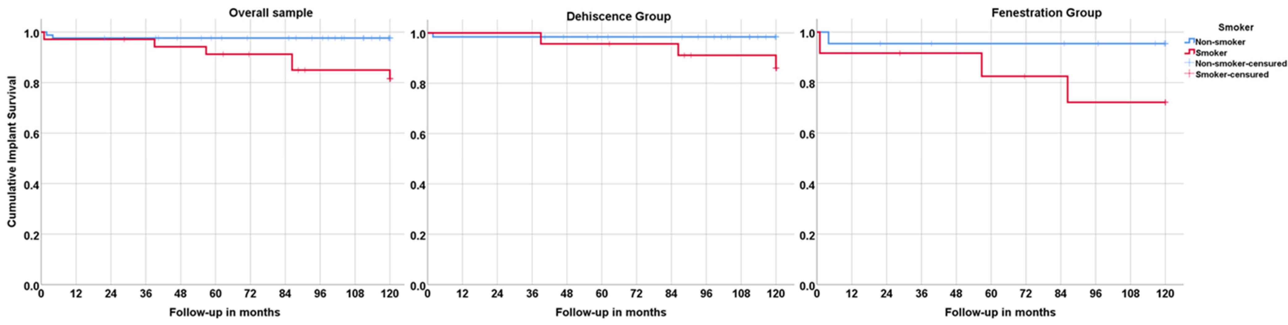
3.2. Lost to Follow-Up Rate; Implant Survival, Success, and Failure; Prosthetic Survival

Twenty-five patients (20%) with 34 implants (17.8%) became unreachable and were lost to follow-up (dehiscence: 19 patients and 27 implants; fenestrations: 5 patients with 5 implants; dehiscence and fenestrations: 1 patient and 2 implants). Comparing demographic variables, a significant difference was registered for age with an increased age for patients lost to follow-up compared to patients with complete follow-up (p = 0.014, Mann–Whitney U test), while no significant difference was registered for sex (p = 0.647, chi-square test). Seven patients lost seven implants (one implant in each patient; dehiscence: three implants; fenestrations: three implants; dehiscence and fenestration: one implant; Table 1 and Table 2). In three patients, new implants were inserted (not accounted for in this study) to replace the failed implants; two patients refused new implants, while the prosthesis remained in function supported by three implants in each patient. In two patients, two prostheses failed (one prosthesis in each patient) following implant failures, rendering a prosthetic survival rate of 98.4% (dehiscence group: 98.9%; fenestration group: 96.4%).
Considering the patient as the unit of analysis, the overall implant cumulative survival at 10 years was 94.1%, with 95.6% and 88.1% for the dehiscence and fenestration groups, respectively (Kaplan–Meier, Table 3). Implant failure was higher in smokers, with an implant cumulative survival of 87.7% for smokers with dehiscence (compared to 98.6% for nonsmokers) and 74.6% for smokers with fenestrations (compared to 95.7% for nonsmokers) (Figure 4), as well as in patients with systemic conditions (93.3% in the dehiscence group compared to 100% in healthy patients; 86.7% in the fenestration group compared to 91.7% in healthy patients).
Considering the implant as the unit of analysis, the overall cumulative survival rate at 10 years was 96.2%, with 97.2% for the dehiscence group and 90.0% for the fenestration group (Table 4). The overall implant cumulative success rate was 93.5%, with 94.6% and 87.6% for the dehiscence and fenestration groups, respectively (Table 5).

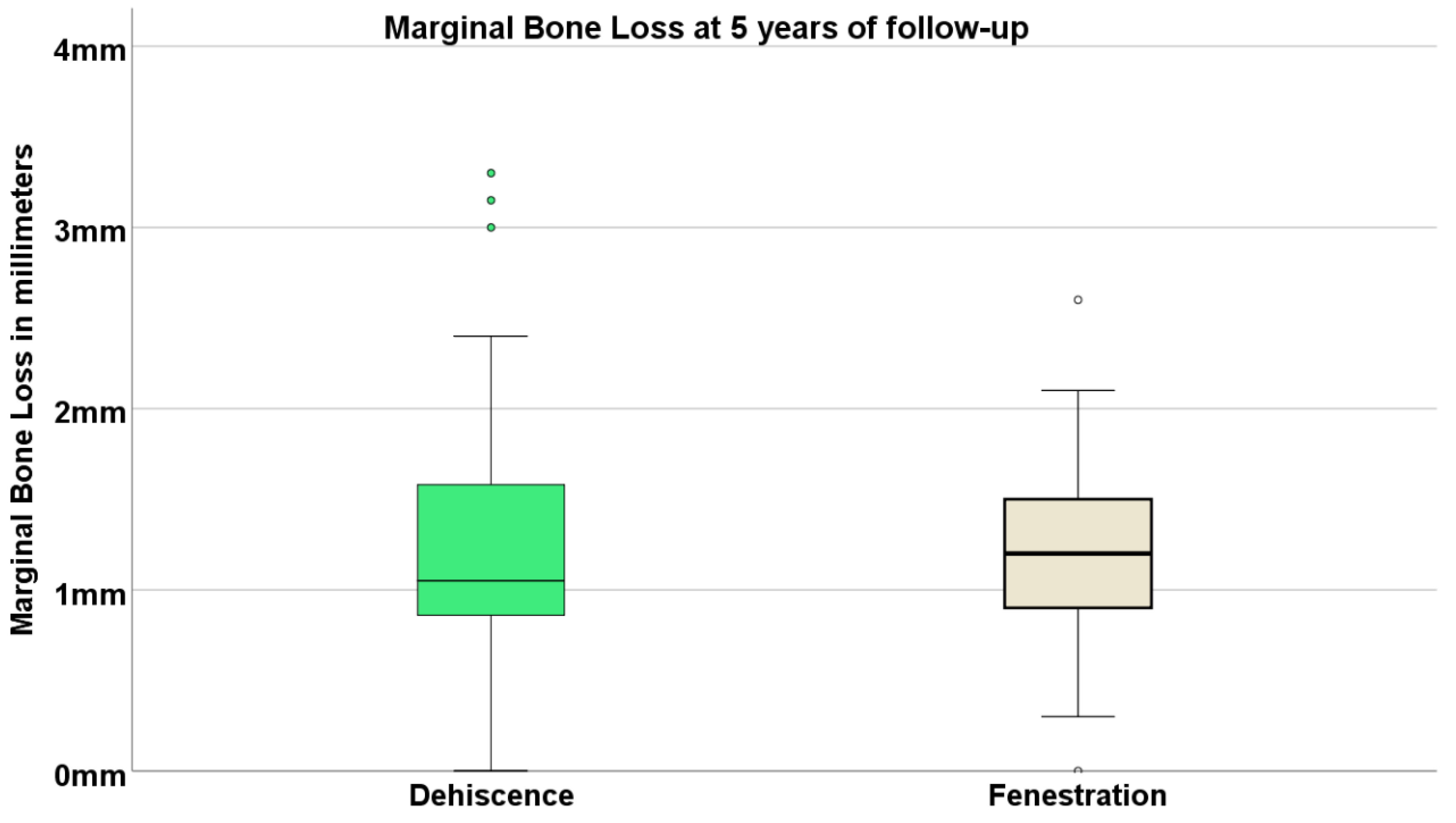
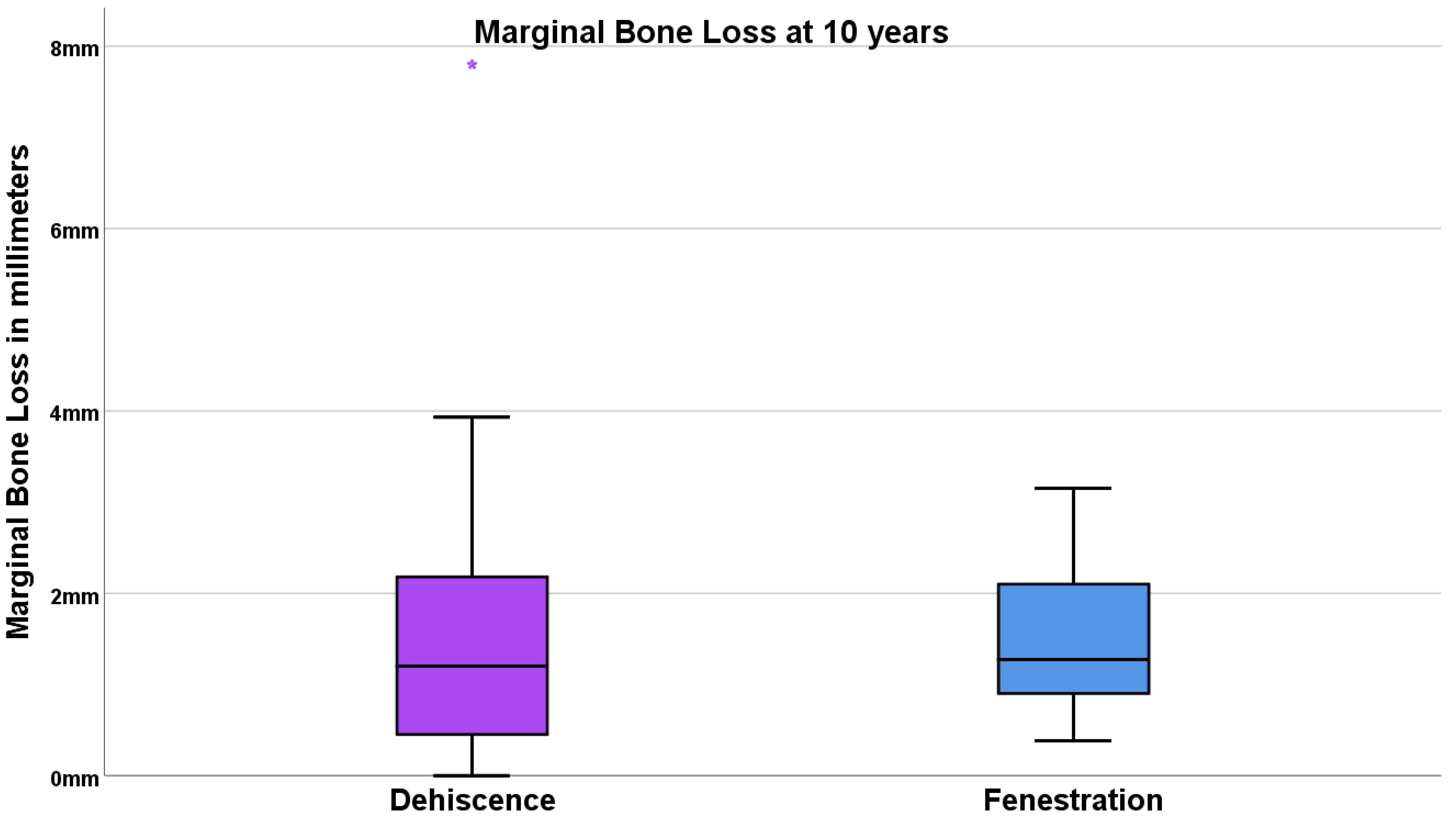
3.3. Marginal Bone Loss

Overall, the percentages of available radiographs for performing the marginal bone resorption measurements were 78% at 5 years and 90% at 10 years. The average (standard deviation) marginal bone loss at 5 years was 1.22 mm (0.66 mm), with 1.25 mm (0.69 mm) for the dehiscence group and 1.22 mm (0.66 mm) for the fenestration group (Figure 5).
The average (standard deviation) marginal bone loss at 10 years was 1.53 mm (0.75 mm), with 1.49 mm (1.34 mm) for the dehiscence group and 1.53 mm (0.75 mm) for the fenestration group (Figure 6).

3.4. Complications

Biological complications occurred for a total of 18 implants (9.4%) in 18 patients (14%): 13 implants (9.1%) in the dehiscence group and 6 implants (12.5%) in the fenestration group (one implant had both dehiscence and fenestration). The complications included suppuration (2 implants), infection (2 implants), fistula (1 implant), and peri-implant pathology (13 implants). The two incidences of suppuration and the incidence of fistula were resolved by utilizing non-surgical therapy (scaling with an ultrasonic scaler device, irrigating with 0.2% chlorhexidine gel, and prescribing the same solution for the patient in daily care, followed by a re-evaluation) and antibiotic administration. One infection was resolved non-surgically, while the other incidence was not resolved (implant considered unsuccessful). The incidences of peri-implant pathology were resolved in five implants through non-surgical intervention and surgically in one implant through an open flap, the removal of granulation tissue, and a decontamination of the implant surface both mechanically using an ultrasonic scaler and chemically through a polish using a brush attached to the contrangle and 0.2% chlorhexidine and saline irrigation; the mucosal flap, apically positioned, was sutured using 4/0 non-resorbable sutures (Braun Silkam4-0, Aesculap, Tuttlinged, Germany), and systemic antibiotics were prescribed to the patient (amoxicillin 875 mg + clavulanic acid 125 mg, Labesfal, Campo de Besteiros, Portugal). In seven implants, peri-implant pathology was not resolved non-surgically or surgically, with two of the implants being removed (considered failures) and five implants remaining in function (considered unsuccessful). Seven of the unresolved biological complications (87.5%) were implants with dehiscence (one implant with both dehiscence and fenestration).

4. Discussion

The present study registered a successful outcome of full-arch restorations with the All-on-4 concept for implants inserted with dehiscence or fenestration after 10 years of follow-up. Previous long-term publications using the same rehabilitation method registered high cumulative implant survival and success rates at the same point of evaluation (10 years). In the maxilla, a study evaluating the outcome between 5 and 13 years of 1072 patients with 4288 implants registered 96.4% and 95.6% of cumulative implant survival and success rates, respectively, together with an average marginal bone loss of 1.7 mm [6]; for the mandible, the evaluation between 10 and 18 years of 471 patients with 1884 implants registered a cumulative survival rate of 96.9%, a cumulative success rate of 95.9%, and an average marginal bone loss of 1.67 mm [7]. The results of the present study imply that full-arch rehabilitations can also be performed with predictability in extreme situations rather than being applied only in the presence of generally accepted inclusion criteria for implant rehabilitation. Nevertheless, despite a favorable marginal bone loss outcome at 5 and 10 years, the overall implant success outcome of the present study was lower when compared to those publications [6,7], which is a result significantly influenced by the fenestration group, with markedly increased failure (10%) and unsuccessful (above 12%) implant outcomes. The occurrence of dehiscence or fenestration is considered an accident [25] or complication [21] of adequate treatment, excluded from the listings of failures and independent from surgical precautions or skills [21]. Usually, the presence of these conditions is attributed to bone-plate thickness deficiency [26], illustrative of challenging conditions for full-arch implant-supported rehabilitations, which may partly explain the lower success outcome. As for the fenestration subgroup, these results concur with previous case reports [27] with a higher incidence of short-term implant losses in these situations. In both conditions, the results follow previously reported pattens for the short-term outcome, with a high survival rate for implants placed with dehiscence, as registered in a clinical trial [15], and a higher incidence of implant failures for implants with fenestration (the majority up to ~2 years postsurgery), as observed in case reports [27]. The type of complementary therapeutic approach (bone augmentation at implants with dehiscence and fenestrations, with or without the use of membranes) does not seem to significantly impact survival outcomes [20], which is a result that is in line with the present study, with high survival rates for implants with dehiscence and no complementary therapeutical approaches, while the outcome of implants with fenestrations was independent of the use of autogenous bone graft.
Smoking was previously registered as a risk indicator for implant failure in a systematic review and meta-analysis, with a 2.92-fold increased odds ratio compared to nonsmokers [28]. Previous publications on the same treatment modality (All-on-4) also registered a significant negative influence of smoking habits with increased implant failure, excess marginal bone loss, or biological complications [6,7]. A clinical trial investigating the outcome of implants with dehiscence placed with simultaneous guided bone regeneration between 22 and 24 years of follow-up registered a significant negative effect of smoking on survival [29]. In the present study, smoking impaired implant survival in both groups with a ~11% decrease for implants with dehiscence and 21% decrease for implants with fenestrations, with a higher percentage of smokers present in the fenestration group where an increased failure rate was registered. Moreover, most smokers in both groups registered late implant failures beyond the osseointegration phase (all three smokers from the dehiscence group and two in three smokers from the fenestration group). It can be hypothesized that the difference in survival could be at least partially due to a confounding effect of smoking rather than the effect of dehiscence or fenestration. Confounding variables are those that may compete with the exposure of interest (in this case, dehiscence and fenestration) in explaining the outcome of the study (implant survival) [30]. To be considered a confounding variable, the variable needs to meet two conditions: it must be correlated with the independent variable (the majority of patients from the fenestration group were smokers) and causally related to the dependent variable (smoking related with implant failure) [28]. Nevertheless, due to a lack of statistical power, it was not possible to assess this evaluation in the present study; therefore, this should be further investigated in studies using statistical models to control for confounding with an adequate sample size.
The biological complication rate was reduced but concentrated on the dehiscence subgroup (mainly peri-implant pathology), particularly the unresolved complications. The presence of dehiscence implies a potential settlement of the bone level around the implant’s middle third at baseline, a condition previously registered as a risk indicator for peri-implant pathology in both inferential [31] and multivariable analysis [23]. A previous study evaluating the impact and accuracy of a risk algorithm for the incidence of peri-implant pathology registered that patients with implants whose bone level was at the middle third have a 14-fold increase in the probability of disease, estimating that the prevention of this condition could potentially suppress 31% of the cases of peri-implant pathology based on the attributable fraction [23]. Furthermore, a clinical trial [15] evaluating the outcome of implants with buccal dehiscence registered a significant increase in clinical attachment loss and probing pocket depths for these implants when compared to implants inserted in healed sites. The implementation of a maintenance protocol, with programmed recall appointments for clinical diagnosis, prophylaxis, oral hygiene instructions, and management of the implant–abutment connection through disinfection [32], is considered paramount for good long-term outcomes. In the present study, the implementation of a maintenance protocol and recall appointments allowed performing close monitoring of the patients that otherwise could have potentially resulted in a further increased implant failure rate.
The dropout rate of 20% was small considering the 10-year span of this clinical study and accounted for good internal validity. Nevertheless, this study had limitations that warrant discussion, including the retrospective evaluation; the convenience sample; the lack of a control group without bone defects; the small sample size that disabled inferential analysis; the lack of stratification for smoking habits; not collecting data on mechanical complications; and being performed in a single center and with one implant system, which imply caution in the extrapolation of the results. Furthermore, the significant difference in age between the sample lost to follow-up and the fully analyzed sample might present a potential bias in the underestimation of the implant and prosthetic survival outcomes, warranting caution in the interpretation of the results.
Future studies with larger samples and multivariable analysis should be performed to control potential confounding effects when evaluating the outcome of these rehabilitations and to allow subsample analytical methods and age and gender discrimination.

5. Conclusions

Considering the limitations of the present study, the long-term outcome of implants inserted with dehiscence and fenestration for full-arch rehabilitations with the All-on-4® concept is possible with overall high survival rates and low average marginal bone resorption; nonetheless, implants with fenestrations yielded lower survival and success rates. Smoking exerted a significant negative effect on implant survival. Implants with dehiscence suffered most from biological complications that could not be resolved.

Author Contributions

Conceptualization, M.d.A.N.; methodology, M.d.A.N.; validation, M.d.A.N., A.L. and E.A.; formal analysis, M.d.A.N.; investigation, M.d.A.N., A.L. and E.A.; data curation, M.d.A.N. and A.L.; writing—original draft preparation, M.d.A.N.; writing—review and editing, M.d.A.N., A.L. and E.A.; visualization, M.d.A.N., A.L. and E.A.; funding acquisition, M.d.A.N. All authors have read and agreed to the published version of the manuscript.

Funding

This research was funded by Nobel Biocare Services AG, grant number 2017.1485.

Institutional Review Board Statement

The study was conducted in accordance with the Declaration of Helsinki and approved by the Ethical Committee for Health (protocol code 002/2017).

Informed Consent Statement

Informed consent was obtained from all subjects involved in the study.

Data Availability Statement

Data are available from a public repository, https://doi.org/10.17605/OSF.IO/E3ZRC, accessed on 17 March 2022.

Acknowledgments

The authors would like to thank Andreia Araújo and Sandro Catarino for all the help in data management and all clinicians at the Maló Clinic for their outstanding clinical support.

Conflicts of Interest

Miguel de Araújo Nobre received previous educational fees from Nobel Biocare Services AG and received previous scientific grant support from Nobel Biocare Services AG; Armando Lopes is currently a consultant for Nobel Biocare, received previous educational fees from Nobel Biocare Services AG, and received previous scientific grant support from Nobel Biocare Services AG; Elsa Antunes received previous scientific grant support from Nobel Biocare Services AG. The funders had no role in the design of the study; in the collection, analyses, or interpretation of data; in the writing of the manuscript; or in the decision to publish the results.

References

  1. Sumie Yaguinuma Gonçalves, G.; Murieli Ferreira de Magalhães, K.; Passos Rocha, E.; Henrique dos Santos, P.; Gonçalves Assunção, W. Oral health-related quality of life and satisfaction in edentulous patients rehabilitated with implant-supported full dentures all-on-four concept: A systematic review. Clin. Oral Investig. 2022, 26, 83–94. [Google Scholar] [CrossRef] [PubMed]
  2. Del Fabbro, M.; Testori, T.; Kekovic, V.; Goker, F.; Tumedei, M.; Wang, H.-L. Clinical Medicine A Systematic Review of Survival Rates of Osseointegrated Implants in Fully and Partially Edentulous Patients Following Immediate Loading. J. Clin. Med. 2019, 8, 2142. [Google Scholar] [CrossRef] [Green Version]
  3. Maló, P.; de Araújo Nobre, M.; Gonçalves, Y.; Lopes, A. Long-Term Outcome of Implant Rehabilitations in Patients with Systemic Disorders and Smoking Habits: A Retrospective Clinical Study. Clin. Implant Dent. Relat. Res. 2016, 18, 649–665. [Google Scholar] [CrossRef]
  4. Maló, P.; De Araújo Nobre, M.; Lopes, A.; Queridinha, B.; Ferro, A.; Gravito, I. Axial implants in immediate function for partial rehabilitation in the maxilla and mandible: A retrospective clinical study evaluating the long-term outcome (Up to 10 Years). Implant Dent. 2015, 24, 557–564. [Google Scholar] [CrossRef]
  5. Maló, P.; de Araújo Nobre, M.; Lopes, A.; Ferro, A.; Gravito, I. Single-Tooth Rehabilitations Supported by Dental Implants Used in an Immediate-Provisionalization Protocol: Report on Long-Term Outcome with Retrospective Follow-Up. Clin. Implant Dent. Relat. Res. 2015, 17, e511-9. [Google Scholar] [CrossRef]
  6. Maló, P.; de Araújo Nobre, M.; Lopes, A.; Ferro, A.; Nunes, M. The All-on-4 concept for full-arch rehabilitation of the edentulous maxillae: A longitudinal study with 5–13 years of follow-up. Clin. Implant Dent. Relat. Res. 2019, 21, 538–549. [Google Scholar] [CrossRef] [PubMed]
  7. Maló, P.; de Araújo Nobre, M.; Lopes, A.; Ferro, A.; Botto, J. The All-on-4 treatment concept for the rehabilitation of the completely edentulous mandible: A longitudinal study with 10 to 18 years of follow-up. Clin. Implant Dent. Relat. Res. 2019, 21, 565–577. [Google Scholar] [CrossRef] [PubMed]
  8. Mattsson, T.; Köndell, P.Å.; Gynther, G.W.; Fredholm, U.; Bolin, A. Implant treatment without bone grafting in severely resorbed edentulous maxillae. J. Oral Maxillofac. Surg. 1999, 57, 281–287. [Google Scholar] [CrossRef]
  9. Bogaerde, L.; Rangert, B.; Wendelhag, I. Immediate/early function of Brånemark System TiUnite implants in fresh extraction sockets in maxillae and posterior mandibles: An 18-month prospective clinical study. Clin. Implant Dent. Relat. Res. 2005, 7, s121–s130. [Google Scholar] [CrossRef]
  10. Malo, P.; de Araújo Nobre, M.; Rangert, B. Implants placed in immediate function in periodontally compromised sites: A five-year retrospective and one-year prospective study. J. Prosthet. Dent. 2007, 97, S86–S95. [Google Scholar] [CrossRef]
  11. Malo, P.; De Araújo Nobre, M.; Lopes, A. Immediate rehabilitation of completely edentulous arches with a four-implant prosthesis concept in difficult conditions: An open cohort study with a mean follow-up of 2 years. Int. J. Oral Maxillofac. Implant. 2012, 27, 1177–1190. [Google Scholar]
  12. Gross, M.D.; Nissan, J. Stress distribution around maxillary implants in anatomic photoelastic models of varying geometry. Part II. J. Prosthet. Dent. 2001, 85, 450–454. [Google Scholar] [CrossRef] [PubMed]
  13. Van Oosterwyck, H.; Duyck, J.; Vander Sloten, J.; Van Der Perre, G.; Naert, I. Peri-implant bone tissue strains in cases of dehiscence: A finite element study. Clin. Oral Implant. Res. 2002, 13, 327–333. [Google Scholar] [CrossRef] [PubMed]
  14. Jung, R.E.; Herzog, M.; Wolleb, K.; Ramel, C.F.; Thoma, D.S.; Hämmerle, C.H.F. A randomized controlled clinical trial comparing small buccal dehiscence defects around dental implants treated with guided bone regeneration or left for spontaneous healing. Clin. Oral Implant. Res. 2017, 28, 348–354. [Google Scholar] [CrossRef] [Green Version]
  15. Siciliano, V.I.; Salvi, G.E.; Matarasso, S.; Cafiero, C.; Blasi, A.; Lang, N.P. Soft tissues healing at immediate transmucosal implants placed into molar extraction sites with buccal self-contained dehiscences. A 12-month controlled clinical trial. Clin. Oral Implant. Res. 2009, 20, 482–488. [Google Scholar] [CrossRef] [PubMed]
  16. Waller, T.; Herzog, M.; Thoma, D.S.; Hüsler, J.; Hämmerle, C.H.F.; Jung, R.E. Long-term clinical and radiographic results after treatment or no treatment of small buccal bone dehiscences at posterior dental implants: A randomized, controlled clinical trial. Clin. Oral Implant. Res. 2020, 31, 517–525. [Google Scholar] [CrossRef]
  17. Boven, G.C.; Meijer, H.J.A.; Slot, W.; Vissink, A.; Raghoebar, G.M. Does a large dehiscent implant surface at placement affect the 5-year treatment outcome? An assessment of implants placed to support a maxillary overdenture. J. Craniomaxillofac. Surg. 2015, 43, 1758–1762. [Google Scholar] [CrossRef]
  18. Chiapasco, M.; Zaniboni, M. Clinical outcomes of GBR procedures to correct peri-implant dehiscences and fenestrations: A systematic review. Clin. Oral Implant. Res. 2009, 20, S113–S123. [Google Scholar] [CrossRef]
  19. Aloy-Prósper, A.; Peñarrocha-Oltra, D.; Peñarrocha-Diago, M.; Peñarrocha-Diago, M. Dental implants with versus without peri-implant bone defects treated with guided bone regeneration. J. Clin. Exp. Dent. 2015, 7, e361–e368. [Google Scholar] [CrossRef]
  20. Merli, M.; Merli, I.; Raffaelli, E.; Pagliaro, U.; Nastri, L.; Nieri, M. Bone augmentation at implant dehiscences and fenestrations. A systematic review of randomised controlled trials. Eur. J. Oral Implantol. 2016, 9, 11–32. [Google Scholar]
  21. Steier, L.; Steier, G. Successful dental implant placement surgeries with buccal bone fenestrations. J. Oral Implantol. 2015, 41, 112–118. [Google Scholar] [CrossRef] [PubMed]
  22. Maló, P.S.; de Araújo Nobre, M.A.; Ferro, A.S.; Parreira, G.G. Five-year outcome of a retrospective cohort study comparing smokers vs. Nonsmokers with full-arch mandibular implant-supported rehabilitation using the All-on-4 concept. J. Oral Sci. 2018, 60, 177–186. [Google Scholar] [CrossRef] [PubMed] [Green Version]
  23. de Araújo Nobre, M.; Mano Azul, A.; Rocha, E.; Maló, P.; Salvado, F. Attributable fractions, modifiable risk factors and risk stratification using a risk score for peri-implant pathology. J. Prosthodont. Res. 2017, 61, 43–53. [Google Scholar] [CrossRef] [PubMed]
  24. World Health Organization. International Classification of Disease, Version 11. Published 2021. Available online: https://icd.who.int/browse11/l-m/en (accessed on 12 October 2021).
  25. Annibali, S.; Ripari, M.; La Monaca, G.; Tonoli, F.; Cristalli, M.P. Local accidents in dental implant surgery: Prevention and treatment. Int. J. Periodontics Restor. Dent. 2009, 29, 325–331. [Google Scholar]
  26. Misch, K.; Wang, H.L. Implant surgery complications: Etiology and treatment. Implant. Dent. 2008, 17, 159–168. [Google Scholar] [CrossRef] [Green Version]
  27. Piattelli, A.; Scarano, A.; Balleri, P.; Favero, G. Clinical and histologic evaluation of an active “implant periapical lesion”: A case report. Int. J. Oral Maxillofac. Implant. 1998, 13, 713–716. [Google Scholar]
  28. Alfadda, S.A. Current Evidence on Dental Implants Outcomes in Smokers and Nonsmokers: A Systematic Review and Meta-Analysis. J. Oral Implantol. 2018, 44, 390–399. [Google Scholar] [CrossRef]
  29. Jung, R.E.; Brügger, L.V.; Bienz, S.P.; Hüsler, J.; Hämmerle, C.H.F.; Zitzmann, N.U. Clinical and radiographical performance of implants placed with simultaneous guided bone regeneration using resorbable and nonresorbable membranes after 22–24 years, a prospective, controlled clinical trial. Clin. Oral Implant. Res. 2021, 32, 1455–1465. [Google Scholar] [CrossRef]
  30. Skelly, A.; Dettori, J.; Brodt, E. Assessing bias: The importance of considering confounding. Evid. Based Spine Care J. 2012, 3, 9–12. [Google Scholar] [CrossRef] [Green Version]
  31. de Araújo Nobre, M.A.; Malo, P.; Oliveira, S. The influence of implant location and position characteristics on peri-implant pathology. Eur. J. Prosthodont. Restor. Dent. 2014, 22, 125–129. [Google Scholar]
  32. Sinjari, B.; D’Addazio, G.; De Tullio, I.; Traini, T.; Caputi, S. Peri-Implant Bone Resorption during Healing Abutment Placement: The Effect of a 0.20% Chlorhexidine Gel vs. Placebo—A Randomized Double Blind Controlled Human Study. Biomed. Res. Int. 2018, 2018, 5326340. [Google Scholar] [CrossRef] [PubMed] [Green Version]
Figure 1. Intraoral photograph illustrating dehiscence: the absence of alveolar bone on the buccal or palatine/lingual aspects, leaving an oval-shaped defect and implant exposure apically from the implant–abutment junction.
Jcm 11 01939 g001
Figure 2. Intraoral photograph illustrating a fenestration, defined as a buccal or lingual window defect leaving marginal bone in situ.
Jcm 11 01939 g002
Figure 3. Clinical and radiographical displays illustrations of patients included in the study: (a) pretreatment intraoral occlusal view of the mandible; (b) perioperative occlusal view following tooth extraction; (c) perioperative clinical photograph illustrating the implants. Note the dehiscence in the two implants of the third quadrant (anterior axial implant and posterior tilted implant on the right pointed by the white arrows) and the fenestration on the implant of the fourth quadrant (posterior tilted implant on the left pointed by the yellow arrow); (d) prosthesis connected on the same day of surgery achieving immediate function; (e) baseline periapical radiographs of the same patient; (f) clinical photograph illustrating the 10 year follow-up of the same patient; (g) 10-year periapical radiographs of the same patient.
Jcm 11 01939 g003
Figure 4. Graphical illustration of the cumulative implant survival estimation using the patient as the unit of analysis. Note the significant negative influence of smoking habits on implant survival overall and in both groups.
Jcm 11 01939 g004
Figure 5. Boxplot illustrating marginal bone loss at 5 years in both groups. The lower box edge represents 25% of the sample; the upper box edge represents 75% of the sample; the horizontal black line represents the median; dots represent outlier values. Note that the median in both groups was localized below 1.25 mm at 5 years.
Jcm 11 01939 g005
Figure 6. Boxplot illustrating the marginal bone loss at 10 years in both groups. The lower box edge represents 25% of the sample; the upper box edge represents 75% of the sample; the horizontal black line represents the median; star represents an outlier value. Note that the median in both groups was localized below 1.3 mm at 10 years.
Jcm 11 01939 g006
Table 1. Overall medical status distribution according to the International Classification of Disease, version 11 (ICD-11) and implant distribution in the sample.
ICD-11 ClassificationICD-11 Group DescriptionExamplesn Patients
1Certain infectious or parasitic diseases(HIV, hepatitis)2
2Neoplasms(Cancer)2
4Diseases of the immune system(Lupus)1
5Endocrine, nutritional, or metabolic diseases(Diabetes, Hyperthyroidism)12
6Mental, behavioral, or neurodevelopmental disorders(Depressive disorder)3
8Diseases of the nervous system(Multiple sclerosis, epilepsy)2
11Diseases of the circulatory system(Hypertension, angina)31
12Diseases of the respiratory system (Asthma)1
13Diseases of the digestive system(Heavy bruxer)3
15Diseases of the musculoskeletal system or connective tissue(Osteoporosis)6
16Diseases of the genitourinary system(Prostate disorder)1
18Pregnancy, childbirth or the puerperium(Hysterectomy)2
24Factors influencing health status(Smoking)42
26Nasal sinusitis disorder(Sinusitis)1
Healthy44
Totals* 25 patients presented with more than a single condition in a total of 79 patients. 123 *
Implant Distribution According to Platform and Length
TypeDiameterLengthn Implants (n Lost; Position; Follow-Up; Group)
Mk III3.7515 mm1 (1; #42; 2 months; dehiscence)
415 mm9
Mk IV415 mm4 (1; #22; 2 months; fenestration)
NobelSpeedy Replace3.5 mm15 mm1
NobelSpeedy Groovy3.310 mm1
11.5 mm4
13 mm1
15 mm1
48.5 mm5 (1; #12; 73 months; dehiscence)
10 mm3
11.5 mm14
13 mm23 (1; #44; 86 months; dehiscence-fenestration)
15 mm72 (1; #44; 4 months; fenestration)
18 mm53 (2; (#15; 39 months; dehiscence) (#25; 57 months; fenestration))
Total 192 (7)
Table 2. Distribution of patients and implants according to the implant’s defect localization.
TotalDehiscenceFenestration
TotalVestibularLingual/PalatalTotalVestibularLingual/Palatal
Number of patients 110995465336289
Number of patients (smokers) 2352514121394
Number of implants 3 (lost)19215271 (4 4)834235 (3)7 (2 4)
1 Eight patients with dehiscence and fenestration and multiple implants with dehiscence on several sites; 2 three patients with dehiscence and fenestration; 3 two implants with a dehiscence and fenestration; 4 one implant with dehiscence in vestibular and fenestration in palatal region.
Table 3. Cumulative implant survival rate using the patient as the unit of analysis: total sample and per group estimations (Kaplan–Meier product limit estimator).
Time (Months)Status
(0 = Survival; 1 = Failure)
Cumulative Proportion Surviving at the TimeN of Cumulative EventsN of Patients at Risk
EstimateStd. Error
Total Sample
00..0123
110.9920.0081122
210.9840.0112121
410.9760.0143120
240..3119
290..3118
3910.9670.0164117
400..4115
480..4113
550..4112
5710.9590.0185111
600..5110
720..5106
7310.9500.0206105
8610.9410.0227104
960..7100
1080..795
1200..790
Dehiscence Group
00..095
210.9890.010194
3910.9790.015293
480..290
600..288
720..285
7310.9670.019384
8610.9560.022483
960..479
1080..475
1110..473
1170..471
1200..470
Fenestration Group
00..036
110.9720.027135
410.9440.038234
240..233
360..232
480..231
5610.9140.048330
720..329
850..328
8610.8810.056427
1170..425
1200..424
Table 4. Life tables for cumulative implant survival rate using the implant as the unit of analysis: dehiscence and fenestration groups.
Dehiscence Group
Time (Months)Number of ImplantsNumber Lost to Follow-UpNumber of FailuresSurvival RateCumulative Survival Rate
Baseline1520199.3%99.3%
1 year15100100%99.3%
2 years15100100%99.3%
3 years1515199.3%98.7%
4 years14540100%98.7%
5 years14130100%98.7%
6 years1380199.3%98.0%
7 years1376199.3%97.2%
8 years13050100%97.2%
9 years12560100%97.2%
10 years11900100%97.2%
Fenestration Group
Time (Months)Number of ImplantsNumber Lost to Follow-UpNumber of FailuresSurvival RateCumulative Survival Rate
Baseline420295.2%95.2%
1 year4010100%95.2%
2 years3910100%95.2%
3 years3810100%95.2%
4 years370197.3%92.7%
5 years3610100%92.7%
6 years3500100%92.7%
7 years351197.1%90.0%
8 years3310100%90.0%
9 years3220100%90.0%
10 years3000100%90.0%
Table 5. Life tables for cumulative implant success rate using the implant as the unit of analysis: dehiscence and fenestration groups.
Dehiscence Group
Time (Months)Number of ImplantsNumber Lost to Follow-UpNumber of FailuresSurvival RateCumulative Survival Rate
Baseline1520199.3%99.3%
1 year1510398.0%97.4%
2 years1480199.3%96.7%
3 years14750100%96.7%
4 years1424199.3%96.0%
5 years13730100%96.0%
6 years1340199.3%95.3%
7 years1336199.2%94.6%
8 years12650100%94.6%
9 years12150100%94.6%
10 years11600100%94.6%
Fenestration Group
Time (Months)Number of ImplantsNumber Lost to Follow-UpNumber of FailuresSurvival RateCumulative Survival Rate
Baseline420295.2%95.2%
1 year4010100%95.2%
2 years391197.4%92.8%
3 years3710100%92.8%
4 years360197.2%90.2%
5 years3500100%90.2%
6 years351197.1%87.6%
7 years3310100%87.6%
8 years3210100%87.6%
9 years3110100%87.6%
10 years3000100%87.6%
Publisher’s Note: MDPI stays neutral with regard to jurisdictional claims in published maps and institutional affiliations.

