Article

Clinical and Genetic Characteristics of Finnish Patients with Autosomal Recessive and Dominant Non-Syndromic Hearing Loss Due to Pathogenic TMC1 Variants

1
Department of Clinical Genetics, Oulu University Hospital, 90029 Oulu, Finland
2
PEDEGO Research Unit, Medical Research Center Oulu, Oulu University Hospital, and University of Oulu, 90014 Oulu, Finland
3
Department of Clinical Genetics, Turku University Hospital, 20521 Turku, Finland
4
Department of Genomics, Turku University Hospital, 20521 Turku, Finland
5
Department of Otorhinolaryngology, Turku University Hospital, 20521 Turku, Finland
6
Department of Otorhinolaryngology, Oulu University Hospital, 90029 Oulu, Finland
*
Author to whom correspondence should be addressed.
J. Clin. Med. 2022, 11(7), 1837; https://doi.org/10.3390/jcm11071837
Submission received: 22 February 2022 / Revised: 20 March 2022 / Accepted: 24 March 2022 / Published: 26 March 2022
(This article belongs to the Special Issue Hearing Disorders: Diagnosis, Management, and Future Opportunities)

Abstract

:
Sensorineural hearing loss (SNHL) is one of the most common sensory deficits worldwide, and genetic factors contribute to at least 50–60% of the congenital hearing loss cases. The transmembrane channel-like protein 1 (TMC1) gene has been linked to autosomal recessive (DFNB7/11) and autosomal dominant (DFNA36) non-syndromic hearing loss, and it is a relatively common genetic cause of SNHL. Here, we report eight Finnish families with 11 affected family members with either recessively inherited homozygous or compound heterozygous TMC1 variants associated with congenital moderate-to-profound hearing loss, or a dominantly inherited heterozygous TMC1 variant associated with postlingual progressive hearing loss. We show that the TMC1 c.1534C>T, p.(Arg512*) variant is likely a founder variant that is enriched in the Finnish population. We describe a novel recessive disease-causing TMC1 c.968A>G, p.(Tyr323Cys) variant. We also show that individuals in this cohort who were diagnosed early and received timely hearing rehabilitation with hearing aids and cochlear implants (CI) have reached good speech perception in noise. Comparison of the genetic data with the outcome of CI rehabilitation increases our understanding of the extent to which underlying pathogenic gene variants explain the differences in CI rehabilitation outcomes.

1. Introduction

Congenital moderate-to-profound bilateral sensorineural hearing loss (SNHL) affects at least 1 in 1000 infants [1], making SNHL is one of the most common sensory deficits worldwide. In developed countries, genetic hearing loss accounts for at least 50–60% of childhood hearing loss cases [2]. Inherited SNHL is often an isolated physical finding (non-syndromic SNHL) [3]. To date, more than 120 genes have been identified in association with monogenic non-syndromic hearing loss (https://hereditaryhearingloss.org/, accessed on 20 February 2022). In addition, hearing loss is associated with at least 400 different syndromes [4].
The transmembrane channel-like protein 1 (TMC1) gene encodes a transmembrane protein that acts as a component of the mechanotransduction channel in the auditory and vestibular hair cells of the inner ear [5]. TMC1/2 mutant mice have no mechanosensitive current [6]. TMC1 is located at the tips of stereocilia of hair cells, where transduction occurs [7]. Pathogenic variants in TMC1 alter the biophysical properties of hair cell mechanosensory transduction [8]. TMC1 has been linked to autosomal recessive non-syndromic severe-to-profound hearing loss (DFNB7/11, OMIM #600974) and autosomal dominant late-onset progressive hearing loss (DFNA36, OMIM #606705) [9]. TMC1 is a relatively common genetic cause of SNHL and accounts for 0.5–8.1% of patients with autosomal recessive SNHL (ARSNHL) in different ethnic populations [10,11,12].
In this study, we report eight Finnish families with 11 affected family members with recessively inherited homozygous, recessively inherited compound heterozygous, or dominantly inherited heterozygous TMC1 variants.

2. Materials and Methods

2.1. Patient Recruitment

All individuals and their family members with pathogenic or likely pathogenic TMC1 variants were contacted about the study, and all patients or their guardians gave their written informed consent for the research. The study was approved by the ethical review committee of Oulu University Hospital (EETTMK: 186/2020).
The patients were recruited from the departments of clinical genetics in Oulu University Hospital (Patients 1–8) and Turku University Hospital (Patients 9–11). In both clinics, genetic testing is offered to all children receiving a diagnosis of hearing loss, and to adults with hearing loss of unknown cause as a part of diagnostics.

2.2. Familial and Clinical Examination

The available medical records, as well as familial and clinical history, were evaluated. Eight (N = 8/11) affected individuals underwent an audiometry at 125–8000 Hz. Otoacoustic emission (OAE) testing (N = 6/11), transient evoked OAE (TEOAE) (N = 2/11), auditory brainstem response (ABR) (N = 6/11), and automated auditory brainstem response (AABR) (N = 2/11) were performed for the young individuals. These were not performed for the older individuals due to availability of the screening and different practices. Seven (N = 7/11) affected individuals underwent a speech-in-noise test. Speech perception was evaluated using spoken language; the adults conducted the Finnish matrix sentence test (FMST) and the children conducted the simplified Finnish matrix sentence test (FINSIMAT). Computed tomography (CT) scans and magnetic resonance imaging (MRI) were performed on those patients who had cochlear implants (CIs). The patients were subsequently referred to the Department of Clinical Genetics for pedigree construction and genetic evaluations.

2.3. Genetic Testing and Data Analysis

Peripheral blood samples were collected from available and consenting individuals, and genomic DNA was extracted using automated QIAsymphony device and Qiagen Qiasymphony DSP DNA Midi kit (Qiagen, Hilden, Germany). Traditionally, GJB2 testing is performed before large gene panels. Six affected individuals were screened for pathogenic variants of GJB2 by Sanger sequencing, with negative results.
Exome based hearing loss gene panels have been conducted over a period of last six years as part of clinical diagnostics. The gene panels performed vary depending on the laboratory where the test is conducted and the year the test is ordered. An exome-based next generation sequencing (NGS) hearing loss gene panels were performed in the Laboratory of Genome Diagnostics in Nijmegen, the Netherlands (gene panel versions DG-2.5 and DG-2.8) for two patients; Blueprint Genetics, Espoo, Finland (Comprehensive Hearing Loss and Deafness gene panel, version 6, 22 February 2020) for two patients; Tyks Genomics, Laboratory medicine, Turku, Finland for two patients, and Nordlab Oulu, Finland for two patients. In three families (Families 3, 5, and 8) with multiple participating affected individuals, targeted variant testing by Sanger sequencing was performed after the pathogenic variant was identified in the proband. In families with compound heterozygous TMC1 variants, Sanger sequencing of parental samples was performed to confirm that the variants are in trans in patients. No other clinically relevant variants were found in the genetic testing.

3. Results

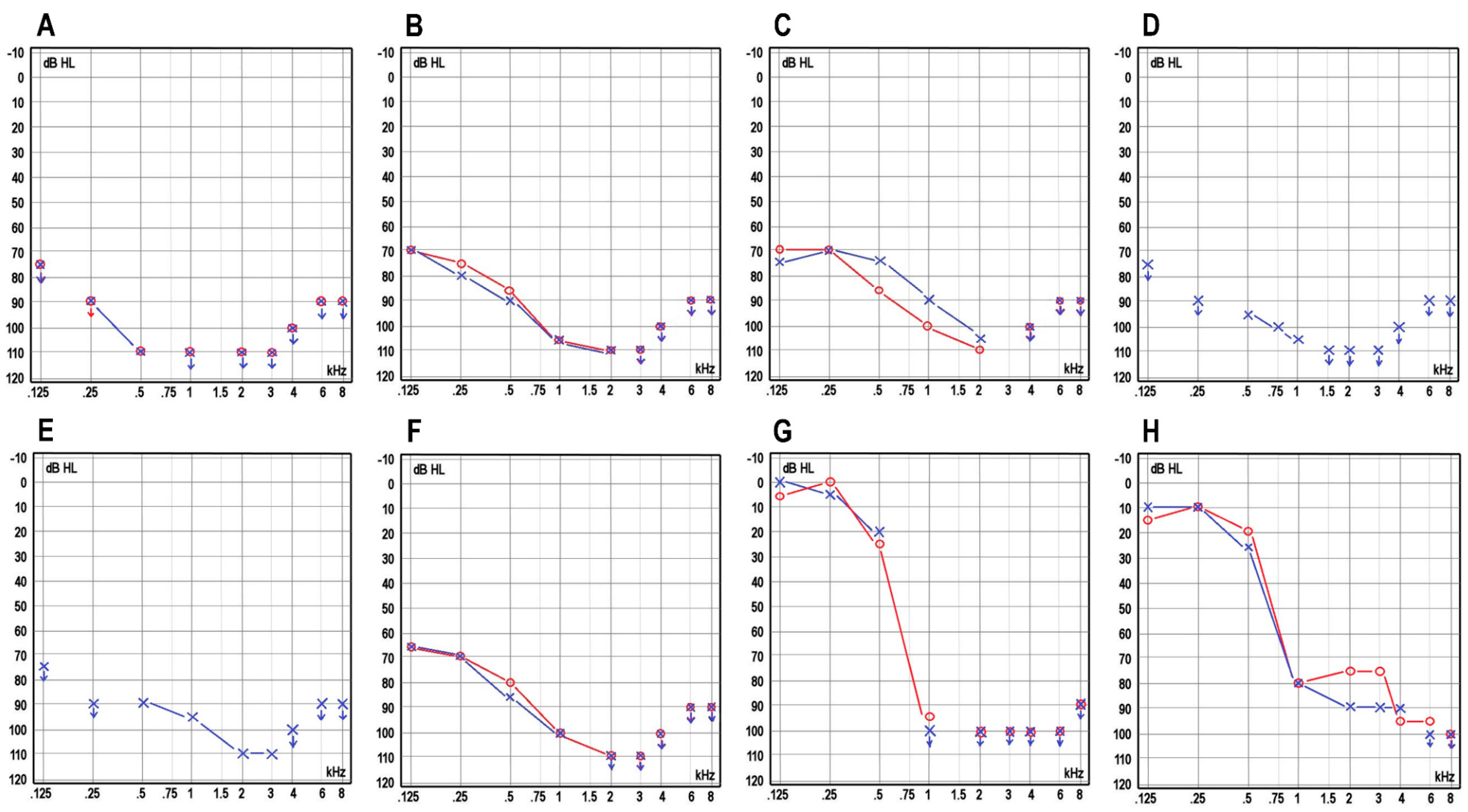
3.1. Clinical Characteristics of the Patients

The clinical characteristics of the affected cases for each family included in this study are summarized in Table 1, and the pedigrees are shown in Figure 1. Eleven affected individuals from eight different Finnish families were recruited for the study. Four were males, and seven were females, and their ages ranged between 0 and 57 years.
In the families with a recessive pattern of inheritance (Families 1–7), the hearing loss was congenital. Three individuals (Patients 3–5 from Families 3 and 4) were born before the start of the newborn hearing screening program, and before CIs were available. Their hearing aid benefit was limited, and they all use sign language as their primary mode of communication. The newborn screenings were abnormal in all tested cases (N = 5/5, 100%), the OAE and TEOAE showed no responses (N = 6/6, 100% and (N = 2/2, 100%, respectively), and the ABR was abnormal in all tested cases (N = 6/6, 100%). An audiogram revealed a bilateral profound SNHL in all the affected individuals (Figure 2A–F). One individual reported a balance problem, but his motor development was normal. The ophthalmological examination revealed one individual with deuteranopia and one individual with esophoria and hyperopia. Imaging studies were unremarkable. Seven individuals were fitted with hearing aids (between 3 months to 1.5 years of age) but obtained limited benefits. Bilateral simultaneous or consecutive CIs were later implanted in five individuals at the ages of 12 months to 2 years, and bilateral simultaneous CIs are planned for the youngest individual at the age of 8–9 months. All implanted patients showed good progress in language acquisition. In the best-aided condition (bilateral CIs), in quiet, the speech recognition scores ranged from 75% to 100%. The two youngest patients, who were tested with the FINSIMAT, had speech reception thresholds (SRTs) of −6.6 dB signal-to-noise ratio (SNR) and −6.2 dB SNR. The best-aided SRTs for the adults, determined with the FMST, ranged from −3.4 dB SNR to −8.6 dB SNR.
In family 8, with dominant inheritance, the onset of hearing loss was postlingual and was diagnosed at the age of 7 years. The hearing loss was progressive, and audiograms revealed down-sloping curves (Figure 2G,H). The older affected individual benefited from CIs, which she received in her left ear at the age of 41 and in her right ear at the age of 51. Her best-aided SRT with FMST was −2.8 dB SNR. The younger affected individual has been offered CIs but currently prefers to use hearing aids. Her best-aided SRT with the FMST was +0.4 dB SNR.

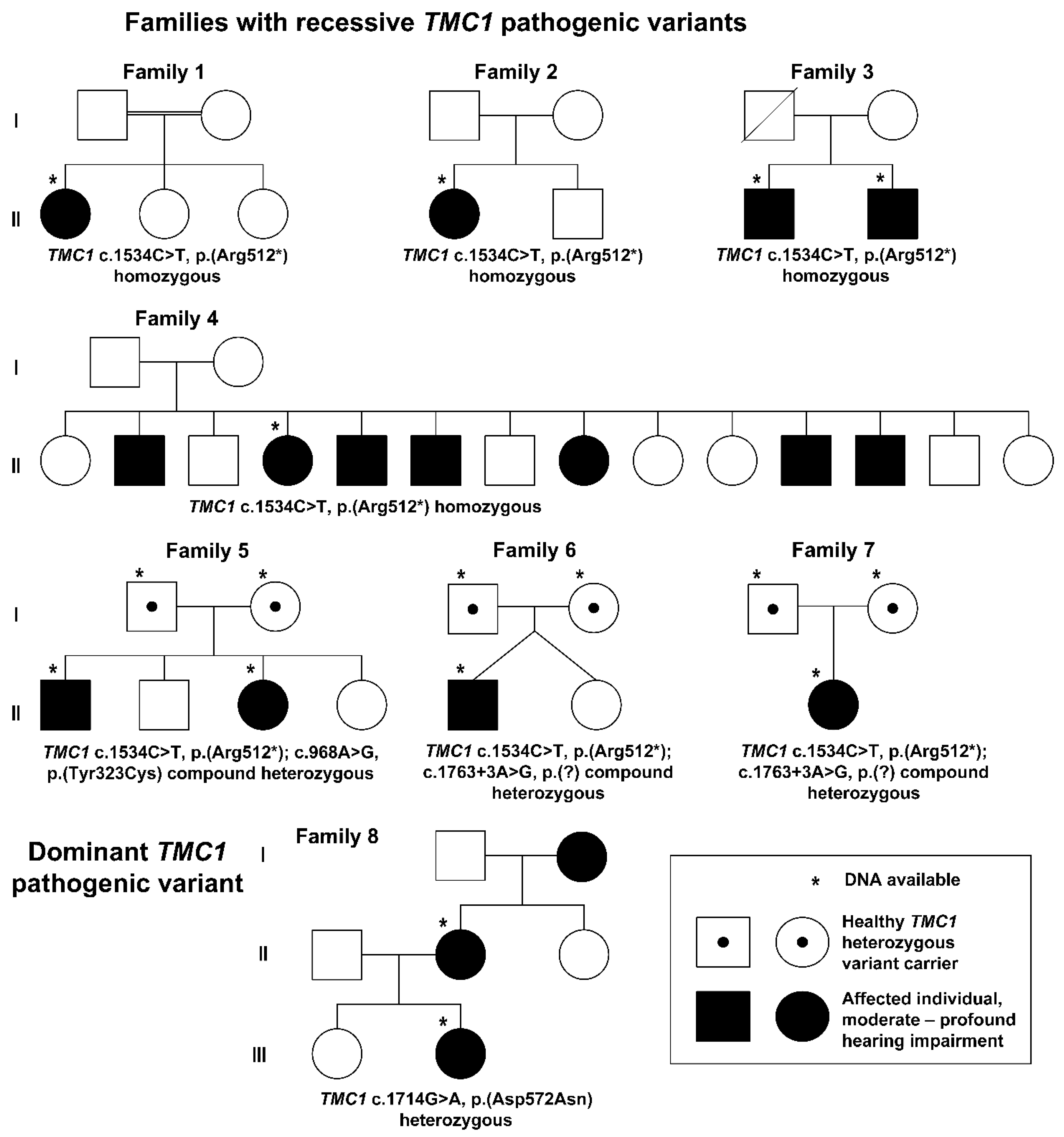
3.2. Genetic Results

Five individuals from families 1 to 4 were homozygous for the TMC1 (NM_138691.3) c.1534C>T, p.(Arg512*) (hg38: chr9:72792320C>T, rs200171616) variant, which was classified as pathogenic according to the ACMG guidelines (PVS1, PM2, PP3, and PP5) [13]. This variant has previously been reported in the medical literature [9,14]. Two individuals from family 5 were compound heterozygous for the TMC1 (NM_138691.3) variants c.1534C>T, p.(Arg512*) and c.968A>G, p.(Tyr323Cys) (hg38: chr9:72788422A>G, rs746724027). The latter is classified as likely pathogenic, according to the ACMG guidelines (PM2, PM3, PP1, and PP3), and, to the authors’ knowledge, this variant has not yet been reported in the literature. Parental testing revealed that the variants were in different alleles. Two affected individuals from families 6 and 7 were compound heterozygous for the TMC1 (NM_138691.3) variants c.1534C>T, p.(Arg512*) and c.1763+3A>G, p.(?) (hg38: chr9:72816213A>G, rs370898981). The latter is classified as pathogenic according to the ACMG guidelines (PS3, PM3, and PM2). TMC1 c.1763+3A>G has previously been reported in the medical literature [15,16]. Parental testing revealed that the variants were in different alleles.
Two individuals from family 8 were heterozygous for the TMC1 (NM_138691.3) c.1714G>A, p.(Asp572Asn), (hg38: chr9:72816161G>A, rs121908072). This heterozygous TMC1 variant and severe hearing loss segregated dominantly in this family. TMC1 c.1714G>A, p.(Asp572Asn) is classified as pathogenic according to the ACMG guidelines (PP5, PM1, PM2, and PM5).

4. Discussion

In this study, we reported nine individuals from seven Finnish families with congenital ARSNHL and biallelic TMC1 variants. We also reported two individuals from one Finnish family with autosomal dominant SNHL (ADSNHL) and a heterozygous TMC1 variant. All the affected individuals with recessively inherited TMC1 variants displayed severe-to-profound congenital hearing loss associated with down-sloping audiograms, where higher frequencies are the most severely affected. This is consistent with previous publications demonstrating that pathogenic recessive TMC1 variants cause moderate-to-profound hearing loss [17,18,19,20]. The individuals affected with ADSNHL displayed progressive postlingual sensorineural hearing loss that led to profound deafness and the need for CIs. In the previous publications, ADSNHL cases showed postlingual-onset progressive hearing loss, with predominant deterioration in the higher frequencies [18,19,20,21]. Only 1 of the 11 affected individuals in the present study had balance problems. This finding is in line with previous studies that found no vestibular symptoms associated with TMC1-associated HL cases [18,19].
Previous work has shown variable auditory outcome in patients with pathogenic biallelic TMC1 variants [19,21,22,23]. Most reports are small case series with heterogeneous samples, which makes it difficult to assess the full effect of a particular genotype. Recent study reported three patients with pathogenic compound heterozygous TMC1 variants demonstrating excellent functional outcome after Cis [22]. Interestingly, one of the patients in this study was compound heterozygous for the Finnish enriched TMC1 c.1534C>T, p.(Arg512*) variant [22]. Another study reported 19 patients from 12 consanguineous Saudi families with pathogenic homozygous TMC1 variants with variable auditory outcome after Cis [19]. In the latter study, both excellent and poor results were reported for individuals with the same variants emphasizing that a significant proportion of the variance in outcomes can be due to social and environmental factors, as well as other known explanatory factors such as the duration of the deafness before the CI operation.
In our study population, the individuals who had access to CIs in early childhood, and received timely hearing rehabilitation with hearing aids and CIs, have reached a good speech perception in quiet and in noise. No age-specific reference values have been established for children for the FINSIMAT, but the two youngest individuals in the present study had SRTs in a range similar to that of typically performing early implanted CI users of their age (Willberg, personal communication). The older, early implanted CI users also reached good speech perception. The average SRT for a bilateral CI user with the FMST was −5.4 dB SNR in a large, unselected group of Finnish CI users [24]. Two of the early implanted individuals in the present study reached an even better SRT, and another individual had a SRT of −3.4 dB, which can still be considered an adequate CI rehabilitation result. Similar to the outcomes from Gallo et al. [22], our outcomes are comparable to the outcomes of individuals with GJB2-associated DFNB1 [25]. Both individuals with ADSNHL had poorer speech perception in noise than the early implanted individuals with ARSNHL. For the younger individual, this was most likely due to insufficient amplification with hearing aids. For the older individual, the poor SRT may be a result of prolonged deafness in the high-frequency region before the CI rehabilitation.
Even though CIs have been highly successful in restoring hearing in severe and profound hearing loss, the rehabilitation results remain highly variable with a significant proportion of the variance still unexplained [26,27]. Implementation of large gene panels and exome sequencing in clinical practice has allowed frequent detection of causative pathogenic variants in patients with hearing loss. Comparing this etiological data systematically with the outcome of CI rehabilitation, especially with speech perception measures in noise, as they are sensitive to minor differences in functional hearing, we can increase our understanding of the extent to which the underlying pathogenic gene variants explain the differences in the CI rehabilitation outcomes.
The TMC1 c.1534C>T, p.(Arg512*) is enriched in the Finnish population, with a minor allele frequency (MAF) 24 times higher in Finns (0.003344) than in non-Finnish Europeans (0.0001395) (gnomAD v.2.1.1, https://gnomad.broadinstitute.org/, accessed on 20 February 2022) [28]. The expected number of TMC1 c.1534C>T, p.(Arg512*) homozygote patients in Finland is (0.003344)2 × 5.5 million or approximately 62, and the carrier frequency of this pathogenic variant is approximately 1/300 in the Finnish population. In this cohort, the TMC1 c.1534C>T, p.(Arg512*) variant was identified in 14 of the 18 alleles (78%) of the ARSNHL patients, and was in either a homozygous or a compound heterozygous state in all patients. Based on the gnomAD and clinical data, we suggest that the TMC1 p.(Arg512*) variant is likely a Finnish founder variant. As hearing loss is genetically highly heterogeneous, additional similar variants will possibly be identified as enriched in the Finnish population.
The TMC1 c.1534C>T, p.(Arg512*) causes a premature stop codon. It is predicted to cause a loss of normal protein function either through protein truncation (512 out of 760 aa) or nonsense-mediated mRNA decay. TMC1 c.1763+3A>G, p.(?) is predicted to cause a splice site defect leading also to a loss-of-function of normal TMC1. TMC1 c.968A>G, p.(Tyr323Cys) was identified in two affected individuals in a compound heterozygote state with a known pathogenic TMC1 c.1534C>T, p.(Arg512*). The mother of these two affected individuals was a healthy heterozygous carrier of this TMC1 c.968A>G, p.(Tyr323Cys) variant suggesting that TMC1 c.968A>G, p.(Tyr323Cys) is a recessive variant leading to a functional null allele.
In contrast, TMC1 c.1714G>A, p.(Asp572Asn) is a well-known dominantly acting TMC1 variant [9,20,29,30]. Amino acid 572 in TMC1 is a critical functional residue for pathogenic dominant variants [9,20,29,30]. Nucleotide 1714 of TMC1 is located within the hypermutable CpG context, thereby explaining its location as a mutational hot spot and its occurrence in different populations [9,29,30]. Dominant variants are likely to confer a specific dominant negative or gain-of-function effect, leading to dominant inheritance [20], whereas pathogenic recessive TMC1 variants create a functional null allele and lead to recessive inheritance through a loss-of-function mechanism. It has been proposed that two mechanisms link pathogenic TMC1 channel variants and deafness: decreased Ca2+ permeability to all pathogenic variants, and decreased resting open probability in low Ca2+ confined to pathogenic dominant variants [31].

5. Conclusions

We identified TMC1 variants c.1534C>T, p.(Arg512*), c.968A>G, p.(Tyr323Cys), and c.1763+3A>G, p.(?) with profound congenital ARSNHL in seven Finnish families, and TMC1 c.1714G>A, p.(Asp572Asn) with postlingual progressive ADSNHL in one Finnish family. To the authors’ knowledge, the TMC1 c.968A>G, p.(Tyr323Cys) variant is a novel variant expanding the knowledge of the genotypic spectrum of pathogenic TMC1 variants. We also showed that the TMC1 c.1534C>T, p.(Arg512*) variant is the most prevalent variant enriched in the Finnish population, suggesting a founder effect. To the authors’ knowledge, these are the first reported cases of TMC1-related SNHL in Finland.
Our study demonstrates that the individuals who had access to CIs in early childhood, and received timely hearing rehabilitation with hearing aids and CIs, have reached a good speech perception in quiet and in noise. Comparison of the genetic data with the outcome of CI rehabilitation increases our understanding of the extent to which underlying pathogenic gene variants explain the differences in CI rehabilitation outcomes. It is even possible that in the future patients can be stratified based on the underlying gene defect into categories that can predict their rehabilitation outcomes.

Author Contributions

Conceptualization: M.K.-T. and E.R.; Formal Analysis: M.K.-T.; Funding acquisition: J.S.M. and E.R.; Investigation: M.K.-T. and S.H.; Resources: M.K.-T., M.K.H., T.W., P.P., R.K.-F., O.K., S.H. and E.R.; Supervision: E.R.; Visualization: S.H. and E.R. Writing—original draft: M.K.-T., M.K.H., T.W. and E.R.; Writing—review and editing: M.K.-T., M.K.H., T.W., P.P., R.K.-F., O.K., J.S.M., S.H. and E.R. All authors have read and agreed to the published version of the manuscript.

Funding

This study was funded by Academy of Finland (grant number 338446), Uolevi Mäki Foundation, and the Competitive State Research Financing of the Expert Responsibility Area of Oulu University Hospital (grant number VTR K36733).

Institutional Review Board Statement

The study was conducted in accordance with the Declaration of Helsinki, and approved by the Ethics Committee of the Northern Ostrobothnia Hospital District (Eettmk § 186, 17 August 2020).

Informed Consent Statement

Written informed consent was obtained from all patients or their guardians.

Data Availability Statement

The reported variants were submitted to the LOVD database hosted at Leiden University Medical Center, The Netherlands.

Acknowledgments

We would like to thank genetic nurse Sirpa Miettinen for recruiting patients and the families for participating in the study.

Conflicts of Interest

All authors declare no conflict of interest.

References

  1. Smith, R.J.H.; Bale, J.F.; White, K.R. Sensorineural Hearing Loss in Children. Lancet 2005, 365, 879–890. [Google Scholar] [CrossRef]
  2. Koffler, T.; Ushakov, K.; Avraham, K.B. Genetics of Hearing Loss: Syndromic. Otolaryngol. Clin. N. Am. 2015, 48, 1041–1061. [Google Scholar] [CrossRef] [PubMed] [Green Version]
  3. Parker, M.; Bitner-Glindzicz, M. Genetic Investigations in Childhood Deafness. Arch. Dis Child. 2015, 100, 271–278. [Google Scholar] [CrossRef]
  4. Shearer, A.E.; Hildebrand, M.S.; Smith, R.J. Hereditary Hearing Loss and Deafness Overview. In GeneReviews®; Adam, M.P., Ardinger, H.H., Pagon, R.A., Wallace, S.E., Bean, L.J., Gripp, K.W., Mirzaa, G.M., Amemiya, A., Eds.; University of Washington, Seattle: Seattle, WA, USA, 1993. [Google Scholar]
  5. Pan, B.; Géléoc, G.S.; Asai, Y.; Horwitz, G.C.; Kurima, K.; Ishikawa, K.; Kawashima, Y.; Griffith, A.J.; Holt, J.R. TMC1 and TMC2 Are Components of the Mechanotransduction Channel in Hair Cells of the Mammalian Inner Ear. Neuron 2013, 79, 504–515. [Google Scholar] [CrossRef] [Green Version]
  6. Kawashima, Y.; Géléoc, G.S.G.; Kurima, K.; Labay, V.; Lelli, A.; Asai, Y.; Makishima, T.; Wu, D.K.; Della Santina, C.C.; Holt, J.R.; et al. Mechanotransduction in Mouse Inner Ear Hair Cells Requires Transmembrane Channel-like Genes. J. Clin. Investig. 2011, 121, 4796–4809. [Google Scholar] [CrossRef]
  7. Kurima, K.; Ebrahim, S.; Pan, B.; Sedlacek, M.; Sengupta, P.; Millis, B.A.; Cui, R.; Nakanishi, H.; Fujikawa, T.; Kawashima, Y.; et al. TMC1 and TMC2 Localize at the Site of Mechanotransduction in Mammalian Inner Ear Hair Cell Stereocilia. Cell Rep. 2015, 12, 1606–1617. [Google Scholar] [CrossRef] [PubMed] [Green Version]
  8. Pan, B.; Akyuz, N.; Liu, X.-P.; Asai, Y.; Nist-Lund, C.; Kurima, K.; Derfler, B.H.; György, B.; Limapichat, W.; Walujkar, S.; et al. TMC1 Forms the Pore of Mechanosensory Transduction Channels in Vertebrate Inner Ear Hair Cells. Neuron 2018, 99, 736–753. [Google Scholar] [CrossRef] [PubMed] [Green Version]
  9. Kurima, K.; Peters, L.M.; Yang, Y.; Riazuddin, S.; Ahmed, Z.M.; Naz, S.; Arnaud, D.; Drury, S.; Mo, J.; Makishima, T.; et al. Dominant and Recessive Deafness Caused by Mutations of a Novel Gene, TMC1, Required for Cochlear Hair-Cell Function. Nat. Genet. 2002, 30, 277–284. [Google Scholar] [CrossRef] [PubMed]
  10. Sirmaci, A.; Duman, D.; Oztürkmen-Akay, H.; Erbek, S.; Incesulu, A.; Oztürk-Hişmi, B.; Arici, Z.S.; Yüksel-Konuk, E.B.; Taşir-Yilmaz, S.; Tokgöz-Yilmaz, S.; et al. Mutations in TMC1 Contribute Significantly to Nonsyndromic Autosomal Recessive Sensorineural Hearing Loss: A Report of Five Novel Mutations. Int. J. Pediatr. Otorhinolaryngol. 2009, 73, 699–705. [Google Scholar] [CrossRef] [PubMed]
  11. Sommen, M.; Schrauwen, I.; Vandeweyer, G.; Boeckx, N.; Corneveaux, J.J.; van den Ende, J.; Boudewyns, A.; De Leenheer, E.; Janssens, S.; Claes, K.; et al. DNA Diagnostics of Hereditary Hearing Loss: A Targeted Resequencing Approach Combined with a Mutation Classification System. Hum. Mutat. 2016, 37, 812–819. [Google Scholar] [CrossRef] [PubMed]
  12. Zazo Seco, C.; Wesdorp, M.; Feenstra, I.; Pfundt, R.; Hehir-Kwa, J.Y.; Lelieveld, S.H.; Castelein, S.; Gilissen, C.; de Wijs, I.J.; Admiraal, R.J.; et al. The Diagnostic Yield of Whole-Exome Sequencing Targeting a Gene Panel for Hearing Impairment in The Netherlands. Eur. J. Hum. Genet. 2017, 25, 308–314. [Google Scholar] [CrossRef] [PubMed] [Green Version]
  13. Richards, S.; Aziz, N.; Bale, S.; Bick, D.; Das, S.; Gastier-Foster, J.; Grody, W.W.; Hegde, M.; Lyon, E.; Spector, E.; et al. Standards and Guidelines for the Interpretation of Sequence Variants: A Joint Consensus Recommendation of the American College of Medical Genetics and Genomics and the Association for Molecular Pathology. Genet. Med. 2015, 17, 405–424. [Google Scholar] [CrossRef] [PubMed] [Green Version]
  14. Bademci, G.; Foster, J.; Mahdieh, N.; Bonyadi, M.; Duman, D.; Cengiz, F.B.; Menendez, I.; Diaz-Horta, O.; Shirkavand, A.; Zeinali, S.; et al. Comprehensive Analysis via Exome Sequencing Uncovers Genetic Etiology in Autosomal Recessive Nonsyndromic Deafness in a Large Multiethnic Cohort. Genet. Med. 2016, 18, 364–371. [Google Scholar] [CrossRef] [PubMed] [Green Version]
  15. de Heer, A.-M.R.; Collin, R.W.J.; Huygen, P.L.M.; Schraders, M.; Oostrik, J.; Rouwette, M.; Kunst, H.P.M.; Kremer, H.; Cremers, C.W.R.J. Progressive Sensorineural Hearing Loss and Normal Vestibular Function in a Dutch DFNB7/11 Family with a Novel Mutation in TMC1. Audiol. Neurootol. 2011, 16, 93–105. [Google Scholar] [CrossRef] [PubMed]
  16. Schrauwen, I.; Sommen, M.; Corneveaux, J.J.; Reiman, R.A.; Hackett, N.J.; Claes, C.; Claes, K.; Bitner-Glindzicz, M.; Coucke, P.; Van Camp, G.; et al. A Sensitive and Specific Diagnostic Test for Hearing Loss Using a Microdroplet PCR-Based Approach and next Generation Sequencing. Am. J. Med. Genet. A 2013, 161A, 145–152. [Google Scholar] [CrossRef] [PubMed]
  17. Imtiaz, A.; Maqsood, A.; Rehman, A.U.; Morell, R.J.; Holt, J.R.; Friedman, T.B.; Naz, S. Recessive Mutations of TMC1 Associated with Moderate to Severe Hearing Loss. Neurogenetics 2016, 17, 115–123. [Google Scholar] [CrossRef] [PubMed] [Green Version]
  18. Nishio, S.-Y.; Usami, S.-I. Prevalence and Clinical Features of Autosomal Dominant and Recessive TMC1-Associated Hearing Loss. Hum. Genet. 2021. [Google Scholar] [CrossRef]
  19. Ramzan, K.; Al-Owain, M.; Al-Numair, N.S.; Afzal, S.; Al-Ageel, S.; Al-Amer, S.; Al-Baik, L.; Al-Otaibi, G.F.; Hashem, A.; Al-Mashharawi, E.; et al. Identification of TMC1 as a Relatively Common Cause for Nonsyndromic Hearing Loss in the Saudi Population. Am. J. Med. Genet. B Neuropsychiatr. Genet. 2020, 183, 172–180. [Google Scholar] [CrossRef]
  20. Wang, H.; Wu, K.; Guan, J.; Yang, J.; Xie, L.; Xiong, F.; Lan, L.; Wang, D.; Wang, Q. Identification of Four TMC1 Variations in Different Chinese Families with Hereditary Hearing Loss. Mol. Genet. Genom. Med. 2018, 6, 504–513. [Google Scholar] [CrossRef]
  21. Makishima, T.; Kurima, K.; Brewer, C.C.; Griffith, A.J. Early Onset and Rapid Progression of Dominant Nonsyndromic DFNA36 Hearing Loss. Otol. Neurotol. 2004, 25, 714–719. [Google Scholar] [CrossRef]
  22. Gallo, S.; Trevisi, P.; Rigon, C.; Caserta, E.; Seif Ali, D.; Bovo, R.; Martini, A.; Cassina, M. Auditory Outcome after Cochlear Implantation in Children with DFNB7/11 Caused by Pathogenic Variants in TMC1 Gene. Audiol. Neurootol. 2021, 26, 157–163. [Google Scholar] [CrossRef] [PubMed]
  23. Lee, S.-Y.; Oh, D.-Y.; Han, J.H.; Kim, M.Y.; Kim, B.; Kim, B.J.; Song, J.-J.; Koo, J.-W.; Lee, J.H.; Oh, S.H.; et al. Flexible Real-Time Polymerase Chain Reaction-Based Platforms for Detecting Deafness Mutations in Koreans: A Proposed Guideline for the Etiologic Diagnosis of Auditory Neuropathy Spectrum Disorder. Diagnostics 2020, 10, 672. [Google Scholar] [CrossRef]
  24. Willberg, T.; Sivonen, V.; Linder, P.; Dietz, A. Comparing the Speech Perception of Cochlear Implant Users with Three Different Finnish Speech Intelligibility Tests in Noise. J. Clin. Med. 2021, 10, 3666. [Google Scholar] [CrossRef]
  25. Black, J.; Hickson, L.; Black, B. Defining and Evaluating Success in Paediatric Cochlear Implantation—An Exploratory Study. Int. J. Pediatr. Otorhinolaryngol. 2012, 76, 1317–1326. [Google Scholar] [CrossRef] [PubMed]
  26. Holden, L.K.; Finley, C.C.; Firszt, J.B.; Holden, T.A.; Brenner, C.; Potts, L.G.; Gotter, B.D.; Vanderhoof, S.S.; Mispagel, K.; Heydebrand, G.; et al. Factors Affecting Open-Set Word Recognition in Adults with Cochlear Implants. Ear Hear. 2013, 34, 342–360. [Google Scholar] [CrossRef] [PubMed] [Green Version]
  27. Zhao, E.E.; Dornhoffer, J.R.; Loftus, C.; Nguyen, S.A.; Meyer, T.A.; Dubno, J.R.; McRackan, T.R. Association of Patient-Related Factors with Adult Cochlear Implant Speech Recognition Outcomes: A Meta-Analysis. JAMA Otolaryngol. Head Neck Surg. 2020, 146, 613–620. [Google Scholar] [CrossRef] [PubMed]
  28. Karczewski, K.J.; Francioli, L.C.; Tiao, G.; Cummings, B.B.; Alföldi, J.; Wang, Q.; Collins, R.L.; Laricchia, K.M.; Ganna, A.; Birnbaum, D.P.; et al. The Mutational Constraint Spectrum Quantified from Variation in 141,456 Humans. Nature 2020, 581, 434–443. [Google Scholar] [CrossRef]
  29. Hilgert, N.; Monahan, K.; Kurima, K.; Li, C.; Friedman, R.A.; Griffith, A.J.; Van Camp, G. Amino Acid 572 in TMC1: Hot Spot or Critical Functional Residue for Dominant Mutations Causing Hearing Impairment. J. Hum. Genet. 2009, 54, 188–190. [Google Scholar] [CrossRef]
  30. Wei, Q.; Zhu, H.; Qian, X.; Chen, Z.; Yao, J.; Lu, Y.; Cao, X.; Xing, G. Targeted Genomic Capture and Massively Parallel Sequencing to Identify Novel Variants Causing Chinese Hereditary Hearing Loss. J. Transl. Med. 2014, 12, 311. [Google Scholar] [CrossRef] [Green Version]
  31. Beurg, M.; Schimmenti, L.A.; Koleilat, A.; Amr, S.S.; Oza, A.; Barlow, A.J.; Ballesteros, A.; Fettiplace, R. New Tmc1 Deafness Mutations Impact Mechanotransduction in Auditory Hair Cells. J. Neurosci. 2021, 41, 4378–4391. [Google Scholar] [CrossRef]
Figure 1. Pedigrees of the families in two (I–II) or three (I–III) generations. In six families, the TMC1 c.1534C>T, p.(Arg512*); c.968A>G, p.(Tyr323Cys), and c.1763+3A>G, p.(?) variants segregated recessively. All the affected individuals showed profound congenital deafness. In one family, a dominantly inherited TMC1 c.1714G>A, p.(Asp572Asn) segregated with the phenotype.
Jcm 11 01837 g001
Figure 2. Pure tone audiograms (PTA) of the affected individuals. (A) Patient 1, (B) Patient 3, (C) Patient 5, (D) Patient 6, (E) Patient 7, and (F) Patient 8. All six of these patients showed severe-to-profound sensorineural hearing loss and mildly down-sloping curves associated with recessive pathogenic TMC1 variants. (G) Patient 10 and (H) Patient 11 had deeply down-sloping curves, demonstrating predominant deterioration of higher frequencies associated with a dominant pathogenic TMC1 variant. In all patients, the last available audiograms are shown. Patients 3, 5, and 11 have not received CIs. In Patients 1, 6, and 7, the audiograms shown are after the first, but before the second CI operation. In Patient 8 the audiogram shown is after bilateral CI operations. In Patient 10, the audiogram shown is before the CI operation. The red circles indicate the right ear and the blue X’s indicate the left ear.
Jcm 11 01837 g002
Table 1. Clinical characteristics of patients. Patients 1–9 had recessively inherited sensorineural hearing loss associated with biallelic pathogenic recessive TMC1 variants. Patients 10 and 11 had a dominantly inherited hearing loss associated with a heterozygous pathogenic TMC1 variant.
Patient 1Patient 2Patient 3Patient 4Patient 5Patient 6Patient 7Patient 8Patient 9Patient 10Patient 11
Family12334556788
Current age2154246532318705721
SexFFMMFMFMFFF
Genotypec.1534C>T, p.(Arg512*), homozygousc.1534C>T, p.(Arg512*), homozygousc.1534C>T, p.(Arg512*), homozygousc.1534C>T, p.(Arg512*), homozygousc.1534C>T, p.(Arg512*), homozygousc.1534C>T, p.(Arg512*) and c.968A>G, p.(Tyr323Cys)c.1534C>T, p.(Arg512*) and c.968A>G, p.(Tyr323Cys)c.1534C>T, p.(Arg512*) and c.1763+3A>Gc.1534C>T, p.(Arg512*) and c.1763+3A>Gc.1714G>A, p.(Asp572Asn) heterozygousc.1714G>A, p.(Asp572Asn) heterozygous
Newborn screeningAbnormalAbnormalNANANANAAbnormalAbnormalAbnormalNANA
OAENo responseNo responseNANANANo responseNo responseNo responseNo responseNANA
TEOAENANo responseNANANANANANANo responseNANA
AABRNANo responseNANANANANANANo responseNANA
ABR (dB)No responsel.dx: no response/l.sin: 90 (4 kHz) and click 85NANANANo responseNo responseNo responseCochlear microphonics at 85, no neural responses at 85NANA
Speech-in-noise testSRS +10 dB SNR l.a. 99%; SRT l.dx. −8.0 dB SNR, SRT l.sin −8.6 dB SNRSRT l.dx −5,9 dB SNR, SRT l.sin −4.9 dB SNR, SRT l.a. −6.6 dB SNRNANANASRS +10 dB SNR l.a. 75%, SRT l.dx −2.3 dB SNR, SRT l.a. −3.4 dB SNRSRS +10 dB SNR l.a. 100%, SRT l.dx −7.6 dB SNR, SRT l.a. −6.6/−7.6 dB SNRSRT l.dx −6.3 dB SNR, SRT l.sin −4.9 dB SNR, SRT l.a. −6.2 dB SNR,NASRS +10 dB SNR l.a. 94%; SRT l.sin. −1.3 dB SNR, SRT l.dx. −2.0 dB SNR, SRT l.a. −2.8 dB SNRSRS +10 dB SNR l.a. 88%, SRT l.a. +0.4 dB SNR
Age at diagnosis7 m2 mFrom birth1 y 8 mFrom birthFrom birth1 m8 mFrom birth7 y7 y
SeverityProfoundProfoundProfoundProfoundProfoundProfoundProfoundProfoundProfoundProfoundSevere
ProgressionNoNoNoNoNoNoNoNoNoYesYes
Hearing aids (from the age)Yes (7 m)Yes (4 m)NoNoYes (1.5 y)Yes (1 y 4 m)Yes (3 m)Yes (9 m)Yes (4 m)NoYes
Cochlear implant (from the age)Yes (1 y 5 m) (right) and 13 y (left)Yes (1 y)NoNoNoYes (2 y 1 m (right) and 20 y 9 m (left)Yes (1 y (right) and 15 y 5 m (left)Yes (1 y 5 m (right) and 1y6m (left)Planned (8–9 m)Yes (41 y (left) and 51 y (right)No
Balance problemsNoNoNoNoNoYesNoNoNANoNo
Language perceptionGood progress with CI useGood progress with CI useSign languageSign languageSign languageGood progress with CI useGood progress with CI useGood progress with CI useNANormal spoken languageNormal spoken language
Abbreviations: OAE, otoacoustic emission; TEOAE, transient evoked OAE; ABR, auditory brainstem response; AABR, automated ABR; SRS, speech recognition score; SNR, signal-to-noise ratio; SRT, speech reception threshold; CI, cochlear implant; l.a., both sides; l.dx., right side; l.sin., left side.
Publisher’s Note: MDPI stays neutral with regard to jurisdictional claims in published maps and institutional affiliations.

