
Affection of Respiratory Muscles in ALS and SMA

 and
and 
Abstract
:1. Introduction
2. Materials and Methods
2.1. Patients and Study Design
2.2. Spirometry and Clinical Routine Data Evaluation
2.3. Ultrasound Examination of the Diaphragm
- Side-separated diaphragm thickness.
- 2.
- Thickening ratio and thickening fraction of the diaphragm.
- 3.
- Side-separated excursion of the diaphragm.
2.4. Statistics
3. Results
3.1. Sociodemographic and Clinical Data
3.2. Diaphragm Ultrasound Is Feasible Even in Advanced MND
3.3. Diaphragm Dysfunction Is More Prevalent in ALS Than SMA
3.4. Respiratory Dysfunction Occurs More Frequently in ALS
3.5. Diaphragm Excursion Correlated Best with Classical Respiratory Measures
4. Discussion
Supplementary Materials
Author Contributions
Funding
Institutional Review Board Statement
Informed Consent Statement
Data Availability Statement
Acknowledgments
Conflicts of Interest
References
- Comley, L.H.; Nijssen, J.; Frost-Nylen, J.; Hedlund, E. Cross-disease comparison of amyotrophic lateral sclerosis and spinal muscular atrophy reveals conservation of selective vulnerability but differential neuromuscular junction pathology. J. Comp. Neurol. 2016, 524, 1424–1442. [Google Scholar] [CrossRef] [PubMed] [Green Version]
- Bourke, S.C.; Tomlinson, M.; Williams, T.L.; Bullock, R.E.; Shaw, P.J.; Gibson, G.J. Effects of non-invasive ventilation on survival and quality of life in patients with amyotrophic lateral sclerosis: A randomised controlled trial. Lancet Neurol. 2006, 5, 140–147. [Google Scholar] [CrossRef]
- Sancho, J.; Martinez, D.; Bures, E.; Diaz, J.L.; Ponz, A.; Servera, E. Bulbar impairment score and survival of stable amyotrophic lateral sclerosis patients after noninvasive ventilation initiation. ERJ Open Res. 2018, 4, 00159–2017. [Google Scholar] [CrossRef] [PubMed] [Green Version]
- Vitacca, M.; Montini, A.; Lunetta, C.; Banfi, P.; Bertella, E.; De Mattia, E.; Lizio, A.; Volpato, E.; Lax, A.; Morini, R.; et al. Impact of an early respiratory care programme with non-invasive ventilation adaptation in patients with amyotrophic lateral sclerosis. Eur. J. Neurol. 2018, 25, 556-e33. [Google Scholar] [CrossRef] [PubMed]
- Hagenacker, T.; Wurster, C.D.; Gunther, R.; Schreiber-Katz, O.; Osmanovic, A.; Petri, S.; Weiler, M.; Ziegler, A.; Kuttler, J.; Koch, J.C.; et al. Nusinersen in adults with 5q spinal muscular atrophy: A non-interventional, multicentre, observational cohort study. Lancet Neurol. 2020, 19, 317–325. [Google Scholar] [CrossRef]
- Ludolph, A.C.; Wurster, C.D. Therapeutic advances in SMA. Curr. Opin. Neurol. 2019, 32, 777–781. [Google Scholar] [CrossRef]
- Boentert, M.; Wenninger, S.; Sansone, V.A. Respiratory involvement in neuromuscular disorders. Curr. Opin. Neurol. 2017, 30, 529–537. [Google Scholar] [CrossRef]
- Roussos, C.; Macklem, P.T. The respiratory muscles. N. Engl. J. Med. 1982, 307, 786–797. [Google Scholar] [CrossRef]
- Simon, N.G.; Turner, M.R.; Vucic, S.; Al-Chalabi, A.; Shefner, J.; Lomen-Hoerth, C.; Kiernan, M.C. Quantifying disease progression in amyotrophic lateral sclerosis. Ann. Neurol. 2014, 76, 643–657. [Google Scholar] [CrossRef]
- Windisch, W.; Dreher, M.; Geiseler, J.; Siemon, K.; Brambring, J.; Dellweg, D.; Grolle, B.; Hirschfeld, S.; Kohnlein, T.; Mellies, U.; et al. Guidelines for Non-Invasive and Invasive Home Mechanical Ventilation for Treatment of Chronic Respiratory Failure—Update 2017. Pneumologie 2017, 71, 722–795. [Google Scholar] [CrossRef]
- Bourke, S.C. Respiratory involvement in neuromuscular disease. Clin. Med. (Lond.) 2014, 14, 72–75. [Google Scholar] [CrossRef] [PubMed]
- Chiang, J.; Mehta, K.; Amin, R. Respiratory Diagnostic Tools in Neuromuscular Disease. Children 2018, 5, 78. [Google Scholar] [CrossRef] [PubMed] [Green Version]
- Simon, N.G.; Kiernan, M.C. Diaphragm ultrasound in amyotrophic lateral sclerosis and other neuromuscular disorders. Clin. Neurophysiol. 2016, 127, 28–30. [Google Scholar] [CrossRef] [PubMed]
- Harlaar, L.; Ciet, P.; van der Ploeg, A.T.; Brusse, E.; van der Beek, N.; Wielopolski, P.A.; de Bruijne, M.; Tiddens, H.; van Doorn, P.A. Imaging of respiratory muscles in neuromuscular disease: A review. Neuromuscul. Disord. 2018, 28, 246–256. [Google Scholar] [CrossRef] [PubMed]
- Criee, C.P.; Baur, X.; Berdel, D.; Bosch, D.; Gappa, M.; Haidl, P.; Husemann, K.; Jorres, R.A.; Kabitz, H.J.; Kardos, P.; et al. Standardization of spirometry: 2015 update. Published by German Atemwegsliga, German Respiratory Society and German Society of Occupational and Environmental Medicine. Pneumologie 2015, 69, 147–164. [Google Scholar] [CrossRef] [PubMed]
- Pera, M.C.; Coratti, G.; Forcina, N.; Mazzone, E.S.; Scoto, M.; Montes, J.; Pasternak, A.; Mayhew, A.; Messina, S.; Sframeli, M.; et al. Content validity and clinical meaningfulness of the HFMSE in spinal muscular atrophy. BMC Neurol. 2017, 17, 39. [Google Scholar] [CrossRef] [PubMed] [Green Version]
- Mazzone, E.S.; Mayhew, A.; Montes, J.; Ramsey, D.; Fanelli, L.; Young, S.D.; Salazar, R.; De Sanctis, R.; Pasternak, A.; Glanzman, A.; et al. Revised upper limb module for spinal muscular atrophy: Development of a new module. Muscle Nerve 2017, 55, 869–874. [Google Scholar] [CrossRef]
- Cedarbaum, J.M.; Stambler, N.; Malta, E.; Fuller, C.; Hilt, D.; Thurmond, B.; Nakanishi, A. The ALSFRS-R: A revised ALS functional rating scale that incorporates assessments of respiratory function. BDNF ALS Study Group (Phase III). J. Neurol. Sci. 1999, 169, 13–21. [Google Scholar] [CrossRef]
- Cardenas, L.Z.; Santana, P.V.; Caruso, P.; Ribeiro de Carvalho, C.R.; Pereira de Albuquerque, A.L. Diaphragmatic Ultrasound Correlates with Inspiratory Muscle Strength and Pulmonary Function in Healthy Subjects. Ultrasound Med. Biol. 2018, 44, 786–793. [Google Scholar] [CrossRef]
- Kabitz, H.J.; Walterspacher, S.; Mellies, U.; Criee, C.P.; Windisch, W. Recommendations for respiratory muscle testing. Pneumologie 2014, 68, 307–314. [Google Scholar] [CrossRef] [Green Version]
- Kabitz, H.J.; Windisch, W.; Schonhofer, B. Understanding ventilator-induced diaphragmatic dysfunction (VIDD): Progress and advances. Pneumologie 2013, 67, 435–441. [Google Scholar] [CrossRef] [PubMed] [Green Version]
- Summerhill, E.M.; El-Sameed, Y.A.; Glidden, T.J.; McCool, F.D. Monitoring recovery from diaphragm paralysis with ultrasound. Chest 2008, 133, 737–743. [Google Scholar] [CrossRef] [PubMed]
- Windisch, W.; Schonhofer, B.; Magnet, F.S.; Stoelben, E.; Kabitz, H.J. Diagnosis and Treatment of Diaphragmatic Dysfunction. Pneumologie 2016, 70, 454–461. [Google Scholar] [CrossRef] [PubMed]
- Kim, W.Y.; Suh, H.J.; Hong, S.B.; Koh, Y.; Lim, C.M. Diaphragm dysfunction assessed by ultrasonography: Influence on weaning from mechanical ventilation. Crit. Care Med. 2011, 39, 2627–2630. [Google Scholar] [CrossRef]
- Spiesshoefer, J.; Herkenrath, S.; Henke, C.; Langenbruch, L.; Schneppe, M.; Randerath, W.; Young, P.; Brix, T.; Boentert, M. Evaluation of Respiratory Muscle Strength and Diaphragm Ultrasound: Normative Values, Theoretical Considerations, and Practical Recommendations. Respiration 2020, 99, 369–381. [Google Scholar] [CrossRef]
- Hiwatani, Y.; Sakata, M.; Miwa, H. Ultrasonography of the diaphragm in amyotrophic lateral sclerosis: Clinical significance in assessment of respiratory functions. Amyotroph. Lateral Scler. Front. Degener. 2013, 14, 127–131. [Google Scholar] [CrossRef]
- Carrie, C.; Bonnardel, E.; Vally, R.; Revel, P.; Marthan, R.; Marthan, R. Vital Capacity Impairment due to Neuromuscular Disease and its Correlation with Diaphragmatic Ultrasound: A Preliminary Study. Ultrasound Med. Biol. 2016, 42, 143–149. [Google Scholar] [CrossRef]
- Buonsenso, D.; Berti, B.; Palermo, C.; Leone, D.; Ferrantini, G.; De Sanctis, R.; Onesimo, R.; Curatola, A.; Fanelli, L.; Forcina, N.; et al. Ultrasound assessment of diaphragmatic function in type 1 spinal muscular atrophy. Pediatr. Pulmonol. 2020, 55, 1781–1788. [Google Scholar] [CrossRef]
- Schroth, M.K. Special considerations in the respiratory management of spinal muscular atrophy. Pediatrics 2009, 123 (Suppl. 4), S245–S249. [Google Scholar] [CrossRef] [Green Version]
- Wang, C.H.; Finkel, R.S.; Bertini, E.S.; Schroth, M.; Simonds, A.; Wong, B.; Aloysius, A.; Morrison, L.; Main, M.; Crawford, T.O.; et al. Consensus statement for standard of care in spinal muscular atrophy. J. Child. Neurol. 2007, 22, 1027–1049. [Google Scholar] [CrossRef]


| Parameter/Score | Outcome | ALS (n = 40) | SMA (n = 23) | p-Value |
|---|---|---|---|---|
| Age [year], median (IQR) | 66 (59–73) | 32 (22–46) | <0.001 † | |
| Sex, n (%) | female male | 18 (45%) 22 (55%) | 14 (61%) 9 (39%) | 0.297 ‡‡ |
| ALS type, n (%) | bulbar spinal unknown | 16 (40%) 23 (57%) 1 (3%) | ||
| SMA type, n (%) | 2 3 | 10 (44%) 13 (56%) | ||
| SMN2copy number, n (%) | 2 3 4 unknown | 1 (4%) 13 (57%) 8 (35%) 1 (4%) | ||
| BMI [kg/m2], median (IQR) | 24 (23–28) | 21 (19–26) | 0.023 § | |
| ALSFRSR [score], median (IQR) | 31 (27–38) | 32 (27–37) | 0.733 † | |
| ALSFRSR respiratory function [ALSFRSR questions 10–12], median (IQR) | 10 (7–12) | 12 (11–12) | <0.001 § | |
| HFMSE [score], median (IQR) | 8 (4–32) | |||
| RULM [score], median (IQR) | 19 (14–35) |
| Parameter | Outcome | ALS (n = 40) | SMA (n = 23) | p-Value § | Multiple Linear Regression Analysis # | |
|---|---|---|---|---|---|---|
| Adjusted Correlation Coefficient (95% CI) | p-Value | |||||
| Diaphragm thickness insp. right | median (IQR) | 2.8 (2.2–3.8) | 2.8 (2.3–3.8) | 0.881 | 0.066 (–0.901–1.033) | 0.892 |
| Diaphragm thickness insp. left | median (IQR) | 2.7 (2.4–4.5) | 3.2 (2.8–3.5) | 0.245 | 0.573 (–0.528–1.674) | 0.301 |
| Diaphragm thickening ratio right | median (IQR) | 1.2 (1.1–1.3) | 1.3 (1.2–1.5) | 0.006 | 0.099 (–0.097–0.294) | 0.317 |
| Diaphragm thickening ratio left | median (IQR) | 1.2 (1.1–1.4) | 1.3 (1.2–1.5) | 0.080 | 0.129 (–0.171–0.428) | 0.393 |
| Diaphragm thickening fraction right | median (IQR) | 15.9 (5.5–26.5) | 31.8 (17.6–45) | 0.006 | 10.006 (–9.698–29.710) | 0.313 |
| Diaphragm thickening fraction left | median (IQR) | 19 (10.0–37.5) | 30.7 (20.2–48.8) | 0.080 | 12.856 (–17.135–42.846) | 0.393 |
| Diaphragm excursion right | median (IQR) | 8.1 (5.1–12.2) | 13.5 (8.0–20.0) | 0.002 | 5.138 (–0.778–11.054) | 0.087 |
| Diaphragm excursion left | median (IQR) | 10.0 (6.3–12.0) | 11.8 (8.6–15.8) | 0.057 | 7.040 (1.458–12.621) | 0.015 |
| Thickness R Insp | Thickness R Exsp | Thickness L Insp | Thickness L Exsp | Thickening Ratio R | Thickening Ratio L | Thickening Fraction R | Thickening Fraction L | Excursion R | Excursion L | ||
|---|---|---|---|---|---|---|---|---|---|---|---|
| ALSFRSR total | −0.17 | −0.26 | −0.29 | −0.31 | 0.22 | −0.05 | 0.23 | −0.05 | 0.32 | 0.48 | 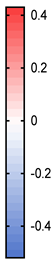 |
| ALSFRSR 10 | −0.11 | −0.16 | −0.41 | −0.38 | 0.09 | −0.10 | 0.10 | −0.10 | 0.28 | 0.42 | |
| ALSFRSR 11 | −0.21 | −0.27 | −0.33 | −0.31 | 0.16 | −0.13 | 0.17 | −0.13 | 0.22 | 0.24 | |
| ALSFRSR 12 | −0.01 | −0.06 | −0.07 | −0.15 | 0.04 | 0.06 | 0.05 | 0.06 | 0.19 | 0.43 | |
| ALSFRSR 10–12 | −0.13 | −0.19 | −0.33 | −0.33 | 0.12 | −0.08 | 0.12 | −0.08 | 0.27 | 0.40 | |
| FEV1 percent | 0.16 | 0.01 | −0.20 | −0.19 | 0.38 | −0.31 | 0.35 | −0.31 | 0.11 | 0.57 | |
| VC percent | 0.07 | −0.14 | 0.16 | 0.10 | 0.47 | 0.02 | 0.47 | 0.02 | 0.25 | 0.48 | |
| pH | −0.15 | −0.21 | −0.04 | −0.12 | 0.13 | 0.24 | 0.14 | 0.24 | 0.19 | 0.18 | |
| pO2 | 0.07 | 0.06 | −0.07 | −0.10 | 0.06 | 0.22 | 0.05 | 0.22 | 0.17 | 0.14 | |
| pCO2 | 0.17 | 0.31 | 0.16 | 0.40 | −0.27 | −0.40 | −0.28 | −0.40 | −0.33 | −0.29 | |
| sO2 | −0.06 | −0.13 | −0.17 | −0.22 | 0.16 | 0.19 | 0.16 | 0.19 | 0.32 | 0.27 | |
| ABE | 0.08 | 0.21 | 0.15 | 0.38 | −0.25 | −0.34 | −0.25 | −0.34 | −0.28 | −0.23 | |
| SBC | 0.12 | 0.25 | 0.18 | 0.41 | −0.26 | −0.34 | −0.26 | −0.34 | −0.28 | −0.23 |
| Thickness R Insp | Thickness R Exsp | Thickness L Insp | Thickness L Exsp | Thickening Ratio R | Thickening Ratio L | Thickening Fraction R | Thickening Fraction L | Excursion R | Excursion L | ||
|---|---|---|---|---|---|---|---|---|---|---|---|
| ALSFRSR total | −0.14 | −0.13 | −0.37 | −0.52 | −0.15 | 0.38 | −0.15 | 0.37 | 0.44 | 0.19 | 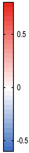 |
| ALSFRSR 10 | 0.23 | 0.16 | 0.04 | −0.03 | 0.29 | 0.13 | 0.29 | 0.13 | 0.26 | 0.07 | |
| ALSFRSR 11 | −0.08 | −0.03 | −0.31 | −0.33 | −0.16 | 0.05 | −0.16 | 0.05 | 0.49 | 0.12 | |
| ALSFRSR 12 | −0.05 | 0.01 | −0.28 | −0.44 | −0.21 | 0.25 | −0.21 | 0.25 | 0.43 | 0.17 | |
| ALSFRSR 10–12 | 0.02 | 0.05 | −0.31 | −0.44 | −0.09 | 0.22 | −0.09 | 0.22 | 0.61 | 0.19 | |
| HFSME | −0.16 | −0.21 | −0.38 | −0.45 | −0.05 | 0.15 | −0.06 | 0.15 | 0.12 | 0.07 | |
| RULM | −0.04 | −0.06 | −0.25 | −0.24 | −0.05 | 0.01 | −0.05 | 0.01 | 0.17 | 0.06 | |
| FEV1 percent | −0.31 | −0.31 | −0.53 | −0.56 | −0.19 | 0.16 | −0.20 | 0.16 | 0.38 | 0.30 | |
| VC percent | −0.23 | −0.28 | −0.57 | −0.51 | −0.08 | 0.00 | −0.08 | 0.00 | 0.38 | 0.21 | |
| pH | 0.61 | 0.80 | 0.56 | 0.55 | −0.05 | −0.11 | −0.05 | −0.11 | 0.02 | −0.02 | |
| pO2 | 0.11 | 0.24 | −0.01 | −0.09 | −0.24 | 0.10 | −0.24 | 0.10 | 0.26 | −0.05 | |
| pCO2 | −0.47 | −0.60 | −0.50 | −0.51 | 0.01 | 0.09 | 0.01 | 0.09 | −0.15 | −0.13 | |
| sO2 | −0.13 | −0.23 | −0.27 | −0.40 | 0.03 | 0.23 | 0.03 | 0.23 | 0.24 | 0.10 | |
| ABE | 0.59 | 0.79 | 0.46 | 0.45 | −0.07 | −0.16 | −0.07 | −0.16 | −0.20 | −0.24 | |
| SBC | 0.23 | 0.28 | 0.22 | 0.22 | 0.02 | −0.07 | 0.02 | −0.07 | −0.50 | −0.15 |
Publisher’s Note: MDPI stays neutral with regard to jurisdictional claims in published maps and institutional affiliations. |
© 2022 by the authors. Licensee MDPI, Basel, Switzerland. This article is an open access article distributed under the terms and conditions of the Creative Commons Attribution (CC BY) license (https://creativecommons.org/licenses/by/4.0/).
Share and Cite
Hermann, W.; Langner, S.; Freigang, M.; Fischer, S.; Storch, A.; Günther, R.; Hermann, A. Affection of Respiratory Muscles in ALS and SMA. J. Clin. Med. 2022, 11, 1163. https://doi.org/10.3390/jcm11051163
Hermann W, Langner S, Freigang M, Fischer S, Storch A, Günther R, Hermann A. Affection of Respiratory Muscles in ALS and SMA. Journal of Clinical Medicine. 2022; 11(5):1163. https://doi.org/10.3390/jcm11051163
Chicago/Turabian StyleHermann, Wiebke, Simona Langner, Maren Freigang, Stefanie Fischer, Alexander Storch, René Günther, and Andreas Hermann. 2022. "Affection of Respiratory Muscles in ALS and SMA" Journal of Clinical Medicine 11, no. 5: 1163. https://doi.org/10.3390/jcm11051163
APA StyleHermann, W., Langner, S., Freigang, M., Fischer, S., Storch, A., Günther, R., & Hermann, A. (2022). Affection of Respiratory Muscles in ALS and SMA. Journal of Clinical Medicine, 11(5), 1163. https://doi.org/10.3390/jcm11051163