
Radiosynovectomy for the Treatment of Chronic Hemophilic Synovitis: An Old Technique, but Still Very Effective

 ,
,
Abstract
1. Introduction
2. When Should a Radiosynovectomy (RS) Be Indicated?
3. RS in Individuals with Inhibitors
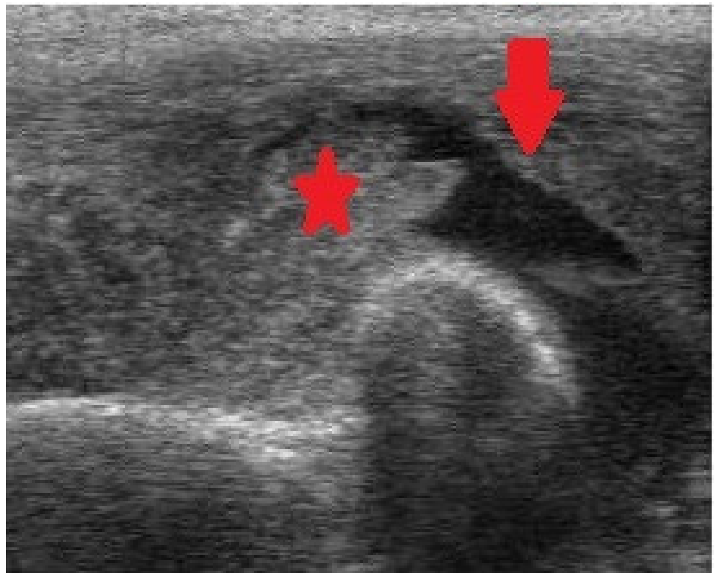
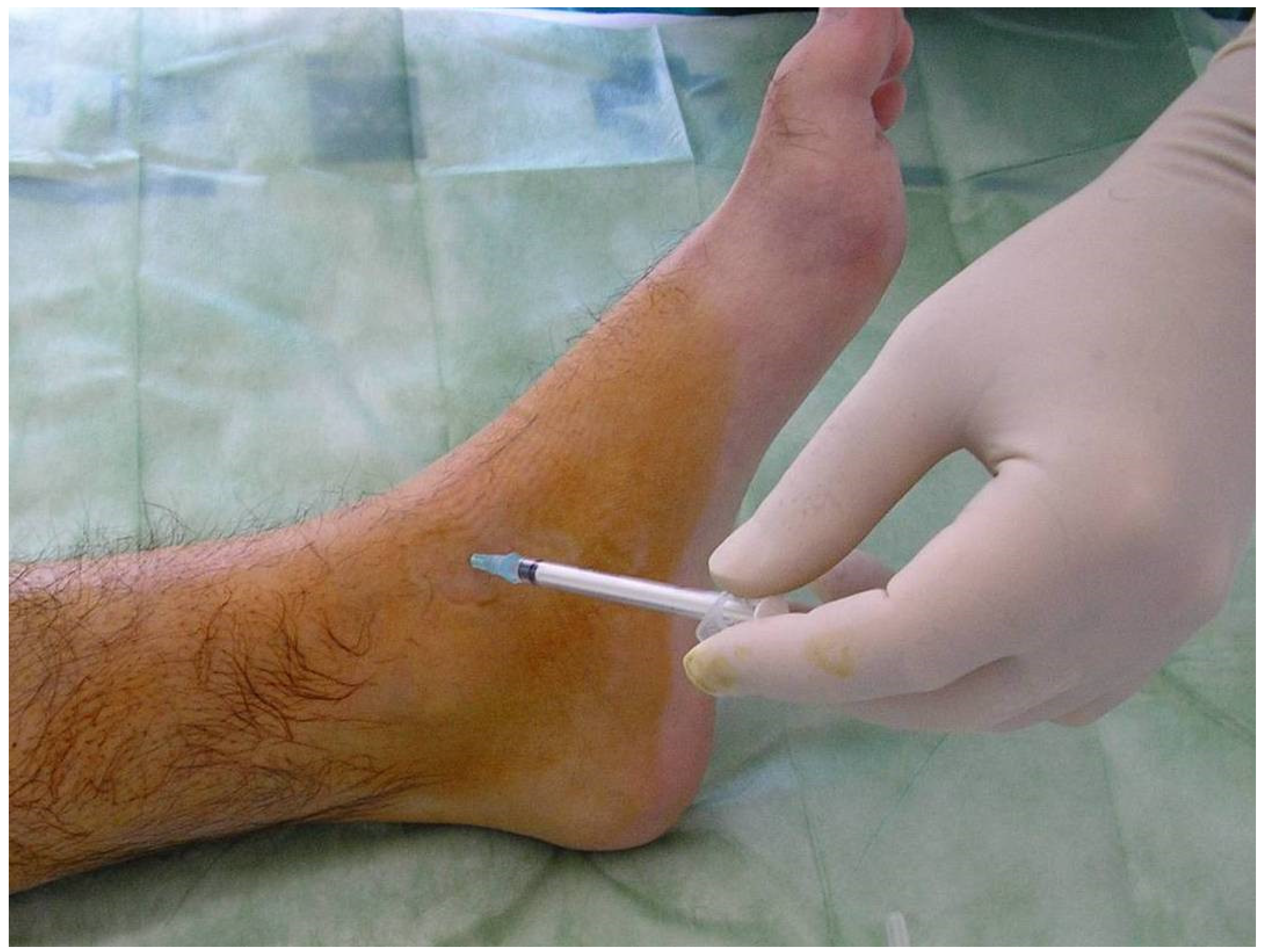
4. Technique of Radiosynovectomy
5. Effectiveness of Radiosynovectomy
6. Complications of Radiosynovectomy
7. Is Radiosynovectomy Safe?
8. Conclusions
Author Contributions
Funding
Institutional Review Board Statement
Informed Consent Statement
Data Availability Statement
Conflicts of Interest
References
- Astermark, J. When to start and when to stop primary prophylaxis in patients with severe haemophilia. Haemophilia 2003, 9 (Suppl. S1), 32–36. [Google Scholar] [CrossRef] [PubMed]
- Manco-Johnson, M.J.; Abshire, T.C.; Shapiro, A.D.; Riske, B.; Hacker, M.R.; Kilcoyne, R.; Ingram, J.D.; Manco-Johnson, M.L.; Funk, S.; Jacobson, L.; et al. Prophylaxis versus episodic treatment to prevent joint disease in boys with severe hemophilia. N. Engl. J. Med. 2007, 357, 535–544. [Google Scholar] [CrossRef] [PubMed]
- Berntorp, E.; Fischer, K.; Miners, A. Models of prophylaxis. Haemophilia 2012, 18 (Suppl. S4), 136–140. [Google Scholar] [CrossRef] [PubMed]
- Ljung, R. Hemophilia and prophylaxis. Pediatr. Blood Cancer 2013, 60 (Suppl. S1), S23–S26. [Google Scholar] [CrossRef] [PubMed]
- Rodriguez-Merchan, E.C.; Valentino, L.A. Emicizumab: Review of the literatura and critical appraisal. Haemophilia 2019, 25, 11–20. [Google Scholar] [CrossRef]
- Kempton, C.; Trask, P.; Parnes, A.; Niggli, M.; Campinha-Bacote, A.; Ucallaghan, M.; O’Connell, N.; Paz-Priel, I.; Mahlangu, J.N. Development and testing of the Satisfaction Questionnaire with Intravenous or Subcutaneous Hemophilia Injection and results from the Phase 3 HAVEN 3 study of emicizumab prophylaxis in persons with haemophilia A without FVIII inhibitors. Haemophilia 2021, 27, 221–228. [Google Scholar] [CrossRef]
- Barg, A.A.; Budnik, I.; Avishai, E.; Brutman-Barazani, T.; Bashari, D.; Misgav, M.; Lubetsky, A.; Kuperman, A.A.; Livnat, T.; Kenet, G. Emicizumab prophylaxis: Prospective longitudinal real-world follow-up and monitoring. Haemophilia 2021, 27, 383–391. [Google Scholar] [CrossRef]
- Mazurkiewicz, Ł.; Czernikiewicz, K.; Rupa-Matysek, J.; Gil, L. Emicizumab: A novel drug in hemophilia A prophylaxis—A narrative review. Expert Rev. Hematol. 2022, 15, 933–942. [Google Scholar] [CrossRef]
- Silva, M.; Luck, J.V.; Siegel, M.E., Jr. 32P chromic phosphate radiosynovectomy for chronic haemophilic synovitis. Haemophilia 2001, 7 (Suppl. S2), 40–49. [Google Scholar] [CrossRef]
- Siegel, H.J.; Luck, J.V.; Siegel, M.E., Jr.; Quinones, C. Phosphate-32 colloid radiosynovectomy in hemophilia: Outcome of 125 procedures. Clin. Orthop. Relat. Res. 2001, 392, 409–417. [Google Scholar] [CrossRef]
- Heim, M.; Goshen, E.; Amit, Y.; Martinowitz, U. Synoviorthesis with radioactive Yttrium in haemophilia: Israel experience. Haemophilia 2001, 7 (Suppl. S2), 36–39. [Google Scholar] [CrossRef]
- Mortazavi, S.M.; Asadollahi, S.; Farzan, M.; Shahriaran, S.; Aghili, M.; Izadyar, S.; Lak, M. (32)P colloid radiosynovectomy in treatment of chronic haemophilic synovitis: Iran experience. Haemophilia 2007, 13, 182–188. [Google Scholar] [CrossRef]
- Calegaro, J.U.; Machado, J.; De Paula, J.C.; De Almeida, J.S.; Casulari, L.A. Clinical evaluation after 1 year of 153-samarium hydroxyapatite synovectomy in patients with haemophilic arthropathy. Haemophilia 2009, 15, 240–246. [Google Scholar] [CrossRef]
- Cho, Y.J.; Kim, K.I.; Chun, Y.S.; Rhyu, K.H.; Kwon, B.K.; Kim, D.Y. Radioisotope synoviorthesis with Holmium-166-chitosan complex in haemophilic arthropath. Radioisotope synoviorthesis with Holmium-166-chitosan complex in haemophilic arthropathy. Haemophilia 2010, 16, 640–646. [Google Scholar]
- De la Corte-Rodriguez, H.; Rodriguez-Merchan, E.C.; Jimenez-Yuste, V. Radiosynovectomy in patients with chronic haemophilic synovitis: When is more than one injection necessary? Eur. J. Haematol. 2011, 86, 430–435. [Google Scholar] [CrossRef]
- De la Corte-Rodriguez, H.; Rodriguez-Merchan, E.C.; Jimenez-Yuste, V. Radiosynovectomy in hemophilia: Quantification of its effectiveness through the assessment of 10 articular parameters. J. Thromb. Haemost. 2011, 9, 928–935. [Google Scholar] [CrossRef]
- De la Corte-Rodriguez, H.; Rodriguez-Merchan, E.C.; Jimenez-Yuste, V. What patient, joint and isotope characteristics influence the response to radiosynovectomy in patients with haemophilia? Haemophilia 2011, 17, e990–e998. [Google Scholar] [CrossRef]
- De la Corte-Rodriguez, H.; Rodriguez-Merchan, E.C.; Jimenez-Yuste, V. Consecutive radiosynovectomy procedures at 6-monthly intervals behave independently in haemophilic synovitis. Blood Transfus. 2013, 11, 254–259. [Google Scholar]
- Rodriguez-Merchan, E.C.; De la Corte-Rodriguez, H.; Jimenez-Yuste, V. Is radiosynovectomy (RS) effective for joints damaged by haemophilia with articular degeneration in simple radiography (ADSR)? Thromb. Res. 2014, 133, 875–879. [Google Scholar] [CrossRef]
- Rodriguez-Merchan, E.C.; De la Corte-Rodriguez, H.; Jimenez-Yuste, V. Radiosynovectomy in haemophilia: Long-term results of 500 procedures performed in a 38-year period. Thromb. Res. 2014, 134, 985–990. [Google Scholar] [CrossRef]
- Knut, L. Radiosynovectomy in the therapeutic management of arthritis. World J. Nucl. Med. 2015, 14, 10–15. [Google Scholar] [CrossRef] [PubMed]
- Savio, E.; Ures, M.C.; Zeledón, P.; Trindade, V.; Paolino, A.; Mockford, V.; Malanga, A.; Fernández, M.; Gaudiano, J. 188Re radiopharmaceuticals for radiosynovectomy: Evaluation and comparison of tin colloid, hydroxyapatite and tin-ferric hydroxide macroaggregates. BMC Nucl. Med. 2004, 4, 1. [Google Scholar] [CrossRef] [PubMed]
- Uğur, O.; Gedik, G.K.; Atilla, B.; Rubello, D. Radiosynovectomy: Current status in the management of arthritic conditions. Nucl. Med. Commun. 2008, 29, 755–758. [Google Scholar] [CrossRef]
- Chojnowski, M.M.; Felis-Giemza, A.; Kobylecka, M. Radionuclide synovectomy—Essentials for rheumatologists. Reumatologia 2016, 54, 108–116. [Google Scholar] [CrossRef] [PubMed]
- Doria, A.S.; Keshava, S.N.; Mohanta, A.; Jarrin, J.; Blanchette, V.; Srivastava, A.; Moineddin, R.; Kavitha, M.L.; Hilliard, P.; Poonnoose, P.; et al. Diagnostic accuracy of ultrasound for assessment of hemophilic arthropathy: MRI correlation. AJR Am. J. Roentgenol. 2015, 204, W336–W347. [Google Scholar] [CrossRef]
- Sierra Aisa, C.; Lucía Cuesta, J.F.; Rubio Martínez, A.; Fernández Mosteirín, N.; Iborra Muñoz, A.; Abío Calvete, M.; Guillén Gómez, M.; Moretó Quintana, A.; Rubio Félix, D. Comparison of ultrasound and magnetic resonance imaging for diagnosis and follow-up of joint lesions in patients with haemophilia. Haemophilia 2014, 20, e51–e57. [Google Scholar] [CrossRef]
- Rodriguez-Merchan, E.C.; Caviglia, H.A.; Magallon, M.; Perez-Bianco, R. Chemical synovectomy vs. Radioactive synovectomy for the treatment of chronic haemophilic synovitis: A prospective short-term study. Haemophilia 1997, 3, 118–122. [Google Scholar] [CrossRef]
- Caviglia, H.A.; Fernandez-Palazzi, F.; Galatro, G.; Perez-Bianco, R. Chemical synoviorthesis with rifampicin in haemophilia. Haemophilia 2001, 7 (Suppl. S2), 26–30. [Google Scholar] [CrossRef]
- Rezazadeh, S.; Haghighat, A.; Mahmoodi, M.; Babanezhad, Z.; Karimi, M. Synoviorthesis induced by rifampicin in hemophilic arthropathy: A report of 24 treated joints. Ann. Hematol. 2011, 90, 963–969. [Google Scholar] [CrossRef]
- Suh, H.C.; Kim, D.K.; Kang, S.H.; Seo, K.M.; Kim, H.S.; Lee, J.Y.; Lee, S.Y.; Yoo, K.Y. Clinical and radiological evaluation after chemical synovectomy with rifampicin in hemophilic arthropathy: Korean experience with a 2-week Interval protocol. Ann. Rehabil. Med. 2018, 42, 449–456. [Google Scholar] [CrossRef]
- Soucie, J.M.; Cianfrini, C.; Janco, R.L.; Kulkarni, R.; Hambleton, J.; Evatt, B.; Forsyth, A.; Geraghty, S.; Hoots, K.; Abshire, T.; et al. Joint range-of-motion limitations among young males with hemophilia: Prevalence and risk factors. Blood 2004, 103, 2467–2473. [Google Scholar] [CrossRef]
- Leissinger, C.A. Prophylaxis in haemophilia patients with inhibitors. Haemophilia 2006, 12 (Suppl. S6), 67–72. [Google Scholar] [CrossRef]
- Morfini, M. Articular status of haemophilia patients with inhibitors. Haemophilia 2008, 14 (Suppl. S6), 20–22. [Google Scholar] [CrossRef] [PubMed]
- Brown, T.M.; Lee, W.C.; Joshi, A.V.; Pashos, C.L. Health-related quality of life and productivity impact in haemophilia patients with inhibitors. Haemophilia 2009, 15, 911–917. [Google Scholar] [CrossRef]
- Hilgartner, M.W.; Makipernaa, A.; Dimichele, D.M. Long-term FEIBA prophylaxis does not prevent progression of existing joint disease. Haemophilia 2003, 9, 261–268. [Google Scholar] [CrossRef]
- Young, G.; McDaniel, M.; Nugent, D.J. Prophylactic recombinant factor VIIa in haemophilia patients with inhibitors. Haemophilia 2005, 11, 203–207. [Google Scholar] [CrossRef]
- Leissinger, C.A.; Becton, D.L.; Ewing, N.P.; Valentino, L.A. Prophylactic treatment with activated prothrombin complex concentrate (FEIBA) reduces the frequency of bleeding episodes in paediatric patients with haemophilia A and inhibitors. Haemophilia 2007, 13, 249–255. [Google Scholar] [CrossRef]
- Rodriguez-Merchan, E.C. Some recent developments regarding arthropathy and inhibitors in haemophilia. Haemophilia 2008, 14, 242–247. [Google Scholar] [CrossRef]
- Fischer, K.; Valentino, L.; Ljung, R.; Blanchette, V. Prophylaxis for severe haemophilia: Clinical challenges in the absence as well as in the presence of inhibitors. Haemophilia 2008, 14 (Suppl. S3), 196–201. [Google Scholar] [CrossRef]
- Jimenez-Yuste, V.; Quintana, M.; Alvarez, M.T.; Martin-Salces, M.; Hernandez-Navarro, F. “Primary prophylaxis” with rFVIIa in a patient with severe haemophilia A and inhibitor. Blood Coagul. Fibrinolysis 2008, 19, 719–720. [Google Scholar] [CrossRef]
- Rodriguez-Merchan, E.C. Prevention of haemophilic arthropathy in haemophilic children with inhibitors. Haemophilia 2008, 14 (Suppl. S6), 1–3. [Google Scholar] [CrossRef] [PubMed]
- Jimenez-Yuste, V.; Rodriguez-Merchan, E.C.; Alvarez, M.T.; Quintana, M.; Martin-Salces, M.; Hernandez-Navarro, F. Experiences in the prevention of arthropathy in haemophila patients with inhibitors. Haemophilia 2008, 14 (Suppl. S6), 28–35. [Google Scholar] [CrossRef] [PubMed]
- Jimenez-Yuste, V.; Alvarez, M.T.; Martín-Salces, M.; Quintana, M.; Rodriguez-Merchan, C.; Lopez-Cabarcos, C.; Velasco, F.; Hernández-Navarro, F. Prophylaxis in 10 patients with severe haemophilia A and inhibitor: Different approaches for different clinical situations. Haemophilia 2009, 15, 203–209. [Google Scholar] [CrossRef] [PubMed]
- Valentino, L.A. The benefits of prophylactic treatment with APCC in patients with haemophilia and high-titre inhibitors: A retrospective case series. Haemophilia 2009, 15, 733–742. [Google Scholar] [CrossRef] [PubMed]
- Franchini, M.; Manzato, F.; Salvagno, G.L.; Montagnana, M.; Zaffanello, M.; Lippi, G. Prophylaxis in congenital hemophilia with inhibitors: The role of recombinant activated factor VII. Semin. Thromb. Hemost. 2009, 35, 814–819. [Google Scholar] [CrossRef]
- Perry, D.; Berntorp, E.; Tait, C.; Dolan, G.; Holme, P.A.; Laffan, M.; Lassila, R.; Mumford, A.; Pasi, J.; Wilde, J.; et al. FEIBA prophylaxis in haemophilia patients: A clinical update and treatment recommendations. Haemophilia 2010, 16, 80–89. [Google Scholar] [CrossRef]
- Ettingshausen, C.E.; Kreuz, W. Early long-term FEIBA prophylaxis in haemophilia A patients with inhibitor after failing immune tolerance induction: A prospective clinical case series. Haemophilia 2010, 16, 90–100. [Google Scholar] [CrossRef]
- Valentino, L.A. Assessing the benefits of FEIBA prophylaxis in haemophilia patients with inhibitors. Haemophilia 2010, 16, 263–271. [Google Scholar] [CrossRef]
- Young, G.; Auerswald, G.; Jimenez-Yuste, V.; Konkle, B.A.; Lambert, T.; Morfini, M.; Santagostino, E.; Blanchette, V. When should prophylaxis therapy in inhibitor patients be considered? Haemophilia 2011, 17, e849–e857. [Google Scholar] [CrossRef]
- Rodriguez-Merchan, E.C.; Jimenez-Yuste, V.; Aznar, J.A.; Hedner, U.; Knobe, K.; Lee, C.A.; Ljung, R.; Querol, F.; Santagostino, E.; Valentino, L.A.; et al. Joint protection in haemophilia. Haemophilia 2011, 17 (Suppl. S2), 1–23. [Google Scholar] [CrossRef]
- Teitel, J.M.; Sholzberg, M. Current status and future prospects for the prophylactic management of hemophilia patients with inhibitor antibodies. Blood Rev. 2013, 27, 103–109. [Google Scholar] [CrossRef]
- Gringeri, A.; Leissinger, C.; Cortesi, P.A.; Jo, H.; Fusco, F.; Riva, S.; Antmen, B.; Berntorp, E.; Biasoli, C.; Carpenter, S.; et al. Health-related quality of life in patients with haemophilia and inhibitors on prophylaxis with anti-inhibitor complex concentrate: Results from the Pro-FEIBA study. Haemophilia 2013, 19, 736–743. [Google Scholar] [CrossRef]
- Stasyshyn, O.; Antunes, S.; Mamonov, V.; Ye, X.; Epstein, J.; Xiong, Y.; Tangada, S. Prophylaxis with anti-inhibitor coagulant complex improves health-related quality of life in haemophilia patients with inhibitors: Results from FEIBA NF Prophylaxis Study. Haemophilia 2014, 20, 644–650. [Google Scholar] [CrossRef]
- Rivard, G.E.; Girard, M.; Cliche, C.L.; Guay, J.P.; Bélanger, R.; Besner, R. Synoviorthesis in patients with hemophilia and inhibitors. Can. Med. Assoc. J. 1982, 127, 41–42. [Google Scholar]
- Löfqvist, T.; Petersson, C. Synoviorthesis in young patients with hemophilia and inhibitory antibodies. Pediatr. Hematol. Oncol. 1992, 9, 167–170. [Google Scholar] [CrossRef]
- Löfqvist, T.; Petersson, C.; Nilsson, I.M. Radioactive synoviorthesis in patients with hemophilia with factor inhibitor. Clin. Orthop. Relat. Res. 1997, 343, 37–41. [Google Scholar]
- Rodriguez-Merchan, E.C.; Valentino, L.; Quintana, M. Prophylaxis and treatment of chronic synovitis in haemophilia patients with inhibitors. Haemophilia 2007, 13 (Suppl. S3), 45–48. [Google Scholar] [CrossRef]
- Pasta, G.; Mancuso, M.E.; Perfetto, O.S.; Solimeno, L.P. Synoviorthesis in haemophilia patients with inhibitors. Haemophilia 2008, 14 (Suppl. S6), 52–55. [Google Scholar] [CrossRef]
- Ozcan, Z. Radiosynovectomy in hemophilic synovitis. Mol. Imaging Radionucl. Ther. 2014, 23, 1–4. [Google Scholar] [CrossRef]
- Rodriguez-Merchan, E.C. Hemophilic synovitis of the knee: Radiosynovectomy or arthroscopic synovectomy? Expert Rev. Hematol. 2014, 7, 507–511. [Google Scholar] [CrossRef]
- Turkmen, C.; Kilicoglu, O.; Dikici, F.; Bezgal, F.; Kuyumcu, S.; Gorgun, O.; Taser, O.; Zulfikar, B. Survival analysis of Y-90 radiosynovectomy in the treatment of haemophilic synovitis of the knee: A 10-year retrospective review. Haemophilia 2014, 20, e45–e50. [Google Scholar] [CrossRef] [PubMed]
- Rodriguez-Merchan, E.C.; Valentino, L.A. Safety of radiation exposure after radiosynovectomy in paediatric patients with haemophilia. Haemophilia 2015, 21, 411–418. [Google Scholar] [CrossRef] [PubMed]
- Rodriguez-Merchan, E.C.; De la Corte-Rodriguez, H. Radiosynovectomy in haemophilic synovitis of elbows and ankles: Is the effectiveness of yttrium-90 and rhenium-186 different? Thromb. Res. 2016, 140, 41–45. [Google Scholar] [CrossRef] [PubMed]
- Zhang, W.Q.; Han, S.Q.; Yuan, Z.; He, Y.T.; Zhang, H.; Zhang, M. Effects of intraarticular (32)P colloid in the treatment of hemophilic synovitis of the knee: A short term clinical study. Indian J. Orthop. 2016, 50, 55–58. [Google Scholar] [CrossRef] [PubMed]
- Wang, Z.; Zhang, Y.; Ge, Y.H.; Liu, H.J.; Liu, Y.H.; Zhao, J.J.; Dou, Y.C.; Lei, P.C. Therapeutic response of radiosynovectomy with p-32 colloid in 326 patients with hemophilic arthropathy. Zhonghua Xue Ye Xue Za Zhi 2017, 38, 39–43. [Google Scholar]
- McGuinn, C.; Cheng, D.; Aschman, D.; Carpenter, S.L.; Sidonio, R.; Soni, A.; Tarantino, M.D.; Wheeler, A.P.; Dunn, A.L.; ATHN3 Working Group. Radionuclide synovectomy/synoviorthesis (RS) in patients with bleeding disorders: A review of patient and procedure demographics and functional outcomes in the ATHNdataset. Haemophilia 2017, 23, 926–933. [Google Scholar] [CrossRef]
- Sabet, A.; Strauss, A.C.; Schmolders, J.; Bornemann, R.; Sabet, A.; Oldenburg, J.; Pennekamp, P.H.; Biersack, H.J.; Ezziddin, S. Radiosynoviorthesis in hemophilic arthropathy: Pathologic blood pool imaging on pre-therapeutic bone scintigraphy is not a predictor of treatment success. Eur. J. Nucl. Med. Mol. Imaging 2017, 44, 461–467. [Google Scholar] [CrossRef]
- Gallant, R.; McNall-Knapp, R.Y.; Khan, O. Remote arterial vasculitis as a possible complication of Phosphorus-32 Radiosynovectomy. Radiol. Case Rep. 2018, 14, 137–140. [Google Scholar] [CrossRef]
- Rodriguez-Merchan, E.C. Radiosynovectomy in haemophilia. Blood Rev. 2019, 35, 1–6. [Google Scholar] [CrossRef]
- Oliveira, S.; Thomas, S.; Dos Santos, C.L.G.; Berdeguez, M.B.T.; de Sa, L.V.; de Souza, S.A.L. Outpatient treatment for haemophilic arthropathy with radiosynovectomy: Radiation dose to family members. Haemophilia 2019, 25, 509–513. [Google Scholar] [CrossRef]
- Kachooei, A.R.; Heidari, A.; Divband, G.; Zandinezhad, M.E.; Mousavian, A.; Farhangi, H.; Aminzadeh, B.; Zarifian, A.; Bagheri, F.; Badiei, Z. Rhenium-188 radiosynovectomy for chronic haemophilic synovitis: Evaluation of its safety and efficacy in haemophilic patients. Haemophilia 2020, 26, 142–150. [Google Scholar] [CrossRef]
- Koc, B.; Kılıcoglu, O.; Turkmen, C.; Zulfikar, B. Prognostic factors of radiosynovectomy in haemophilia patients with inhibitors: Survival analysis in a 19-year period. Haemophilia 2020, 26, 855–860. [Google Scholar] [CrossRef]
- Ebrahimpour, A.; Ebrahiminasab, M.; Kaseb, M.; Asadollahi, S.; Mortazavi, S.J. Chromic phosphate-32 colloid radiosynovectomy for the treatment of haemophilic synovitis: A long-term follow-up study. Haemophilia 2020, 26, 136–141. [Google Scholar] [CrossRef]
- Magalhães, A.F.; de Oliveira, L.C.O.; Pitella, F.A.; Wichert-Ana, L.; Engel, E.E.; Barbieri, C.H. Yttrium-90 radiosynovectomy in knees and ankles (25 joints in 22 hemophilic patients). Short-term results. Hematol. Transfus. Cell Ther. 2021, 43, 15–20. [Google Scholar] [CrossRef]
- Szerb, I.; Gál, T.; Mikó, I.; Hangody, L. Radiosynoviorthesis in the treatment of posttraumatic joint bleedings of hemophilic patients (concerning hip, knee and ankle joints)-Hungarian experience. Injury 2021, 52 (Suppl. S1), S53–S56. [Google Scholar] [CrossRef]
- Rodriguez-Merchan, E.C.; de la Corte-Rodriguez, H.; Jimenez-Yuste, V. Efficacy of celecoxib in the treatment of joint pain caused by advanced haemophilic arthropathy in adult patients with haemophilia A. Haemophilia 2014, 20, e225–e227. [Google Scholar] [CrossRef]
- Rodriguez-Merchan, E.C. Treatment of musculo-skeletal pain in haemophilia. Blood Rev. 2018, 32, 116–121. [Google Scholar] [CrossRef]
- Turkmen, C.; Ozturk, S.; Unal, S.N.; Zulfikar, B.; Taser, O.; Sanli, Y.; Cefle, K.; Kilicoglu, O.; Palanduz, S. The genotoxic effects in lymphocyte cultures of children treated with radiosynovectomy by using yttrium-90 citrate colloid. Cancer Biother. Radiopharm. 2007, 22, 393–399. [Google Scholar] [CrossRef]
- Infante-Rivard, C.; Rivard, G.E.; Derome, F.; Cusson, A.; Winikoff, R.; Chartrand, R.; Guay, J.P. A retrospective cohort study of cancer incidence among patients treated with radiosynoviorthesis. Haemophilia 2012, 18, 805–809. [Google Scholar] [CrossRef]


| Authors [Reference] | Year | Results | Conclusions |
|---|---|---|---|
| Ozcan [59] | 2014 | This review focused on the practical aspects of RS in PWH. | RS rendered elimination of inflamed synovial membrane. |
| Rodriguez-Merchan [60] | 2014 | RS was the best approach for PWH with unremitting CHS of the knee irresponsive to a 3-month trial of hematological prophylaxis | No neoplastic changes were encountered. |
| Turkmen et al. [61] | 2014 | These authors reported their 10-year experience (2002–2012) of Yttrrium-90 RS in 82 knee joints of PWH (N = 67) with hemophilic synovitis. The mean age was 17 years, and the mean follow-up period was 40 months. | Y-90 RS in knee joint markedly diminished joint bleeding and long-run durability. |
| Rodriguez-Merchan and Valentino [62] | 2015 | This review analyzed the safety of RS in children with hemophilia and rendered a risk-benefit evaluation. Children undergoing knee RS receive a radiation dose of around 0.74 mSv (90 megabecquerels-MBq) and elbow and ankle RSs a dose of about 0.32 mSv (30–40 MBq). | RS must be indicated in children with inhibitors or in patients without inhibitors when bleeding is recurrent despite adequate factor replacement. |
| Rodriguez-Merchan et al. [63] | 2016 | Seventy RSs were carried out in 70 articulations (44 elbows, 26 ankles) of 70 PWH diagnosed with chronic synovitis. The mean patient age was 20 years. RS led to substantial improvement in the three variables analyzed (six months prior to RS vs. six months after RS), namely in the number of episodes of hemarthrosis (67.8% amelioration); the size of the synovial membrane as measured by means of a clinical scale (43.8% improvement) and imaging techniques in millimeters (26.7% amelioration). | Yttrium-90 RS and Rhenium-186 RS were equally efficacious in diminishing the number of hemarthroses and the size of the synovial membrane in ankles and elbows in the short-run (6 months). |
| Zhang et al. [64] | 2016 | In 24 knees assessed in PWH (N = 22), there was a substantial decrease in the number of hemarthroses after Phosporus-32 colloid treatment, along with substantial pain alleviation. | The frequency of hemarthroses was substantially diminished in the short-run by RS |
| Wang et al. [65] | 2017 | RS with Phosporus-was carried out in PWH (N = 326, 405 joints). Synovial volumes diminished after 6 months when compared with baseline. | RS was a safe and efficacious technique. |
| McGuinn et al. [66] | 2017 | In the ATHNdataset these authors found 19 539 control-patients and 196 case-patients treated with RS. | Case-patients had worse joint ROM compared to control-patients. |
| Sabet et al. [67] | 2017 | PWH (N = 34) experienced RS after failure of conservative treatment in 34 joints (8 knees, 5 elbows, 21 ankles). Joint bleeding frequency (hemarthrosis) diminished from 4.5 to 2.1 during the first 6 months after RS. No significant amelioration was found for ROM. | Hemophilic synovitis can be effectively treated with RS. |
| Gallant et al. [68] | 2018 | These authors described a boy with severe hemophilia A, who suffered arterial vasculitis and perivasculitis targeting the brachiocephalic, right common carotid, and right subclabvian arteries happening within few days after Phosporus-32 RS. | This complication was possibly due to RS. |
| Rodriguez-Merchan [69] | 2019 | This review article stated that RS was a simple, efficacious and safe technique for the restrain of CHS that produces recurrent hemarthrosis. RS should be the first invasive option (instead of arthroscopic synovectomy) for treatment of CHS. | RS must be carried out under factor coverage as soon as possible. |
| Oliveira et al. [70] | 2019 | This study analyzed 119 family members’ safety (16.7% pregnant women). Results demonstrated that family members should be recommended to stay at 1 m from PWH to diminish accumulated dose by 98%. | RS was a safe procedure for family members. |
| Kachooei et al. [71] | 2020 | PWH (N = 20) were assessed before RS, and at 1, 3, 6 and 12 months after RS with Rhenium-188. Minor adverse events, including temporary pain and swelling happened in 20% of PWH, and no serious adverse events were found after Rhenium-188 RS. | Rhenium-188 was a good treatment for PWH with recurrent hemarthrosis. |
| Koc et al. [72] | 2020 | RS was carried out in 51 articulations of 22 PWH with inhibitors diagnosed with CHS. The mean bleeding frequency of the joints was 11 within the last 6 months in the pre-injection assessment. After the injection, the mean bleeding frequency of the joints diminished to 1 for first 6 months. | RS was an effective and safe technique in PWH with inhibitors. |
| Ebrahimpour et al. [73] | 2020 | These authors presented the long-term follow-up of 32 Phosporus-32 RS performed in 44 patients (52 RSs). The mean follow-up was 15 years. The joint bleeding frequency was not statistically significant at the latest follow-compared with 31 months (0.8 vs. 0.4 per week). | The bleeding control effect of Phosporus-32 RS on the target joint remained over time. |
| Magalhaes et al. [74] | 2021 | In a one year follow-up, 22 PWH (25 joints) who presented 3 hemarthroses or more in the same joint over the last 6 months experiencedYttrium-90 RS. | The volume of the synovium was diminished after the RS. |
| Szerb et al. [75] | 2021 | This study analyzed the role of RS in the restraint of hemarthroses in PWH (N = 54). Mean age of the patients was 32 years. The RS led to a 95% decrease in hemarthroses per year and eliminated the incidence of further hemarthroses in 55% of the treated articulations. | The findings of this study supported the view that RS can be considered as the first choice treatment for posttraumatic joint bleedings of PWH. |
| Natural Sources | 2 mSv |
|---|---|
| Advised limit for patients (apart from natural sources) | 1 mSv per year |
| Chest X-ray | 0.1 mSv |
| Body CT scan | 10 mSv |
| Bone scintigraphy (Tc-99) in individual of 70 kg | 5.6 mSv (700 MBq) |
| Increased lifetime cancer risk about 0.5% | 100 mSv per year |
| The individual could suffer from radiation illness. It is often deadly and can cause bleeding, shedding of the lining on the gastrointestinal tract, and increase cancer risk (DNA mutation) | 2000 mSv |
| Knee RS | Adulthood: 1.48 mSv (185 MBq) |
| Childhood: 0.74 mSv (90 MBq) | |
| Elbow–ankle RSs | Adulthood: 0.54 mSv (56–74 MBq) |
| Childhood: 0.32 mSv (30–40 MBq) |
Publisher’s Note: MDPI stays neutral with regard to jurisdictional claims in published maps and institutional affiliations. |
© 2022 by the authors. Licensee MDPI, Basel, Switzerland. This article is an open access article distributed under the terms and conditions of the Creative Commons Attribution (CC BY) license (https://creativecommons.org/licenses/by/4.0/).
Share and Cite
Rodriguez-Merchan, E.C.; De la Corte-Rodriguez, H.; Alvarez-Roman, M.T.; Gomez-Cardero, P.; Jimenez-Yuste, V. Radiosynovectomy for the Treatment of Chronic Hemophilic Synovitis: An Old Technique, but Still Very Effective. J. Clin. Med. 2022, 11, 7475. https://doi.org/10.3390/jcm11247475
Rodriguez-Merchan EC, De la Corte-Rodriguez H, Alvarez-Roman MT, Gomez-Cardero P, Jimenez-Yuste V. Radiosynovectomy for the Treatment of Chronic Hemophilic Synovitis: An Old Technique, but Still Very Effective. Journal of Clinical Medicine. 2022; 11(24):7475. https://doi.org/10.3390/jcm11247475
Chicago/Turabian StyleRodriguez-Merchan, Emerito Carlos, Hortensia De la Corte-Rodriguez, Maria Teresa Alvarez-Roman, Primitivo Gomez-Cardero, and Victor Jimenez-Yuste. 2022. "Radiosynovectomy for the Treatment of Chronic Hemophilic Synovitis: An Old Technique, but Still Very Effective" Journal of Clinical Medicine 11, no. 24: 7475. https://doi.org/10.3390/jcm11247475
APA StyleRodriguez-Merchan, E. C., De la Corte-Rodriguez, H., Alvarez-Roman, M. T., Gomez-Cardero, P., & Jimenez-Yuste, V. (2022). Radiosynovectomy for the Treatment of Chronic Hemophilic Synovitis: An Old Technique, but Still Very Effective. Journal of Clinical Medicine, 11(24), 7475. https://doi.org/10.3390/jcm11247475