Abstract
Limb muscle strength asymmetry affects many physical abilities. The present study (1) quantified limb muscle asymmetry in patients with adolescent idiopathic scoliosis (AIS); (2) compared AIS patients with major thoracolumbar/lumbar (TL/L) or major thoracic (MT) curves; (3) examined correlations between limb muscle asymmetry and radiographic parameters. Patients with AIS with major TL/L curves (Lenke type 5C) and MT curves (Lenke Type 1A) who underwent posterior spinal fusion at our university hospitals were included. Patients with left hand dominance were excluded. Body composition was measured using whole-body dual-energy X-ray absorptiometry and asymmetry of left and right side skeletal muscles were evaluated. Upper extremity skeletal muscles on the dominant side were significantly larger than those on the nondominant side in both Lenke1A and 5C groups. The asymmetry of upper extremity skeletal muscles was significantly greater in the Lenke1A group than in the Lenke5C group. Additionally, the size of the asymmetry did not correlate with the magnitude of the major curve and rotational deformation but did correlate with a right shoulder imbalance in the Lenke1A group. These results suggest that in AIS with a constructive thoracic curve, right shoulder imbalance is an independent risk factor for upper extremity skeletal muscle asymmetry.
1. Introduction
Asymmetry in limb muscle strength affects a variety of physical abilities. Lower-limb muscle power asymmetry has been reported to be related to gait speed and knee joint health [1,2,3]. Additionally, asymmetry of handgrip strength is associated with physical problems such as lower cognitive function, neurodegenerative disorders, functional disability, future falls, fractures, and shortened time to mortality in elderly people [4,5,6,7,8,9]. Therefore, further studies of asymmetry in limb muscles are required to develop well-targeted strategies for preventing mobility limitation in older people. However, the precise etiology of limb muscle asymmetry is still unknown, although trauma, sports, and stroke have been reported [1,10].
Adolescent idiopathic scoliosis (AIS) is a structural and lateral curvature with rotation of the spine that affects 1–3% of children and arises around puberty [11]. Various left-right imbalances, such as shoulder and body balance, have been observed in AIS. Many studies of AIS-related muscle asymmetry involve paravertebral, psoas, and masticatory muscles [12,13,14,15]. In contrast, asymmetry in limb muscles in patients with AIS remains unclear.
The purposes of the present study were to (1) quantify limb muscle asymmetry in patients with AIS; (2) compare AIS patients with major lumbar and major thoracic curves; (3) examine correlations between limb muscle asymmetry and radiographic parameters.
2. Materials and Methods
The study was approved for ethics by the institutional review board at the corresponding author’s institution. The study was approved for ethics by the institutional review board at the corresponding author’s institution (approved number 2556; the data of approval; 14 April 2018). Written informed consent was obtained from the parents or guardians of all subjects in the form.
2.1. Patient Population
The medical records of 54 consecutive AIS patients, including those with sports experiences, were reviewed retrospectively. This study included eligible patients with AIS with major thoracolumbar/lumbar (TL/L) curves (Lenke type 5C) or MT curves (Lenke Type 1A) who underwent posterior spinal fusion surgery between July 2018 and August 2022 at our university hospitals. Patients with left-hand dominance were excluded.
2.2. Radiographic Parameters
Body composition was measured using whole-body dual-energy X-ray absorptiometry (DXA, QDR-DELPHIW scanner DPX-NT; Hologic, Waltham, MA, USA), which assessed lean muscle mass, soft tissue, fat, and whole-body bone minerals for all appendicular limb regions. Appendicular skeletal muscle mass was calculated as the sum of skeletal muscle mass in the arms and legs, assuming that the lean mass of muscle and soft tissue was representative of the skeletal muscle mass. The skeletal muscle mass index (SMI) was calculated as the sum of upper and lower limb soft tissue mass (kg/m2). The ratio of right to left upper arm skeletal muscle mass was defined as the asymmetry ratio (rt/lt). The difference in right and left upper arm skeletal muscle mass was defined as the asymmetry difference (rt-lt).
The Lenke classification defines a major T/TL/L curve with nonstructural thoracic curves (Cobb’s angle <25° on a side bending film). Standing whole spine posterior-anterior and lateral standing radiographs were evaluated by two surgeons (TO and GG). The magnitudes of the MT and TL/L curves were measured using Cobb’s method for the curve parameters. Additionally, clavicle angle (Cla-A) and radiographic shoulder height (RSH) were measured. Apical vertebral rotation was measured from preoperative computed tomography (CT). Radiographic measurements were obtained by two board-certified spine surgeons (Authors 1 and 2) to determine the interobserver error. The mean values of their measurements were used to calculate an intraclass coefficient of 0.893, indicating that the inter-rater reliability was almost ideal.
2.3. Statistical Analysis
Radiographic parameters were compared between the 1A and 5C groups. Mean ± SD values were reported for continuous variables, and number (percentage) values were used for categorical variables. Student’s t-tests, Mann-Whitney tests, or Fisher’s exact tests were used to compare the mean values between pre- and postsurgical patients, assuming normal distributions for continuous variables. We used Prism (version 9.0; GraphPad Software, La Jolla, CA, USA) to calculate summary statistics and perform t-tests. Asterisks indicate statistical significance (p < 0.05).
3. Results
3.1. Overall Data
There was no significant difference in age, gender, height, weight, body mass index (BMI), or SMI between the 1A and 5C groups. The mean (± standard error) Cobb angles of the MT and TL/L before surgery were 46.3° ± 8.8° and 39.3° ± 8.6°, respectively. Both Cla-A and RSH were significantly smaller in group 1A than in group 5C, meaning that group 1A had more cases of the right shoulder up.
3.2. Asymmetry in Skeletal Muscles of the Upper Arm in AIS Patients
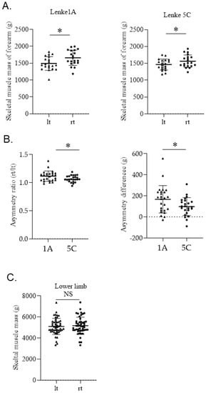
In both 1A and 5C groups, forearm skeletal muscle mass was significantly greater on the right side, which was the dominant side (Figure 1A). Both the asymmetry ratio and the asymmetry difference of upper arm skeletal muscles were significantly larger in group 1A than in group 5C (Figure 1B). There was no significant difference in skeletal muscle mass between the left and right lower limbs in AIS patients (Figure 1C).

Figure 1.
(A) Comparison of left and right skeletal muscles of the upper arm in Lenke1A or 5C groups. (* p < 0.005), (B) Comparison of asymmetry ratio (rt/lt) and asymmetry difference (rt−lt) for Lenke1A and 5C groups. (* p < 0.005), (C) Comparison of left and right skeletal muscles of the lower limb in all AIS cases. (NS; not significant).
3.3. Sports
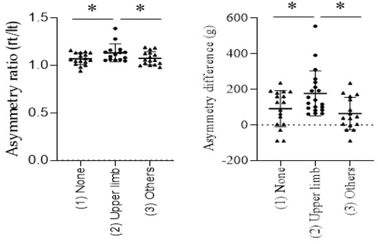
Based on their sporting experience, patients were divided into three groups: (1) no sports experience, (2) sports that primarily use the upper limb, (3) Other sports. There were no significant differences in the frequency of sports experience between the Lenke1A and 5C groups (Table 1). The asymmetry ratio and asymmetry difference of upper arm skeletal muscle mass in group (2) were significantly larger than in groups (1) and (3) (Figure 2).

Table 1.
Preoperative patient characteristics, radiographic measurements.

Figure 2.
Comparison of asymmetry ratio (rt/lt) and asymmetry difference for (1) No sports experience, (2) Sports that primarily use the upper limb, and (3) Other sports in all AIS cases. (* p < 0.005).
3.4. Correlation between Asymmetry in Skeletal Muscles of the Upper Arm and Coronal Parameters in the Lenke1A Group
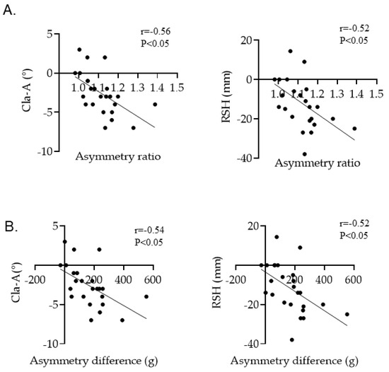
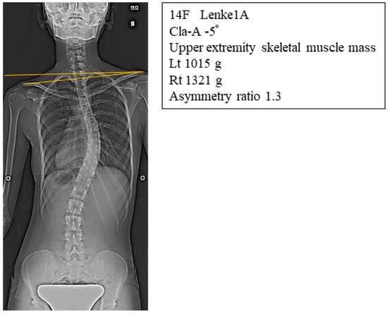
Cla-A and RSH were significantly correlated with both asymmetry ratio and asymmetry difference in skeletal muscles of the upper arm (Figure 3A,B). In contrast, the Cobb angle and AVR were not correlated in group 1A (Table 2). Figure 4 shows a representative radiograph of a Lenke1A AIS patient with the right shoulder up, who had a large asymmetry ratio of upper arm skeletal muscle mass. There were no significant correlations between asymmetry in skeletal muscles of the upper arm and coronal parameters (data not shown).

Figure 3.
(A) Correlation between Cla-A, RSH, and asymmetry ratio in the Lenke1A group, (B) Correlation between Cla-A, RSH, and asymmetry difference in the Lenke1A group.

Table 2.
Correlations between asymmetry in skeletal muscles of the upper arm and coronal parameters in group 1A.

Figure 4.
Representative radiograph of a Lenke1A AIS patient with a right shoulder imbalance who had a large asymmetry ratio of upper arm skeletal muscle mass.
4. Discussion
The present study showed that upper extremity skeletal muscles on the dominant side were significantly larger than those on the nondominant side in both Lenke1A and 5C groups. Interestingly, we found that both the asymmetry ratio and asymmetry difference of upper extremity skeletal muscles were significantly greater in the Lenke1A group than in the Lenke5C group. Additionally, the size of the asymmetry did not correlate with the magnitude of the major curve or rotational deformation but did correlate with the right shoulder up in the Lenke1A group. These results suggest that in AIS with a constructive thoracic curve, right shoulder up is an independent risk factor for upper extremity skeletal muscle asymmetry. There are many reports focusing on muscle asymmetry in scoliosis involving paravertebral, psoas, and masticatory muscles [12,13,14]. However, to our knowledge, this is the first study to show asymmetry in upper extremity muscle mass in patients with AIS.
Currently, treatment for AIS focuses on the patient’s self-appearance, low back pain, and respiratory function. This study is the first to suggest that functional disabilities due to upper extremity skeletal muscle asymmetry in the elderly are a new target for AIS treatment. A recent study revealed that shoulder imbalance in AIS patients had a very large impact on their satisfaction with appearance [16]. Because postoperative shoulder imbalance (PSI) commonly causes dissatisfaction among AIS patients, numerous reports have focused on techniques and fixation ranges to reduce PSI [17,18,19,20]. Further studies are needed to clarify whether the asymmetry of the skeletal muscles of the upper extremity could be improved by correcting the shoulder balance through spinal corrective surgery.
One limitation of this study was that it did not identify the mechanism by which the right shoulder imbalance is involved in upper extremity skeletal muscle asymmetry. We believe that imaging analysis of the upper extremity skeletal muscle mass will be necessary in the future. A relationship has been demonstrated between bilateral strength imbalances and sports in which the dominant side is used more frequently, such as volleyball and basketball [21,22]. The association between sports experience and AIS also has been described [23]. The present study showed there might be a relationship between sports that primarily use the upper extremity and upper extremity skeletal muscle asymmetry. The possibility that the right shoulder up with a thoracic structural curve may result in the more frequent use of the right side compared to the left side should be considered in the future. This study suggests that even if 5C cases have shoulder balance, this can be compensated for in normal life by the nonstructural curve of the thoracic spine. In contrast, 1A cases with a right shoulder up due to a structural thoracic spine have a greater risk of left-right differences in upper limb use resulting in upper extremity skeletal muscle mass asymmetry.
5. Conclusions
The asymmetry ratio and asymmetry difference of upper extremity skeletal muscles were significantly greater in the Lenke1A group than in the Lenke5C group. The size of the asymmetry did not correlate with the magnitude of the major curve or rotational deformation but did correlate with the right shoulder up in the Lenke1A group.
Author Contributions
Methodology, T.O., K.O., H.T., K.K. and H.O.; software, G.G.; validation, M.K. and H.H.; formal analysis, T.O., G.G., K.O. and K.K.; data curation, T.O., N.T., K.O. and M.K.; writing—original draft, T.O.; writing—review & editing, T.O.; visualization, M.K. and H.T.; supervision, H.H.; project administration, N.T. and H.O.; funding acquisition, T.O. All authors have read and agreed to the published version of the manuscript.
Funding
The manuscript submitted does not contain information about medical device (s)/drug (s). There were no relevant financial activities outside the submitted work. We did not receive any specific grant from funding agencies in the public, commercial, or not-for-profit sectors for this research.
Institutional Review Board Statement
The study was approved for ethics by the institutional review board at the corresponding author’s institution (approved number 2556; the data of approval; 14 April 2018).
Informed Consent Statement
Informed consent was obtained from all subjects involved in the study.
Data Availability Statement
The data presented in this study are available on request from the corresponding author.
Conflicts of Interest
The authors declare no conflict of interest.
References
- Ithurburn, M.P.; Thomas, S.; Paterno, M.V.; Schmitt, L.C. Young athletes after ACL reconstruction with asymmetric quadriceps strength at the time of return-to-sport clearance demonstrate drop-landing asymmetries two years later. Knee 2021, 29, 520–529. [Google Scholar] [CrossRef] [PubMed]
- Portegijs, E.; Sipila, S.; Alen, M.; Kaprio, J.; Koskenvuo, M.; Tiainen, K.; Rantanen, T. Leg Extension Power Asymmetry and Mobility Limitation in Healthy Older Women. Arch. Phys. Med. Rehabil. 2005, 86, 1838–1842. [Google Scholar] [CrossRef] [PubMed]
- Iwasaka, C.; Mitsutake, T.; Horikawa, E. The Independent Relationship Between Leg Skeletal Muscle Mass Asymmetry and Gait Speed in Community-Dwelling Older Adults. J. Aging Phys. Act. 2020, 28, 943–951. [Google Scholar] [CrossRef] [PubMed]
- McGrath, R.; Cawthon, P.M.; Cesari, M.; Al Snih, S.; Clark, B.C. Handgrip Strength Asymmetry and Weakness Are Associated with Lower Cognitive Function: A Panel Study. J. Am. Geriatr. Soc. 2020, 68, 2051–2058. [Google Scholar] [CrossRef]
- McGrath, R.; Tomkinson, G.R.; LaRoche, D.P.; Vincent, B.M.; Bond, C.W.; Hackney, K.J. Handgrip Strength Asymmetry and Weakness May Accelerate Time to Mortality in Aging Americans. J. Am. Med. Dir. Assoc. 2020, 21, 2003–2007. [Google Scholar] [CrossRef]
- McGrath, R.; Clark, B.C.; Cesari, M.; Johnson, C.; Jurivich, D.A. Handgrip strength asymmetry is associated with future falls in older Americans. Aging Clin. Exp. Res. 2020, 33, 2461–2469. [Google Scholar] [CrossRef]
- Chen, Z.; Ho, M.; Chau, P.H. Handgrip strength asymmetry is associated with the risk of neurodegenerative disorders among Chinese older adults. J. Cachex- Sarcopenia Muscle 2022, 13, 1013–1023. [Google Scholar] [CrossRef]
- McGrath, R.; Blackwell, T.L.; E Ensrud, K.; Vincent, B.M.; Cawthon, P.M. The Associations of Handgrip Strength and Leg Extension Power Asymmetry on Incident Recurrent Falls and Fractures in Older Men. J. Gerontol. Ser. A 2021, 76, e221–e227. [Google Scholar] [CrossRef]
- McGrath, R.; Vincent, B.; A Jurivich, D.; Hackney, K.J.; Tomkinson, G.R.; Dahl, L.J.; Clark, B.C. Handgrip Strength Asymmetry and Weakness Together Are Associated with Functional Disability in Aging Americans. J. Gerontol. Ser. A 2020, 76, 291–296. [Google Scholar] [CrossRef]
- Hart, N.H.; Nimphius, S.; Weber, J.; Spiteri, T.; Rantalainen, T.; Dobbin, M.; Newton, R.U. Musculoskeletal Asymmetry in Football Athletes: A Product of Limb Function over Time. Med. Sci. Sport. Exerc. 2016, 48, 1379–1387. [Google Scholar] [CrossRef]
- Weinstein, S.L.; Dolan, L.A.; Cheng, J.C.; Danielsson, A.; Morcuende, J.A. Adolescent idiopathic scoliosis. Lancet 2008, 371, 1527–1537. [Google Scholar] [CrossRef] [PubMed]
- Park, Y.; Ko, J.Y.; Jang, J.Y.; Lee, S.; Beom, J.; Ryu, J.S. Asymmetrical activation and asymmetrical weakness as two different mechanisms of adolescent idiopathic scoliosis. Sci. Rep. 2021, 11, 17582. [Google Scholar] [CrossRef] [PubMed]
- Uçar, I.; Batın, S.; Arık, M.; Payas, A.; Kurtoğlu, E.; Karartı, C.; Seber, T.; Çöbden, S.B.; Taşdemir, H.; Unur, E. Is scoliosis related to mastication muscle asymmetry and temporomandibular disorders? A cross-sectional study. Musculoskelet. Sci. Pract. 2022, 58, 102533. [Google Scholar] [CrossRef] [PubMed]
- Shafaq, N.; Suzuki, A.; Matsumura, A.; Terai, H.; Toyoda, H.; Yasuda, H.; Ibrahim, M.; Nakamura, H. Asymmetric Degeneration of Paravertebral Muscles in Patients with Degenerative Lumbar Scoliosis. Spine 2012, 37, 1398–1406. [Google Scholar] [CrossRef]
- Kim, H.; Lee, C.-K.; Yeom, J.S.; Lee, J.H.; Cho, J.H.; Shin, S.I.; Lee, H.-J.; Chang, B.-S. Asymmetry of the cross-sectional area of paravertebral and psoas muscle in patients with degenerative scoliosis. Eur. Spine J. 2013, 22, 1332–1338. [Google Scholar] [CrossRef]
- Lee, S.Y.; Ch’Ng, P.Y.; Wong, T.S.; Ling, X.W.; Chung, W.H.; Chiu, C.K.; Chan, C.Y.W.; Lean, M.L.; Kwan, M.K. Patients’ Perception and Satisfaction on Neck and Shoulder Imbalance in Adolescent Idiopathic Scoliosis. Glob. Spine J. 2021, 1–12. [Google Scholar] [CrossRef]
- Oba, H.; Takahashi, J.; Kobayashi, S.; Ohba, T.; Ikegami, S.; Kuraishi, S.; Uehara, M.; Takizawa, T.; Munakata, R.; Hatakenaka, T.; et al. Upper instrumented vertebra to the right of the lowest instrumented vertebra as a predictor of an increase in the main thoracic curve after selective posterior fusion for the thoracolumbar/lumbar curve in Lenke type 5C adolescent idiopathic scoliosis: Multicenter study on the relationship between fusion area and surgical outcome. J. Neurosurg. Spine 2019, 31, 857–864. [Google Scholar] [CrossRef]
- Smith, P.L.; Donaldson, S.; Hedden, D.; Alman, B.; Howard, A.; Stephens, D.; Wright, J.G. Parents’ and Patients’ Perceptions of Postoperative Appearance in Adolescent Idiopathic Scoliosis. Spine 2006, 31, 2367–2374. [Google Scholar] [CrossRef]
- Tang, X.; Luo, X.; Liu, C.; Fu, J.; Yao, Z.; Du, J.; Wang, Y.; Zhang, Y.; Zheng, G. The Spontaneous Development of Cosmetic Shoulder Balance and Shorter Segment Fusion in Adolescent Idiopathic Scoliosis with Lenke I Curve: A Consecutive Study Followed Up for 2 to 5 Years. Spine 2016, 41, 1028–1035. [Google Scholar] [CrossRef]
- Chung, W.H.; Chiu, C.K.; Ng, S.J.; Goh, S.H.; Chan, C.Y.W.; Kwan, M.K. How Common Is Medial and Lateral Shoulder Discordance in Lenke 1 and 2 Curves?: A Preoperative Analysis of Medial and Lateral Shoulder Balance Among 151 Lenke 1 and 2 Adolescent Idiopathic Scoliosis Patients. Spine 2019, 44, E480–E486. [Google Scholar] [CrossRef]
- Thomas, C.; Comfort, P.; Dos’Santos, T.; Jones, P.A. Determining Bilateral Strength Imbalances in Youth Basketball Athletes. Int. J. Sport. Med. 2017, 38, 683–690. [Google Scholar] [CrossRef] [PubMed]
- Ahmadi, S.; Gutierrez, G.L.; Uchida, M.C. Asymmetry in glenohumeral muscle strength of sitting volleyball players: An isokinetic profile of shoulder rotations strength. J. Sports Med. Phys. Fit. 2020, 60, 395–401. [Google Scholar] [CrossRef] [PubMed]
- Watanabe, K.; Michikawa, T.; Yonezawa, I.; Takaso, M.; Minami, S.; Soshi, S.; Tsuji, T.; Okada, E.; Abe, K.; Takahashi, M.; et al. Physical Activities and Lifestyle Factors Related to Adolescent Idiopathic Scoliosis. J. Bone Jt. Surg. 2017, 99, 284–294. [Google Scholar] [CrossRef] [PubMed]
Publisher’s Note: MDPI stays neutral with regard to jurisdictional claims in published maps and institutional affiliations. |
© 2022 by the authors. Licensee MDPI, Basel, Switzerland. This article is an open access article distributed under the terms and conditions of the Creative Commons Attribution (CC BY) license (https://creativecommons.org/licenses/by/4.0/).