
Procedural Outcomes of a Self-Expanding Transcatheter Heart Valve in Small Annuli
 , , ,
, , ,
Abstract
:1. Introduction
2. Methods
2.1. Multidetector Computed Tomography
2.2. Assessment of PPM and Implantation Depth
2.3. Outcomes of Interest
2.4. Statistical Analysis
3. Results
3.1. Baseline Data
3.2. Procedural Data and Outcomes
3.3. Hemodynamics and PPM
4. Discussion
4.1. Prosthesis–Patient Mismatch and PVL
4.2. Pacemaker
4.3. Procedural and 30-Day Outcome
4.4. Limitations
5. Conclusions
Author Contributions
Funding
Institutional Review Board Statement
Informed Consent Statement
Data Availability Statement
Acknowledgments
Conflicts of Interest
Abbreviations
| Ca-density | calcium density |
| CI | cover index |
| LVOT | left ventricular outflow tract |
| MDCT | multidetector computed tomography |
| PVL | paravalvular leakage |
| STJ | sinotubular junction |
| TAVR | transcatheter aortic valve replacement |
| THV | transcatheter heart valve |
| PPM | Prosthesis–patient mismatch |
| iAVA | indexed aortic valve area |
References
- Popma, J.J.; Deeb, G.M.; Yakubov, S.J.; Mumtaz, M.; Gada, H.; O’Hair, D.; Bajwa, T.; Heiser, J.C.; Merhi, W.; Kleiman, N.S.; et al. Transcatheter Aortic-Valve Replacement with a Self-Expanding Valve in Low-Risk Patients. N. Engl. J. Med. 2019, 380, 1706–1715. [Google Scholar] [CrossRef] [PubMed]
- Mack, M.J.; Leon, M.B.; Thourani, V.H.; Makkar, R.; Kodali, S.K.; Russo, M.; Kapadia, S.R.; Malaisrie, S.C.; Cohen, D.J.; Pibarot, P.; et al. Transcatheter Aortic-Valve Replacement with a Balloon-Expandable Valve in Low-Risk Patients. N. Engl. J. Med. 2019, 380, 1695–1705. [Google Scholar] [CrossRef] [PubMed]
- Writing Committee Members; Otto, C.M.; Nishimura, R.A.; Bonow, R.O.; Carabello, B.A.; Erwin, J.P.; Gentile, F.; Jneid, H.; Krieger, E.V.; Mack, M.; et al. 2020 ACC/AHA guideline for the management of patients with valvular heart disease: A report of the American College of Cardiology/American Heart Association Joint Committee on Clinical Practice Guidelines. J. Am. Coll. Cardiol. 2021, 77, e25–e197. [Google Scholar] [CrossRef] [PubMed]
- Siontis, G.C.M.; Jüni, P.; Pilgrim, T.; Stortecky, S.; Büllesfeld, L.; Meier, B.; Wenaweser, P.; Windecker, S. Predictors of permanent pacemaker implantation in patients undergoing transcatheter aortic valve replacement-a systematic review and meta-analysis. J. Am. Coll. Cardiol. 2014, 64, 129–140. [Google Scholar] [CrossRef] [PubMed]
- Grube, E.; Sinning, J.M. The “Big Five” complications after transcatheter aortic valve replacement: Do we still have to be afraid of them? JACC Cardiovasc. Interv. 2019, 12, 370–372. [Google Scholar] [CrossRef]
- Möllmann, H.; Walther, T.; Siqueira, D.; Diemert, P.; Treede, H.; Grube, E.; Nickenig, G.; Baldus, S.; Rudolph, T.; Kuratani, T.; et al. Transfemoral TAVI using the self-expanding ACURATE neo prosthesis: One-year outcomes of the multicentre “CE-approval cohort”. EuroIntervention 2017, 13, e1040–e1046. [Google Scholar] [CrossRef]
- Möllmann, H.; Holzhey, D.M.; Hilker, M.; Toggweiler, S.; Schäfer, U.; Treede, H.; Joner, M.; Søndergaard, L.; Christen, T.; Allocco, D.J.; et al. The ACURATE neo2 valve system for transcatheter aortic valve implantation: 30-day and 1-year outcomes. Clin. Res. Cardiol. 2021, 110, 1912–1920. [Google Scholar] [CrossRef]
- Achenbach, S.; Delgado, V.; Hausleiter, J.; Schoenhagen, P.; Min, J.K.; Leipsic, J.A. SCCT expert consensus document on computed tomography imaging before transcatheter aortic valve implantation (TAVI)/transcatheter aortic valve replacement (TAVR). J. Cardiovasc. Comput. Tomogr. 2012, 6, 366–380. [Google Scholar] [CrossRef]
- Agatston, A.S.; Janowitz, W.R.; Hildner, F.J.; Zusmer, N.R.; Viamonte, M., Jr.; Detrano, R. Quantification of coronary artery calcium using ultrafast computed tomography. J. Am. Coll. Cardiol. 1990, 15, 827–832. [Google Scholar] [CrossRef]
- Kim, W.-K.; Blumenstein, J.; Liebetrau, C.; Rolf, A.; Gaede, L.; Van Linden, A.; Arsalan, M.; Doss, M.; Tijssen, J.G.P.; Hamm, C.W.; et al. Comparison of outcomes using balloon-expandable versus self-expanding transcatheter prostheses according to the extent of aortic valve calcification. Clin. Res. Cardiol. 2017, 106, 995–1004. [Google Scholar] [CrossRef]
- Kim, W.-K.; Bhumimuang, K.; Renker, M.; Fischer-Rasokat, U.; Möllmann, H.; Walther, T.; Choi, Y.-H.; Nef, H.; Hamm, C.W. Determinants of paravalvular leakage following transcatheter aortic valve replacement in patients with bicuspid and tricuspid aortic stenosis. Eur. Heart J.-Cardiovasc. Imaging 2021, 22, 1387–1396. [Google Scholar] [CrossRef]
- VARC-3 Writing Committee; Généreux, P.; Piazza, N.; Alu, M.C.; Nazif, T.; Hahn, R.T.; Pibarot, P.; Bax, J.J.; Leipsic, J.A.; Blanke, P.; et al. Valve Academic Research Consortium 3: Updated endpoint definitions for aortic valve clinical research. Eur. Heart J. 2021, 42, 1825–1857. [Google Scholar] [CrossRef] [PubMed]
- Kim, W.; Möllmann, H.; Walther, T.; Hamm, C.W. Predictors of permanent pacemaker implantation after ACURATE neo transcatheter heart valve implantation. Pacing Clin. Electrophysiol. 2020, 44, 410–415. [Google Scholar] [CrossRef] [PubMed]
- Regazzoli, D.; Chiarito, M.; Cannata, F.; Pagnesi, M.; Miura, M.; Ziviello, F.; Picci, A.; Reifart, J.; De Marco, F.; Francesco Bedogni, F.; et al. Transcatheter self-expandable valve implantation for aortic stenosis in small aortic annuli: The TAVI-SMALL registry. Cardiovasc. Interv. 2020, 13, 196–206. [Google Scholar]
- Voigtländer, L.; Kim, W.-K.; Mauri, V.; Goßling, A.; Renker, M.; Sugiura, A.; Linder, M.; Schmidt, T.; Schofer, N.; Westermann, D.; et al. Transcatheter aortic valve implantation in patients with a small aortic annulus: Performance of supra-, intra- and infra-annular transcatheter heart valves. Clin. Res. Cardiol. 2021, 110, 1957–1966. [Google Scholar] [CrossRef] [PubMed]
- Pibarot, P.; Weissman, N.J.; Stewart, W.J.; Hahn, R.T.; Lindman, B.R.; McAndrew, T.; Kodali, S.K.; Mack, M.J.; Thourani, V.H.; Miller, D.C.; et al. Incidence and sequelae of prosthesis-patient mismatch in transcatheter versus surgical valve replacement in high-risk patients with severe aortic stenosis: A PARTNER Trial Cohort-A Analysis. J. Am. Coll. Cardiol. 2014, 64, 1323–1334. [Google Scholar] [CrossRef]
- Mauri, V.; Kim, W.K.; Abumayyaleh, M.; Walther, T.; Moellmann, H.; Schaefer, U.; Conradi, L.; Hengstenberg, C.; Hilker, M.; Wahlers, T.; et al. Short-term outcome and hemodynamic performance of next-generation self-expanding versus balloon-expandable transcatheter aortic valves in patients with small aortic annulus: A multicenter propensity-matched comparison. Circ. Cardiovasc. Interv. 2017, 10, e005013. [Google Scholar] [CrossRef]
- Liao, Y.-B.; Li, Y.-J.; Jun-Li, L.; Zhao, Z.-G.; Wei, X.; Tsauo, J.-Y.; Xiong, T.-Y.; Xu, Y.-N.; Feng, Y.; Chen, M. Incidence, Predictors and Outcome of Prosthesis-Patient Mismatch after Transcatheter Aortic Valve Replacement: A Systematic Review and Meta-analysis. Sci. Rep. 2017, 7, 15014. [Google Scholar] [CrossRef]
- Reardon, M.J.; Van Mieghem, N.M.; Popma, J.J.; Kleiman, N.S.; Søndergaard, L.; Mumtaz, M.; Adams, D.H.; Deeb, G.M.; Maini, B.; Gada, H.; et al. Surgical or Transcatheter Aortic-Valve Replacement in Intermediate-Risk Patients. N. Engl. J. Med. 2017, 376, 1321–1331. [Google Scholar] [CrossRef]
- De Torres-Alba, F.; Kaleschke, G.; Diller, G.P.; Vormbrock, J.; Orwat, S.; Radke, R.; Reinke, F.; Fischer, D.; Reinecke, H.; Baumgartner, H. Changes in the pacemaker rate after transition from Edwards SAPIEN XT to SAPIEN 3 transcatheter aortic valve implantation: The critical role of valve implantation height. JACC Cardiovasc. Interv. 2016, 9, 805–813. [Google Scholar] [CrossRef]
- Nadeem, F.; Tsushima, T.; Ladas, T.P.; Thomas, R.B.; Patel, S.M.; Saric, P.; Patel, T.; Lipinski, J.; Li, J.; Costa, M.A.; et al. Impact of Right Ventricular Pacing in Patients Who Underwent Implantation of Permanent Pacemaker After Transcatheter Aortic Valve Implantation. Am. J. Cardiol. 2018, 122, 1712–1717. [Google Scholar] [CrossRef] [PubMed]
- Pagnesi, M.; Kim, W.-K.; Conradi, L.; Barbanti, M.; Stefanini, G.G.; Schofer, J.; Hildick-Smith, D.; Pilgrim, T.; Abizaid, A.; Zweiker, D.; et al. Impact of Predilatation Prior to Transcatheter Aortic Valve Implantation With the Self-Expanding Acurate neo Device (from the Multicenter NEOPRO Registry). Am. J. Cardiol. 2020, 125, 1369–1377. [Google Scholar] [CrossRef] [PubMed]

| Variable | Off-Label Sizing | On-Label Sizing | p Value |
|---|---|---|---|
| n = 125 | n = 529 | ||
| Age, years | 82.0 [79.9; 85.5] | 82.0 [79.0; 85.6] | 0.677 |
| Female gender | 119 (95.2%) | 495 (93.6%) | 0.635 |
| BMI, kg/m2 | 26.6 [22.8; 30.1] | 25.8 [23.4; 29.8] | 0.928 |
| EuroScore I, % | 16.3 [10.7; 25.7] | 16.7 [10.1; 24.3] | 0.489 |
| EuroScore II, % | 3.3 [2.5; 4.9] | 3.4 [2.3; 5.0] | 0.931 |
| eGFR, mL/min/1,73 m2 | 59.0 [42.0; 74.0] | 55.0 [42.0; 75.0] | 0.526 |
| Peripheral artery disease | 13 (10.4%) | 60 (11.3%) | 0.886 |
| Prior stroke | 8 (6.4%) | 65 (12.3%) | 0.085 |
| Atrial fibrillation | 43 (34.4%) | 170 (32.1%) | 0.704 |
| Coronary artery disease | 61 (48.8%) | 303 (57.3%) | 0.106 |
| Electrocardiographic data | |||
| Previous right bundle branch block | 10 (8.0%) | 43 (8.1%) | 1.000 |
| Previous left bundle branch block | 8 (6.4%) | 39 (7.4%) | 0.852 |
| Previous atrioventricular block | 11 (8.8%) | 76 (14.4%) | 0.133 |
| Echocardiographic data | |||
| Ejection fraction, % | 65.0 [60.0; 65.0] | 65.0 [60.0; 65.0] | 0.666 |
| Mean gradient, mmHg | 43.0 [34.0; 49.0] | 43.0 [35.0; 54.0] | 0.173 |
| AVA, cm2 | 0.7 [0.6; 0.8] | 0.7 [0.6; 0.8] | 0.080 |
| MDCT data | |||
| Perimeter derived annulus diameter, mm | 20.4 [20.1; 20.7] | 22.0 [21.6;22.4] | <0.001 |
| LVOT, mm | 19.4 [18.1; 20.2] | 21.2 [20.1;22.2] | <0.001 |
| STJ, mm | 24.9 [23.3; 26.5] | 25.9 [24.7;27.1] | <0.001 |
| Aortic valve calcification, AU | 1372.0 [875.0; 2042.0] | 1670.0 [1088.0; 2486.8] | 0.001 |
| Calcium density, AU/m2 | 427.7 [276.7; 684.1] | 481.9 [317.4;732.1] | 0.050 |
| Calcification in LVOT | 5 (4.0%) | 24 (4.5%) | 0.983 |
| Eccentric calcification | 11 (8.8%) | 75 (14.2%) | 0.146 |
| Variable | Off-Label Sizing | On-Label Sizing | p Value |
|---|---|---|---|
| n = 125 | n = 529 | ||
| Procedural parameter | |||
| THV cover index (perimeter), % | 10.9 [9.9; 12.6] | 4.5 [2.6; 6.3] | <0.001 |
| Procedural duration, min | 47.0 [35.0; 61.0] | 45.0 [36.0; 60.0] | 0.691 |
| Contrast agent, mL | 80.0 [55.0; 106.0] | 80.0 [55.8; 110.0] | 0.627 |
| Pre-dilatation, % | 73 (58.4%) | 385 (72.8%) | 0.002 |
| Post-dilatation, % | 21 (16.8%) | 138 (26.4%) | 0.033 |
| Depth NCC, mm | 6.0 [4.0; 6.5] | 6.0 [4.0; 6.5] | 0.966 |
| Depth LCC, mm | 6.0 [5.0; 7.0] | 6.0 [4.0; 7.0] | 0.665 |
| Echocardiographic outcome | |||
| Ejection fraction, % | 65.0 [63.0;65.0] | 65.0 [60.0;65.0] | 0.873 |
| Mean gradient, mmHg | 10.0 [6.0;12.0] | 9.0 [6.8;13.0] | 0.834 |
| AVA, cm2 | 1.5 [1.3; 1.7] | 1.6 [1.4;1.8] | <0.001 |
| iAVA, cm2/m2 | 0.87 [0.7; 1.0] | 0.92 [0.8;1.1] | 0.029 |
| Clinical outcome | |||
| Technical success | 111 (88.8%) | 479 (90.5%) | 0.671 |
| Device success at 30 days | 100 (80.0%) | 444 (83.9%) | 0.355 |
| Early safety at 30 days | 51 (40.8%) | 220 (41.6%) | 0.952 |
| In-hospital death | 7 (5.6%) | 9 (1.7%) | 0.020 |
| All-cause death at 30 days | 8 (6.5%) | 12 (2.3%) | 0.036 |
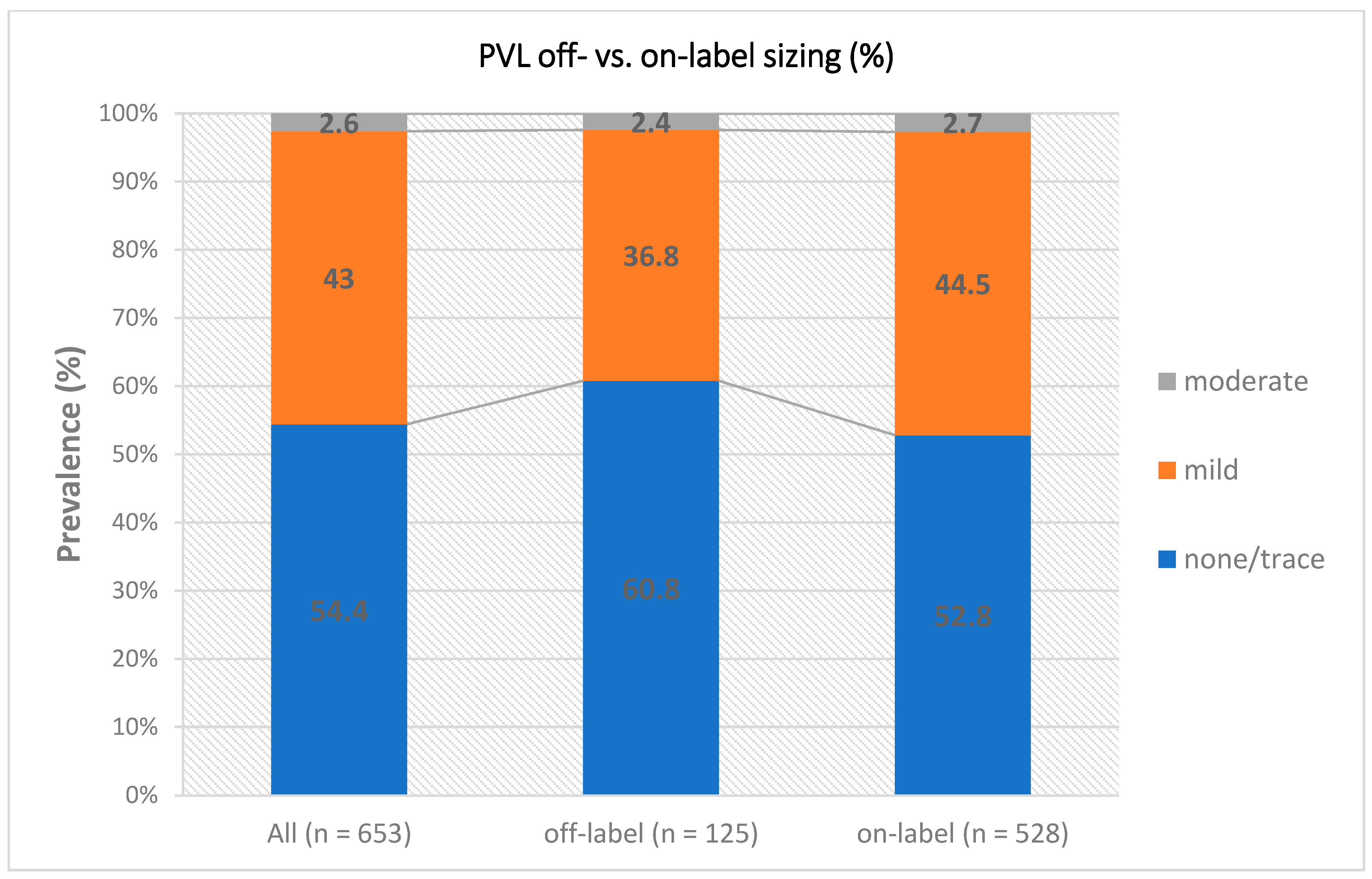
| Relevant PVL (>mild/trace) | 4 (3.2%) | 15 (2.8%) | 0.770 |
| Moderate to severe PPM | 29 (35.4%) | 111 (26.5%) | 0.113 |
| Severe PPM | 4 (4.9%) | 16 (3.8%) | 0.552 |
| Conversion to sternotomy | 2 (1.6%) | 3 (0.6%) | 0.244 |
| Multiple valves (ViV) | 2 (1.6%) | 3 (0.6%) | 0.244 |
| Device embolization | 4 (3.2%) | 7 (1.3%) | 0.235 |
| Major vascular complication | 12 (9.6%) | 46 (8.7%) | 0.885 |
| Bleeding (type 2–4) | 28 (22.6%) | 116 (21.9%) | 0.970 |
| Overt CNS injury | 5 (4.0%) | 15 (2.8%) | 0.561 |
| Cardiac structural complication | 2 (1.6%) | 8 (1.5%) | 1.000 |
| AKI (type 2–4) | 8 (6.4%) | 24 (4.5%) | 0.524 |
| New permanent pacemaker 1 | 10 (8.4%) | 38 (8.1%) | 1.000 |
| Off-Label Sizing | On-Label Sizing | p Value | |
|---|---|---|---|
| n = 8 | n = 12 | ||
| All-cause death | 0.900 | ||
| Cardiac/procedural related | 5 (62.5%) | 5 (41.7%) | |
| Inflammatory/sepsis | 2 (25.0%) | 3 (25.0%) | |
| Multiorgan failure (MOF) | 1 (12.5%) | 2 (16.7%) | |
| Stroke | 0 (0.0%) | 2 (16.7%) |
| no PPM | PPM | p Value | |
|---|---|---|---|
| n = 361 | n = 140 | ||
| Procedural parameter | |||
| THV cover index (perimeter), % | 5.0 [3.1; 7.5] | 4.7 [3.1; 8.2] | 0.637 |
| THV cover index (STJ), % | −0.2 [−9.1; −0.1] | −0.1 [−0.2; −0.1] | <0.001 |
| Procedural duration, min | 45.0 [35.0; 58.0] | 40.0 [33.0; 48.0] | 0.001 |
| Contrast agent, mL | 77.0 [48.8; 104.2] | 74.0 [50.0; 100.0] | 0.647 |
| Pre-dilatation, % | 239 (66.2%) | 94 (67.1%) | 0.925 |
| Post-dilatation, % | 96 (26.9%) | 32 (22.9%) | 0.417 |
| Depth NCC, mm | 5.7 [4.0; 6.5] | 6.0 [5.0; 6.5] | 0.232 |
| Depth LCC, mm | 6.0 [4.0; 7.0] | 6.0 [5.0; 7.0] | 0.527 |
| Echocardiographic outcome | |||
| Ejection fraction, % | 65.0 [61.0; 65.0] | 65.0 [65.0; 65.0] | 0.092 |
| Mean gradient, mmHg | 9.0 [6.0; 12.0] | 13.0 [9.0; 15.0] | <0.001 |
| AVA, cm2 | 1.7 [1.5; 1.8] | 1.3 [1.2; 1.4] | <0.001 |
| Clinical outcome (VARC 3) | |||
| Technical success | 338 (93.6%) | 131 (93.6%) | 1.000 |
| Device success at 30 days | 324 (89.8%) | 114 (81.4%) | 0.018 |
| Early safety at 30 days | 150 (41.6%) | 37 (26.4%) | 0.002 |
| All-cause death at 30 days | 2 (0.6%) | 1 (0.7%) | 1.000 |
| In-hospital death | 1 (0.3%) | 1 (0.7%) | 0.481 |
| Relevant PVL | 11 (3.0%) | 5 (3.6%) | 0.780 |
| Conversion to sternotomy | 2 (0.6%) | 0 (0.0%) | 1.000 |
| Multiple valves (ViV) | 1 (0.3%) | 3 (2.1%) | 0.068 |
| Embolization | 4 (1.1%) | 3 (2.1%) | 0.405 |
| Major vascular complication | 27 (7.5%) | 10 (7.1%) | 1.000 |
| Bleed (type 2–4) | 70 (19.4%) | 23 (16.5%) | 0.546 |
| Overt CNS injury | 7 (1.9%) | 3 (2.1%) | 1.000 |
| AKI (type 2–4) | 16 (4.4%) | 3 (2.1%) | 0.346 |
| New permanent pacemaker 1 | 27 (7.5%) | 7 (5.0%) | 0.428 |
| Univariate | p Value | Multivariate | p Value | |
|---|---|---|---|---|
| Predictors | ||||
| Age (>84 years) | 1.23 (0.82, 1.83) | 0.316 | ||
| CAD (%) | 1.08 (0.73, 1.60) | 0.690 | ||
| Annulus area (>3.38 mm2) | 0.22 (0.12, 0.40) | <0.001 | excluded 1 | |
| Cover index, STJ ( >−0.19%) | 2.95 (1.88, 4.63) | <0.001 | 3.26 (2.03–5.23) | <0.001 |
| Cover index, Perimeter (>4.74%) | 0.81 (0.55, 1.20) | 0.302 | ||
| BMI (>22.77 kg/m2) | 2.90 (1.59, 5.30) | <0.001 | excluded 1 | |
| EuroScore II (>2.49) | 1.27 (0.82, 1.96) | 0.278 | ||
| Depth LCC (>5.7 mm) | 1.64 (1.09, 2.47) | 0.017 | 2.25 (1.44–3.53) | <0.001 |
| LVOT calcification | 1.18 (0.40, 3.45) | 0.767 | ||
| Mean transaortic pressure (mmHg) | 2.12 (1.33, 3.37) | 0.002 | 1.49 (0.85–2.61) | 0.157 |
| Severe calcification (>1690 HU) | 2.01 (1.35, 3.00) | <0.001 | 2.07 (1.29–3.33) | 0.002 |
| Post dilatation | 0.81 (0.51, 1.27) | 0.351 | ||
| Ejection fraction (<55%) | 1.33 (0.73, 2.43) | 0.358 | ||
Publisher’s Note: MDPI stays neutral with regard to jurisdictional claims in published maps and institutional affiliations. |
© 2022 by the authors. Licensee MDPI, Basel, Switzerland. This article is an open access article distributed under the terms and conditions of the Creative Commons Attribution (CC BY) license (https://creativecommons.org/licenses/by/4.0/).
Share and Cite
Eckel, C.; Sötemann, D.; Kim, W.-K.; Grothusen, C.; Tiyerili, V.; Dohmen, G.; Renker, M.; Charitos, E.; Hamm, C.W.; Choi, Y.-H.; et al. Procedural Outcomes of a Self-Expanding Transcatheter Heart Valve in Small Annuli. J. Clin. Med. 2022, 11, 5313. https://doi.org/10.3390/jcm11185313
Eckel C, Sötemann D, Kim W-K, Grothusen C, Tiyerili V, Dohmen G, Renker M, Charitos E, Hamm CW, Choi Y-H, et al. Procedural Outcomes of a Self-Expanding Transcatheter Heart Valve in Small Annuli. Journal of Clinical Medicine. 2022; 11(18):5313. https://doi.org/10.3390/jcm11185313
Chicago/Turabian StyleEckel, Clemens, Dagmar Sötemann, Won-Keun Kim, Christina Grothusen, Vedat Tiyerili, Guido Dohmen, Matthias Renker, Efstratios Charitos, Christian W. Hamm, Yeong-Hoon Choi, and et al. 2022. "Procedural Outcomes of a Self-Expanding Transcatheter Heart Valve in Small Annuli" Journal of Clinical Medicine 11, no. 18: 5313. https://doi.org/10.3390/jcm11185313
APA StyleEckel, C., Sötemann, D., Kim, W.-K., Grothusen, C., Tiyerili, V., Dohmen, G., Renker, M., Charitos, E., Hamm, C. W., Choi, Y.-H., Möllmann, H., & Blumenstein, J. (2022). Procedural Outcomes of a Self-Expanding Transcatheter Heart Valve in Small Annuli. Journal of Clinical Medicine, 11(18), 5313. https://doi.org/10.3390/jcm11185313