Article

Leisure-Time Physical Activity Has a More Favourable Impact on Carotid Artery Stiffness Than Vigorous Physical Activity in Hypertensive Human Beings

1
Cardiac Centre, King Faisal Specialist Hospital and Research Center, Riyadh 11211, Saudi Arabia
2
School of Medicine, Alfaisal University, Riyadh 11533, Saudi Arabia
3
Department of Cardiology, San Antonio Hospital, 33038 San Daniele del Friuli, Italy
4
Department of Medicine, University of Padova, 35128 Padua, Italy
*
Author to whom correspondence should be addressed.
J. Clin. Med. 2022, 11(18), 5303; https://doi.org/10.3390/jcm11185303
Submission received: 18 August 2022 / Revised: 5 September 2022 / Accepted: 7 September 2022 / Published: 8 September 2022

Abstract

:
Aim. To assess the effect of leisure time versus vigorous long-term dynamic physical activity (PA) on carotid stiffness in normotensive versus hypertensive subjects. Methods. The study was conducted on 120 leisure-time exercisers and 120 competitive athletes. One hundred and twenty sedentary subjects served as controls. In addition, participants were classified according to whether their systolic blood pressure was ≥130 mmHg (hypertensives, n = 120) or normal (normotensives, n = 240) according to the ACC/AHA 2017 definition. Carotid artery stiffness was assessed with an echo-tracking ultrasound system, using the pressure-strain elastic modulus (EP) and one-point pulse wave velocity (PWVβ) as parameters of stiffness. Results. The effect of the two levels of PA differed in the normotensives and the hypertensives. Among the normotensives, there was an ongoing, graded reduction in EP and PWVβ from the sedentary subjects to the athletes. By contrast, among the hypertensives, the lowest levels of EP and PWVβ were found among the leisure-time PA participants. EP and PWVβ did not differ between the hypertensive sedentary subjects and the athletes. A significant interaction was found between PA and BP status on EP (p = 0.03) and a borderline interaction on PWVβ (p = 0.06). In multiple regression analyses, PA was a negative predictor of EP (p = 0.001) and PWVβ (p = 0.0001). The strength of the association was weakened after the inclusion of heart rate in the models (p = 0.04 and 0.007, respectively). Conclusions. These data indicate that in people with hypertension, leisure-time PA has beneficial effects on carotid artery stiffness, whereas high-intensity chronic PA provides no benefit to vascular functions.

1. Introduction

A large number of studies have shown that regular aerobic physical activity (PA) is beneficial for both the prevention and treatment of hypertension [1], diabetes [2], and ischemic heart disease [3]. Thus, most international guidelines recommend that people engage in at least 150 to 300 min/wk of moderate PA, 75 to 150 min/wk of vigorous PA or a balanced combination of the two [1,2,3]. The protective role of regular PA in cardiovascular diseases has been attributed to several different factors, including a decrease in blood pressure (BP), heart rate, and body mass index (BMI), an improvement in the lipid profile and an increase in insulin sensitivity [4,5,6]. These mechanisms also seem to be responsible for the beneficial effects of PA on arterial distensibility [7,8]. Indeed, several cross-sectional and longitudinal studies have demonstrated that regular PA is associated with a decrease in large artery stiffness. In a meta-analysis including 42 clinical trials (n = 1627 participants), Ashor et al. [9] showed that aerobic exercise caused a significant reduction in pulse wave velocity and augmentation index, an effect that was larger in participants with stiffer arteries. However, there are concerns about the potentially harmful effects of an excessive amount of high-intensity PA on the cardiovascular system [10]. In particular, whether engaging in vigorous competitive aerobic sports provides a greater benefit than leisure-time PA on cardiovascular health or whether it may be even more harmful is not known. It might well be that a high level of chronic PA causes a detrimental effect on vascular structure and function in the long run in people with hypertension. Thus, the aim of this study was to assess the effect of leisure-time, long-term aerobic PA versus vigorous PA on carotid artery stiffness in relation to blood pressure status. A secondary objective was to evaluate whether, and to what extent, the effect of PA on carotid stiffness was mediated by the PA-induced heart rate reduction.

2. Methods

2.1. Study Population

The present study was a cross-sectional analysis of data collected from 2009 to 2017 [11]. Subjects were selected from among those referred to the outpatient clinic of the Cardiology Department of the San Antonio Hospital, San Daniele del Friuli, Udine (Italy), for work eligibility or competitive athletics participation and from healthy subjects who wanted a cardiologic work-up. Apparently healthy subjects without important cardiovascular risk factors or overt cardiovascular disease, taking no medications, were also included. Among these, 120 consecutive athletes engaging in aerobic competitive sports and 120 subjects of similar age who performed leisure-time aerobic PA for at least 2 years were considered. One hundred and twenty sedentary subjects were used as controls. To define the PA category, we used previously published criteria [4] using a standardized questionnaire. Briefly, activities were classified on the basis of relative intensity. Individuals were categorized as sedentary (class 0) if they did not regularly perform any sports activity; leisure-time exercisers (class 1) if they performed leisure physical activities such as running, jogging, cycling, swimming, soccer, tennis and so on at least once a week without participating in competitions; and athletes (class 2) if they performed aerobic sports and participated in competitions requiring an eligibility check-up. The exclusion criteria covered all cardiovascular diseases, diabetes, chronic kidney disease, other serious diseases, hypertension on antihypertensive treatment, and any other condition requiring chronic medication. All participants underwent a standard initial evaluation, including a thorough medical history inquiry and careful physical examination, using the same procedures for all [11,12]. Data on medical history, dietary habits, and cigarette smoking (yes or no) were collected from all participants. All methods were performed in accordance with the relevant guidelines and regulations according to the Declaration of Helsinki. Ethical approval was obtained from the local Ethics Committee, and informed consent was provided by each participant.

2.2. Procedures

Office brachial BP and heart rates were measured 6 times over two visits using a validated oscillometric semiautomatic sphygmomanometer on the dominant arm, in the supine position, and a quiet room [4]. On the basis of the mean of the 6 BP readings, participants were classified according to whether their systolic BP was ≥130 mmHg (hypertensives, n = 120) or normal (n = 240) according to the definition of hypertension by 2017 ACC/AHA guidelines [13]. The procedures used to assess local carotid stiffness were previously published [11,12]. Briefly, measurements were made at the level of the left common carotid artery, at one point between 1 and 2 cm, before its bifurcation using a high-definition echo-tracking ultrasound system (Alpha 10, Aloka, Tokyo, Japan) with a multifrequency 5–13 MHz linear probe, which uses raw radiofrequency signals that are based on video signals with an accuracy of 0.01 mm. The optimal angle between the ultrasound beam and the vessel wall for diameter change measurements by echo-tracking is 90°, but as the blood-flow velocity is perpendicular to the beam, it cannot be detected. Thus, a different independent steerable ultrasound beam was used. Both beams were steered to intersect at the center of the range gate. The ultrasound beam steering angle can be changed from −30° to +30° with 5° angular increments. The echo-tracking gates were manually set at the boundaries between the intima and media of the anterior and posterior walls. The rate gate for velocity measurements was automatically positioned at the center of the diameter using echo-tracking gates. Flow velocity was corrected for the angle between the ultrasound beam and the blood flow velocity vector. Brachial cuff pressure was measured just before starting ultrasound imaging and was entered into the system for the calculation of carotid stiffness indices [14]. At least five consecutive beats were averaged to obtain a representative waveform. The two parameters of stiffness taken into account were the pressure-strain elastic modulus (EP) and one-point pulse wave velocity (PWVβ). The formulas used for the calculation of the carotid arterial stiffness parameters were:
EP = (Ps − Pd)/[(Ds − Dd)/Dd] and PVWβ = ln(Ps/Pd)/[(Ds − Dd)/Dd]
where Ds and Dd refer to carotid arterial systolic and diastolic diameter; Ps and Pd refer to systolic and diastolic brachial BP (used as a surrogate of carotid systolic and diastolic BP). Inter and intra variability and repeatability of carotid stiffness parameters were previously published [15].

2.3. Statistics

All continuous data were normally distributed according to the Shapiro–Wilk normality test and were presented as means (±SD). Comparisons between the three groups were carried out using a two-way analysis of covariance, using the BP status and PA category as fixed factors and age, sex, BMI, diastolic BP and smoking as covariates. Variables with a p < 0.15 in univariate analysis were entered into a multivariate linear regression, which was used to investigate the association of carotid stiffness with PA (yes or no) and heart rate, thereby controlling for confounders. To compare regression model fit, we used the Akaike Information Criteria (AIC, −2Log-likelihood +2k, where k is the number of parameters estimated) and Schwarz’s Bayesian Information Criterion (BIC, −2∗Log-likelihood + k∗log (n)) [16,17]. For both AIC and BIC criteria, the model providing the smallest values is considered to have the best fit [16,17]. In addition, a measure that can be used to compare survival models is the delta AIC (Δi). This is a measure of each model’s AIC relative to the model with the smallest AIC. As a rule for interpretation, a Δi < 2 suggests substantial evidence for the model, as values between 3 and 7 indicate that the model has considerably less support, whereas a Δi > 10 indicates that the model is very unlikely [18]. Categorical data were compared with the chi2 test or Fisher’s exact test and were presented as a percentage. Analyses were performed using a significance level of α = 0.05 (2-sided).

3. Results

The main characteristics of the study participants are shown in Table 1. Compared with sedentary subjects, athletes were younger (p = 0.03). No age difference was found between the two active groups (p = 0.36). No differences in sex distribution, systolic BP and diastolic BP were found between the three groups. The prevalence of smokers was lower among the athletes than the leisure-time PA group. BMI and heart rate progressively decreased from the sedentary subjects to the athletes (Table 1).

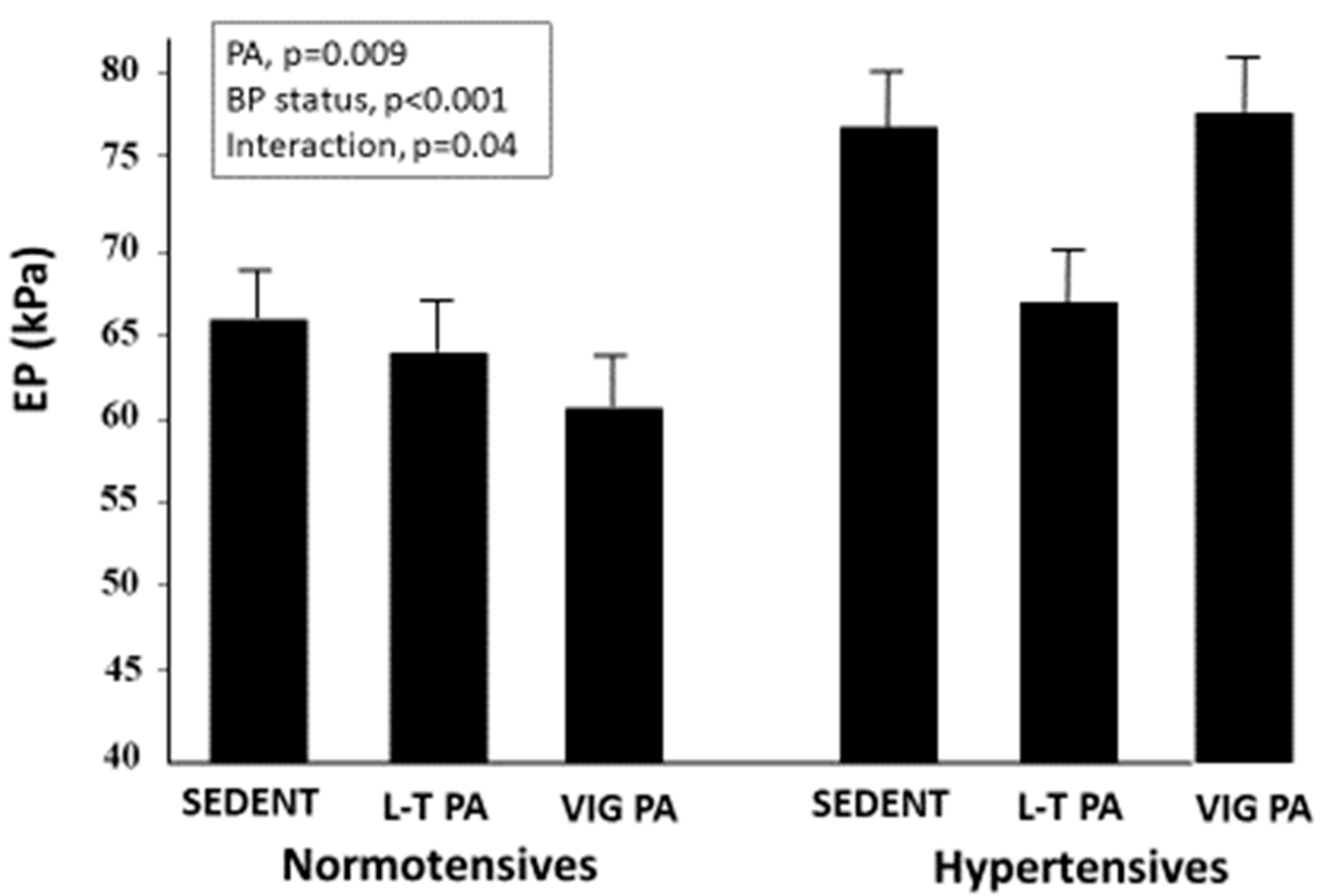
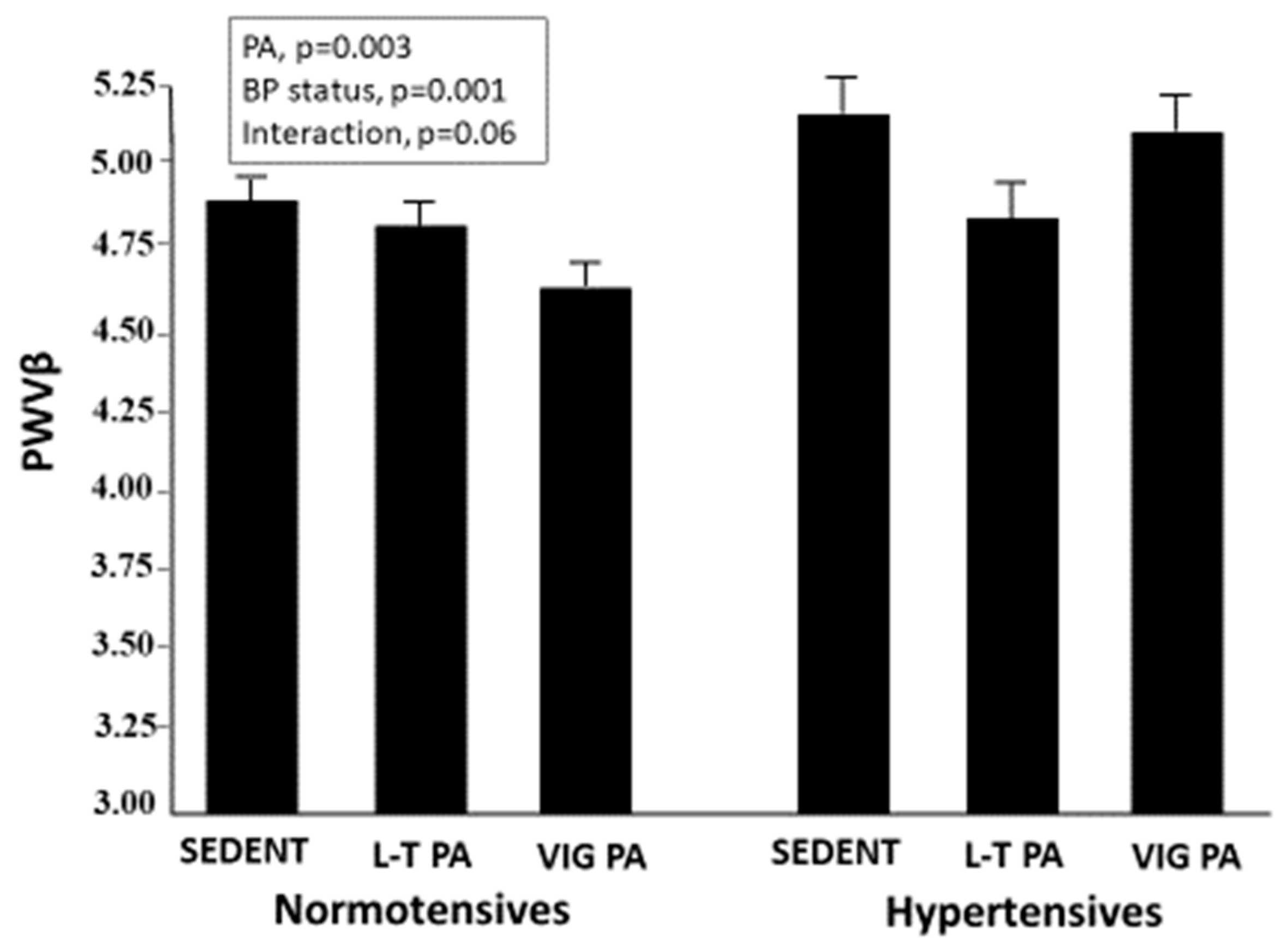
3.1. Carotid Stiffness Parameters

EP was lower in both active groups than in the sedentary subjects (p < 0.03) and in the normotensives than the hypertensives (p < 0.001). However, in a two-way ANCOVA adjusted for age, sex, BMI, diastolic blood pressure and smoking, a different pattern was observed among the normotensives and the hypertensives (Figure 1). In the former, a graded decrease in EP was observed with increasing PA levels, whereas, in the latter, the lowest EP values were observed in the leisure-time activity group. A significant effect on EP was found for both blood pressure status (p < 0.001) and PA (p = 0.009) with a significant interactive effect of the two factors (p = 0.04).
Similar results were found for PWVβ. PWVβ was lower in both athletes and leisure-time PA participants than in sedentary subjects (p < 0.02) and was lower in the normotensives than the hypertensives (p < 0.001). Again, in a two-way ANCOVA, a linear relationship was found in the normotensive group, and a U-shaped relationship was found in the hypertensive participants (Figure 2). A significant effect was found for both blood pressure status (p = 0.001) and PA (p = 0.003) on PWVβ, with a borderline interactive effect of the two factors (p = 0.06).

3.2. Multiple Regression Analysis

In a linear multivariable regression analysis, PA, when considered as a two-class variable (sedentary vs. active), was an independent predictor of EP (p = 0.001). After the inclusion of heart rate in the regression, PA remained a significant predictor of EP, but the association was weakened with a p-value = 0.04 (Table 2). Moreover, the association of PA with PWVβ (p < 0.001) was attenuated after adjusting for heart rate (p = 0.007, Table 3). In both regressions, heart rate was a strong predictor of carotid stiffness parameters (p < 0.001 for both EP and PWVβ) and improved the fit of the models (Table 4). The models, including heart rate, had the smallest AIC with a Delta AIC of 9.4 and 9.2, respectively, compared to the base model with covariates. Compared to the base model, both models, including heart rate, also had a smaller BIC, lending further support to the latter as being the most informative model.

4. Discussion

Our data confirm that aerobic PA has beneficial effects on carotid stiffness parameters and extend previous observations by showing that the negative association between the level of PA and carotid stiffness is linear in normotensive individuals and U-shaped in hypertensive patients. Indeed, within the hypertensive subgroup, the lowest stiffness parameters were observed in the participants performing leisure-time PA.
An association between PA and large artery stiffness has been previously shown in cross-sectional [19,20,21] as well as intervention studies [22,23], but has never been evaluated in relation to the intensity of exercise training before. In keeping with our results, Sugawara et al. showed that the carotid stiffness parameter β significantly decreased after moderate or vigorous intensity cycling exercise training in a small number of apparently healthy sedentary or recreationally active women [21]. However, due to the small number of participants, no comparison was made between the two exercise modalities, and no data were available for the hypertensive subjects. A beneficial effect of a 24-week lifestyle intervention, including aerobic exercise and Mediterranean-style diet, on carotid stiffness parameter β was found by Aizawa et al. in 63 subjects with metabolic syndrome [24]. However, in that study, no information was available as to the effect of different exercise intensities on carotid stiffness. In addition, all intervention studies were conducted over a short period of time and thus may not be representative of the chronic effects of PA on carotid artery properties. In the present study, the participation of our active subjects in the respective PA category lasted for at least 2 years, making it likely that the association with carotid stiffness parameters was the result of chronic exposure to exercise.
The beneficial effects of regular PA on the cardiovascular system have been documented by strong experimental and epidemiological evidence [25,26]. A lifestyle of regular exercise and training has been shown to reduce the risk of cardiovascular events and mortality [27,28]. Several studies have found a graded association between PA and outcomes, showing no additional benefits after reaching a certain threshold [10,29], and some studies even showed a U-shaped association between PA and mortality [30]. In a study of 55,000 participants from the Aerobics Center Longitudinal Study, substantial benefits were obtained with PA doses much lower than those recommended by current major exercise guidelines [31]. In addition, it is still largely unknown whether PA dosage should differ for certain groups of individuals [32]. It is thus important to identify the level of PA that can provide the optimal effect on vascular structure and function in different settings of health and disease.
To this end, we chose two clear-cut groups of individuals trained in aerobic sports. Considering how anamnestic information on PA is often unreliable, to reduce the common biases observed in previous studies of PA [33], competitive athletes were enrolled because they train at a higher intensity and volume than individuals involved in recreational non-competitive sports. The results obtained in the normotensive athletes are reassuring because they indicate that participation in competitive athletics is beneficial in healthy individuals with a normal BP.
Hypertension is often associated with vascular dysfunction. An increased BP leads to vasoconstriction and alterations in the morphology of the small arteries and arterioles due to changes in vascular smooth muscle and endothelial cells, extracellular matrix, and perivascular tissues [34,35]. This leads to the impairment of vascular distensibility [34,35] with reduced flow reserve at the level of the coronary [36] and cerebral microcirculation [37]. Capillary rarefaction and remodeling are other morphological changes observed in hypertensive animals and human beings, which contribute to increased vascular resistance [38,39]. In this milieu, it is possible that a high level of chronic PA provides no benefit or even causes a detrimental effect on vascular structure and function. An exercise-induced exacerbation of oxidative stress has been shown to occur in hypertensive patients compared to healthy controls, and reactive oxygen species have an important role in the development and time-course of hypertension [40]. Previous studies have shown that aortic stiffness appears to be resistant to change in response to short-term aerobic exercise interventions among subjects with pre- or stage 1 hypertension, such as those investigated in the present study [41]. In a study by Kraft et al., aortic stiffness was significantly lower in the fit, normotensive participants versus the unfit ones [42]. By contrast, in the hypertensive cohort, stiffness was not significantly different between the fit and unfit subgroups. However, in this as well as other previous studies, no information was provided as to the effect of different levels of exercise intensity or physical fitness on large artery stiffness. Our results show that high-intensity training had virtually no effect on carotid stiffness parameters, which in the hypertensive athletes were similar to those in the sedentary subjects. In contrast, a beneficial effect was observed in the hypertensive participants performing recreational PA.

Role of Heart Rate

In the present study, heart rate was a strong determinant of carotid artery stiffness, in agreement with the results of previous investigations. Experimental studies have shown that a progressive increase in heart rate with pacing in human beings was associated with a progressive increase in pulse wave velocity (PWV), indicating a cross-sectional association of heart rate with arterial stiffness [43,44]. According to Tan et al., there is a 0.17 m/s increase in PWV for each 10 beat/min increment in heart rate by pacing [44]. A direct association between heart rate and PWV was also found by Tomiyama et al. [45] and by Benetos et al. [46] in longitudinal studies both in normotensive and hypertensive subjects. It is thus likely that the decrease in heart rate caused by regular aerobic training is a main determinant of improved arterial distensibility in physically active subjects. Indeed, in the present study, the inclusion of heart rate in the regression models improved the model fit substantially and attenuated, but did not abolish, the relationship between PA and stiffness parameters. This suggests that low heart rate and PA independently exert beneficial effects on arterial elasticity and that PA-induced bradycardia only partially accounts for vascular function improvement. A regular PA may improve arterial elasticity through mechanisms other than heart rate reduction by protecting the arterial tree from the adverse effects of traditional risk factors for cardiovascular disease. The regression of microvascular remodeling, normalization of capillary density and the improvement of endothelial functions have been reported in hypertensive animals and humans after exercise training [47,48,49,50,51].

5. Limitations

A limitation of our study is that we used only two PA categories and could not provide a more graded relationship between the PA dose and its effects on carotid stiffness. Another major limitation is the cross-sectional nature of our study design that precludes us from making inferences on temporality. Third, we used brachial BP for the calibration of carotid diameter changes, which usually overestimates the central BP in an age-dependent manner. However, mean age was similar in the three PA groups.

6. Conclusions

There is still uncertainty as to the optimal exercise regimen required to achieve the greatest benefits in terms of cardiovascular health. The data of the present study indicate that in hypertension, there is a U-shaped relationship between the PA level and carotid stiffness, suggesting that leisure-time aerobic PA is more beneficial than high-intensity PA on arterial function. A high level of chronic PA provides no benefit to vascular structure and function in people with mildly elevated BP levels and might even cause a detrimental effect in patients with more severe hypertension [40,52]. The effect of exercise training on carotid stiffness seems to be partly, but not entirely, mediated by the reduction in heart rate that accompanies physical training. Future studies are required to validate our data in a larger series of subjects with a wider BP range and to explore the mechanisms underpinning these findings.

Author Contributions

Conceptualization, P.P.; Data curation, O.V.; Funding acquisition, L.M. All authors have contributed to the work. All authors have read and agreed to the published version of the manuscript.

Funding

This research received no external funding.

Institutional Review Board Statement

The study protocol followed the principles of the Declaration of Helsinki and had obtained both approval from its local research ethics committee and informed consent from each subject.

Informed Consent Statement

Written informed consent has been obtained from the patients to publish this paper.

Data Availability Statement

The data that support the findings of this study are available on request from the corresponding author.

Conflicts of Interest

Authors are not directly or indirectly related to the work submitted for publication. The authors have no relevant financial or non-financial interests to disclose.

References

  1. Williams, B.; Mancia, G.; Spiering, W.; Agabiti Rosei, E.; Azizi, M.; Burnier, M.; Burnier, M.; Clement, D.; Coca, A.; De Simone, G.; et al. 2018 ESC/ESH Guidelines for the management of arterial hypertension. Eur. Heart J. 2018, 39, 3021–3104. [Google Scholar] [CrossRef] [PubMed]
  2. American Diabetes Association. Standards of Medical Care in Diabetes-2016. Diabetes Care 2016, 39 (Suppl. S1), S1–S111. [Google Scholar]
  3. Fihn, S.D.; Blankenship, J.C.; Alexander, K.P.; Bittl, J.A.; Byrne, J.G.; Fletcher, B.J.; Fonarow, G.C.; Lange, R.A.; Levine, G.N.; Maddox, T.M.; et al. 2014 ACC/AHA/AATS/PCNA/SCAI/STS focused update of the guideline for the diagnosis and management of patients with stable ischemic heart disease: A report of the American College of Cardiology/American Heart Association Task Force on Practice Guidelines, and the American Association for Thoracic Surgery, Preventive Cardiovascular Nurses Association, Society for Cardiovascular Angiography and Interventions, and Society of Thoracic Surgeons. J. Am. Coll. Cardiol. 2014, 64, 1929–1949. [Google Scholar] [CrossRef] [PubMed]
  4. Palatini, P.; Graniero, G.; Mormino, P.; Nicolosi, L.; Mos, L.; Visentin, P.; Pessina, A.C. Relation between physical training and ambulatory blood pressure in stage I hypertensive subjects. Results of the HARVEST trial. Circulation 1994, 90, 2870–2876. [Google Scholar] [CrossRef] [PubMed]
  5. Jurik, R.; Stastny, P.J. Role of Nutrition and Exercise Programs in Reducing Blood Pressure: A Systematic Review. Clin. Med. 2019, 8, 1393. [Google Scholar] [CrossRef]
  6. Liang, M.; Pan, Y.; Zhong, T.; Zeng, Y.; Cheng, A.S.K. Effects of aerobic, resistance, and combined exercise on metabolic syndrome parameters and cardiovascular risk factors: A systematic review and network meta-analysis. Rev. Cardiovasc. Med. 2021, 22, 1523–1533. [Google Scholar] [CrossRef]
  7. Palatini, P.; Puato, M.; Rattazzi, M.; Pauletto, P. Effect of regular physical activity on Carotid Intima-Media Thickness. Results from a 6-year Prospective Study in the early stage of hypertension. Blood Press 2011, 20, 37–44. [Google Scholar] [CrossRef]
  8. Saladini, F.; Benetti, E.; Mos, L.; Mazzer, A.; Casiglia, E.; Palatini, P. Regular physical activity is associated with improved small artery distensibility in young to middle-age stage 1 hypertensives. Vasc. Med. 2014, 19, 458–464. [Google Scholar] [CrossRef]
  9. Ashor, A.W.; Lara, J.; Siervo, M.; Celis-Morales, C.; Mathers, J.C. Effects of exercise modalities on arterial stiffness and wave reflection: A systematic review and meta-analysis of randomized controlled trials. PLoS ONE 2014, 9, e110034. [Google Scholar]
  10. Lee, D.H.; Rezende, L.F.M.; Joh, H.; Keum, N.; Ferrari, G.; Rey-Lopez, J.P.; Rimm, E.B.; Tabung, F.K.; Giovannucci, E.L. Long-Term Leisure-Time Physical Activity Intensity and All-Cause and Cause-Specific Mortality: A Prospective Cohort of US Adults. Circulation 2022, 146, 523–534. [Google Scholar] [CrossRef]
  11. Vriz, O.; Magne, J.; Jarosh, J.; Bossone, E.; Aboyans, V.; Palatini, P. Local carotid arterial stiffness is an independent determinant of left ventricular remodeling in never-treated hypertensive patients. Blood Press 2019, 28, 23–33. [Google Scholar] [CrossRef] [PubMed]
  12. Vriz, O.; Favretto, S.; Jaroch, J.; Wojciech, R.; Bossone, E.; Driussi, C.; Antonini-Canterin, F.; Palatini, P.; Loboz-Grudzien, K. Left Ventricular Function Assessed by One-Point Carotid Wave Intensity in Newly Diagnosed Untreated Hypertensive Patients. J. Ultrasound Med. 2017, 36, 25–35. [Google Scholar] [CrossRef] [PubMed]
  13. Whelton, P.K.; Carey, R.M.; Aronow, W.S.; Casey, D.E., Jr.; Collins, K.J.; Dennison Himmelfarb, C.; DePalma, S.M.; Gidding, S.; Jamerson, K.A.; Jones, D.W.; et al. 2017 ACC/AHA/AAPA/ABC/ACPM/AGS/APhA/ASH/ASPC/NMA/PCNA Guideline for the Prevention, Detection, Evaluation, and Management of High Blood Pressure in Adults: A Report of the American College of Cardiology/American Heart Association Task Force on Clinical Practice Guidelines. Hypertension 2018, 71, e13–e115. [Google Scholar] [PubMed]
  14. Tanaka, M.; Sugawara, M.; Ogasawara, Y.; Suminoe, I.; Izumi, T.; Niki, K.; Kajiya, F. Noninvasive evaluation of left ventricular force-frequency relationships by measuring carotid arterial wave intensity during exercise stress. J. Med. Ultrason. 2015, 42, 65–70. [Google Scholar] [CrossRef] [PubMed]
  15. Vriz, O.; Zito, C.; di Bello, V.A.; La Carrubba, S.; Driussi, S.; Carerj, S.; Bossone, E.; Antonini-Canterin, F. Non-invasive one-point carotid wave intensity in a large group of healthy subjects: A ventricular-arterial coupling parameter. Heart Vessel. 2016, 31, 360–369. [Google Scholar] [CrossRef]
  16. Akaike, H. A new look at the statistical model identification. IEEE Trans. Automat. Contr. 1974, 19, 716–723. [Google Scholar] [CrossRef]
  17. Schwarz, G. Estimating the dimension of a model. Ann. Stat. 1978, 6, 461–464. [Google Scholar] [CrossRef]
  18. Burnham, K.P.; Anderson, D.R. Model Selection and Multimodel Inference: A Practical Information-Theoretic Approach, 2nd ed.; Springer: New York, NY, USA, 2002. [Google Scholar]
  19. Tomoto, T.; Liu, J.; Tseng, B.Y.; Pasha, E.P.; Cardim, D.; Tarumi, T.; Hynan, L.S.; Munro Cullum, C.; Zhang, R. One-Year Aerobic Exercise Reduced Carotid Arterial Stiffness and Increased Cerebral Blood Flow in Amnestic Mild Cognitive Impairment. J. Alzheimers. Dis. 2021, 80, 841–853. [Google Scholar] [CrossRef]
  20. Saz-Lara, A.; Cavero-Redondo, I.; Álvarez-Bueno, C.; Notario-Pacheco, B.; Reina-Gutiérrez, S.; Sequí-Domínguez, I.; Ruiz, J.R.; Martínez-Vizcaíno, V. What type of physical exercise should be recommended for improving arterial stiffness on adult population? A network meta-analysis. Eur. J. Cardiovasc. Nurs. 2021, 20, 696–716. [Google Scholar] [CrossRef]
  21. Sugawara, J.; Otsuki, T.; Tanabe, T.; Hayashi, K.; Maeda, S.; Matsuda, M. Physical activity duration, intensity, and arterial stiffening in postmenopausal women. Am. J. Hypertens 2006, 19, 1032–1036. [Google Scholar] [CrossRef]
  22. Zhang, Y.; Qi, L.; Xu, L.; Sun, X.; Liu, W.; Zhou, S.; van de Vosse, F.; Greenwald, S.E. Effects of exercise modalities on central hemodynamics, arterial stiffness and cardiac function in cardiovascular disease: Systematic review and meta-analysis of randomized controlled trials. PLoS ONE 2018, 13, e0200829. [Google Scholar] [CrossRef] [PubMed]
  23. Kollet, D.P.; Marenco, A.B.; Bellé, N.L.; Barbosa, E.; Boll, L.; Eibel, B.; Waclawovsky, G.; Lehnen, A.M. Aerobic exercise, but not isometric handgrip exercise, improves endothelial function and arterial stiffness in patients with myocardial infarction undergoing coronary intervention: A randomized pilot study. BMC Cardiovasc. Disord. 2021, 21, 101. [Google Scholar] [CrossRef] [PubMed]
  24. Aizawa, K.; Shoemaker, J.K.; Overend, T.J.; Petrella, R.J. Effects of lifestyle modification on central artery stiffness in metabolic syndrome subjects with pre-hypertension and/or pre-diabetes. Diabetes Res. Clin. Pract. 2009, 83, 249–256. [Google Scholar] [CrossRef]
  25. Palatini, P.; Visentin, P.; Dorigatti, F.; Guarnieri, C.; Santonastaso, M.; Cozzio, S.; Pegoraro, F.; Bortolazzi, A.; Vriz, O.; Mos, L.; et al. Regular physical activity prevents development of left ventricular hypertrophy in hypertension. Eur. Heart J. 2009, 30, 225–232. [Google Scholar] [CrossRef] [PubMed]
  26. Zhu, L.Y.; Li, M.Y.; Li, K.H.; Yang, X.; Yang, Y.Y.; Zhao, X.X.; Yan, T.; Li, M.M.; Luo, S.Q.; Zhang, M.L.; et al. Effect of Exercise Prescription Implementation Rate on Cardiovascular Events. Front. Cardiovasc. Med. 2022, 8, 753672. [Google Scholar] [CrossRef]
  27. Rossi, A.; Dikareva, A.; Bacon, S.L.; Daskalopoulou, S.S. The impact of physical activity on mortality in patients with high blood pressure: A systematic review. J. Hypertens 2012, 30, 1277–1288. [Google Scholar] [CrossRef]
  28. Joseph, G.; Marott, J.L.; Torp-Pedersen, C.; Biering-Sørensen, T.; Nielsen, G.; Christensen, A.E.; Johansen, M.B.; Schnohr, P.; Sogaard, P.; Mogelvang, R. Dose-Response Association between Level of Physical Activity and Mortality in Normal, Elevated, and High Blood Pressure. Hypertension 2019, 74, 1307–1315. [Google Scholar] [CrossRef]
  29. Arem, H.; Moore, S.C.; Patel, A.; Hartge, P.; De Gonzalez, A.B.; Visvanathan, K.; Campbell, P.T.; Freedman, M.; Weiderpass, E.; Adami, H.O. Leisure time physical activity and mortality: A detailed pooled analysis of the dose-response relationship. JAMA Intern. Med. 2015, 175, 959–967. [Google Scholar] [CrossRef]
  30. Schnohr, P.; O’Keefe, J.H.; Lavie, C.J.; Holtermann, A.; Lange, P.; Jensen, G.B.; Marott, J.L. U-Shaped Association between Duration of Sports Activities and Mortality: Copenhagen City Heart Study. Mayo Clin. Proc. 2021, 96, 3012–3020. [Google Scholar] [CrossRef]
  31. Lee, D.C.; Sui, X.; Artero, E.G.; Lee, I.M.; Church, T.S.; McAuley, P.A.; Stanford, F.C.; Kohl, H.W., III; Blair, S.N. Long-term effects of changes in cardiorespiratory fitness and body mass index on all-cause and cardiovascular disease mortality in men: The Aerobics Center Longitudinal Study. Circulation 2011, 124, 2483–2490. [Google Scholar] [CrossRef]
  32. Hou, Z.; Zhang, X.; Gao, F. Prospective Advances in Beneficial Effects of Exercise on Human Health. Adv. Exp. Med. Biol. 2020, 1228, 455–459. [Google Scholar]
  33. Lee, D.H.; Rezende, L.F.; Ferrari, G.; Aune, D.; Keum, N.; Tabung, F.K.; Giovannucci, E.L. Physical activity and all-cause and cause-specific mortality: Assessing the impact of reverse causation and measurement error in two large prospective cohorts. Eur. J. Epidemiol. 2021, 36, 275–285. [Google Scholar] [CrossRef] [PubMed]
  34. Laurent, S.; Boutouyrie, P. The structural factor of hypertension: Large and small artery alterations. Circ. Res. 2015, 116, 1007–1021. [Google Scholar] [CrossRef] [PubMed]
  35. Saladini, F.; Palatini, P. Arterial Distensibility, Physical Activity, and the Metabolic Syndrome. Curr. Hypertens Rep. 2018, 20, 39–46. [Google Scholar] [CrossRef]
  36. Völz, S.; Svedlund, S.; Andersson, B.; Li-Ming, G.; Rundqvist, B. Coronary flow reserve in patients with resistant hypertension. Clin. Res. Cardiol. 2017, 106, 151–157. [Google Scholar] [CrossRef] [PubMed]
  37. Rizzoni, D.; De Ciuceis, C.; Porteri, E.; Paiardi, S.; Boari, G.E.; Mortini, P.; Cornali, C.; Cenzato, M.; Rodella, L.F.; Borsani, E.; et al. Altered structure of small cerebral arteries in patients with essential hypertension. J. Hypertens 2009, 27, 838–845. [Google Scholar] [CrossRef]
  38. Paiardi, S.; Rodella, L.F.; De Ciuceis, C.; Porteri, E.; Boari, G.E.; Rezzani, R.; Rizzardi, N.; Platto, C.; Tiberio, G.A.; Giulini, S.M.; et al. Immunohistochemical evaluation of microvascular rarefaction in hypertensive humans and in spontaneously hypertensive rats. Clin. Hemorheol. Microcirc. 2009, 42, 259–268. [Google Scholar] [CrossRef] [PubMed]
  39. Gliemann, L.; Buess, R.; Nyberg, M.; Hoppeler, H.; Odriozola, A.; Thaning, P.; Hellsten, Y.; Baum, O.; Mortensen, S.P. Capillary growth, ultrastructure remodelling and exercise training in skeletal muscle of essential hypertensive patients. Acta Physiol. 2015, 214, 210–220. [Google Scholar] [CrossRef]
  40. Dekleva, M.; Lazic, J.; Arandjelovic, A.; Mazic, S. Beneficial and harmful effects of exercise in hypertensive patients: The role of oxidative stress. Hypertens Res. 2017, 40, 15–20. [Google Scholar] [CrossRef]
  41. Pierce, G.L. Aortic Stiffness in Aging and Hypertension: Prevention and Treatment with Habitual Aerobic Exercise. Curr. Hypertens Rep. 2017, 19, 90–99. [Google Scholar] [CrossRef]
  42. Kraft, K.A.; Arena, R.; Arrowood, J.A.; Fei, D.Y. High aerobic capacity does not attenuate aortic stiffness in hypertensive subjects. Am. Heart J. 2007, 154, 976–982. [Google Scholar] [CrossRef] [PubMed]
  43. Lantelme, P.; Mestre, C.; Lievre, M.; Gressard, A.; Milon, H. Heart rate: An important confounder of pulse wave velocity assessment. Hypertension 2002, 39, 1083–1087. [Google Scholar] [CrossRef] [PubMed]
  44. Tan, I.; Spronck, B.; Kiat, H.; Barin, E.; Reesink, K.D.; Delhaas, T.; Avolio, A.P.; Butlin, M. Heart rate dependency of large artery stiffness. Hypertension 2016, 68, 236–242. [Google Scholar] [CrossRef] [PubMed] [Green Version]
  45. Tomiyama, H.; Hashimoto, H.; Tanaka, H.; Matsumoto, C.; Odaira, M.; Yamada, J.; Yoshida, M.; Shiina, K.; Nagata, M.; Yamashina, A. Synergistic relationship between changes in the pulse wave velocity and changes in the heart rate in middle-aged Japanese adults: A prospective study. J. Hypertens 2010, 28, 687–694. [Google Scholar] [CrossRef]
  46. Benetos, A.; Adamopoulos, C.; Bureau, J.M.; Temmar, M.; Labat, C.; Bean, K.; Thomas, F.; Pannier, B.; Asmar, R.; Zureik, M.; et al. Determinants of accelerated progression of arterial stiffness in normotensive subjects and in treated hypertensive subjects over a 6-year period. Circulation 2002, 105, 1202–1207. [Google Scholar] [CrossRef]
  47. Tanaka, H. Antiaging effects of aerobic exercise on systemic arteries. Hypertension 2019, 74, 237–243. [Google Scholar] [CrossRef]
  48. Pedralli, M.L.; Marschner, R.A.; Kollet, D.P.; Neto, S.G.; Eibel, B.; Tanaka, H.; Lehnen, A.M. Different exercise training modalities produce similar endothelial function improvements in individuals with prehypertension or hypertension: A randomized clinical trial Exercise, endothelium and blood pressure. Sci. Rep. 2020, 10, 7628. [Google Scholar] [CrossRef]
  49. Huang, J.; Zhang, H.; Tan, X.; Hu, M.; Shen, B. Exercise restores impaired endothelium-derived hyperpolarizing factor-mediated vasodilation in aged rat aortic arteries via the TRPV4-KCa2.3 signaling complex. Clin. Interv. Aging 2019, 14, 1579–1587. [Google Scholar] [CrossRef]
  50. Wang, S.; Li, J.; Zhang, C.; Xu, G.; Tang, Z.; Zhang, Z.; Liu, Y.; Wang, Z. Effects of aerobic exercise on the expressions and activities of nitric oxide synthases in the blood vessel endothelium in prediabetes mellitus. Exp. Ther. Med. 2019, 17, 4205–4212. [Google Scholar] [CrossRef] [PubMed]
  51. Borges, J.P.; Nascimento, A.R.; Lopes, G.O.; Medeiros-Lima, D.J.M.; Coelho, M.P.; Nascimento, P.M.C.; Kopiler, D.A.; Matsuura, C.; Mediano, M.F.F.; Tibirica, E. The impact of exercise frequency upon microvascular endothelium function and oxidative stress among patients with coronary artery disease. Clin. Physiol. Funct. Imaging 2018, 38, 840–846. [Google Scholar] [CrossRef]
  52. O’Keefe, J.H.; Patil, H.R.; Lavie, C.J.; Magalski, A.; Vogel, R.A.; McCullough, P.A. Potential adverse cardiovascular effects from excessive endurance exercise. Mayo Clin. Proc. 2012, 87, 587–895. [Google Scholar] [CrossRef] [PubMed] [Green Version]
Figure 1. Pressure-strain elastic modulus (EP) in the study participants stratified according to physical activity level and blood pressure status. Sedent indicates sedentary subjects; L-T PA indicates leisure-time physical activity; VIG PA indicates vigorous physical activity (competitive athletes).
Jcm 11 05303 g001
Figure 2. One-point pulse wave velocity in the study participants stratified according to physical activity level and blood pressure status. Legends as in Figure 1.
Jcm 11 05303 g002
Table 1. Characteristics of the 3 groups of study participants.
SedentaryLeisure-Time PACompetitive Athletesp-Value
N120120120
Age, years33.9 (13.1)31.8 (12.8)29.6 (14.1)0.042
Sex, men77.5%77.5%83.3%0.435
BMI, kg/m224.4 (3.7)23.7 (3.7)22.8 (2.8)0.002
Office Systolic BP, mmHg123.8 (12.5)123.9 (12.8)122.2 (12.4)0.57
Office Diastolic BP, mmHg75.5 (10.2)73.8 (10.3)72.2 (9.8)0.19
Heart rate, bpm70.6 (11.1)66.1 (9.3)59.5 (12.5)<0.001
Smoking, yes10.0%17.5%7.5%0.043
Data are mean (SD). PA indicates physical activity; BP indicates blood pressure.
Table 2. Predictors of pressure-strain elastic modulus in a multivariable linear regression analysis. Data from 360 subjects.
PredictorsCoefficientStandard ErrorTp-ValueR Partial
Age, years1.12250.088612.664<0.00010.5595
Sex3.62212.77031.3070.190.0695
Body mass index, Kg/m20.55220.34821.5860.110.0842
Smoking, yes/no3.41673.17231.0770.280.0573
Systolic blood pressure, mmHg0.53640.08965.986<0.00010.3040
Physical activity, no/yes−4.70262.3142−2.0320.042−0.1077
Heart rate, bpm0.31890.09473.3670.00080.1766
Table 3. Predictors of one-point pulse wave velocity in a multivariable linear regression analysis. Data from 360 subjects.
PredictorsCoefficientStandard ErrorTp-ValueR Partial
Age, years0.04170.002914.337<0.00010.6072
Sex0.17000.09091.8700.0620.0992
Body mass index, Kg/m20.02600.01142.2730.0240.1203
Smoking, yes/no0.10500.10411.0080.310.0537
Systolic blood pressure, mmHg0.01840.00296.251<0.00010.3161
Physical activity, no/yes−0.20540.0760−2.7030.007−0.1426
Heart rate, bpm0.01040.00313.3380.00090.1752
Table 4. Akaike information criterion (AIC), Delta AIC, and Bayesian information criterion (BIC) for the linear regression analyses having EP and PWVβ as dependent variables.
ModelsAICDelta AICBIC
EP
Model including PA3386.7 3398.4
Base Model * (M0)3163.9222.83195.0
Model 1 (M0 + Heart rate)3154.59.43189.5
PWVβ
Model including PA968.3 978.0
Base Model * (M0)703.9264.4735.0
Model 1 (M0 + Heart rate)694.79.2729.6
* Including: PA, Age, Gender, Body mass index, Systolic blood pressure, and Smoking. AIC indicates Akaike Information Criterion; Delta AIC is a measure of each model relative to the model with the smallest AIC; BIC indicates Bayesian Information Criterion. EP indicates pressure-strain elastic modulus. PWVβ indicates one-point pulse wave velocity. PA indicates physical activity level (two-class category).
Publisher’s Note: MDPI stays neutral with regard to jurisdictional claims in published maps and institutional affiliations.

