Article

Sex-Specific Cut-Off Values for Low Skeletal Muscle Mass to Identify Patients at Risk for Treatment-Related Adverse Events in Head and Neck Cancer

by
Aniek T. Zwart
1,2,3,*,†,
Wolf Pörtzgen
3,†,
Irene van Rijn-Dekker
4,
Grigory A. Sidorenkov
1,
Rudi A. J. O. Dierckx
2,
Roel J. H. M. Steenbakkers
4,
Inge Wegner
3,
Anouk van der Hoorn
2,
Geertruida H. de Bock
1 and
Gyorgy B. Halmos
3
1
Department of Epidemiology, University Medical Centre Groningen, 9700 RB Groningen, The Netherlands
2
Department of Radiology, University Medical Centre Groningen, 9700 RB Groningen, The Netherlands
3
Department of Otolaryngology and Head and Neck Surgery, University Medical Centre Groningen, 9700 RB Groningen, The Netherlands
4
Department of Radiotherapy, University Medical Centre Groningen, 9700 RB Groningen, The Netherlands
*
Author to whom correspondence should be addressed.
These authors have equally contributed to the study.
J. Clin. Med. 2022, 11(16), 4650; https://doi.org/10.3390/jcm11164650
Submission received: 6 May 2022 / Revised: 15 July 2022 / Accepted: 27 July 2022 / Published: 9 August 2022
(This article belongs to the Special Issue Head and Neck Cancer—Updates in Research and Treatment)

Abstract

:
A low skeletal muscle index (SMI), defined with cut-off values, is a promising predictor for adverse events (AEs) in head and neck squamous cell cancer (HNSCC) patients. The aim was to generate sex-specific SMI cut-off values based on AE to diagnose low SMI and to analyse the relationship between low SMI and AEs in HNSCC patients. In this present study, HNSCC patients were prospectively included in a large oncological data-biobank and SMI was retrospectively measured using baseline neck scans. In total, 193 patients were included and were stratified according to treatment modality: (chemo-)radiotherapy ((C)RT) (n = 135) and surgery (n = 61). AE endpoints were based on the occurrence of clinically relevant toxicities (Common Terminology Criteria for Adverse Events grade ≥ III) and postoperative complications (Clavien–Dindo Classification grade ≥ II). Sex-specific SMI cut-off values were generated with receiver operating characteristic curves, based on the AE endpoints. The relationship of the baseline characteristics and AEs was analysed with logistic regression analysis, with AEs as the endpoint. Multivariable logistic analysis showed that low SMI (OR 3.33, 95%CI 1.41–7.85) and tumour stage (OR 3.45, 95%CI 1.28–9.29) were significantly and independently associated to (C)RT toxicity. Low SMI was not related to postoperative complications. To conclude, sex-specific SMI cut-off values, were generated based on the occurrence of AEs. Low SMI and tumour stage were independently related to (C)RT toxicity in HNSCC patients.

1. Introduction

Worldwide, head and neck cancer incidence affects 650,000 people and leads to 330,000 deaths annually [1]. In general, head and neck cancer is a heterogeneous group with upper aero-digestive-tract malignancies, with most patients having squamous cell carcinoma of the head and neck (HNSCC) [2]. Surgery, radiotherapy and/or systemic treatment are the cornerstones of HNSCC treatment. As most patients with HNSCC are diagnosed with advanced stages of disease, intensive treatment is necessary, which is associated with a high prevalence of adverse events (AEs) [3,4]. AEs can lead to early treatment termination, resulting in higher mortality, and may impact patients’ quality of life [5]. Therefore, it is essential to predict which HNSCC patients will develop adverse events, to make a proper treatment selection and/or to optimise patients before treatment.
In the last decade, a low amount of skeletal muscle mass has been found to be a promising radiological biomarker for adverse events in HNSCC patients including (chemo)radiotherapy ((C)RT) toxicity, postoperative complications and mortality [6,7,8,9]. Decline of muscle is a biological process, but HNSCC patients are susceptible to muscle wasting due to (pre-existent) inadequate nutrition and cancer cachexia [10]. The latter is defined as a complex metabolic condition, characterised as an inflammatory process, with muscle loss, which is most prevalent in metastatic disease. Skeletal muscle mass is measured with skeletal muscle index (SMI) using abdominal imaging in oncology patients [11,12]. However, HNSCC patients generally do not have abdominal imaging, except in exceptional cases in the setting of a metastatic workup. Therefore, SMI has been recently measured using neck imaging [13,14]. This enables measuring SMI in an HNSCC population without abdominal imaging. To define which HNSCC patients have low SMI, SMI cut-off values have to be applied. Although no consensus has been reached about which SMI cut-off value to use, it is recommended to apply sex- and region-specific SMI cut-off values, as SMI is known to differ between sex, ethnicity and cancer types [15,16,17,18,19]. The current literature addressing SMI cut-off values based on the occurrence of toxicities or postoperative complications in HNSCC has some limitations, as these are not sex-specific [20,21] or were they generated in an Asian population [6,22,23,24,25]. Moreover, previously reported sex-specific SMI cut-off values in a Western cohort of HNSCC of van Rijn-Dekker et al. and Karavolia et al. were based on the lowest interquartile range [26,27]. This method is seen as inferior, as it may underestimate the effect of low SMI on the occurrence of adverse events.
A more population-fitted SMI cut-off value using sex-specific SMI cut-off values, based on the occurrence of toxicities and postoperative complications to diagnose low SMI, could help identify HNSCC patients at risk for toxicity and postoperative complications. Moreover, when low SMI is found to be a good predictor, this may lead to (nutritional or exercise) interventions to prevent muscle wasting and, therefore, reduce the occurrence of toxicity and postoperative complications in HNSCC patients. The latter is underscored by a previous interventional study, as it was possible to maintain skeletal muscle mass with preoperative exercise in patients with pancreatic cancer [28]; however, such interventional studies in HNSCC patients does not exist yet. Thus, the role of interventional studies in maintaining or improving skeletal muscle mass in HNSCC remains unclear, although pre-treatment interventions were found to be related to an improved quality of life [29] and were found to be feasible [30]. Therefore, the aim of the present study was to generate sex-specific SMI cut-off values based on toxicity and postoperative complications, to diagnose low SMI and to analyse the relationship between low SMI and AEs in HNSCC patients.

2. Materials and Methods

2.1. Study Design and Ethical Approval

This retrospective cohort study is registered at the University Medical Center Groningen (UMCG) Research Register (number 201900587). Data were extracted from the OncoLifeS data-biobank with permission of the scientific committee of OncoLifeS [31]. The OncoLifeS data-biobank is approved by the by the Medical Ethical Committee of the University Medical Centre of Groningen and is registered in the Dutch Trial Register, registration number: NL7839. The OncoLifeS data-biobank collects data on adult oncological patients, including patient, cancer and treatment characteristics. Patients were prospectively included into the data-biobank, after patients’ informed consent.

2.2. Patient Population

HNSCC patients with primary tumours of the oral cavity, larynx, oropharynx, nasopharynx and hypopharynx, regardless of tumour stage, with pre-treatment neck imaging and with treatment with curative intent, were included. Modalities of primary treatment were: radiotherapy with or without neoadjuvant chemotherapy and surgery. In case of post-operative (C)RT, surgery was selected as primary treatment modality, and only the postoperative complications were included. Exclusion criteria were patients with a history of previous HNSCC, multiple simultaneous (head and neck and non-head and neck) malignant tumours, early-stage tumours (including stage I–II HNSCC and carcinoma in situ) treated by transoral laser surgery (because of its minimally invasive nature), treatment outside the UMCG and an interval of more than 30 days between pre-treatment imaging and treatment. SMI measurements were found to be unreliable in the case of insufficient imaging and, therefore, were excluded from the analysis. Insufficient imaging was categorised as too much angulation of the neck, insufficient signal to noise ratio or imaging artefacts, tumour muscle infiltration and (partially) not imaging the relevant anatomy. Patients were divided into groups according to their primary treatment: (C)RT or surgery. Surgical procedures were further stratified into minor and major surgery. Transoral surgery with or without sentinel node procedure were considered minor. Major surgery was in the case of commando procedure, total laryngectomy (TLE), hemi- or partial maxillectomy and transoral surgery with any type of neck dissection or flap reconstruction. In general, type of (C)RT treatment depended on oncological stage and patients’ age or fitness. Treatment options according to this protocol were: conventional fractionated radiation therapy (5 fractions per week); accelerated radiation therapy (6 fractions per week); accelerated radiation therapy with weekly cetuximab (400 mg/m2 1 week before radiation therapy, followed by weekly 250 mg/m2 during radiation therapy); and concomitant chemoradiation (3 courses of carboplatin on day 1, 300–350 mg/m2 and 5-fluorouracil on day 1–4, 600 mg/m2/24 h every 3 weeks or 3 courses of cisplatin (100 mg/m2) on day 1, 22 and 43) [32]. HNSCC patients in the here-presented study were treated according to (inter)national guidelines within the multidisciplinary head and neck tumour board [4].

2.3. Baseline Characteristics

Baseline characteristics were derived from the OncoLifeS data-biobank, and, if needed, supplemented with data from the patients’ electronic medical files. The following characteristics were retrieved: age (years), sex, tumour site and stage, human papillomavirus (HPV) status, treatment modality, Body Mass Index (BMI, kg/m2), severity of comorbidities, frailty status, history of smoking and alcohol consumption. The 7th edition of American Joint Committee on Cancer Manual was active at time of prospective inclusion of patients and was used for oncological staging [33]. Comorbidities were evaluated with the Adult Comorbidity Evaluation-27 (ACE-27). Frailty was assessed with Geriatrics 8 (G8) and Groningen Frailty Indicator (GFI). Patients were defined as frail when G8 ≤ 14 or GFI ≥ 4 [34,35]. Smoking was expressed in categories “current/history of smoking” or “never smoked”. Alcohol was measured in terms of “abuse” or “no abuse”. Abuse was defined as >3 units per day for men and >2 units a day for women [36].

2.4. Skeletal Muscle Mass Analysis

Both CT and MRI neck scans were used to quantify skeletal muscle mass according to validated methods, as described earlier [13,14]. Shortly, delineation of skeletal muscle was done at level of the third cervical vertebrae (C3) including the right and left sternocleidomastoid muscle and paravertebral muscles, resulting in the cross-sectional area (CSA, cm2) at C3 level. Using CSA at C3 level, CSA at L3 level was estimated according to the prediction model of Swartz et al., and, with that, the patient’s SMI was calculated (cm2/m2) [13]. Delineation was performed with Aquarius workstation iNtuition edition program (ver. 4.4.13.P6, Terarecon Inc., Foster City, CA, USA). The main observer (W.P.), a medical student, performed all skeletal muscle measurements and was first trained in a training set, and the observer’s performance was tested before doing measurements in this study cohort. This was done according to previous published methods [14,37].
In case of both available imaging modalities, CT imaging was preferred because delineation of skeletal muscle is semi-automatic for CT. For CT, to ensure a good differentiation between muscle and other structures, scans were preferably post-contrast (n = 44), reconstructed with a 1 mm slice thickness (n = 25), and a soft-tissue kernel (n = 18). For MRI, neck scans were generated by 1.5 or 3 Tesla MRI scanners, and multiple sequences were available. Preference was made to perform skeletal muscle mass analysis on T2 weighted images, as this sequence has the highest agreement with CT neck imaging and has excellent observer reliability [14].

2.5. Endpoints for Adverse Events

After inclusion, patients were systematically followed up and the occurrence of AEs were prospectively recorded into the OncoLifeS data-biobank. Clinically relevant (C)RT toxicity was classified according to the Common Terminology Criteria for Adverse Events (CTCAE), with grade ≥ III as clinically relevant [38]. Toxicity was scored 12 weeks after the start of treatment. Domains scored by the CTCAE were: xerostomia, taste, throat pain, oral pain, general pain, dysphagia, hoarseness and mucositis. All-cause (C)RT toxicity was defined as CTCAE toxicity grade ≥ III, irrespective of domain. Dysphagia toxicity with CTCAE grade ≥ III was also scored separately.
Postoperative complications were classified according to the Clavien–Dindo classification (CDC), with grade ≥ II as clinically relevant [39]. Postoperative complications were measured as all-cause postoperative complications and as wound infection, independently. When multiple postoperative complications or toxicities occurred, the highest-graded postoperative complication or toxicity was selected as AE endpoint.

2.6. Statistical Analysis

Kolmogorov–Smirnov normality test was used for continuous data. Baseline descriptive statistics were presented as mean with standard deviation (SD) or median with interquartile range (IQR) for normal and non-normal distributed variables, respectively, and frequencies with percentages for categorical variables. Additionally, differences between patients treated with primary (C)RT and surgery were evaluated by the Student’s t-test, Mann–Whitney U test, chi-squared test and Fisher’s exact test, depending on the normality and type of data. Inter-observer reliability was determined by the inter-class correlation. To determine sex-specific SMI cut-off values based on the adverse events endpoints, a Receiver Operator Characteristic (ROC) test was performed, and the area under the curve (AUC), specificity and sensitivity of the cut-off value were provided. An AUC of ≥0.7 was considered as diagnostic acceptable [40]. Based on the four AE endpoints, eight sex-specific SMI cut-off values were generated. The Youden index was used to identify the optimal SMI cut-off values. To analyse the relationship of the baseline characteristics on the occurrence of adverse events endpoints, univariable and multivariable binary logistic regression were performed. Odds Ratios (OR) and 95% Confidence Intervals (CI) were provided. Clinically relevant and significant high impact variables from the univariable logistic regression were selected as covariates for the multivariable logistic regression, and covariates were excluded from the multivariable model in a backwards manner. A two-sided p-value of <0.05 was considered significanttistical analysis was performed using IBM SPSS Statistics (IBM Corp. Released 2021. IBM SPSS Statistics for Windows, Version 28.0. Armonk, NY, USA: IBM Corp).

3. Results

3.1. Patients’ and Baseline Characteristics

Between June 2013 and November 2019, 557 head and neck patients treated with curative intent at the UMCG were enrolled in the OncoLifeS data-biobank [31]. Of these patients, 224 were initially found suitable for inclusion in this retrospective cohort study, based on in- and exclusion criteria (Figure 1). Thereafter, patients were excluded as
Some patients had to be excluded from the initial study cohort due to imaging or muscle delineation difficulties, including too much angulation of the neck (n = 16), insufficient signal to noise ratio or artefacts (n = 11), tumour muscle infiltration (n = 12), and relevant anatomy was not or partially imaged (n = 2). Moreover, we had to excluded an additional nine patients as there were no data available on the endpoints. In total, 196 patients were included in the present study, of which 49 patients were also included in previous studies [14,37]. Of patients, 61 and 135 patients were respectively treated with surgery and (C)RT. Of the surgical group, 29 patients had minor surgery (transoral procedure with or without sentinel node excision n = 29) and 32 patients had major surgery (commando procedure n = 15, transoral procedure with any kind of neck dissection or flap reconstruction n = 10, total laryngectomy with or without neck dissection n = 4 and hemi- or partial maxillectomy n = 3). Of the (C)RT group, 74 patients received radiotherapy alone, 54 patients received chemoradiation and 7 patients had accelerated radiotherapy with cetuximab.
Baseline characteristics of the total cohort and differences between the two treatment modalities are presented in Table 1. Most patients were male (65.3%), and the mean age of the cohort was 64 (±11) years. The most common anatomical site of the primary tumour was the oropharynx (35.2%), followed by the oral cavity (30.6%), larynx (28.6%), hypopharynx (5.1%) and nasopharynx (0.5%). The cohort had a relatively advanced stage of disease, as most common tumour stage was stage IV (57.6%). Differences in baseline characteristics between the two treatment modalities are presented in Table 1.

3.2. Occurrence of Adverse Events

Occurrence of the adverse endpoints are displayed in Table 2. Thirty-three (24.4%) patients experienced clinically relevant toxicity (CTCAE ≥ III), irrespective of toxicity domain. On all toxicity domains, no grade IV or V (death) toxicity occurred. Twenty-nine (21.5%) patients suffered dysphagia grade III or more. Prevalence of CTCAE grade ≥ III on other toxicity domains were 3.0% xerostomia, 3.0% hoarseness, 2.3% mucositis, 2.3% general pain, 0.8% oral pain and 0.8% throat pain. No grade ≥ III toxicity on the domain of taste occurred.
Clinically relevant postoperative complications (CDC ≥ II) occurred in 21 (34.4%) patients. The highest scored grade was grade III, no grade IV or V (death) complication occurred. Prevalence of post-operative wound infection with CDC ≥ II was 16.9%.

3.3. Observer Analysis, Skeletal Muscle Quantification and SMI Cut-Off Values

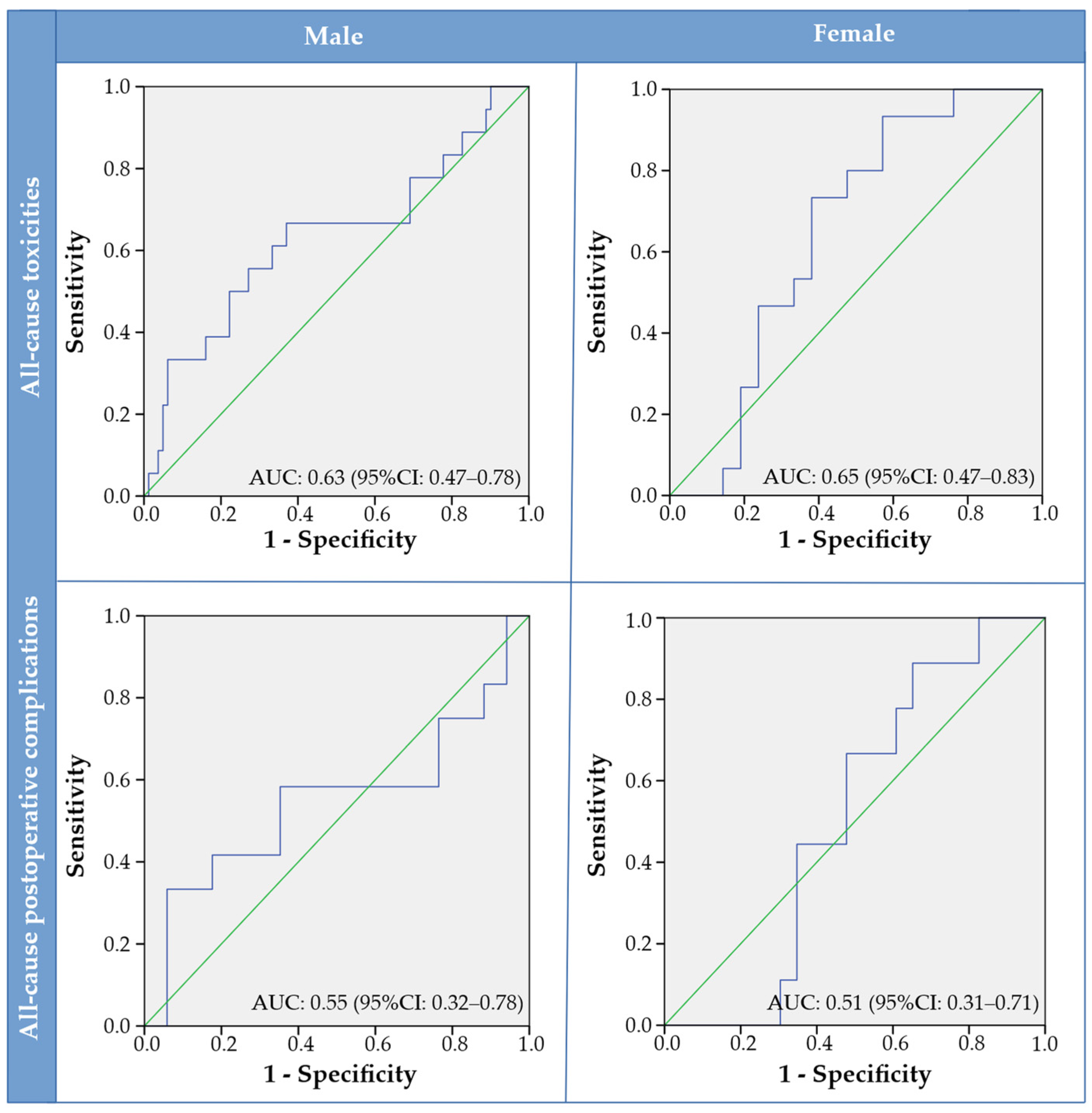
Performance of the main observer for skeletal-muscle quantification in the teaching file was determined to be excellent as ICCs were r = 0.991 (p < 0.001) and r = 0.975 (p < 0.001) for MRI (n = 10) and CT (n = 10), respectively. Delineation of skeletal muscle used for further analysis was more often performed on CT (78.2%) than MRI (21.8%). For the total cohort, male patients had significantly higher SMI than female patients (47.6 ± 5.7 cm2/m2 versus 35.3 ± 6.5 cm2/m2, p < 0.001). Sex-specific SMI cut-off values were generated to identify patients having low SMI based on the adverse events endpoints. In total, eight SMI cut-off values were generated and sensitivity, specificity and AUCs were provided per SMI cut-off value (Table 3). AUCs were all <0.7 (0.425–0.688). ROC curves showing the diagnostic accuracy for sex-specific SMI and the occurrence of all-cause toxicities and postoperative complications are shown in Figure 2.

3.4. Relationship between Low Skeletal Muscle Mass and Adverse Events

For total cohort, prevalence of low SMI was 34.7% (3.9% of males, 92.6% of females), 34.7% (3.9% of males, 92.6% of females), 33.2% (4.7% of males, 86.8% of females) and 29.6% (0% of males, 85.3% of females) for SMI cut-off values based on all-cause toxicity, dysphagia, all-cause postoperative complications and wound infection, respectively. Although AUCs were <0.7 for all generated SMI cut-off values, a logistic-regression analysis was performed to analyse the relationship between low SMI and the adverse events endpoints.
An univariate logistic regression was performed with the AE endpoints as dependent variable (Table 4 and Table 5). For the multivariable regression analyses for toxicity, tumour site, oncological stage and low SMI were selected from the univariate logistic-regression analysis, according to their OR and p-values, and age was added to the model as a clinically relevant variable. Other potential important clinically relevant variables such as G8, GFI and ACE-27 were not included to the multivariable model due to a relatively high rate of missing’s. Moreover, BMI and gender were also not selected as these variables we used to calculate SMI. Tumour site and age were excluded from the model when backward selection was applied. Stage IV disease (OR 3.45, 95% CI 1.28–9.29) and low SMI (OR 3.33, 95% CI 1.41–7.82) remained independently and significantly related to all-cause relevant (C)RT toxicities (Table 6). The separate univariable and multivariable regression analysis (Table 4 and Table 6, respectively) for grade III dysphagia had similar results compared to the analysis with all clinically relevant (C)RT toxicities, as stage IV disease (OR 3.47, 95% CI 1.21–9.92) and low SMI (OR 2.50, 95% CI 1.02–6.14) were also independently and significantly related to grade III dysphagia. The univariable logistic regression for all-cause postoperative complications (CDC ≥ II) did not identified variables significantly associated with the endpoint (Table 5). Tumour site was the only variable significantly associated with the occurrence of wound infections (CDC ≥ II). Compared to the oral cavity, tumours located in the hypopharynx/larynx had the highest risk (OR 10.29, 95% CI 1.45–72.81) of developing postoperative wound infection.

4. Discussion

This is the first study that established both sex and treatment modality stratified SMI cut-off values based on clinically relevant adverse outcomes to diagnose low SMI in HNSCC patients. Analysis showed that SMI was significantly higher in males than females, in both the (C)RT group and the surgery group, supporting the hypothesis that sex-specific SMI cut-off values are mandatory to diagnose low SMI. Sex-specific SMI cut-off values were generated with ROC curves for the different AE endpoints. AUCs were all <0.7, indicating insufficient discriminative power of low SMI on the endpoints. Nevertheless, low SMI and stage IV disease were independently and significantly related to all-cause toxicities and dysphagia. However, no other characteristics significantly related to postoperative complications could be identified. Low SMI, according to the established sex and modality specific SMI cut-off values, is, therefore, related to all-cause toxicities and dysphagia but not to postoperative complications in this present cohort with HNSCC.
Currently, no consensus has been reached to diagnose low SMI in HNSCC patients. Skeletal muscle mass can be measured using different modalities including dual-energy X-ray absorptiometry, bioelectric impedance analysis and CT and MRI imaging [15]. CT and MRI imaging are often used in oncology as these images are already part of the normal oncological work-up, and, therefore, no additional scans have to be made. In CT and MRI imaging, the gold standard is to measure SMI at the L3 level [15]. However, abdominal imaging is often lacking in HNSCC, and neck CT or MRI at the level C3 were found to be a valid alternative to measure SMI [13,14]. However, other possible alternatives exist to measure SMI in HNSCC patients, for instance measurements on the masticatory muscles [41]. In general, SMI cut-off values can be generated using different methods including cohort mean (using -2SD), cohort median (using lowest interquartile range) and optimal stratification based on clinical outcome (using ROC curves or log likelihood models). The ROC method is the most optimal method of defining a clinical implacable cut-off value, since this method searches for the point where the sensitivity and specificity is the highest [40].
SMI cut-off values are often applied from Prado et al. and Martin et al. in oncology patients [42,43]. These cut-offs are based on the log likelihood of survival, and both studies included patients with solid tumours of the respiratory or gastrointestinal tract. SMI cut-off values of Prado et al. are remarkably higher compared to this present study. The difference can be explained as the cohort of Prado et al. consisted of mostly men, sarcopenic obese patients and a different oncological population. Apart from sex-specific SMI cut-off values, Martin et al. also introduced BMI-specific SMI cut-off values. The SMI cut-off values of Martin et al. are quite similar, especially for underweight and normal weight patients, indicating that HNSCC patients are normal- to underweight compared to their study population. This difference emphasises that HNSCC patients are in need of their own SMI cut-off value to diagnose low SMI.
Nevertheless, already published SMI cut-off values generated in a HNSCC population have several limitations as most are not sex-specific [21,44], are based on the median SMI of the given cohort [26,27], or have a specific (Asian) ethnicity [6,22,23,24,25]. Only three studies concerning toxicity in HNSCC patients have a Western study population [21,26,27] and are, therefore, comparable with the SMI cut-off values as generated in this present study, as SMI will differ between ethnicities. Wendrich et al. created a non-sex-specific cut-off value of <43.2 cm2/m2, using the lowest log-likelihood value based on chemotherapy dose-limiting toxicity in a Dutch population, arguing no difference in chemotherapy dose-limiting toxicity between males and females [21]. This seems controversial as female patients have lower SMI, resulting in low SMI of almost all female patients. As of today, Karavolia et al. provided sex-specific SMI cut-off values (SMI <42.0 cm2/m2 and SMI <31.2 cm2/m2 for males and females, respectively) in the largest published HNSCC population undergoing (C)RT [27]. Van Rijn et al. used very similar SMI cut-off values using the same strategy to generate SMI cut-off values; however, the cohorts of these studies are overlapping. Nevertheless, the established sex-specific SMI cut-off values of Karavolia et al. and van Rijn et al. are based on the lowest quartile, and this method is inferior, as it may underestimate the effect of low SMI on clinical outcome.
Currently, low SMI is gaining more interest and is increasingly recognised for its predictive value for adverse events in oncology. However, the relationship of low SMI and the occurrence of toxicity seems to be less investigated in HNSCC patients, as only four studies could be identified [26,27,45,46]. The study of Lere-Chevaleyre et al. showed that pre-treatment SMI was the only predictive factor of all-cause toxicity (CTCAE ≥ III) in head and neck cancer patients receiving induction chemotherapy by docetaxel, cisplatin and 5-fluorouracil [45]. However, Thureau et al. could not link low SMI with toxicity in HNSCC patients treated with chemotherapy or chemoradiation [46] and is, therefore, not in line with the results of this present study. The association between low SMI and dysphagia is supported by Karavolia et al. and van Rijn et al. [26,27], since both found a significant association between low SMI with respectively acute dysphagia (CTCAE ≥ III) and late dysphagia (CTCAE ≥ II) in a large cohort of HNSCC patients treated with definitive (C)RT [26,27]. Several underlying mechanisms could explain the relationship between low SMI and toxicity. In the case of chemotherapy, low lean body mass (which largely corresponds to low SMI) results in less body volume, consequently risking chemotherapy overdose [47]. Therefore, to prevent chemotherapy-related toxicities, others advocate to adjust chemotherapy doses based on lean body mass instead of body surface area or weight [45,47]. Increased radiation-induced toxicities in patients with low SMI could also be due to a pro-inflammatory state [48,49] or due to malnutrition as a result of dysphagia [50,51]. Furthermore, low SMI is associated with impaired muscle function [15] and may also influence the swallowing muscles [51]. Accordingly, pre-treatment low SMI seems to have sufficient discriminative power to identify patients at risk for the occurrence of all-cause toxicities and dysphagia in HNSCC patients.
The present study could not significantly link low SMI and all-cause postoperative complications and wound infection (both defined as CDC grade ≥ II). Bril et al. included patients undergoing a total laryngectomy and found that low SMI was related to pharyngocutaneous fistulas and overall survival but not to all-grade (CDC ≥ I) or severe postoperative complications (CDC ≥ III) [52], which is in line with this present study. However, Bril et al. applied the non-sex-specific SMI cut-off values based on the occurrence of chemotherapy dose-limiting toxicity from Wendrich et al., to identify patients with low SMI in a surgical population, and may have resulted in the lack of a significant relationship between low SMI and postoperative complications, as graded with the CDC. Although two other studies, also using CDC (grade ≥ III), did find a relationship between low SMI and severe postoperative complication, though it was in patients undergoing mandibular reconstruction with a free fibula flap [53] and autologous head and neck free tissue reconstruction [54]. Outcomes of three the above-mentioned studies were further analysed in a meta-analysis and showed that low SMI was indeed related to postoperative complications [8]. Discrepancy between this meta-analysis and our study could be explained by differences in applied SMI cut-off values and/or endpoints for postoperative complications. We also performed additional analysis on infectious postoperative complications, as it has been found to be related to low SMI in other studies, so muscle loss is thought to be the result of reduced immune function, due to a decrease in skeletal muscle mass and increase in adipocytes [23]. However, this present study failed to find a significant relationship. Moreover, the relation between SMI and postoperative complications after less-invasive surgical interventions (e.g., transoral robotic surgery) remains unclear in HNSCC [55].
This present cohort had some obvious limitations. The oncological stage was determined according to 7th instead of 8th edition of American Joint Committee on Cancer Manual, as during the prospective inclusion of patients the newest 8th edition did not exist yet. It was not possible to stage according to the 8th edition, due to lacking data including invasion depth of oral cavity tumours, which could be seen as a limitation. Moreover, this study is very likely underpowered due to the limited sample size, especially in the surgical group. For some variables in the logistic regression with postoperative complications as the endpoint, ORs were unrealistically high or could not be calculated, as a result of too few patients within the category. Translation of the results regarding the surgical group is, therefore, difficult and is potentially biased due to heterogeneity in the tumour site and surgical procedure. Interpreting these data may have led to low AUCs in the surgical group and, therefore, underestimation of the effect and insignificant results. Unfortunately, performing subgroup analysis on specific types of surgery was not possible in this study, due to the low number of cases. Although higher AUCs were achieved in the (C)RT group, AUCs were still <0.7, indicating insufficient diagnostic discriminative power of SMI on toxicities. Low AUCs could also be due to heterogeneity within the (C)RT group as multiple treatment types were included, i.e., accelerated and not accelerated RT, CRT and RT with cetuximab. Moreover, a limited sample of variables could be included in the multivariable regression analysis due to the risk of overfitting. Ideally, more determinants such as frailty, comorbidities and treatment specifications (e.g., chemotherapy type and dosage) were included in the multivariable regression to analyse potential confounding. Nevertheless, low SMI, according to the generated SMI cut-off values, was found to be independently and significantly related to all-cause toxicity and dysphagia. Future research should be focused on the external validation of the here generated sex-specific SMI cut-off values.
Compared to prior publications regarding SMI cut-off values in HNSCC patients, the main strength is that this present study provided sex-specific cut-off values based on the occurrence of adverse events using ROC-curves, which is seen as the optimal method for defining clinically implacable cut-off values. Not only sex- and modality-specific SMI thresholds were generated in this present study but also the relationship of low SMI with adverse outcome was analysed, which is highly clinically relevant. Moreover, SMI measurements made for this analysis were reliable, as observer analysis showed excellent performance of the main observer. Furthermore, patients were prospectively included and assessed with a geriatric assessment at baseline, using a broad range of validated scales and tools. After inclusion, patients were systematically followed-up to assess the occurrence of adverse events, as measured with the CTCAE and CDC, resulting in a well-documented and reliable data-biobank.

5. Conclusions

To conclude, identifying HNSCC patients at risk for adverse events is highly clinically relevant. The sex-specific SMI cut-off values to diagnose low SMI, as generated in the (C)RT group, were able to identify patients at risk for all-cause toxicity and dysphagia. More research is necessary to establish SMI cut-off values based on postoperative complications, ideally in a larger and more homogenous HNSCC population. These outcomes help with constructing a practical and clinically relevant biomarker for adverse events in HNSCC patients.

Author Contributions

Conceptualization, A.T.Z., W.P., I.v.R.-D., G.A.S., R.J.H.M.S., I.W., A.v.d.H., G.H.d.B. and G.B.H.; supervision methodology, A.T.Z., G.A.S., R.J.H.M.S., I.W., A.v.d.H., G.H.d.B. and G.B.H.; software, R.A.J.O.D.; validation, A.T.Z. and W.P.; formal analysis, A.T.Z. and W.P.; investigation, A.T.Z., W.P., I.v.R.-D., G.A.S., R.A.J.O.D., R.J.H.M.S., I.W., A.v.d.H., G.H.d.B. and G.B.H.; software, R.A.J.O.D.; resources, R.A.J.O.D., R.J.H.M.S., G.H.d.B. and G.B.H.; data curation, A.T.Z., W.P., I.v.R.-D., G.A.S., R.J.H.M.S., I.W., A.v.d.H., G.H.d.B. and G.B.H.; writing—original draft preparation, A.T.Z., W.P. and G.B.H.; writing—review and editing, A.T.Z., W.P., I.v.R.-D., G.A.S., R.A.J.O.D., R.J.H.M.S., I.W., A.v.d.H., G.H.d.B. and G.B.H.; investigation, A.T.Z., W.P., I.v.R.-D., G.A.S., R.A.J.O.D., R.J.H.M.S., I.W., A.v.d.H., G.H.d.B. and G.B.H. All authors have read and agreed to the published version of the manuscript.

Funding

The first author received a Ph.D. scholarship for excellent master students from the Graduate School of Medical Sciences (GSMS), University of Groningen and University Medical Centre Groningen.

Institutional Review Board Statement

The study was conducted according to the guidelines of the Declaration of Helsinki. Data was extracted from the OncoLifeS data-biobank with permission of the scientific committee of OncoLifeS (protocol number 201900587). The OncoLifeS data-biobank is approved by the by the Medical Ethical Committee of the University Medical Centre of Groningen and is registered in the Dutch Trial Register, registration number: NL7839. The OncoLifeS data-biobank collects data on adult oncological patients, including, e.g., patients, cancer and treatment characteristics. Patients were prospectively included into the data-biobank, after their informed consent was obtained.

Informed Consent Statement

Informed consent was obtained from all subjects involved in the study.

Data Availability Statement

Data are available on request due to privacy restrictions.

Conflicts of Interest

The authors declare no conflict of interest.

References

  1. Bray, F.; Ferlay, J.; Soerjomataram, I.; Siegel, R.L.; Torre, L.A.; Jemal, A. Global cancer statistics 2018: GLOBOCAN estimates of incidence and mortality worldwide for 36 cancers in 185 countries. CA Cancer J. Clin. 2018, 68, 394–424. [Google Scholar] [CrossRef] [PubMed] [Green Version]
  2. Crozier, E.; Sumer, B.D. Head and neck cancer. Med. Clin. N. Am. 2010, 94, 1031–1046. [Google Scholar] [CrossRef] [PubMed]
  3. Mascarella, M.A.; Richardson, K.; Mlynarek, A.; Forest, V.-I.; Hier, M.; Sadeghi, N.; Mayo, N. Evaluation of a Preoperative Adverse Event Risk Index for Patients Undergoing Head and Neck Cancer Surgery. JAMA Otolaryngol. Neck Surg. 2019, 145, 345–351. [Google Scholar] [CrossRef] [PubMed]
  4. Pfister, D.G.; Spencer, S.; Adelstein, D.; Adkins, D.; Anzai, Y.; Brizel, D.M.; Bruce, J.Y.; Busse, P.M.; Caudell, J.J.; Cmelak, A.J.; et al. Head and Neck Cancers, Version 2.2020, NCCN Clinical Practice Guidelines in Oncology. J. Natl. Compr. Cancer Netw. 2020, 18, 873–898. [Google Scholar] [CrossRef] [PubMed]
  5. Niska, J.R.; Thorpe, C.S.; Halyard, M.Y.; Tan, A.D.; Atherton, P.J.; Dueck, A.C.; Patel, S.H.; Sloan, J.A. Patient-reported quality-of-life outcomes in relation to provider-assessed adverse events during head and neck radiotherapy. J. Patient-Rep. Outcomes 2020, 4, 60. [Google Scholar] [CrossRef]
  6. Hua, X.; Liao, J.-F.; Huang, X.; Huang, H.-Y.; Wen, W.; Long, Z.-Q.; Guo, L.; Yuan, Z.-Y.; Lin, H.-X. Sarcopenia is associated with higher toxicity and poor prognosis of nasopharyngeal carcinoma. Ther. Adv. Med. Oncol. 2020, 12, 1758835920947612. [Google Scholar] [CrossRef]
  7. Findlay, M.; White, K.; Lai, M.; Luo, D.; Bauer, J.D. The Association Between Computed Tomography–Defined Sarcopenia and Outcomes in Adult Patients Undergoing Radiotherapy of Curative Intent for Head and Neck Cancer: A Systematic Review. J. Acad. Nutr. Diet. 2020, 120, 1330–1347.e8. [Google Scholar] [CrossRef]
  8. Surov, A.; Wienke, A. Low skeletal muscle mass predicts relevant clinical outcomes in head and neck squamous cell carcinoma. A Meta Analysis. Ther. Adv. Med. Oncol. 2021, 13, 17588359211008844. [Google Scholar] [CrossRef]
  9. Wong, A.; Zhu, D.; Kraus, D.; Tham, T. Radiologically Defined Sarcopenia Affects Survival in Head and Neck Cancer: A Meta-Analysis. Laryngoscope 2020, 131, 333–341. [Google Scholar] [CrossRef]
  10. Couch, M.E.; Dittus, K.; Toth, M.J.; Willis, M.S.; Guttridge, D.C.; George, J.R.; Barnes, C.A.; Gourin, C.G.; Der-Torossian, H. Cancer cachexia update in head and neck cancer: Definitions and diagnostic features. Head Neck 2014, 37, 594–604. [Google Scholar] [CrossRef]
  11. Mitsiopoulos, N.; Baumgartner, R.N.; Heymsfield, S.B.; Lyons, W.; Gallagher, D.; Ross, R. Cadaver validation of skeletal muscle measurement by magnetic resonance imaging and computerized tomography. J. Appl. Physiol. 1998, 85, 115–122. [Google Scholar] [CrossRef] [PubMed]
  12. Mourtzakis, M.; Prado, C.M.; Lieffers, J.R.; Reiman, T.; McCargar, L.J.; Baracos, V.E. A practical and precise approach to quantification of body composition in cancer patients using computed tomography images acquired during routine care. Appl. Physiol. Nutr. Metab. 2008, 33, 997–1006. [Google Scholar] [CrossRef] [PubMed]
  13. Swartz, J.E.; Pothen, A.J.; Wegner, I.; Smid, E.J.; Swart, K.M.; de Bree, R.; Leenen, L.P.; Grolman, W. Feasibility of using head and neck CT imaging to assess skeletal muscle mass in head and neck cancer patients. Oral Oncol. 2016, 62, 28–33. [Google Scholar] [CrossRef] [PubMed]
  14. Zwart, A.T.; Becker, J.N.; Lamers, M.J.; Dierckx, R.A.J.O.; de Bock, G.H.; Halmos, G.B.; van der Hoorn, A. Skeletal muscle mass and sarcopenia can be determined with 1.5-T and 3-T neck MRI scans, in the event that no neck CT scan is performed. Eur. Radiol. 2020, 31, 4053–4062. [Google Scholar] [CrossRef] [PubMed]
  15. Cruz-Jentoft, A.J.; Bahat, G.; Bauer, J.; Boirie, Y.; Bruyere, O.; Cederholm, T.; Cooper, C.; Landi, F.; Rolland, Y.; Sayer, A.A.; et al. Sarcopenia: Revised European consensus on definition and diagnosis. Age Ageing 2019, 48, 16–31. [Google Scholar] [CrossRef] [Green Version]
  16. Cruz-Jentoft, A.J.; Baeyens, J.P.; Bauer, J.M.; Boirie, Y.; Cederholm, T.; Landi, F.; Martin, F.C.; Michel, J.-P.; Rolland, Y.; Schneider, S.M.; et al. Sarcopenia: European consensus on definition and diagnosis: Report of the European Working Group on Sarcopenia in Older People. Age Ageing 2010, 39, 412–423. [Google Scholar] [CrossRef] [Green Version]
  17. Hilmi, M.; Jouinot, A.; Burns, R.; Pigneur, F.; Mounier, R.; Gondin, J.; Neuzillet, C.; Goldwasser, F. Body composition and sarcopenia: The next-generation of personalized oncology and pharmacology? Pharmacol. Ther. 2019, 196, 135–159. [Google Scholar] [CrossRef]
  18. Shachar, S.S.; Williams, G.R.; Muss, H.B.; Nishijima, T.F. Prognostic value of sarcopenia in adults with solid tumours: A meta-analysis and systematic review. Eur. J. Cancer 2016, 57, 58–67. [Google Scholar] [CrossRef]
  19. Cruz-Jentoft, A.J.; Landi, F.; Schneider, S.M.; Zuniga, C.; Arai, H.; Boirie, Y.; Chen, L.K.; Fielding, R.A.; Martin, F.C.; Michel, J.P.; et al. Prevalence of and interventions for sarcopenia in ageing adults: A systematic review. Report of the International Sarcopenia Initiative (EWGSOP and IWGS). Age Ageing 2014, 43, 748–759. [Google Scholar] [CrossRef]
  20. Huiskamp, L.F.J.; Chargi, N.; Devriese, L.A.; May, A.M.; Huitema, A.D.R.; De Bree, R. The Predictive Value of Low Skeletal Muscle Mass Assessed on Cross-Sectional Imaging for Anti-Cancer Drug Toxicity: A Systematic Review and Meta-Analysis. J. Clin. Med. 2020, 9, 3780. [Google Scholar] [CrossRef]
  21. Wendrich, A.W.; Swartz, J.E.; Bril, S.I.; Wegner, I.; de Graeff, A.; Smid, E.J.; de Bree, R.; Pothen, A.J. Low skeletal muscle mass is a predictive factor for chemotherapy dose-limiting toxicity in patients with locally advanced head and neck cancer. Oral Oncol. 2017, 71, 26–33. [Google Scholar] [CrossRef] [PubMed]
  22. Chang, S.; Hsu, C.; Tsai, Y.; Chang, G.; Tsai, M.; Huang, E.I.; Tsai, Y. Prognostic Value of Third Cervical Vertebra Skeletal Muscle Index in Oral Cavity Cancer: A Retrospective Study. Laryngoscope 2021, 131, E2257–E2265. [Google Scholar] [CrossRef] [PubMed]
  23. Nakamura, H.; Makiguchi, T.; Yamaguchi, T.; Suzuki, K.; Yokoo, S. Impact of sarcopenia on postoperative surgical site infections in patients undergoing flap reconstruction for oral cancer. Int. J. Oral Maxillofac. Surg. 2020, 49, 576–581. [Google Scholar] [CrossRef] [PubMed]
  24. Nishikawa, D.; Hanai, N.; Suzuki, H.; Koide, Y.; Beppu, S.; Hasegawa, Y. The Impact of Skeletal Muscle Depletion on Head and Neck Squamous Cell Carcinoma. ORL 2018, 80, 1–9. [Google Scholar] [CrossRef]
  25. Shodo, R.; Yamazaki, K.; Ueki, Y.; Takahashi, T.; Horii, A. Sarcopenia predicts a poor treatment outcome in patients with head and neck squamous cell carcinoma receiving concurrent chemoradiotherapy. Eur. Arch. Oto-Rhino-Laryngol. 2020, 278, 2001–2009. [Google Scholar] [CrossRef]
  26. van Rijn-Dekker, M.I.; Bosch, L.V.D.; Hoek, J.G.V.D.; Bijl, H.P.; van Aken, E.; van der Hoorn, A.; Oosting, S.F.; Halmos, G.B.; Witjes, M.; van der Laan, H.P.; et al. Impact of sarcopenia on survival and late toxicity in head and neck cancer patients treated with radiotherapy. Radiother. Oncol. 2020, 147, 103–110. [Google Scholar] [CrossRef]
  27. Karavolia, E.; van Rijn-Dekker, M.I.; Bosch, L.V.D.; Hoek, J.G.V.D.; Oldehinkel, E.; Meijer, T.W.; Halmos, G.B.; Witjes, M.J.; Oosting, S.F.; van der Hoorn, A.; et al. Impact of sarcopenia on acute radiation-induced toxicity in head and neck cancer patients. Radiother. Oncol. 2022, 170, 122–128. [Google Scholar] [CrossRef]
  28. Parker, N.H.; Gorzelitz, J.; Ngo-Huang, A.; Caan, B.J.; Prakash, L.; Garg, N.; Petzel, M.Q.B.; Schadler, K.; Basen-Engquist, K.; Katz, M.H.G. The Role of Home-Based Exercise in Maintaining Skeletal Muscle During Preoperative Pancreatic Cancer Treatment. Integr. Cancer Ther. 2021, 20, 1534735420986615. [Google Scholar] [CrossRef]
  29. Capozzi, L.C.; McNeely, M.L.; Lau, H.Y.; Reimer, R.A.; Giese-Davis, J.; Fung, T.S.; Culos-Reed, S.N. Patient-reported outcomes, body composition, and nutrition status in patients with head and neck cancer: Results from an exploratory randomized controlled exercise trial. Cancer 2016, 122, 1185–1200. [Google Scholar] [CrossRef] [Green Version]
  30. Grote, M.; Maihöfer, C.; Weigl, M.; Davies-Knorr, P.; Belka, C. Progressive resistance training in cachectic head and neck cancer patients undergoing radiotherapy: A randomized controlled pilot feasibility trial. Radiat. Oncol. 2018, 13, 215. [Google Scholar] [CrossRef]
  31. Sidorenkov, G.; Nagel, J.; Meijer, C.; Duker, J.J.; Groen, H.J.M.; Halmos, G.B.; Oonk, M.H.M.; Oostergo, R.J.; van der Vegt, B.; Witjes, M.J.H.; et al. The OncoLifeS data-biobank for oncology: A comprehensive repository of clinical data, biological samples, and the patient’s perspective. J. Transl. Med. 2019, 17, 374. [Google Scholar] [CrossRef] [PubMed]
  32. Bosch, L.V.D.; van der Laan, H.P.; van der Schaaf, A.; Oosting, S.F.; Halmos, G.B.; Witjes, M.J.; Oldehinkel, E.; Meijer, T.W.; Hoek, J.G.V.D.; Steenbakkers, R.J.; et al. Patient-Reported Toxicity and Quality-of-Life Profiles in Patients with Head and Neck Cancer Treated with Definitive Radiation Therapy or Chemoradiation. Int. J. Radiat. Oncol. 2021, 111, 456–467. [Google Scholar] [CrossRef]
  33. Sobin, L.; Gospodarowicz, M.; Wittekind, C. (Eds.) TNM Classification of Malignant Tumours, 7th ed.; Wiley-Blackwell: Oxford, UK, 2009. [Google Scholar]
  34. Soubeyran, P.; Bellera, C.A.; Gregoire, F.; Blanc, J.; Ceccaldi, J.; Blanc-Bisson, C.; Mertens, C.; Mathoulin-Pélissier, S.; Fonck, M.; Rainfray, M. Validation of a screening test for elderly patients in oncology. J. Clin. Oncol. 2008, 26, 20568. [Google Scholar] [CrossRef]
  35. Steverink, N.; Slaets, J.; Schuurmans, H.; van Lis, M. Measuring frailty: Developing and testing of the Groningen Frailty Indicator (GFI). Gerontologist 2001, 41, 236–237. [Google Scholar]
  36. Riper, H.; Kramer, J.; Smit, F.; Conijn, B.; Schippers, G.; Cuijpers, P. Web-based self-help for problem drinkers: A pragmatic randomized trial. Addiction 2008, 103, 218–227. [Google Scholar] [CrossRef] [PubMed]
  37. Zwart, A.T.; van der Hoorn, A.; van Ooijen, P.M.A.; Steenbakkers, R.J.H.M.; de Bock, G.H.; Halmos, G.B. CT-measured skeletal muscle mass used to assess frailty in patients with head and neck cancer. J. Cachexia Sarcopenia Muscle 2019, 10, 1060–1069. [Google Scholar] [CrossRef] [Green Version]
  38. Common Terminology Criteria for Adverse Events (CTCAE) Version 4.0. In: Health NIo, editor. Sep 2009 ed: U.S.DEPARTMENT OF HEALTH AND HUMAN SERVICES. 2009. Available online: https://evs.nci.nih.gov/ftp1/CTCAE/CTCAE_4.03/Archive/CTCAE_4.0_2009-05-29_QuickReference_8.5x11.pdf (accessed on 5 May 2022).
  39. Dindo, D.; Demartines, N.; Clavien, P.A. Classification of surgical complications: A new proposal with evaluation in a cohort of 6336 patients and results of a survey. Ann. Surg. 2004, 240, 205–213. [Google Scholar] [CrossRef]
  40. Mandrekar, J.N. Receiver Operating Characteristic Curve in Diagnostic Test Assessment. J. Thorac. Oncol. 2010, 5, 1315–1316. [Google Scholar] [CrossRef] [PubMed] [Green Version]
  41. Chang, S.-W.; Tsai, Y.-H.; Hsu, C.-M.; Huang, E.I.; Chang, G.-H.; Tsai, M.-S.; Tsai, Y.-T. Masticatory muscle index for indicating skeletal muscle mass in patients with head and neck cancer. PLoS ONE 2021, 16, e0251455. [Google Scholar] [CrossRef]
  42. Prado, C.M.; Lieffers, J.R.; McCargar, L.J.; Reiman, T.; Sawyer, M.B.; Martin, L.; Baracos, V.E. Prevalence and clinical implications of sarcopenic obesity in patients with solid tumours of the respiratory and gastrointestinal tracts: A population-based study. Lancet Oncol. 2008, 9, 629–635. [Google Scholar] [CrossRef]
  43. Martin, L.; Birdsell, L.; Macdonald, N.; Reiman, T.; Clandinin, M.T.; McCargar, L.J.; Murphy, R.; Ghosh, S.; Sawyer, M.B.; Baracos, V.E. Cancer cachexia in the age of obesity: Skeletal muscle depletion is a powerful prognostic factor, independent of body mass index. J. Clin. Oncol. 2013, 31, 1539–1547. [Google Scholar] [CrossRef] [PubMed]
  44. Huiskamp, L.F.J.; Chargi, N.; Devriese, L.A.; De Jong, P.A.; De Bree, R. The predictive and prognostic value of low skeletal muscle mass for dose-limiting toxicity and survival in head and neck cancer patients receiving concomitant cetuximab and radiotherapy. Eur. Arch. Oto-Rhino-Laryngol. 2020, 277, 2847–2858. [Google Scholar] [CrossRef] [PubMed] [Green Version]
  45. Lere-Chevaleyre, A.; Bernadach, M.; Lambert, C.; Cassagnes, L.; Puechmaille, M.; Mom, T.; Gilain, L.; Lapeyre, M.; Boirie, Y.; Biau, J.; et al. Toxicity of induction chemotherapy in head and neck cancer: The central role of skeletal muscle mass. Head Neck 2021, 44, 681–690. [Google Scholar] [CrossRef] [PubMed]
  46. Thureau, S.; Lebret, L.; Lequesne, J.; Cabourg, M.; Dandoy, S.; Gouley, C.; Lefebvre, L.; Mallet, R.; Mihailescu, S.-D.; Moldovan, C.; et al. Prospective Evaluation of Sarcopenia in Head and Neck Cancer Patients Treated with Radiotherapy or Radiochemotherapy. Cancers 2021, 13, 753. [Google Scholar] [CrossRef]
  47. Prado, C.M.; Baracos, V.E.; McCargar, L.J.; Reiman, T.; Mourtzakis, M.; Tonkin, K.; Mackey, J.R.; Koski, S.; Pituskin, E.; Sawyer, M.B. Sarcopenia as a Determinant of Chemotherapy Toxicity and Time to Tumor Progression in Metastatic Breast Cancer Patients Receiving Capecitabine Treatment. Clin. Cancer Res. 2009, 15, 2920–2926. [Google Scholar] [CrossRef] [Green Version]
  48. Yamahara, K.; Mizukoshi, A.; Lee, K.; Ikegami, S. Sarcopenia with inflammation as a predictor of survival in patients with head and neck cancer. Auris Nasus Larynx 2021, 48, 1013–1022. [Google Scholar] [CrossRef]
  49. Murimwa, G.Z.; Venkat, P.S.; Jin, W.; Leuthold, S.; Latifi, K.; Almhanna, K.; Pimiento, J.M.; Fontaine, J.P.; Hoffe, S.E.; Frakes, J.M. Impact of sarcopenia on outcomes of locally advanced esophageal cancer patients treated with neoadjuvant chemoradiation followed by surgery. J. Gastrointest. Oncol. 2017, 8, 808–815. [Google Scholar] [CrossRef] [Green Version]
  50. Crowder, S.L.; Douglas, K.G.; Pepino, M.Y.; Sarma, K.P.; Arthur, A.E. Nutrition impact symptoms and associated outcomes in post-chemoradiotherapy head and neck cancer survivors: A systematic review. J. Cancer Surviv. 2018, 12, 479–494. [Google Scholar] [CrossRef]
  51. Karsten, R.T.; Al-Mamgani, A.; Bril, S.I.; Msc, S.T.; Molen, L.; Boer, J.P.; Hilgers, F.J.M.; Smeele, L.E.; Brekel, M.W.M.; Stuiver, M.M. Sarcopenia, a strong determinant for prolonged feeding tube dependency after chemoradiotherapy for head and neck cancer. Head Neck 2019, 41, 4000–4008. [Google Scholar] [CrossRef]
  52. Bril, S.I.; Pezier, T.F.; Tijink, B.M.; Janssen, L.M.; Braunius, W.W.; De Bree, R. Preoperative low skeletal muscle mass as a risk factor for pharyngocutaneous fistula and decreased overall survival in patients undergoing total laryngectomy. Head Neck 2019, 41, 1745–1755. [Google Scholar] [CrossRef] [Green Version]
  53. Ansari, E.; Chargi, N.; Gemert, J.; Es, R.; Dieleman, F.; Rosenberg, A.J.W.P.; Cann, E.; Bree, R. Low skeletal muscle mass is a strong predictive factor for surgical complications and a prognostic factor in oral cancer patients undergoing mandibular reconstruction with a free fibula flap. Oral Oncol. 2020, 101, 104530. [Google Scholar] [CrossRef] [PubMed]
  54. Alwani, M.M.; Jones, A.J.; Novinger, L.J.; Pittelkow, E.; Bonetto, A.; Sim, M.W.; Moore, M.G.; Mantravadi, A.V. Impact of Sarcopenia on Outcomes of Autologous Head and Neck Free Tissue Reconstruction. J. Reconstr. Microsurg. 2020, 36, 369–378. [Google Scholar] [PubMed]
  55. Meccariello, G.; Maniaci, A.; Bianchi, G.; Cammaroto, G.; Iannella, G.; Catalano, A.; Sgarzani, R.; De Vito, A.; Capaccio, P.; Pelucchi, S.; et al. Neck dissection and trans oral robotic surgery for oropharyngeal squamous cell carcinoma. Auris Nasus Larynx 2021, 49, 117–125. [Google Scholar] [CrossRef] [PubMed]
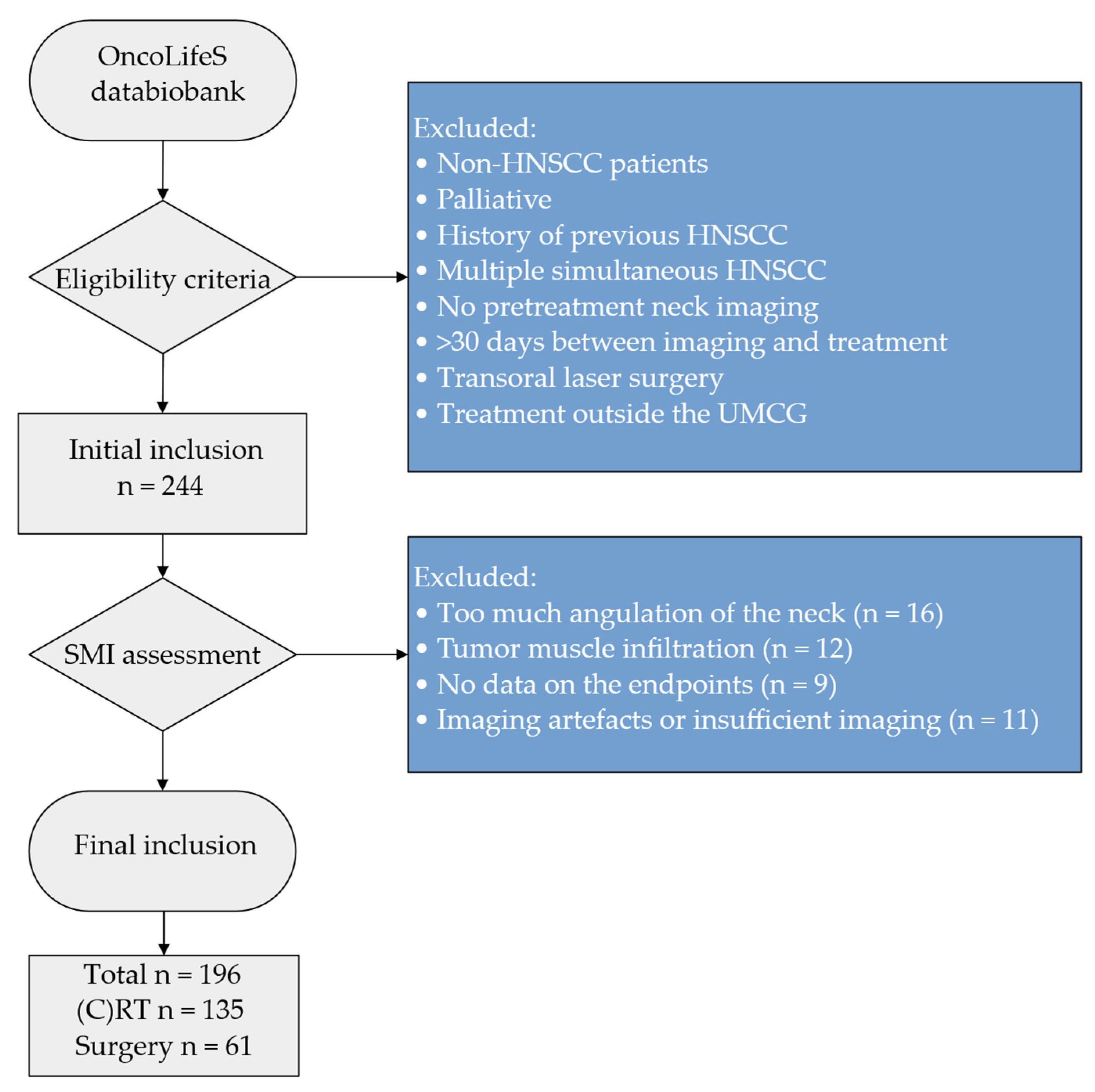
Figure 1. Flowchart of in- and excluded patients. Abbreviations: HNSCC = head and neck squamous cell carcinoma, (C)RT = (Chemo-)Radiotherapy, SMI = skeletal muscle index, UMCG = University Medical Center Groningen.
Jcm 11 04650 g001
Figure 2. Receiver operator characteristic curves stratified for males versus females and all-cause toxicities versus all-cause postoperative complications. Abbreviations: AUC = area under the curve, CI = confidence interval.
Jcm 11 04650 g002
Table 1. Baseline characteristics of total cohort and differences between patients treated with (chemo-)radiotherapy and surgery.
CharacteristicsTotal(Chemo-)
Radiotherapy
Surgery p-Value
n = 196n = 135n = 61
Sex
        Male128 (65.3%)99 (73.3%)29 (47.5%)<0.001 *
        Female68 (34.7%)36 (26.7%)32 (52.5%)
Age (years)64.2 (±11.2)63.5 (±10.5)65.8 (±12.5)0.176 **
BMI (kg2/m2)25.8 (±5.3)25.4 (±5.4)26.5 (±5.0)0.184 **
Tumour site
        Oropharynx69 (35.2%)67 (49.6%)2 (3.3%)<0.001 ***
        Oral cavity60 (30.6%)5 (3.7%)55 (90.2%)
        Larynx56 (28.6%)52 (38.5%)4 (6.6%)
        Hypopharynx10 (5.1%)10 (7.4%)0 (0%)
        Nasopharynx1 (0.5%)1 (0.7%)0 (0%)
Stage
        I23 (11.7%)3 (2.2%)20 (32.8%)<0.001 *
        II24 (12.2%)17 (12.6%)7 (11.5%)
        III37 (18.0%)31 (23.0%)6 (9.8%)
        IV112 (57.1%)84(62.2%)28 (45.9%)
HPV (Oropharynx)
        +22 (40.7%)21 (39.6%)1 (100%)0.223 *
        −32 (59.3%)32 (60.4%)0 (0%)
        Missing15 14 1
ACE-27
        None32 (28.3%)24 (28.6%)8 (37.6%)0.113 *
        Mild37 (32.7%)32 (38.1%)5 (17.2%)
        Moderate30 (26.5%)20 (23.8%)10 (34.5%)
        Severe14 (12.4%)8 (9.5%)6 (20.7%)
        Missing83 51 32
Smoking
        Never9 (8.8%)3 (4.0%)6 (22.2%)0.004 *
        Active/quit93 (91.2%)72 (96.0%)21 (77.8%)
        Missing94 60 34
Alcohol
        No abuse72 (77.4%)51 (73.9%)21 (87.5%)0.170 *
        Abuse21 (22.6%)18 (26.1%)3 (12.5%)
        Missing103 66 37
GFI
        Non-frail67 (69.1%)50 (71.4%)18 (64.3%)0.419 *
        Frail30 (30.9%)20 (28.6%)10 (35.7%)
        Missing99 65 34
G8
        Non-frail41 (42.7%)32 (46.4%)9 (33.3%)0.245 *
        Frail55 (57.3%)37 (53.6%)18 (66.7%)
        Missing10066 34
Significant p-values (<0.05) are indicated in bold. * Chi-squared test. ** Independent t-test. *** Chi-squared test was performed on a reorganised variable (oral cavity, nasopharynx/oropharynx and hypopharynx/larynx). Abbreviations: ACE-27 = Adult Comorbidity Evaluation-27, BMI = Body Mass Index, G8 = Geriatrics 8, GFI = Groningen Frailty Indicator, HPV = herpes papillomavirus.
Table 2. Occurrence of (chemo)radiotherapy toxicities and postoperative complications as measured with CTCAE grade ≥ III and Clavien–Dindo grade ≥II, respectively.
Characteristics(Chemo-)Radiotherapy Toxicities
All-cause (n = 135)Dysphagia (n = 135)
CTCAE
        None8 (5.9%)40 (29.6%)
        Grade I45 (33.3%)36 (26.7%)
        Grade II49 (36.3%)30 (22.2%)
        ≥Grade III33 (24.4%)29 (21.5%)
CharacteristicsPostoperative complications
All-cause (n = 61)Wound infection (n = 61)
Clavien–Dindo
        None32 (52.5%)51 (78.5%)
        Grade I8 (13.1%)3 (4.6%)
        Grade II12 (19.7%)7 (10.8%)
        Grade III9 (14.8%)4 (6.2%)
        ≥Grade II21 (34.4.0%)11 (16.9%)
Abbreviations: CTCAE = Common Terminology Criteria for Adverse Events.
Table 3. SMI cut-off values based on the occurrence of adverse events endpoints. AUC, sensitivity and specificity per SMI cut-off value are provided.
SMI Cut-OffSensitivitySpecificityAUC95%CIp-Value
Toxicities
All-cause
Male46.490.670.630.6320.47–0.780.082
Female37.900.930.430.6480.47–0.830.136
Dysphagia
Male46.490.650.620.6100.45–0.770.156
Female34.910.750.580.6420.46–0.830.169
Postoperative complications
All-cause
Male41.970.330.940.5490.32–0.780.658
Female39.090.890.650.5120.31–0.710.917
Wound infection
Male40.790.140.960.4090.15–0.670.476
Female35.641.000.540.6070.42–0.790.494
Abbreviations: AUC = area under the curve, CI = confidence interval, SMI = skeletal muscle index (cm2/m2).
Table 4. Univariable logistic regression for individual predictors of all-cause toxicities and dysphagia toxicity according to CTCAE classification (grade ≥ III).
Characteristics(Chemo-)Radiotherapy Toxicities
All-CauseDysphagia
OR95% CIp-ValueOR95% CIp-Value
Sex
        MaleRef Ref
        Female0.310.14–0.720.0060.420.17–0.990.047
Age (years)0.970.93–1.000.0760.970.93–1.010.126
BMI (kg/m2)0.840.75–0.930.0010.850.77–0.950.003
Tumour site
        Oral cavityRef 0.007Ref 0.011
        Nasopharynx/Oropharynx0.830.13–5.24 1.910.20–18.14
        Hypopharynx/Larynx0.190.03–1.35 0.430.04–4.48
Stage
        I–IIIRef 0.010Ref 0.014
        IV3.551.35–9.34 3.681.30–10.38
HPV (oropharynx)
        +Ref Ref
        −0.730.23–2.310.5930.760.26–2.650.833
ACE-27
        NoneRef 0.704Ref 0.837
        Mild1.240.20–7.67 1.000.16–6.35
        Moderate1.170.20–6.94 1.000.14–5.99
        Severe0.530.07–3.98 0.540.07–3.98
GFI
        Non-frailRef 0.265Ref 0.482
        Frail1.910.61–5.95 1.520.47–4.88
G8
        Non-frailRef 0.621Ref 0.810
        Frail1.320.44–4.01 1.150.37–3.54
Smoking
        NeverRef Ref
        Active/quit0.670.06–7.800.7470.620.05–7.250.702
Alcohol
        No abuseRef Ref
        Abuse0.530.13–2.110.3360.590.15–2.350.449
SMI (cm2/m2)0.910.86–0.96<0.0010.920.87–0.970.002
Low SMI
        NoRef Ref
        Yes3.861.69–8.860.0012.701.12–6.470.026
Significant p-Values (<0.05) are indicated in bold. Abbreviations: ACE-27 = Adult Comorbidity Evaluation-27, BMI = Body Mass Index, CI = confidence interval, G8 = Geriatrics 8, GFI = Groningen Frailty Indicator, HPV = herpes papillomavirus, OR = odds ratio, SMI = skeletal muscle index.
Table 5. Univariable logistic regression for individual predictors of all-cause postoperative complications and postoperative wound infection according to Clavien–Dindo classification (grade ≥ II).
CharacteristicsPostoperative Complications
All-CauseWound Infection
OR95%CIp-ValueOR95%CIp-Value
Sex
        MaleRef Ref
        Female1.800.62–5.250.2792.230.58–8.590.245
Age (years)0.970.93–1.020.1880.990.94–1.040.616
BMI (kg/m2)0.960.86–1.070.4791.010.89–1.150.830
Tumour site
        Oral cavityRef 0.827Ref 0.026
        Nasopharynx/Oropharynx2.440.14–41.40 6.860.38–122.52
        Hypopharynx/Larynx** 20.571.87–226.32
Stage
        I–IIIRef 0.463Ref 0.060
        IV1.490.52–4.30 4.000.95–16.93
HPV (oropharynx)
        +* *
        −
ACE-27
        NoneRef 0.303Ref 0740
        Moderate0.670.07–6.87 1.670.12–24.26
        Severe5.330.50–127.90 1.250.06–26.87
        Missing1.330.24–16.36 3.330.28–40.29
GFI
        Non-frailRef 0.517Ref 0.974
        Frail1.690.35–8.22 0.970.18–537
G8
        Non-frailRef 0.069Ref 0.454
        Frail5.500.88–34.46 1.750.28–11.15
Smoking
        Never** **
        Active/quit
Alcohol
        No abuse** **
        Abuse
SMI (cm2/m2)1.020.96–1.080.5291.040.96–1.110.344
Low SMI (sex-specific)
        NoRef 0.377Ref
        Yes0.620.21–1.81 0.670.17–2.580.562
Surgical intensity
        MinorRef 0.287Ref 0.148
        Major1.800.61–5.28 2.890.69–12.17
Significant p-values (<0.05) are indicated in bold. * No regression was performed due to too few patients within the category. Abbreviations: ACE-27 = Adult Comorbidity Evaluation-27, BMI = Body Mass Index, CI = confidence interval, G8 = Geriatrics 8, GFI = Groningen Frailty Indicator, HPV = herpes papillomavirus, OR = odds ratio, SMI = skeletal muscle index.
Table 6. Multivariable regression analysis for determining predictors for all-cause (C)RT toxicities and dysphagia toxicity.
Characteristics(Chemo-)Radiotherapy Toxicities
All-CauseDysphagia
OR95%CIp-ValueOR95%CIp-Value
Low SMI
        NoRef 0.006Ref 0.046
        Yes3.331.41–7.82 2.501.02–6.14
Stage
        I–IIIRef 0.014Ref 0.020
        IV3.451.28–9.29 3.471.21–9.92
Age, tumour site, oncological stage and low SMI were included into the model. Tumour site and age were excluded when backward selection was applied. Significant p-values (<0.05) are indicated in bold. Abbreviations: BMI = Body Mass Index, CI = confidence interval, (C)RT = (chemo)radiotherapy, OR = odds ratio.
Publisher’s Note: MDPI stays neutral with regard to jurisdictional claims in published maps and institutional affiliations.