Article

Trigeminal Nerve Affection in Patients with Neuro-Sjögren Detected by Corneal Confocal Microscopy

1
Department of Neurology, Hannover Medical School, 30625 Hannover, Germany
2
Department of Ophthalmology, Hannover Medical School, 30625 Hannover, Germany
3
Department of Otolaryngology, Hannover Medical School, 30625 Hannover, Germany
4
Department of Rheumatology & Immunology, Hannover Medical School, 30625 Hannover, Germany
*
Author to whom correspondence should be addressed.
These authors contributed equally to this work.
J. Clin. Med. 2022, 11(15), 4484; https://doi.org/10.3390/jcm11154484
Submission received: 6 July 2022 / Revised: 28 July 2022 / Accepted: 30 July 2022 / Published: 1 August 2022
(This article belongs to the Special Issue Corneal Confocal Microscopy and the Nervous System)

Abstract

:
Background: Patients with Sjögren’s syndrome and polyneuropathy more frequently develop cranial nerve affection when compared to patients with chronic inflammatory demyelinating polyneuropathy (CIDP). We therefore aimed to analyze trigeminal corneal nerve fibre characteristics in both patient groups. Methods: A total of 26 patients with Sjögren’s syndrome associated neuropathy and 29 patients with CIDP were recruited at our university hospital and compared to 6 healthy controls. Dry eye symptoms and signs were assessed via clinical examination and the Ocular Disease Surface Index questionnaire. Trigeminal corneal nerve fibres were analyzed via corneal confocal microscopy (CCM) as a non-invasive in vivo microscopy. Results: CCM revealed significantly reduced corneal nerve fibre density and corneal nerve fibre main branch density in the Neuro-Sjögren group when compared with healthy controls. There were no significant group differences between the Neuro-Sjögren and the CIDP group for any of the microscopic parameters. Dry eye assessment showed similarly reduced scores for both patient groups, while healthy controls showed better results for objective dry eye signs. There was no correlation between microscopic parameters of the corneal confocal microscopy and parameters of dry eye assessment. Conclusions: Our data revealed trigeminal corneal nerve affection in patients with neuropathy associated with Sjögren’s syndrome and patients with CIDP detected by CCM. No difference was found between both neuropathy groups indicating that CCM is not able to distinguish between both entities.

1. Introduction

Sjögren’s syndrome has become increasingly recognized for its neurological involvement in recent years [1,2,3,4], forming the entity of Neuro-Sjögren. Peripheral nervous system impairment is most commonly found in affected patients, but the disease can also manifest at the central nervous system or at muscular structures in the sense of myositis [5,6,7]. Still, many clinical characteristics of Neuro-Sjögren are yet to be investigated.
Previous studies have found many similarities between the neuropathy associated with Sjögren’s syndrome and other autoimmune-mediated neuropathies such as chronic inflammatory demyelinating polyneuropathy. Both entities are associated with, for example, focal thickening of peripheral nerves and/or prominent fascicles [8]. In a comparison of CIDP patients with and without Sjögren’s syndrome, only cranial nerve impairment and female sex were more common in the subgroup with additional Sjögren’s syndrome, while clinical presentation as well as electrophysiological and laboratory findings of cerebrospinal fluid were similar [9].
The cornea is the most densely innervated tissue in the human body, which accounts for its extreme sensitivity. It measures about 12 mm in diameter, has a thickness of about 0.5 mm centrally and consists of five distinct layers namely: the epithelium, Bowman’s layer, the stroma, Descemet’s membrane and the endothelium. Its nerve fibres originate from the ophthalmic division of the trigeminal nerve, which eventually terminates in branched nerve fibres that form the subbasal nerve plexus (SBNP) located between the epithelium and Bowman’s layer [10]. Among the components of this avascular tissue, the nerves are of particular interest, as changes in their morphology and/or quantification serve as pointers towards understanding the presentation and progression of a wide range of not only keratopathies, but also neurovascular and neurodegenerative diseases. The characteristics of corneal nerves in systemic conditions associated with peripheral neuropathies such as diabetes, Fabry’s disease, Behçet’s and Parkinson’s disease have been studied [11,12,13,14,15,16].
Corneal confocal microscopy is increasingly becoming a valuable non-invasive tool in both diagnostics and research, by which corneal structure in systemic medical conditions can be examined on a cellular level [17,18]. Most importantly, this method has already been shown to reliably detect trigeminal nerve fibre damage in patients with CIDP [19,20,21].
We therefore aimed to characterize the corneal trigeminal nerve affection in patients with Sjögren’s syndrome associated neuropathy in comparison to patients with chronic inflammatory demyelinating polyneuropathy (CIDP) and to evaluate possible connections with dry eye disease symptoms and signs.

2. Materials and Methods

2.1. Study Design

Ophthalmological examination by corneal confocal microscopy as well as clinical evaluation of dry eye symptoms and signs were prospectively performed among three groups: patients with Sjögren’s syndrome and clinical impairment of peripheral nerves (Neuro-Sjögren), patients with CIDP and a control group of healthy volunteers. Data were analyzed for in-between group differences and correlation between parameters of the corneal confocal microscopy and the clinical evaluation of dry eye symptoms and signs.
Patients were recruited between 04/2019 and 09/2021 at the Hannover Medical School (departments of neurology and ophthalmology) from both outpatient clinics and wards. Written informed consent to study participation and data analysis was obligatory. Exclusion criteria were ophthalmological conditions technically interfering with the corneal confocal microscopy (such as corneal ulcers and recently performed ophthalmological invasive procedures) and advanced neurological invalidity that would prohibit participants from complementing the laborious testing. Additionally, patients with topical ophthalmic treatment other than tear substitutes were not included.

2.2. Diagnosis of Sjögren’s Syndrome and Evaluation of Disease and Symptom Severity

Patients in the Neuro-Sjögren group had to fulfill the current classification criteria of the American College of Rheumatology/European League Against Rheumatism (ACR/EULAR) as previously described [4,22]. Every patient was therefore evaluated for the established Focus Score derived by summation of the following items: objective xerophthalmia (1 point) and xerostomia (1 point) on formal testing via Schirmer’s test and Saxon test, respectively; Anti-SSA/Ro-antibody-positivity (3 points) and sialadenitis with ≥one evident lymphocytic focus/mm2 (3 points) [23]. Sjögren’s syndrome was diagnosed if the Focus Score resulted in values ≥4.
For patients with Sjögren’s syndrome, disease severity was surveyed by the EULAR Sjögren’s Syndrome Disease Activity Index (ESSDAI), while symptom severity concerning Sjögren’s syndrome was evaluated by the EULAR Sjögren’s Syndrome Patient Report Index (ESSPRI) [24]. The ESSDAI evaluates organ manifestations of Sjögren’s syndrome through domain subscores: “constitutional”, “lymphadenopathy”, glandular”, “articular”, “cutaneous”, “pulmonary”, “renal”, “muscular”, “peripheral nervous system”, “central nervous system”, “haematological” and “biological”. The cumulative score ranges from 0 (no organ involvement) to 123 (maximum involvement in all domains). The ESSPRI comprises of three items (dryness, fatigue and pain) and ranges from 0 (no complaints) to 10 (maximal complaints). Patients of the CIDP group had to fulfill the current diagnostic criteria of the European Federation of Neurological Societies/Peripheral Nerve Society (EFNS/PNS) [25] while diagnostic workup of Sjögren’s syndrome had to be unambiguously negative. Serological biomarkers of the CIDP group were not evaluated [26].

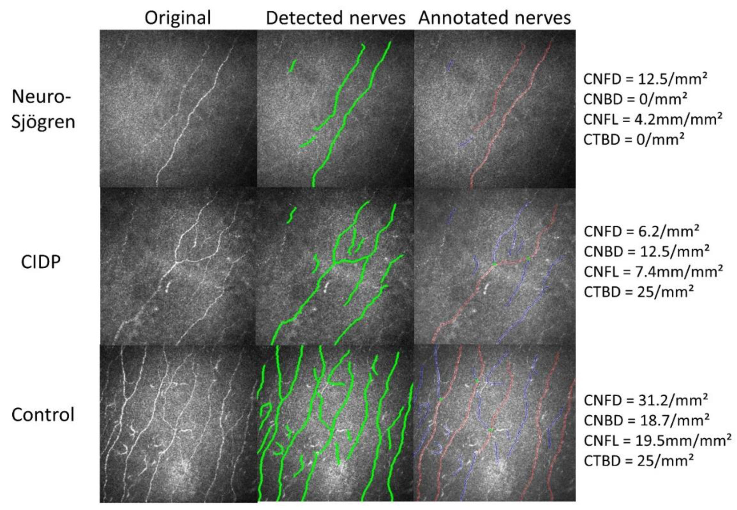
2.3. Corneal Confocal Microscopy

The corneal confocal microscopy is a highly resolving laser microscopic technique that allows an accurate, non-invasive assessment of the corneal nerve fibres in vivo [27] and was conducted by experienced investigators for this study. Microscopic corneal image acquisition was carried out using the Rostock Cornea Module (RCM) mounted on the HRT III tomograph (Heidelberg Engineering GmbH, Heidelberg, Germany, while using the recommended resolution of 400/384 = 1.0417 µm. Each eye was first anaesthetized using a drop of Proparacaine (Proxymetacaine Hydrochloride 0.5%, Ursapharm Arzneimittel GmbH, Saarbrücken, Germany) followed by the instillation of a gel tear substitute (Vidisic Gel, Carbomer 2 mg per 1 g, Bausch and Lomb, Brunsbuetteler Damm 165/173, Berlin, Germany) which served as a protective layer over the corneal epithelium layer. A sterile TomoCap (Heidelberg Engineering GmbH) was placed over the RCM objective lens before contact was made with the anaesthetized corneal surface.
Image analysis was conducted by the ACC Metrics Software© V1 2015, Manchester, UK.
The following parameters were automatically calculated:
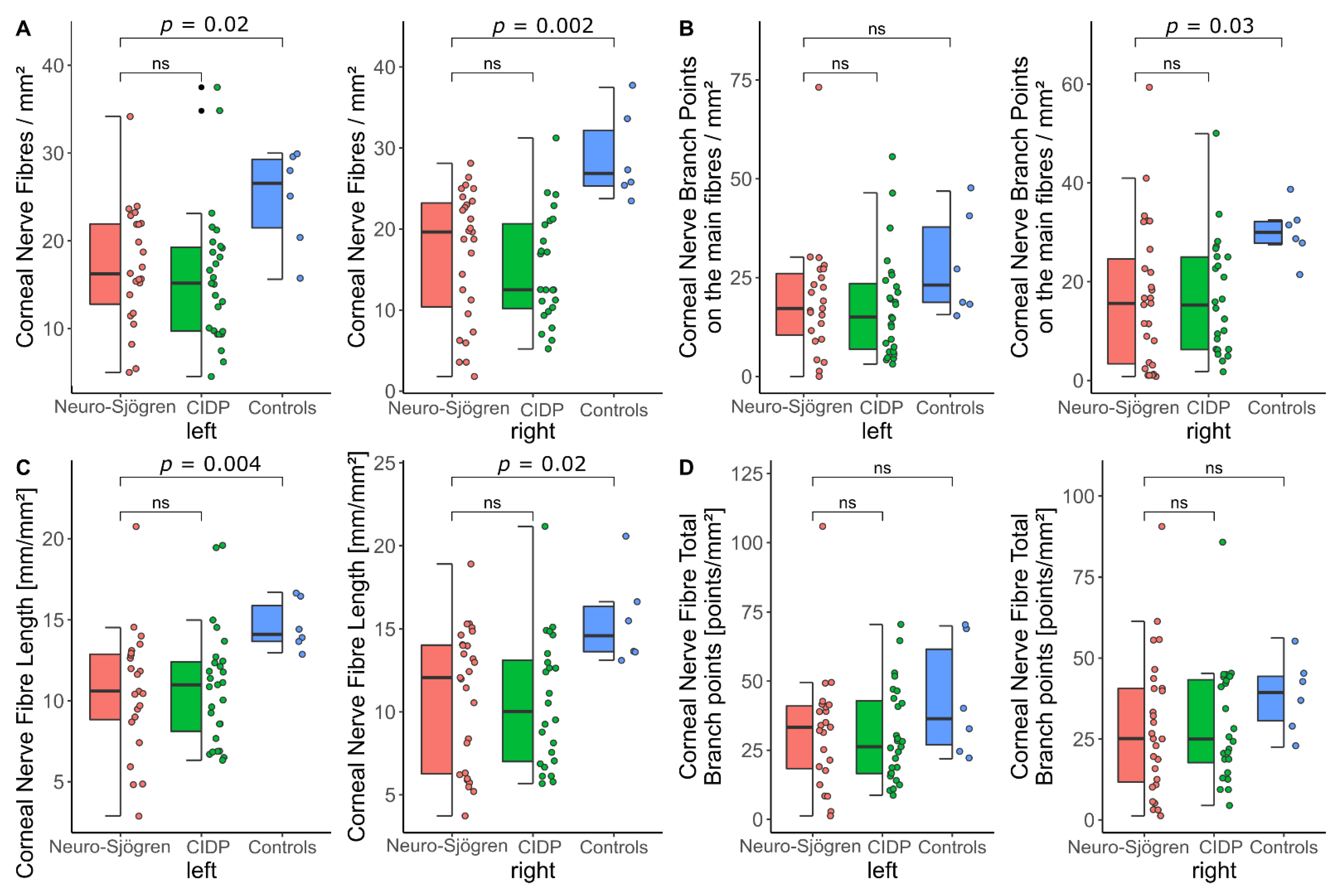
  • Corneal Nerve Fibre Density (CNFD): The number of detected fibres per mm2
  • Corneal Nerve Fibre Main Branch Density (CNBD): The number of detected branch points on the main fibres per mm2
  • Corneal Nerve Fibre Length (CNFL): The total length of detected nerve fibres in mm per mm2
  • Corneal Nerve Fibre Total Branch Density (CTBD): The total number of branch points per mm2

2.4. Clinical Evaluation of Dry Eye Symptoms and Signs

Dry eye symptoms and signs were evaluated by general ophthalmological examination quantified by the predefined “clinical score of the dry eye” and the established Ocular Surface Disease Index Questionnaire (OSDI):

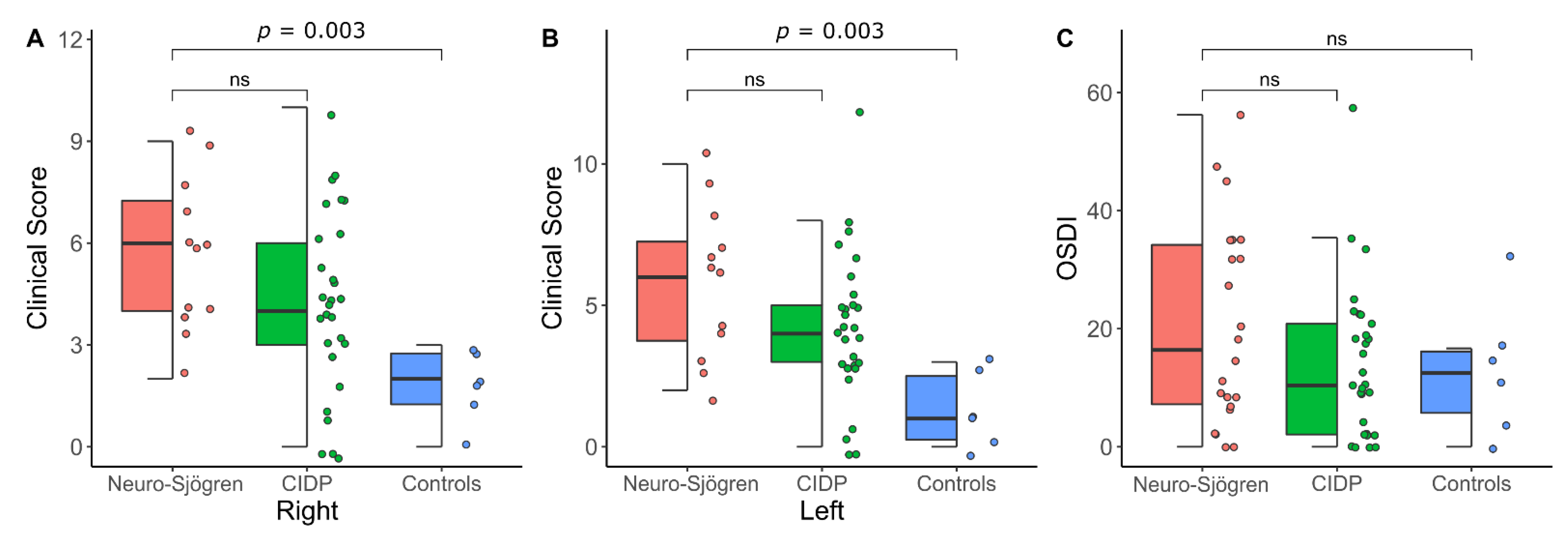
2.4.1. Clinical Score of the Dry Eye

Each eye underwent a comprehensive examination of the anterior segment of the eye with the Slit lamp, conducted by experienced ophthalmologists. Ocular surface integrity was assessed with the cobalt blue filter following fluorescein staining with a drop of Thilorbin (4 mg Oxybuprocaine Hydrochloride + 0.8 mg Fluorescein Sodium), OmniVision GmbH. The ophthalmological examination was quantified by the “Clinical score of the dry eye”, a modified version of existing recommendations for dry eye assessment [28,29,30]. This score comprises evaluation of conjunctival injection, tear film break-up time, blink frequency [31,32], the presence and extent of superficial punctate keratitis and the presence of meibomian gland dysfunction. The scoring system is shown in Table 1. Eventually, the sum score for each eye ranged from 0 (no evidence of dry eye disease) to 13 (severe manifestation in all aspects).

2.4.2. Ocular Surface Disease Index (OSDI)

The dry eye associated symptoms with impact on the quality of daily life were evaluated by the OSDI [36,37]. This index consists of 12 questions concerning ocular symptoms, vision-related function and environmental triggers during the previous week, while scores range from 0 (none of the time) to 4 (all of the time), respectively. The total OSDI sum score is subsequently formulated through the sum of scores × 25 divided by the number of answered questions. It therefore ranges from 0 (no complaints) to 100 (all symptoms all the time).

2.5. Statistical Analysis and Data Visualization

Decimal variables were tested for parametrical distribution via the Shapiro–Wilk test. Parametrical data were described as mean ± standard deviation, whereas non-parametrical data were described as median (interquartile range). After descriptive analysis as designated, group comparison was achieved via the Wilcoxon Rank sum Test for decimal data and via Chi² test for binary data. Correlation analysis was performed using Spearman’s rank correlation. p-values < 0.05 were considered significant. Statistical analysis was performed by STATA® V16.1 (StataCorp LLC, 4905 Lakeway Dr, College Station, TX, USA). Figures were created by RStudio Desktop V1.4.1717 (RStudio Inc, 250 Northern Ave, Boston, MA, USA).

3. Results

3.1. Patients’ Characteristics

A total of 26 patients with Sjögren’s syndrome (according to the current ACR/EULAR classification criteria) and evident neuropathy (Neuro-Sjögren), as well as 29 patients with previously diagnosed chronic inflammatory demyelinating polyneuropathy (CIDP) and without Sjögren’s syndrome were included in the analysis. Additionally, six control patients without Sjögren’s syndrome or CIDP underwent identical testing. Patients in the Neuro-Sjögren group were female in 58% and showed a median age at evaluation of 64 years, while patients in the CIDP group were female in 21% and showed a median age of 67 years. Controls were female in 83% and aged 61 years (median). The cohort characteristics concerning the ACR/EULAR classification criteria of Sjögren’s syndrome, the disease duration, as well as disease and symptom severity are shown in detail in Table 2.
The serological and clinical characteristics of patients with neuropathy associated with Sjögren’s syndrome are displayed in Table 3.

3.2. Confocal Microscopy of the Corneal Nerve Fibres

Confocal microscopy of the corneal nerve fibres revealed no significant group differences between the Neuro-Sjögren and the CIDP group for any of the microscopic parameters (values represent mean ± standard deviation of median (interquartile range) as appropriate):
Corneal Nerve Fibre Density (fibres/mm2): Neuro-Sjögren group 17.1 ± 6.8 (left) and 16.9 ± 8 (right); CIDP group 15.9 ± 7.7 (left, p = 0.27) and 15.2 ± 6.8 (right, p = 0.29).
Corneal Nerve Fibre Main Branch Density (main branch points/mm2): Neuro-Sjögren group 17.2 (9.4–27.1) (left) and 15.6 (3.1–26.6) (right); CIDP group 15 (6.3–24.3) (left, p = 0.6) and 15.3 (6.3–25) (right, p = 0.68).
Corneal Nerve Fibre Length (total length of nerves) (mm/mm2): Neuro-Sjögren group 10.6 ± 3.9 (left) and 12.1 (6.2–14.1) (right); CIDP group 10.9 ± 3.6 (left, p = 0.93) and 10 (7–13.3) (right, p = 0.73).
Corneal Nerve Fibre Total Branch Density [total branch points/mm2]: Neuro-Sjögren group 33.3 (17.7–41.4) (left) and 25.2 (10.9–40.6) (right); CIDP group 26.3 (16.5–43,8) (left, p = 0.88) and 25 (16.7–43.6) (right, p = 0.67).
Control participants showed significantly better results with the corneal nerve fibre density (24.8 ± 5.8/mm2, p = 0.02 (left) and 28.9 ± 5.4/mm2, p = 0.002 (right)) and the corneal nerve fibre length (total length of nerves) (14.6 ± 1.5 mm/mm2, p = 0.004 (left) and 14.6 (13.6–16.6) mm/mm2, p = 0.02 (right). For the corneal nerve fibre main branch density (when compared to the Neuro-Sjögren group), significantly better results were evident only in the right eyes (23.1 (18.7–41.2)/mm2, p = 0.15 (left) and 30 (27.5–32.5), p = 0.02 (right)). Analysis of the corneal nerve fibre total branch density did not reveal significant group differences between patients with Neuro-Sjögren and controls (36.4 (25–68.7), p = 0.3 (left) and 39.4 (28.7–45), p = 0.15 (right)).
The detailed workup for the confocal microscopy results is illustrated in Figure 1. Exemplary findings in Neuro-Sjögren patients and rarefied corneal nerve fibres versus control participants are shown in Figure 2.

3.3. Clinical Evaluation of Dry Eye Symptoms and Signs

Considering the clinical score of the dry eye and the Ocular Surface Disease Index, there were no significant in-between-group differences between patients with Neuro-Sjögren and CIDP. Nevertheless, controls reached significantly better results at the clinical score of the dry eye when compared with the Neuro-Sjögren group (values are stated as median (interquartile range)): Neuro-Sjögren group 6 (4–8) (left) and 6 (4–8) [right]; CIDP group 4 (3–5) (left, p = 0.12) and 4 (3–6) (right, p = 0.12); controls 1 (0–3) (left, p = 0.003) and 2 (1–3) (right, p = 0.003).
However, the OSDI score was similar for all three groups (values are stated as median (interquartile range)): Neuro-Sjögren group 16.4 (6.8–35); CIDP group 10.4 (2.1–20.8) (p = 0.26), controls 12.5 (4.2–16.7 (p = 0.38).
The results of the Clinical evaluation of dry eye symptoms and signs are graphically demonstrated in Figure 3.

3.4. Correlation Analysis between Parameters of the Corneal Confocal Microscopy and the Clinical Evaluation of Dry Eye Symptoms and Signs

There was no significant correlation between any of the parameters of the corneal confocal microscopy and the final sum score results of the Clinical score of the dry eye or the Ocular Surface Disease Index (Table 4). Nevertheless, there was a significant correlation between the clinical score of the dry eye and the Ocular Surface Disease Index (p = 0.005 (left) and p = 0.002 (right)).

4. Discussion

4.1. Corneal Trigeminal Nerve Affection Is Similar in Neuro-Sjögren and CIDP

Analysis of the corneal nerve parameters on corneal confocal microscopy did not show significant group differences between patients with Neuro-Sjögren and patients with CIDP. This finding aligns with previously published data suggesting a similar phenotype of these two entities, even though cranial nerve affection had been found to occur more frequently in patients with Neuro-Sjögren [9].
Although there are no concretely established normal values or cutoff recommendations for the parameters of the corneal confocal microscopy, efforts have been made towards establishing reference values against which corneal confocal microscopy findings can be measured [38]. In this study, the absolute values of the corneal nerve fibre density and the corneal nerve fibre length were significantly reduced in patients with Neuro-Sjögren when compared to healthy controls. The same effect has been previously described for patients with Behçet’s disease [39], non-Sjögren dry eye disease [40], Sjögren’s syndrome without focus on neurological involvement [41,42,43,44,45], diabetes [43], hereditary [46] and autoimmune neuropathies other than Sjögren’s syndrome associated neuropathy [19,47]. Interestingly, the published absolute values for corneal nerve fibre density and length vary depending on the disease. A literature review of comparable measurements is included in Table 5. Still, there is a slight variability previously published on the respective values for control patients and the values for the right and left eye also differing slightly within our control group. This effect might be explained by differences for age and size of the control cohorts, but possibly also by the technical nature of the testing procedure. For example, it is essential for patients to remain very still in order to obtain representative corneal images, which might be difficult in case of additionally limiting physical disabilities or conditions such as general or truncal muscle weakness or disrupted oculomotor functions. Nevertheless, the automated nature of image analysis through the provided ACC Metrics Software© offers a standardized data assessment with great validity.
It is noteworthy that only one male patient was included in the control group to align the ratio of women and men for the control group and for patients with Sjögren’s syndrome. However, no sex-specific influence on corneal confocal microscopy could be found in previous works [38,48]. Therefore, a resulting relevant confounding effect seems unlikely.

4.2. Dry eye Disease in Both Patient Groups

Clinical evaluation of dry eye symptoms and signs by ophthalmological examination showed significantly worse results for patients with Neuro-Sjögren and CIDP when compared to control participants. However, results of the OSDI questionnaire did not differ between the three groups due to a great variation of subjective suffering in all groups. We deduced that the clinical examination is more precise in quantifying dry eye symptoms and signs than the OSDI, which also includes general aspects (i.e., having eye problems that limit TV watching) possibly affected by confounding factors (i.e., refraction anomalies etc.).
Nevertheless, there was a significant correlation between scores of the clinical examination and the OSDI supporting a general connection between the objective and subjective evaluation of dry eye symptoms and signs as reported by previous studies [40].
Another aspect of these results is that patients with Neuro-Sjögren and patients with CIDP had similar ophthalmological findings concerning signs of dry eye disease. This supports the fact that dry eye disease is by no means a specific characteristic of Sjögren’s syndrome, even though lymphocytic infiltration of the lacrimal glands is a known hallmark for this entity. On the contrary, sicca symptoms have been previously described in chronical diseases [50,51]. It is also possible that neurological involvement of corneal nerve fibres in patients with Sjögren’s syndrome precedes more severe sicca symptoms as has been shown for other neurological manifestations of Sjögren’s syndrome [52,53].

4.3. Notional Connection between Dry Eye Disease and Corneal Trigeminal Nerve Affection

Both patient groups showed a similar severity of clinical ophthalmological sicca syndrome and corneal nerve fibre affection. Still, parameters of the corneal confocal microscopy did not show a significant correlation with either of the clinical scores. Therefore, a potential reciprocal impact of both conditions remains notional.

5. Conclusions

Our data revealed trigeminal nerve affection at the cornea in patients with neuropathy associated with Sjögren’s syndrome and patients with CIDP. No difference was found between both neuropathy groups, indicating that measurement of corneal nerve fibre/branch density and fibre length is not able to distinguish between both entities.

Author Contributions

Conceptualization: T.S. (Tabea Seeliger), T.S. (Thomas Skripuletz), A.B. and E.D.; methodology: S.G., D.E., N.K.P. and M.A.G.; formal analysis and investigation: T.S. (Tabea Seeliger), M.A.G., I.O.-P., A.B., M.W., E.D., M.H. and F.F.K.; writing—original draft preparation: T.S. (Tabea Seeliger), M.A.G. and I.O.-P., writing—review and editing: A.P., C.F., T.W. and D.E.; resources: A.P., N.K.P., C.F., T.W. and T.S. (Thomas Skripuletz); supervision: T.S. (Tabea Seeliger), T.S. (Thomas Skripuletz) and A.B. All authors have read and agreed to the published version of the manuscript.

Funding

Tabea Seeliger was funded by the Ellen Schmidt Scholarship of the Hannover Medical School. There was no other external funding for this study.

Institutional Review Board Statement

The study was conducted in accordance with the Declaration of Helsinki and approved by the Local Ethics Committee of the Hannover Medical School (No. 8297_BO_S_2019).

Informed Consent Statement

Informed consent was obtained from all subjects involved in the study.

Data Availability Statement

Data are available upon reasonable request.

Conflicts of Interest

The authors have no relevant financial or non-financial interests to disclose. The funders had no role in the design of the study; in the collection, analyses, or interpretation of data; in the writing of the manuscript; or in the decision to publish the results. Outside the submitted work, the authors received honoraria for lectures, travel grants or research grants: T.S. (Tabea Seeliger) received financial support for congress fees by Abbvie. A.P. participated in Advisory Boards of Bayer, Novartis and Roche and received honoraria for lectures and presentations by Novartis, Bayer and Roche as well as consulting fees from the Roche National Study Committee. S.G. received a grant from the German Research Foundation and honoraria for preparation of articles/presentations bei Thieme Medical Publishers, Merck and Alnylam. D.E. received presentation honoraria from Abbvie, Amgen, BMS, Chugai, Cilag-Janssen, Galapagos, GSK, Medac, Lilly, Pfizer, Novartis, Roche and participated in advisory boards for Abbvie, Galapagos, Amgen and Novartis. T.W. received honoraria for lectures by Abbvie, BMS, Chugai, Galapagos, Janssen, Lilly, Pfizer, UCB and Roche. T.S. (Thomas Skripuletz) received honoraria for lectures by Alexion, Alnylam, Bayer Vital, Biogen, Celgene, CSL Behring, Euroimmun, Janssen, Merck Serono, Novartis, Pfizer, Roche, Sanofi and Siemens. N.K.P., E.D., M.W., C.F., A.B., M.A.G., M.H., I.O.-P. and F.F.K. have nothing to disclose.

References

  1. Seeliger, T.; Bönig, L.; Witte, T.; Thiele, T.; Lesinski-Schiedat, A.; Stangel, M.; Lenarz, T.; Prenzler, N.C.; Skripuletz, T. Hearing dysfunction in patients with Neuro-Sjögren: A cross-sectional study. Ann. Transl. Med. 2020, 8, 1069. [Google Scholar] [CrossRef]
  2. Seeliger, T.; Jacobsen, L.; Hendel, M.; Bönig, L.; Prenzler, N.K.K.; Thiele, T.; Ernst, D.; Witte, T.; Stangel, M.; Kopp, B.; et al. Cognitive impairment in patients with Neuro-Sjögren. Ann. Clin. Transl. Neurol. 2020, 7, 1352–1359. [Google Scholar] [CrossRef] [PubMed]
  3. Butryn, M.; Neumann, J.; Rolfes, L.; Bartels, C.; Wattjes, M.P.; Mahmoudi, N.; Seeliger, T.; Konen, F.F.; Thiele, T.; Witte, T.; et al. Clinical, Radiological, and Laboratory Features of Spinal Cord Involvement in Primary Sjögren’s Syndrome. J. Clin. Med. 2020, 9, 1482. [Google Scholar] [CrossRef]
  4. Seeliger, T.; Prenzler, N.K.; Gingele, S.; Seeliger, B.; Körner, S.; Thiele, T.; Bönig, L.; Sühs, K.-W.; Witte, T.; Stangel, M.; et al. Neuro-Sjögren: Peripheral Neuropathy With Limb Weakness in Sjögren’s Syndrome. Front. Immunol. 2019, 10, 1600. [Google Scholar] [CrossRef] [PubMed] [Green Version]
  5. Shiboski, C.H.; Shiboski, S.C.; Seror, R.; Criswell, L.A.; Labetoulle, M.; Lietman, T.M.; Rasmussen, A.; Scofield, H.; Vitali, C.; Bowman, S.J.; et al. 2016 American College of Rheumatology/European League Against Rheumatism classification criteria for primary Sjögren’s syndrome. Ann. Rheum. Dis. 2017, 76, 9–16. [Google Scholar] [CrossRef] [PubMed]
  6. Grant, I.A.; Hunder, G.G.; Homburger, H.A.; Dyck, P.J. Peripheral neuropathy associated with sicca complex. Neurology 1997, 48, 855–892. [Google Scholar] [CrossRef]
  7. Pavlakis, P.; Alexopoulos, H.; Kosmidis, M.; Mamali, I.; Moutsopoulos, H.; Tzioufas, A.; Dalakas, M. Peripheral neuropathies in Sjögren’s syndrome: A critical update on clinical features and pathogenetic mechanisms. J. Autoimmun. 2012, 39, 27–33. [Google Scholar] [CrossRef]
  8. Seeliger, T.; Bönig, L.; Gingele, S.; Prenzler, N.K.; Thiele, T.; Ernst, D.; Witte, T.; Stangel, M.; Skripuletz, T.; Körner, S. Nerve ultrasound findings in Sjögren’s syndrome-associated neuropathy. J. Neuroimaging 2021, 31, 1156–1165. [Google Scholar] [CrossRef]
  9. Seeliger, T.; Gingele, S.; Bönig, L.; Konen, F.F.; Körner, S.; Prenzler, N.; Thiele, T.; Ernst, D.; Witte, T.; Stangel, M.; et al. CIDP associated with Sjögren’s syndrome. J. Neurol. 2021, 268, 2908–2912. [Google Scholar] [CrossRef]
  10. Yang, A.Y.; Chow, J.; Liu, J. Corneal innervation and sensation: The eye and beyond. Yale J. Biol. Med. 2018, 91, 13–21. [Google Scholar]
  11. Petropoulos, I.N.; Alam, U.; Fadavi, H.; Marshall, A.; Asghar, O.; Dabbah, M.A.; Chen, X.; Graham, J.; Ponirakis, G.; Boulton, A.J.M.; et al. Rapid Automated Diagnosis of Diabetic Peripheral Neuropathy With In Vivo Corneal Confocal Microscopy. Investig. Opthalmology Vis. Sci. 2014, 55, 2071–2078. [Google Scholar] [CrossRef] [PubMed]
  12. Lim, S.H.; Ferdousi, M.; Kalteniece, A.; Kass-Iliyya, L.; Petropoulos, I.N.; Malik, R.A.; Kobylecki, C.; Silverdale, M. Corneal confocal microscopy detects small fibre neurodegeneration in Parkinson’s disease using automated analysis. Sci. Rep. 2020, 10, 20147. [Google Scholar] [CrossRef] [PubMed]
  13. Shen, F.; Dong, X.; Zhou, X.; Yan, L.; Wan, Q. Corneal subbasal nerve plexus changes in patients with episodic migraine: An in vivo confocal microscopy study. J. Pain Res. 2019, ume 12, 1489–1495. [Google Scholar] [CrossRef] [Green Version]
  14. Flockerzi, E.; Daas, L.; Seitz, B. Structural changes in the corneal subbasal nerve plexus in keratoconus. Acta Ophthalmol. 2020, 98, e928–e932. [Google Scholar] [CrossRef] [Green Version]
  15. Grossman, S.A.; Krabak, M.J. Leptomeningeal carcinomatosis. Cancer Treat. Rev. 1999, 25, 103–119. [Google Scholar] [CrossRef]
  16. Che, N.N.; Yang, H.Q. Potential use of corneal confocal microscopy in the diagnosis of Parkinson’s dis-ease associated neuropathy. Transl. Neurodegener. 2020, 9, 28. [Google Scholar] [CrossRef]
  17. Bohn, S.; Stahnke, T.; Sperlich, K.; Linke, S.J.; Farrokhi, S.; Klemm, M.; Allgeier, S.; Köhler, B.; Reichert, K.-M.; Witt, M.; et al. In Vivo Histology of the Cornea—From the “Rostock Cornea Module” to the “Rostock Electronic Slit Lamp”—A Clinical “Proof of Concept” Study. Klin. Mon. Augenheilkd. 2020, 237, 1442–1454. [Google Scholar] [CrossRef] [PubMed]
  18. Lee, O.L.; Tepelus, T.C.; Huang, J.; Irvine, A.G.; Irvine, C.; Chiu, G.B.; Sadda, S.R. Evaluation of the corneal epithelium in non-Sjögren’s and Sjögren’s dry eyes: An in vivo confocal microscopy study using HRT III RCM. BMC Ophthalmol. 2018, 18, 309. [Google Scholar] [CrossRef] [PubMed]
  19. Stettner, M.; Hinrichs, L.; Guthoff, R.; Bairov, S.; Petropoulos, I.N.; Warnke, C.; Hartung, H.-P.; Malik, R.A.; Kieseier, B.C. Corneal confocal microscopy in chronic inflammatory demyelinating polyneuropathy. Ann. Clin. Transl. Neurol. 2015, 3, 88–100. [Google Scholar] [CrossRef]
  20. Schneider, C.; Bucher, F.; Cursiefen, C.; Fink, G.R.; Heindl, L.M.; Lehmann, H.C. Corneal confocal microscopy detects small fiber damage in chronic inflammatory demyelinating polyneuropathy (CIDP). J. Peripher. Nerv. Syst. 2014, 19, 322–327. [Google Scholar] [CrossRef] [PubMed]
  21. Pitarokoili, K.; Sturm, D.; Labedi, A.; Greiner, T.; Eitner, L.; Kumowski, N.; Enax-Krumova, E.K.; Fisse, A.L.; Maier, C.; Gold, R.; et al. Neuroimaging markers of clinical progression in chronic inflammatory demyelinating polyradiculoneuropathy. Ther. Adv. Neurol. Disord. 2019, 12. [Google Scholar] [CrossRef] [PubMed]
  22. Shiboski, C.H.; Shiboski, S.C.; Seror, R.; Criswell, L.A.; Labetoulle, M.; Lietman, T.M.; Rasmussen, A.; Scofield, H.; Vitali, C.; Bowman, S.J.; et al. 2016 American College of Rheumatology/European League Against Rheumatism Classification Criteria for Primary Sjögren’s Syndrome: A Consensus and Data-Driven Methodology Involving Three International Patient Cohorts. Arthritis Rheum. 2017, 76, 9–16. [Google Scholar] [CrossRef]
  23. Chisholm, D.M.; Mason, D.K. Labial salivary gland biopsy in Sjogren’s disease. J. Clin. Pathol. 1968, 21, 656–660. [Google Scholar] [CrossRef] [PubMed]
  24. Seror, R.; Theander, E.; Brun, J.G.; Ramos-Casals, M.; Valim, V.; Dörner, T.; Bootsma, H.; Tzioufas, A.; Solans-Laqué, R.; Mandl, T.; et al. Validation of EULAR primary Sjögren’s syndrome disease activity (ESSDAI) and patient indexes (ESSPRI). Ann. Rheum. Dis. 2015, 74, 859–866. [Google Scholar] [CrossRef] [PubMed]
  25. Van den Bergh, P.Y.K.; van Doorn, P.A.; Hadden, R.D.M.; Avau, B.; Vankrunkelsven, P.; Allen, J.A.; Attarian, S.; Blomkwist-Markens, P.H.; Cornblath, D.R.; Eftimov, F.; et al. European Academy of Neurology/Peripheral Nerve Society guideline on diagnosis and treatment of chronic inflammatory demyelinating polyradiculoneuropathy: Report of a joint Task Force—Second revision. Eur. J. Neurol. 2021, 28, 3556–3583. [Google Scholar] [CrossRef] [PubMed]
  26. Delmont, E.; Manso, C.; Querol, L.; Cortese, A.; Berardinelli, A.; Lozza, A.; Belghazi, M.; Malissart, P.; Labauge, P.; Taieb, G.; et al. Autoantibodies to nodal isoforms of neurofascin in chronic inflammatory demyelinating polyneuropathy. Brain 2017, 140, 1851–1858. [Google Scholar] [CrossRef] [Green Version]
  27. Oliveira-Soto, L.; Efron, N. Morphology of Corneal Nerves Using Confocal Microscopy. Cornea 2001, 20, 374–384. [Google Scholar] [CrossRef] [PubMed]
  28. Baudouin, C.; Aragona, P.; Van Setten, G.; Rolando, M.; Irkeç, M.; Del Castillo, J.B.; Geerling, G.; Labetoulle, M.; Bonini, S.; ODISSEY European Consensus Group members. Diagnosing the severity of dry eye: A clear and practical algorithm. Br. J. Ophthalmol. 2014, 98, 1168–1176. [Google Scholar] [CrossRef] [PubMed]
  29. Sowmya, S.; Jayachander, D.; Kamath, V.; Rao, M.S.; Tonse, M.R.; Baliga, M.S. Evaluation of the prevalence and severity of xerophthalmia in head and neck cancers patients undergoing curative radiotherapy. Asian J. Ophthalmol. 2016, 14, 154–165. [Google Scholar] [CrossRef]
  30. Lemp, M.A.; Baudouin, C.; Baum, J. The definition and classification of dry eye disease: Report of the definition and classification subcommittee of the international Dry Eye WorkShop. Ocul. Surf. 2007, 5, 75–92. [Google Scholar]
  31. Abusharha, A.A. Changes in blink rate and ocular symptoms during different reading tasks. Clin. Optom. 2017, ume 9, 133–138. [Google Scholar] [CrossRef] [Green Version]
  32. Doughty, M.J. Spontaneous eyeblink activity under different conditions of gaze (eye position) and visual glare. Graefe’s Arch. Clin. Exp. Ophthalmol. 2014, 252, 1147–1153. [Google Scholar] [CrossRef] [PubMed]
  33. DIAGNOSIS—Clinical TESTS—Ocular Surface Center Berlin. Available online: https://oscb-berlin.org/e-diagnosis-overview#Conjunctival-Redness-Index. (accessed on 26 September 2021).
  34. Abelson, M.B.; Smith, L.M.; Hollander, D.A.; Usner, D. How Do You Quantify The Qualitative? Rev. Ophthalmol. 2016, 13, 48–51. [Google Scholar]
  35. Xiao, J.; Adil, M.Y.; Chen, X.; Utheim, A.; Ræder, S.; Tønseth, K.A.; Lagali, N.S.; Dartt, D.A.; Utheim, T.P. Functional and Morphological Evaluation of Meibomian Glands in the Assessment of Meibomian Gland Dysfunction Subtype and Severity. Am. J. Ophthalmol. 2020, 209, 160–167. [Google Scholar] [CrossRef] [PubMed] [Green Version]
  36. Walt, J.G.; Rowe, M.; Stern, K. Evaluating the functional impact of dry eye: The Ocular Surface Disease Index. Drug Inf. J. 1997, 31, 1436. [Google Scholar]
  37. Schiffman, R.M.; Christianson, M.D.; Jacobsen, G.; Hirsch, J.D.; Reis, B.L. Reliability and Validity of the Ocular Surface Disease Index. Arch. Ophthalmol. 2000, 118, 615–621. [Google Scholar] [CrossRef]
  38. Tavakoli, M.; Ferdousi, M.; Petropoulos, I.N.; Morris, J.; Pritchard, N.; Zhivov, A.; Ziegler, D.; Pacaud, D.; Romanchuk, K.; Perkins, B.A.; et al. Normative Values for Corneal Nerve Morphology Assessed Using Corneal Confocal Microscopy: A Multinational Normative Data Set. Diabetes Care 2015, 38, 838–843. [Google Scholar] [CrossRef] [Green Version]
  39. Bitirgen, G.; Turkmen, K.; Malik, R.A.; Ozkagnici, A.; Zengin, N. Corneal confocal microscopy detects corneal nerve damage and increased dendritic cells in Fabry disease. Sci. Rep. 2018, 8, 12244. [Google Scholar] [CrossRef]
  40. Labbé, A.; Liang, Q.; Wang, Z.; Zhang, Y.; Xu, L.; Baudouin, C.; Sun, X. Corneal Nerve Structure and Function in Patients With Non-Sjögren Dry Eye: Clinical Correlations. Investig. Opthalmology Vis. Sci. 2013, 54, 5144–5150. [Google Scholar] [CrossRef] [Green Version]
  41. Villani, E.; Galimberti, D.; Viola, F.; Mapelli, C.; Ratiglia, R. The Cornea in Sjögren’s Syndrome: An In Vivo Confocal Study. Investig. Opthalmology Vis. Sci. 2007, 48, 2017–2022. [Google Scholar] [CrossRef] [PubMed]
  42. McNamara, N.A.; Ge, S.; Lee, S.M.; Enghauser, A.M.; Kuehl, L.; Chen, F.Y.-T.; Gallup, M.; McKown, R.L. Reduced Levels of Tear Lacritin Are Associated With Corneal Neuropathy in Patients With the Ocular Component of Sjögren’s Syndrome. Investig. Opthalmology Vis. Sci. 2016, 57, 5237–5243. [Google Scholar] [CrossRef]
  43. Perkins, B.A.; Lovblom, L.E.; Bril, V.; Scarr, D.; Ostrovski, I.; Orszag, A.; Edwards, K.; Pritchard, N.; Russell, A.; Dehghani, C.; et al. Corneal confocal microscopy for identification of diabetic sensorimotor polyneuropathy: A pooled multinational consortium study. Diabetologia 2018, 61, 1856–1861. [Google Scholar] [CrossRef] [Green Version]
  44. Li, F.; Zhang, Q.; Ying, X.; He, J.; Jin, Y.; Xu, H.; Cheng, Y.; Zhao, M. Corneal nerve structure in patients with primary Sjögren’s syndrome in China. BMC Ophthalmol. 2021, 21, 211. [Google Scholar] [CrossRef] [PubMed]
  45. Tepelus, T.C.; Chiu, G.B.; Huang, J.; Huang, P.; Sadda, S.R.; Irvine, J.; Lee, O.L. Correlation between corneal innervation and inflammation evaluated with confocal microscopy and symptomatology in patients with dry eye syndromes: A preliminary study. Graefe’s Arch. Clin. Exp. Ophthalmol. 2017, 255, 1771–1778. [Google Scholar] [CrossRef]
  46. Sturm, D.; Schmidt-Wilcke, T.; Greiner, T.; Maier, C.; Schargus, M.; Tegenthoff, M.; Vorgerd, M. Confocal Cornea Microscopy Detects Involvement of Corneal Nerve Fibers in a Patient with Light-Chain Amyloid Neuropathy Caused by Multiple Myeloma: A Case Report. Case Rep. Neurol. 2016, 8, 134–139. [Google Scholar] [CrossRef] [PubMed]
  47. Bucher, F.; Schneider, C.; Blau, T.; Cursiefen, C.; Fink, G.R.; Lehmann, H.C.; Heindl, L.M. Small-Fiber Neuropathy Is Associated With Corneal Nerve and Dendritic Cell Alterations. Cornea 2015, 34, 1114–1119. [Google Scholar] [CrossRef] [PubMed]
  48. Niederer, R.L.; Perumal, D.; Sherwin, T.; McGhee, C.N.J. Age-related differences in the normal human cornea: A laser scanning in vivo confocal microscopy study. Br. J. Ophthalmol. 2007, 91, 1165–1169. [Google Scholar] [CrossRef] [Green Version]
  49. Cardigos, J.; Barcelos, F.; Carvalho, H.; Hipólito, D.; Crisóstomo, S.; Vaz-Patto, J.; Alves, N. Tear Meniscus and Corneal Sub-basal Nerve Plexus Assessment in Primary Sjögren Syndrome and Sicca Syndrome Patients. Cornea 2019, 38, 221–228. [Google Scholar] [CrossRef] [PubMed]
  50. Yu, K.; Bunya, V.; Maguire, M.; Asbell, P.; Ying, G.-S. Systemic Conditions Associated with Severity of Dry Eye Signs and Symptoms in the Dry Eye Assessment and Management Study. Ophthalmology 2021, 128, 1384–1392. [Google Scholar] [CrossRef] [PubMed]
  51. Ismail, O.M.; Poole, Z.B.; Bierly, S.L.; Van Buren, E.D.; Lin, F.-C.; Meyer, J.J.; Davis, R.M. Association Between Dry Eye Disease and Migraine Headaches in a Large Population-Based Study. JAMA Ophthalmol. 2019, 137, 532–536. [Google Scholar] [CrossRef] [PubMed]
  52. Delalande, S.; de Seze, J.; Fauchais, A.-L.; Hachulla, E.; Stojkovic, T.; Ferriby, D.; Dubucquoi, S.; Pruvo, J.-P.; Vermersch, P.; Hatron, P.-Y. Neurologic manifestations in primary Sjögren syndrome: A study of 82 pa-tients. Medicine 2004, 83, 280–291. [Google Scholar] [CrossRef] [PubMed]
  53. Teixeira, F.; Moreira, I.; Silva, A.M.; Vasconcelos, C.; Farinha, F.; Santos, E. Neurological involvement in Primary Sjögren Syndrome. Acta Reum. Port. 2013, 38, 29–36. [Google Scholar] [CrossRef]
Figure 1. Confocal microscopy parameters with significance levels at in-between group differences for patients with Neuro-Sjögren (red), chronic inflammatory demyelinating polyneuropathy (CIDP, green) and controls (blue): (A) corneal nerve fibre density; (B) corneal nerve fibre main branch density; (C) corneal nerve fibre length (total length of nerves); (D) corneal nerve fibre total branch density; ns: not significant.
Jcm 11 04484 g001
Figure 2. Exemplary findings in one patient with Neuro-Sjögren, one with CIDP and one participant of the control group. Image analysis is displayed for the automatically detected and colored nerves (green) and the annotated nerve fibres (red: main fibres, blue: nerve branches, green dots–branch points at the main fibres). Abbreviations; CIDP: chronic inflammatory demyelinating polyneuropathy; CNFD: corneal nerve fibre density; CNBD: corneal nerve fibre main branch density; CNFL: corneal nerve fibre length (total length of nerves); CTBD: corneal nerve fibre total branch density.
Jcm 11 04484 g002
Figure 3. Results of the Clinical Evaluation of dry eye symptoms and signs with significance levels at in-between group differences for patients with Neuro-Sjögren (red), chronic inflammatory demyelinating polyneuropathy (CIDP, green) and controls (blue): clinical score of the dry eye for the right (A) and left side (B); ocular surface disease index (OSDI, C); ns: not significant.
Jcm 11 04484 g003
Table 1. Clinical score of the dry eye, applied for each eye separately *, grey color indicating non-applicable cells.
0123
Conjunctival Injection [33]nonemildmoderatesevere
Tear Film Breakup Time>10 s≤10 s≤5 simmediate
Blink Frequency ≤21/min>21/min
Schirmer’s test (mm/5 min)>10≤10≤5≤2
Superficial Punctate Keratitis (National Eye Institute Score) [34]Grade 0Grade 1Grade 2Grade 3
Meibomian Gland Dysfunction [35]nonemildmoderatesevere
* Modified version of existing recommendations for dry eye assessment [28,29,30].
Table 2. Baseline parameters for the analyzed subgroups: Neuro-Sjögren, chronic inflammatory demyelinating polyneuropathy (CIDP) and controls.
Neuro-SjögrenCIDPControls
n26296
Age at evaluation, median (IQR) (years)64 (56–72)67 (60–74)61 (60–64)
Female, n (%)15 (58%)6 (21%)5 (83%)
Disease duration, median (IQR) (years)7 (2–18)9 (4–10)n/a
ACR/ EULAR classification criteria
 Objective xerophthalmia, n (%)20 (77%)7 (24%)n/a
 Objective xerostomia, n (%)12 (46%)8 (28%)n/a
 SSA(Ro) antibody positivity, n (%)11 (42%)0 (0%)n/a
 Sialadenitis grade 3/4 by Chisholm and Mason, n (%)21/22 (95%)0/10 (0%)n/a
Focus Score, median (IQR)4 (4–5)0 (0–1)n/a
ESSDAI, median (IQR)17 (11–22)n/an/a
ESSPRI, median (IQR)3.2 (2.3–4.8)n/an/a
Abbreviations: CIDP: chronic inflammatory demyelinating polyneuropathy; n: number of patients; IQR: interquartile range; ACR/EULAR: American College of Rheumatology/European League Against Rheumatism; n/a: not applicable.
Table 3. Additional serological and clinical features of the subgroup of patients with Sjögren’s syndrome and associated neuropathy.
n (%)
Clinical feature of peripheral nerve affection
 Cranial nerve impairment9 (35%)
 Small Fibre Neuropathy2 (8%)
 Motor impairment21 (81%)
 Sensory deficits21 (81%)
 ● Pain 4 (19%)
 ● Paresthesia 12 (57%)
 ● Sensory ataxia 13 (62%)
 Autonomic dysfunction4 (16%)
Nerve damage pattern on pathological nerve conduction studies (n = 24)
 SSB(La)-antibody positivity3 (12%)
 Cryoglobulins0 (0%)
Additional serological parameters
 SSB(La)-antibody positivity3 (12%)
 Cryoglobulins0 (0%)
 Free Kappa Light Chains, median (IQR) (mg/L)19 (21.1–21.5)
 Free Lambda Light Chains, median (IQR) (mg/L)19.7 (11.4–22.3)
 Kappa/Lambda ratio, median (IQR)(0.81–1.21)
Table 4. Correlation analysis for the full cohort between parameters of the corneal confocal microscopy and the clinical evaluation of dry eye symptoms and signs with significance levels (green color-significant group differences).
Analysis without Subgroup DivisionLeftRight
Clinical score, mean ± standard deviation4 (3–6.5)4 (3–6.5)
OSDI12.5 (4.2–25)
Correlation analysisp-values (left)p-values (right)
Clinical Score vs. OSDI0.0050.002
Clinical Score vs.
 Corneal Nerve Fibre Density0.340.6
 Corneal Nerve Fibre Main Branch Density0.440.88
 Corneal Nerve Fibre Length (total length of nerves)0.130.41
 Corneal Nerve Fibre Total Branch Density0.660.74
OSDI vs.
 Corneal Nerve Fibre Density0.500.68
 Corneal Nerve Fibre Main Branch Density0.400.48
 Corneal Nerve Fibre Length (total length of nerves)0.140.42
 Corneal Nerve Fibre Total Branch Density0.080.30
Table 5. Literature review on published data concerning corneal confocal microscopy parameters in different cohorts.
ReferencePatientsControls
Patients’
Condition, n
Median AgeCorneal Nerve Fibre Density, Mean ± SD (fibres/mm2)Corneal Nerve Fibre Length, Mean ± SD (mm/mm2)nMedian AgeCorneal Nerve Fibre Density, Mean ± SD (fibres/mm2)Corneal Nerve Fibre Length, Mean ± SD (mm/mm2)
[39]Behçet’s disease, 4939.927.7 ± 8.616.3 ± 4.63041.235.6 ± 1018.5 ± 4.1
[40]Dry eye disease, 4346.234.9 ± 8.116.26 ± 3.51445.445.9 ± 4.221.86 ± 2.1
[42]Sjögren’s syndrome, 1058.221.7 ± 18.94.18 ± 3.41056.531.8 ± 9.36.54 ± 2.47
[49]Sjögren’s syndrome with dry eye disease, 5457.828.1 ± 12.210.3 ± 6.62050.943.9 ± 12.915.4 ± 5.1
[43]Diabetes, 9985220.6 ± 9.812.5 ± 4.6----
[19]CIDP, 88n/a19 ± 712 ± 385n/a29 ± 617 ± 3
MMN, 6n/a18 ± 1111 ± 5
MGUSN, 12n/a20 ± 512 ± 2
Publisher’s Note: MDPI stays neutral with regard to jurisdictional claims in published maps and institutional affiliations.

