Abstract
Background: The aim of this study was to assess the effectiveness of 38 supervised postoperative physiotherapy (SVPh) visits conducted between 1 and 20 weeks after SSATOM on the values of 3D gait parameters measured at 10 and 20 weeks after surgery. Material: Group I comprised male patients (n = 22) after SSATOM (SVPh x = 38 visits) and Group II comprised male patients (n = 22) from the control group. Methods: A non-randomized, open-label, controlled clinical trial was performed in the two groups to obtain the following values: Step length (cm), stride length (cm), step width (cm), next stance phase (%), swing phase (%), double support (%), gait velocity (m/s), and walking frequency (step/min). The measurements were carried out using the BTS SMART system (Italy). Results: Orthopedic examination showed no pain, a negative result of Thompson and Matles tests, and proper healing of Achilles tendon (ultrasound image). In Group I, between 10 and 20 weeks after SSATOM, there was a statistically significant improvement in all tested gait parameter values (p ≤ 0.001 to 0.009). Conclusions: Conducting 38 SVPh visits significantly improved the values of the analyzed kinematic and spatiotemporal gait parameters in patients in the twentieth week after SSATOM, which were mostly close to the non-operated side and the results of the control group. However, the gait speed and stride length were not close to the results of the control group.
1. Introduction
The contraction of human triceps muscles of the Achilles tendon (AT) during locomotion, generates plantar flexion and propulsion of the foot [1]. In recent years, the number of AT lesions has been increasing, reaching up to 70 cases per 100,000 people in the population [2]. The problem often concerns middle-aged and young men who are physically active [3]. In the case of AT rupture, the orthopedist conducts a thorough history and clinical test as well as pain assessment of the patient [4]. An ultrasound examination of AT is considered as a standard procedure in these cases, and if necessary, additional examinations are performed [5,6]. Achilles tendon injuries are treated surgically or conservatively [7,8]. A review of the literature indicates the predominance of surgical treatment over the conservative treatment [7,8,9,10]. The goal of postoperative physiotherapy is to gradually restore the function of the tendon, ankle joint, the entire limb, and then independent walking. Subsequent stages of supervised postoperative physiotherapy (SVPh) should restore the patients’ optimal physical activity [11]. At present, there are as yet, no unequivocal answers to the following question: What is the impact of postoperative physiotherapy on the behavior of kinematic and spatiotemporal gait parameters using an open method (SSATOM), which is conducted and supervised by a physiotherapist in patients after surgical suturing of the Achilles tendon [12,13,14].
In the literature, only a few publications were found, that meet the requirements of a standardized description of conducted and supervised physiotherapy after SSATOM [11,15,16,17,18,19,20]. Moreover, some interesting studies concerning the discussed research problem were conducted on a small number of patients after AT injury [13,21]. According to Tengman et al. (2013), 2 to 5 years after the surgical treatment of AT, patients had signs of tendon elongation, as well as low muscle strength and function, especially during walking [22]. Sun et al. (2020) conducted a study of gait analysis in patients 2 years following the surgical suturing of AT. The authors found that a restraint of plantar flexion of the foot along with a greater deficit in muscle strength is responsible for plantar flexion of the foot on the operated side, resulting in the increased risk in knee injury during gait [23]. Jadacka et al. (2017) [24] had similar conclusions as Sun et al. (2020) [23].
Heikkinen et al. (2017) showed that the non-surgical treatment of AT rupture resulted in significantly greater soleus muscle atrophy and AT elongation compared with the surgical treatment. Patients treated surgically had on average 10 to 80% greater calf muscle strength compared with patients treated conservatively [25]. Speedtsberg et al. (2019) observed that even 4.5 years after the non-operative treatment of the injured AT, patients had impaired peak dorsiflexion values, reduced total strength of the concentric and plantar flexor, as well as decreased quasi-stiffness in the initial and final range of dorsal flexion of the foot, especially during walking [26]. Aufwerber et al. (2021) showed that the more accelerated rehabilitation regimen with early functional mobilization did not lead to a more symmetrical gait pattern neither at 8 weeks nor at 6 months after SSATOM [12].
Lins et al. (2013) noted that 12 months after AT surgery a crucial improvement was observed in the values of walking speed, walking frequency, stride length, and percentage share of the support phase compared with the control group. Finally, the authors found few gait disturbances during walking compared with 6 months after surgery [21]. Nordenholm et al. (2022) showed that performing a surgery after the AT rupture and postoperative rehabilitation may be an effective way to improve the values of the biomechanical parameters of walking after the incident [27]. However, even 12 months after surgery, patients showed significantly lower values of gait speed, step width, shorter stride length, longer relative stance phase, and power disturbances during walking compared with the control group [27]. Chan et al. (2011) conducted an analysis of gait in two groups of patients after the surgical suturing of the AT, using the open and the mini-open methods. At the twelve month after surgery, no significant differences were found between the study groups in the time of support and swing, and the length of a single step [28]. Don et al. (2007) evaluated the clinical condition of muscle strength and gait analysis in patients who had undergone surgical suturing of a complete AT rupture at 3, 6, 12, and 24 months. At 24 months, a deficit in calf-muscle eccentric strength was present, determining adaptive changes in gait strategy [14]. Alvit et al. (2017) showed that the double support on the non-operated limbs and the support time on the operated limbs were significantly lower in the sixth month after the surgery compared with the control group [29].
The authors of this study assumed that in order to improve the kinematic and spatiotemporal parameters of gait in patients after SSATOM, early and careful postoperative physiotherapy, should be initiated based on a specific protocol of and SVPh conducted by a physiotherapist in consultation with the treating physician [11]. Subsequently, the duration, number, and frequency of SVPh visits with the patient should be taken into account. Subsequently, the duration, number and frequency of SVPh visits with the patient should be taken into account, as well as the patient’s exercises performed at home [11,16,30,31].
The aim of the study was to evaluate the effectiveness of 38 supervised postoperative physiotherapy visits, that were performed between the first and twentieth week after surgical suturing of the Achilles tendon, on the 3D gait parameters measured at 10 and 20 weeks after surgery.
2. Materials and Methods
The research was conducted at the Center of Rehabilitation and Medical Education and the College of Physiotherapy in Wrocław, in accordance with the guidelines of the Declaration of Helsinki. The participants and the Ethics Committee at the College of Physiotherapy in Wrocław No. 1/2019, the Ethics Committee at the College of Physiotherapy in Wrocław No. 1/2012, and the Senate Committee of The Ethics of Research at the Academy of Physical Education in Wrocław 2006 (18 May 2006) provided written consent for the research. The study was designed as an open-label, non-randomized, controlled clinical trial and was conducted between 2010 and 2019. Short title: “Evaluation of the Effectiveness of Physiotherapy on 3D gait after Achilles Tendon Surgery”.
2.1. Participants
Initially, 55 patients (n = 48 men and n = 7 women) were included in the study, after surgical suturing of the Achilles tendon using an open method (SSATOM). The following inclusion criteria were applied: Unilateral surgical suturing of Achilles tendon (AT) using the open method, conducting postoperative supervised physiotherapy in patients by one physiotherapist, in accordance with one physiotherapy protocol, absence of concomitant lesions of the lower limbs, spine and concomitant diseases, and individuals between 20 and 60 years of age. Physiotherapy was carried out in the same rehabilitation center, using the same devices and experimental apparatus. On the basis of history and medical records, some patients were excluded from the initial group due to the following reasons: Inflammation (n = 2), coexisting injuries to the lower limbs (n = 4), angiologic diseases (n = 1), previous ankle sprains (n = 3), ankle cartilage reconstruction (n = 1), and ankle ligament reconstruction (n = 1). Subsequently, patients with pain in the lumbar spine (n = 1), patients who were more than 60 years old (n = 3) and less than 20 years old (n = 0), as well as those who did not undergo 20 weeks of the postoperative protocol of supervised physiotherapy, carried out by one physiotherapist, in the same rehabilitation center (n = 10 men and n = 7 women) (Figure 1) were excluded from the study [11]. Finally, 22 men with no AT rupture or other concomitant lower limb injuries were qualified for Group II (Figure 1).
Figure 1.
Flow diagram of the study; n–number of individuals.
Furthermore, in Group I, the men (n = 22) after SSATOM, who had an operation on 10 right lower limbs and 12 left lower limbs, were qualified for further analysis. Patients in Group I had 21 right dominant lower limbs and one left dominant lower limb. The men in Group II of the control group, with no AT injuries (n = 22), had 20 right dominant lower limbs and 2 left dominant limbs. In Group I, patients after a unilateral complete rupture of the AT had an open method of surgical AT suturing using Keesler’s approach and immediately after surgery, they had anticoagulant treatment, and analgesics [32]. The characteristics of the dominant lower limbs, including those operated on in Group I, and the dominant limbs in Group II are presented in Table 1. Both groups were not significantly different in age, body height and weight.
Table 1.
The between-group comparison of the values corresponding to age, body mass, body height, the duration of postoperative physiotherapy, the number of postoperative physiotherapy sessions presented between 1–10 weeks and 1–20 weeks after surgical suturing of Achilles tendon, time to start of supervised physiotherapy after surgical suturing of Achilles tendon, average weekly frequency visits of postoperative physiotherapy sessions presented between 1–10 weeks and 1–20 weeks after surgical suturing of Achilles tendon, dominant limb, and operated limb between Group I and Group II (control).
2.2. Supervised Postoperative Physiotherapy (SVPh)
General assumptions of the physiotherapeutic procedure: When planning, SVPh visits had to be performed between the first to twentieth week after surgical suturing of Achilles tendon using an open method (SSATOM) in the outpatient Rehabilitation Center, instituted three visits per week, conducted by an experienced physiotherapist, during the subsequent stages of this therapy, for patients who, after the AT surgery, planned to return to their daily activities and successively to recreational physical activity. Each SVPh visit lasted for 2 h at the Rehabilitation Center. For professional athletes, five SVPh visits per week were accepted. Supervised postoperative physiotherapy was carried out based on the protocol described above [11]. This section presented a shorter scheme of the physiotherapeutic protocol, as reported in Table 2 [11]. In addition, each patient was instructed by a physiotherapist on how to perform the therapeutic exercises at home, depending on the stage of postoperative physiotherapy. In Group I, the patients completed an average of 37.82 SVPh visits (it was hypothesized that they completed 38 SVPh visits). During the first 10 weeks after SSATOM, the patients completed an average of 14 SVPh visits (individually from 1 to 32 visits) (Table 1). The average time to start SVPh after SSATOM was 5.14 ± 2.14 weeks (the earliest individual visit was from the first week after SSATOM). The mean frequency of SVPh visits per week was 1.36 between 1–10 weeks after SSATOM, and 1.89 between 1–20 weeks after SSATOM (Table 1). The physiotherapist did not have an influence on the regularity of patients’ participation in this therapy, who ultimately made their own decisions about the number and regularity of the visits. Additional factors that could influence the process of SVPh in the first 10 weeks are as follows: Excessive pain and swelling, problems with postoperative scar, the patients’ motivation to treatment, varied level of physical fitness before Achilles tendon injury, compliance with the specific recommendations made by a physician, conducting the exercises recommended by the physiotherapist at home or non-adherence to the recommended amount and intensity of exercises to be performed individually. The exclusion criteria we adopted excluded patients who had excessive pain and inflammation (Figure 1). However, 10% of patients pursuing SVPh had periodic swelling and pain, which, however, resolved in the course of the treatment. The factors described above resulted in a periodic reduction in the frequency and total number of visits.
Table 2.
Characteristics of the five-stage supervised postoperative physiotherapy (SVPh) program for patients after surgical suturing of Achilles tendon (SSATOM).
Figure 8.
(a,b) Exercise with progressive load on the platform substrate with a lower limb operated for vGRF values, up to 1.4 bodyweight values.
Figure 7.
The individual result obtained by the patient for the value of the ground reaction forces (N) for the vertical component, while standing on one leg, and alternately, on the operated and non-operated lower limb. After conversion to kilograms, this corresponds approximately to the patient’s bodyweight during the exercise sessions on a strain gauge platform, within 8 weeks of physical therapy after surgical suturing of Achilles tendon, (red line—operated leg, blue line—non-injured leg).
Figure 6.
Record of the report from one session of exercises individually, to which the patient could partially load the platform substrate with a limb operated for vGRF values, 6 weeks after SSATOM, (red line—operated leg, blue line—non-injured leg).
Figure 5.
(a,b) Record of the report from one session of exercises individually selected with a value of 180 N, to which the patient could partially load the platform substrate with a limb operated for vGRF values, 3 weeks after SSATOM. This value was determined in the baseline measurement (Figure 3b) (red line—operated leg, blue line—non-injured leg).
Figure 4.
(a) The individual measurement of the ground reaction force values for the vertical component (vGRF) measured in Newtons (N) in a lax free-state on both legs; (b) the measurement of both legs (red line—operated leg, blue line—non-injured leg).
Figure 3.
(a) Single-leg standing test on non-injured leg; (b) the individual measurement of the ground reaction force values for the vertical component (vGRF) measured in Newtons (N), blue line—non-injured leg.
Figure 2.
Exercise of continuing passive movements of plantar flexion of the ankle joint on the operated side, with limitation of dorsal flexion of the foot after SSATOM.
Table 2.
Characteristics of the five-stage supervised postoperative physiotherapy (SVPh) program for patients after surgical suturing of Achilles tendon (SSATOM).
| Stage of SVPh | Weeks after SSATOM | Short Protocol of SVPh [11] |
|---|---|---|
| I | Up to 2 weeks after the surgery | Exercises for shank and foot muscles:
|
Exercises for other parts of the body:
| ||
Physical therapy treatment:
| ||
| II | 3 to 4 weeks after the surgery | The physiotherapeutic procedure from the previous stage was continued and additionally: Exercises for shank and foot muscles:
Additionally, the physiotherapist instructed the patient on how to perform the exercises at home and cool the Achilles tendon area every 6 h. |
Exercises for other parts of the body:
| ||
Physical therapy treatment:
| ||
Manual therapy:
| ||
| 5 to 6 weeks after the surgery | The physiotherapeutic procedure from the previous stage was continued and additionally: Exercises for shank and foot muscles:
| |
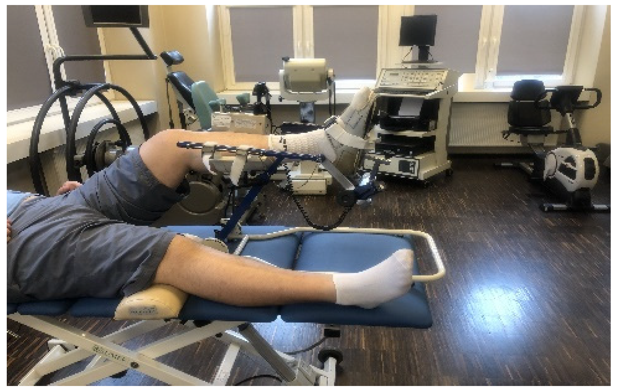
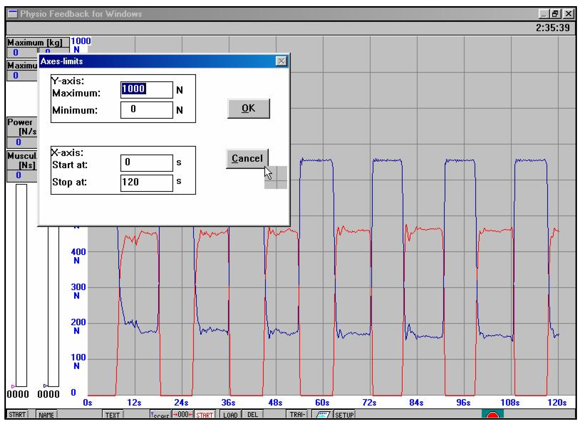
| III | 7 to 12 weeks after the surgery | The physiotherapeutic procedure from the previous stage was continued and additionally: Exercises for shank and foot muscles: Progressive pressure of the foot on the ground, with a load on the operated lower limb during exercises on platforms up to 80% of the bodyweight value. Successively, the load was gradually increased up to 100% of bodyweight values (without an orthopedic shoe) between 8 and 9 weeks after surgery. For example, if the patient’s bodyweight value is 67 kg, then when standing on one leg on the platform the value of the reaction force of the vertical component of the platform of about 670 N (Figure 7) is triggered (the word concerning results from the unstable equilibrium state in humans). It should be hypothesized that a value of 670 N corresponds to a bodyweight of 67 kg (1.0 bodyweight). Successively, the load was gradually increased every 3–4 days with a value of 0.1 bodyweight without a shoe,
|
Exercises for other parts of the body:
| ||
| Physical therapy treatment: Electrodiagnostic and electrostimulation of the triceps surae muscles of the injured leg. | ||
Manual therapy of injured tissues:
| ||
| Additional comments: The physiotherapist instructed the patient on how to perform the exercises at home. | ||
| 13 to 16 weeks after the surgery | The physiotherapeutic procedure from the previous stage was continued and additionally: Exercises for shank and foot muscles:
| |
Exercises for other parts of the body:
| ||
| IV | 17 to 24 weeks after SSATOM | The physiotherapeutic procedure from the previous stage was continued and additionally: Exercises for shank and foot muscles:
|
Exercises for other parts of body:
| ||
| Additional comments: The physiotherapeutic procedure from the previous stage was continued. | ||
| V | 24 to 28 weeks after SSATOM | Exercises for shank and foot muscles: The physiotherapist performed and supervised the patient’s exercises including running with maximum speed, changing movement directions, and specific exercises aimed at improving power, speed, and agility, adapted for a particular sport and the individual patient’s capabilities. Additional comments:
|
2.3. Orthopedic Examination
Concerning the subjects in Groups I and II, prior to the registration of the values of kinematic and spatiotemporal gait parameters, a specialist orthopedic physician, conducted a medical examination, which included the patient’s history, pain assessment using a visual analogue scale (VAS), and AT efficiency values with the Matles and Thompson tests. Subsequently, the doctor assessed the ankle joint structure, its stability, and continuity of AT through palpation and ultrasound [33]. Whereas, the subjects in Group I, prior to proceeding with the following stages of postoperative physiotherapy and before the last registration and measurement of the kinematic and spatiotemporal gait parameters in the twentieth week after SSATOM, had another orthopedic examination carried out in accordance with the schemes mentioned above. All of the subjects in Groups I and II obtained regular orthopedic results and were admitted to the next stage of the study.
2.4. Three-Dimensional Gait Analysis Registration of Kinematic and Spatiotemporal Parameter Values
In Group I, the registration and subsequent evaluation of the kinematic and spatiotemporal gait parameters on hard ground were carried out at 10 and 20 weeks after surgical suturing of AT, using the optoelectronic three-dimensional recording system (BTS Bioengineering, Milan, Italy, 2006) [34]. Six infrared camcorders (with a recording frequency of 120 Hz) and two piezoelectric Kistler platforms (with a recording frequency of 960 Hz) were used. Calibration was performed prior to each test, in accordance with the manufacturer’s instructions [34]. Anthropometric measurements were carried out, using a diameter compass for the measurement of both lower limbs, pelvis, such as pelvic width (cm) measured from the anterior superior iliac spine, pelvic depth (cm) measured from the anterior superior iliac spine to the greater trochanter of the femur on both sides of the body, width of the knee joints (cm) measured between the condyles of the femur, width of the ankle joints (cm) measured from the lateral to the medial malleolus, and the relative length of the lower limbs (cm). Additionally, the height and bodyweight of the subject were measured. The data obtained were entered into the computer system (BTS Bioengineering, Milan, Italy, 2006), in accordance with the manufacturer’s instructions [34]. The subject was wearing a swimsuit, and after disinfection of the skin, 22 markers were glued on both lower limbs, starting from the fifth head of the metatarsal bone, on the lateral malleolus, the posterior loftiness of the calcaneus, the middle part of the fibula, the head of the fibula, the lateral condyle of the femur, half the length of the femur, in the place of the greater trochanter, the anterior superior iliac spines, and at the level of S1 vertebra. Moreover, three markers were glued in the upper body at the height of the C7 vertebra and on the right and left shoulder appendage [35]. The measurement started by standing on both legs on a strain gauge platform for 10 s with the upper limbs positioned along the trunk. In the further part of the measurement, the participants were asked to walk 7 m along a path starting with each step from the left-lower limb, after hearing the Go command. Six measurements of walking along the path in the gait laboratory were performed, and each of them was carried out under identical conditions. After the test, the report was prepared using BTS capture, tracker, and analyzer programs [33,36]. For further analysis, two measurements were selected using the programs mentioned above, which contain 100% of the gait cycle of the first and last heels in contact with each lower limb [37]. Additionally, during the gait analysis, the patient had to perform a contact of each foot with one of the two Kistler platforms. The following values were measured: Step length (cm); stride length (cm); step width (cm), stance phase (%), swing phase (%), double support (%), gait velocity (m/s), and walking frequency (step/min) using the six-phase gait cycle [37]. In the control subjects of Group II, measurements of the kinematic and spatiotemporal gait parameters were performed using the same method in Group I, but were carried out only once.
2.5. Test of the Covered Distance When Walking on Unstable Treadmill Ground
To assess the ability to cover the walking distance on unstable ground, the subjects in Groups I and II had to walk 4 km on a treadmill at a speed of 6 km/h (1.66 m/s). The speed of 6 km/h corresponded to the fast pace of walking by the men in both groups, taking into account their body height, weight, and age, as presented in Table 1. The patients in Group I performed this test at week 20 after surgical suturing of Achilles tendon. The condition for passing this test was to cover a distance of 4 km without pain and swelling on the day after the end of the test, as well as on the following day. Covering the distance of 4 km of marching without rest on unstable ground allowed the patients to take 7000 to 8000 steps (single step from 50 to 60 cm), which are recommended by the American Heart Association to ensure adequate health-promoting activities for adults [38].
2.6. Statistical Analysis
For statistical analysis, Microsoft Office Excel 2007 (Microsoft, Redmond, WA, USA) and IBM SPSS 23 (IBM, Armonk, NY, USA) were used. We started our analysis with the calculation of mean values (M) and standard deviations (SD). The Shapiro–Wilk test was used to verify the normality of the distribution of the tested variables. Independent tests were used for the comparison of age, body height, and weight as well as the between-group values. For intra-group comparisons, tests for dependent groups were used. Repeated-measures ANOVA with Tukey’s HSD as post-hoc test determined the intra-group and inter-group values of spatiotemporal and kinematic gait parameters in Group I in 10 and 20 weeks after surgical suturing of Achilles tendon using an open method. One-way ANOVA was used for the comparison of the values obtained for the right operated limb in Group I and the left limb in Group II including step length (cm), stride length (cm), step width (cm), stance phase (%), swing phase (%), double support (%), and gait velocity (m/s). When the significance level was at p < 0.05, Tukey’s post-hoc test was performed for a group with the same number of people. Pearson’s linear correlation coefficient (r) was calculated for the strength and direction of the linear relationship between the number of visits to the supervised physiotherapy and the results of kinematic and spatiotemporal gait parameters. The values corresponding to all of the two-dimensional associations were classified as negligible (0.00–0.30), low (0.31–0.50), moderate (0.51–0.70), high (0.71–0.90), and very high (0.901–1.00) [39].
3. Results
3.1. Evaluation of Kinematic and Spatiotemporal Parameter Values Using a Three-Dimensional Gait Analysis (3D) System
In Group I, at the tenth week of postoperative supervised physiotherapy after surgical suturing of Achilles tendon using an open method (SSATOM), statistically significant disturbances in values of step length, stance phase, and swing phase were noted between the involved and uninvolved lower limbs (p < 0.001). In addition, 10 weeks after SSATOM in Group I, the values of step length, stride length, gait velocity, and walking frequency values, were statistically significantly lower in involved and uninvolved legs compared with the study conducted at the twentieth week after surgery, respectively for the involved and uninvolved legs (p < 0.001). Then, between the tenth and twentieth weeks after SSATOM in Group I, there was a statistically significant improvement in weeks for all of the analyzed biomechanical gait parameters (from p ≤ 0.009 to p ≤ 0.001), as shown in Table 3. Comparison of the particular week of each of the examined lower limbs (weeks x limb) obtained statistical significant improved parameters in 20 weeks after SSATOM of step length, stance phase, swing phase, and double support (Table 3).
Table 3.
Comparison of the intra-group and inter-group values of spatiotemporal and kinematic gait parameters in Group I in 10 and 20 weeks after SSATOM.
On the basis of analysis of variance, statistically significantly lower values of stride length, swing phase, and gait velocity of the operated limbs were shown in Group I at 20 weeks after SSATOM compared with the right and left limbs of Group II (from p ≤ 0.001 to p = 0.014, Table 4). Tukey’s test revealed statistically significantly lower values of stride length, swing phase, and gait velocity of the operated limb in Group I compared with the right and left limbs in Group II (from p ≤ 0.001 to p = 0.039, Table 4). In all of the presented values of gait parameters, no statistically significant differences were found between the right and left limbs in Group II (Table 4).
Table 4.
Inter-group comparison of spatiotemporal and kinematic gait parameters between the involved leg of Group I compared with the right and left leg of Group II, measured at 20 weeks after SSATOM.
3.2. Test of the Covered Distance When Walking on Unstable Treadmill Ground
In Group I, all of the patients after SSATOM walked a distance of 4 km on a treadmill at a speed of 1.66 m/s (6 km/h), without pain (VAS = 0) and the occurrence of AT swelling after the test and during the next day. In Group II, the patients walked a distance of 4 km on a treadmill at a speed of 1.66 m/s (6 km/h), without pain and the appearance of AT swelling after the test and during the next day.
3.3. Last Orthopedic Examination Results
The latest results of the orthopedic examination preceded the last recording and measurement of the kinematic and spatiotemporal gait parameters, as well as the gait test on a treadmill over a distance of 4 km in Group I in the twentieth week after SSATOM. In Group II (control), the study was conducted once. The results showed no pain on the VAS scale, negative Thompson and Matles test results in both study groups.
3.4. Association of Number of Supervised Postoperative Physiotherapy Visits with Kinematic and Spatiotemporal Parameter Values
A significantly positive and moderate correlation was noted between the higher number of visits during the first to the tenth week of supervised postoperative physiotherapy and higher gait velocity values for operated and non-operated limbs, respectively (r = 0.581; p = 0.005 and r = 0.542; p = 0.009) and stride length (r = 0.544; p = 0.009 and r = 0.544; p = 0.009), as presented in Table 5 A significantly negative and low correlation was noted between the higher number of visits up to 10 weeks after SSATOM of postoperative physiotherapy and lower step width values for operated and non-operated limbs (r = −0.475; p = 0.025, r = −0.475; p = −0.475; p = 0.025) and for the stance phase of the non-operated limb (r = −0.430; p = 0.046). A significantly positive and moderate correlation was found between the greater number of supervised postoperative physiotherapy (SVPh) visits in the period from the first to the tenth week for the swing phase of the non-operated limb (r = 0.503; p = 0.017), as shown in Table 5.
Table 5.
Correlation between the number of postoperative physiotherapy visits conducted between 1–10 weeks, after surgical suturing of Achilles tendon to the obtained spatiotemporal and kinematic gait values in Group I.
4. Discussion
For the purpose of this study, the authors have shown that conducting 38 supervised postoperative physiotherapy visits between 1 and 20 weeks after surgical suturing of the Achilles tendon resulted in significant improvements in the analyzed kinematic and spatiotemporal gait parameters obtained at week 20 compared with the results obtained 10 weeks after surgery. In addition, at 20 weeks after surgery, these gait parameters were mostly similar to the parameters obtained on the non-operated side and to the results of the control group without Achilles tendon injury. However, the gait velocity, stride length and, to a lesser extent, the swing phase of the operated limbs were lower than the results obtained in the control group.
In this study, the authors aimed to capture the interest of other researchers to the problem of retainment of 3D gait parameters, while conducting the complex treatment that included surgery after a complete AT rupture, immobilization, postoperative pharmacotherapy, and successively postoperative physiotherapy that is conducted and supervised by a physiotherapist. In particular, the authors paid attention to the latter element of treatment. Therefore, a detailed protocol and the total number of supervised postoperative physiotherapy (SVPh) visits, weekly frequency, duration of physiotherapy, precise procedures of applied treatment, and exercises for each 2-h physiotherapy visit were described above. Moreover, for each stage of physiotherapy, the authors specified recommendations for the home-based exercises that should be performed by the patients.
The literature analysis indicates that some of the authors presented a biomechanical assessment of gait in patients treated conservatively [22,26,40,41,42] and surgically [21,27,43] after AT rupture. Moreover, studies indicate that the authors used various methods and surgical techniques [44,45,46]. Different physiotherapy protocols and the level of accuracy of the description of these protocols, as well as the duration of physiotherapy, are known [11,16,28]. Some publications do not contain a description of physiotherapeutic procedures or are limited to providing basic schemas of early postoperative management [15,22,43].
In reference to the results obtained by other authors, who assessed the behavior of the biomechanical gait parameters over a period of 6 to 12 months, as well as in a longer period of time after AT surgery [12,13,14,21,27,28,29], our study showed a significant improvement in the twentieth week after surgical suturing of Achilles tendon using an open method (SSATOM) in most of the analyzed values of 3D gait parameters. However, the authors of the current study did not achieve the restoration of all gait parameters in patients after SSATOM, which would be similar to the non-operated side and the results of the control group. In addition, other studies showed a correlation between the higher number of SVPh visits in the first 10 weeks after SSATOM, which may indicate a favorable behavior of the analyzed biomechanical gait parameters between the operated and non-operated lower limb. However, the evaluation of the results obtained on the basis of correlation is not a sufficient tool and should be extended in the future to include an analysis, e.g., linear regression.
Several factors may affect the obtained results of the study of the complex treatment of patients after SSATOM, including the result of the performed surgery, the type and time of immobilization after the operation, pharmacological support, and individual patients’ predisposition for tissues’ healing of AT after surgery. Other conditions include adaptation to postoperative recommendations, age, gender, patients’ motivation for the conducted treatment or possible concomitant diseases. The last stage of complex treatment after acute rupture of AT is postoperative physiotherapy. In this study, the authors hypothesized that the average frequency of postoperative SVPh would be three visits per week with a physiotherapist. However, these proposed assumptions were not fully implemented by the patients. In addition, the authors’ recommendations did not have any influence on the systematicity of the patients’ planned participation process in postoperative physiotherapy. Moreover, we did not reach a valid answer to the following question: Is it possible to better improve the values of analyzed 3D gait parameters in Group I patients by conducting an average of three visits per week, in accordance with the planned SVPh protocol, and thus with a higher total number of SVPh visits in the 20 weeks after SSATOM? On the other hand, other studies have shown that only time after treatment has a full influence on the return of symmetrical and normative 3D gait parameters of patients after Achilles tendon rupture [12,13,14,21,27,28,29].
The presented research entails some limitations. Initially, our research showed that individually, patients differed in the timing of the early initiation of individual physiotherapy that is conducted and supervised by a physiotherapist. Moreover, the analysis showed that our patients had different regularities and frequencies of conducted and supervised postoperative physiotherapy after SSATOM. Despite our recommendations, we had no influence on the number and frequency of these visits due to the individually varying regularity of patients’ participation in postoperative physiotherapy. In the future, this problem must be resolved. The solution to the presented problem will probably allow us to finally determine whether a higher number and frequency of conducted physiotherapy, and its early but also safe implementation after surgery SVPh, will have an impact on the dynamics and improvement of the 3D biomechanical gait parameters in patients after SSATOM. The current studies should be treated as an early evaluation and these studies were intended to be used for the ongoing monitoring of changes in gait biomechanics in relation to the clinical evaluation in the current physiotherapeutic treatment. Furthermore, in the future, it would be necessary to continue the presented research at a distant date after the AT surgery. Then, prior to the registration and measurement of 3D gait parameter values, the patient should be advised to cover the walking distance as quickly but as safely as possible, and not only to walk the distance “at his own pace”.
The reason for obtaining a lower walking speed during the measurement of the values of 3D biomechanical gait parameters on hard ground in patients after SSATOM, compared with a much higher walking speed on a treadmill, was the adopted study methodology, not the lack of ability to walk faster.
As well known, it is difficult to compare gait results on stable and unstable grounds. On the other hand, the authors wanted to point out that the gait assessment should be carried out in both of the presented ground conditions and should also take into account the supplementation of the 3D gait assessment with practical tests similar to the patients’ daily needs. In addition, covering the distance of 4 km of marching without rest on unstable ground allowed the patients to take 7000 to 8000 steps, which are recommended by the American Heart Association to ensure health-promoting activities for adults [38].
In the future, the retainment of the values of the 3D gait parameters presented during this treatment should be compared with other surgical techniques [47], possible complications, and ways of preventing these complications [48]. Another limitation is the lack of participation of women in the current research. Seven female patients were excluded from the study as they did not meet the hypothesized 20 weeks of SVPh. In addition, 10 male patients were excluded from further studies for the same reason. Silbernagel et al. (2015) suggested that the small number of women with Achilles tendon injuries is due to the fact that they have better blood supply conditions of AT and are less physically active than men [49].
We would like to emphasize that the description and subsequent step-by-step implementation of SVPh visits, in accordance with the therapeutic protocol described above, is as important as describing in detail the surgical technique, immobilization and selection, and dosage of the recommended drugs. This comprehensive treatment approach may allow for better standardization of treatment in the future. Moreover, the standardization of the SVPh protocol will allow for a real comparison of the results of our research, using the presented physiotherapy protocol, especially in terms of the number of visits, and their frequency, for different groups of patients (athletes, professional athletes, people practising sports for recreational purposes, and the elderly). Furthermore, the standardization of the last stage of treatment, which is postoperative physiotherapy in patients after complete rupture to the AT, would allow for a comparison of the results obtained in our research with the results obtained by other authors.
Nevertheless, conducting an open-label, non-randomized, controlled clinical trial research can be susceptible to bias. Therefore, this type of study should be carried out as a randomized clinical trial to increase the value of the research. Furthermore, it would be necessary to continue the research with more participants in the study groups.
5. Conclusions
Our research demonstrated that conducting 38 supervised physiotherapy visits in patients between 1 and 20 weeks after surgical suturing of the Achilles tendon, resulted in significant improvements of all the analyzed kinematic and spatiotemporal gait parameters at week 20 compared with the results obtained 10 weeks after surgery. In addition, at 20 weeks after surgery, these gait parameters were mostly similar to the non-operated side and to the results of the control group without Achilles tendon injury.
However, the gait velocity, stride length and, to a lesser extent, the swing phase of the operated limbs were lower than the results obtained in the control group.
Author Contributions
Conceptualization, A.C.; methodology, A.C. and Ł.S.; formal analysis, A.C. and Ł.S.; investigation, A.C. and Ł.S.; data curation, A.C. and Ł.S.; writing—original draft preparation, A.C.; writing—review and editing, A.C. and Ł.S.; visualization, Ł.S.; supervision, A.C.; project administration, A.C.; funding acquisition, A.C. All authors have read and agreed to the published version of the manuscript.
Funding
This research received no external funding. All of the expenses of the research were covered by the Center of Rehabilitation and Medical Education in Wroclaw, Poland and by the College of Physiotherapy in Wroclaw, Poland, where the studies were conducted as part of their research. The other institutions or companies and our institutions did not influence the obtained results.
Institutional Review Board Statement
The study was conducted in accordance with the guidelines of the Declaration of Helsinki and approved by the Ethics Committee at the College of Physiotherapy in Wroclaw (1/2019, 11 February 2019 and 1/2012, 18 November 2012) and the Senate Committee of The Ethics of Research at the Academy of Physical Education in Wroclaw 2006 (18 May 2006).
Informed Consent Statement
Informed consent was obtained from all subjects involved in the study.
Data Availability Statement
Results are available upon request to the editors and reviewers.
Conflicts of Interest
The authors declare no conflict of interest.
References
- Zelik, K.E.; Adamczyk, P. A unified perspective on ankle push-off in human walking. J. Exp. Biol. 2016, 219, 3676–3683. [Google Scholar] [CrossRef] [PubMed] [Green Version]
- Lerch, T.D.; Schwinghammer, A.; Schmaranzer, F.; Anwander, H.; Ecker, T.M.; Schmid, T.; Weber, M.; Krause, F. Return to Sport and Patient Satisfaction at 5-Year Follow-up after Nonoperative Treatment for Acute Achilles Tendon Rupture. Foot Ankle Int. 2020, 41, 784–792. [Google Scholar] [CrossRef] [PubMed]
- Huttunen, T.; Kannus, P.; Rolf, C.; Felländer-Tsai, L.; Mattila, V.M. Acute Achilles Tendon Ruptures: Incidence of injury and sur-gery in Sweden between 2001 and 2012. Am. J. Sports Med. 2014, 42, 2419–2423. [Google Scholar] [CrossRef] [PubMed]
- Thevendran, G.; Sarraf, K.M.; Patel, N.K.; Sadri, A.; Rosenfeld, P. The ruptured Achilles tendon: A current overview from biology of rupture to treatment. Musculoskelet. Surg. 2013, 97, 9–20. [Google Scholar] [CrossRef] [PubMed] [Green Version]
- Biz, C.; Cerchiaro, M.; Belluzzi, E.; Bragazzi, N.L.; De Guttry, G.; Ruggieri, P. Long Term Clinical–Functional and Ultrasound Outcomes in Recreational Athletes after Achilles Tendon Rupture: Ma and Griffith versus Tenolig. Med. Kaunas 2021, 57, 1073. [Google Scholar] [CrossRef] [PubMed]
- Romero-Morales, C.; Bravo-Aguilar, M.; Ruiz-Ruiz, B.; Almazán-Polo, J.; López-López, D.; Blanco-Morales, M.; Téllez-González, P.; Lobo, C.C. Current advances and research in ultrasound imaging to the assessment and management of musculoskeletal disorders. Dis. Mon. 2020, 67, 101050. [Google Scholar] [CrossRef]
- Lantto, I.; Heikkinen, J.; Flinkkila, T.; Ohtonen, P.; Siira, P.; Laine, V.; Leppilahti, J. A Prospective Randomized Trial Comparing Surgical and Nonsurgical Treatments of Acute Achilles Tendon Ruptures. Am. J. Sports Med. 2016, 44, 2406–2414. [Google Scholar] [CrossRef]
- Deng, S.; Sun, Z.; Zhang, C.; Chen, G.; Li, J. Surgical Treatment versus Conservative Management for Acute Achilles Tendon Rupture: A Systematic Review and Meta-Analysis of Randomized Controlled Trials. J. Foot Ankle Surg. 2017, 56, 1236–1243. [Google Scholar] [CrossRef]
- Maffulli, G.; Del Buono, A.; Richards, P.; Oliva, F.; Maffulli, N. Conservative, minimally invasive and open surgical repair for management of acute ruptures of the Achilles tendon: A clinical and functional retrospective study. Muscle Ligaments Tendons J. 2017, 7, 46–52. [Google Scholar] [CrossRef]
- Meulenkamp, B.; Woolnough, T.; Cheng, W.; Shorr, R.; Stacey, D.; Richards, M.; Gupta, A.; Fergusson, D.; Graham, I.D. What Is the Best Evidence to Guide Management of Acute Achilles Tendon Ruptures? A Systematic Review and Network Meta-Analysis of Randomized Controlled Trials. Clin. Orthop. Relat. Res. 2021, 479, 2119–2131. [Google Scholar] [CrossRef]
- Czamara, A. Physiotherapeutic treatments after surgery of total Achilles tendon rupture. J. Ortopaedics Trauma Surg. Relat. Res. 2007, 1, 75–93. [Google Scholar]
- Pt, S.A.; Naili, J.E.; Silbernagel, K.G.; Ackermann, P.W. No effects of early functional mobilization on gait patterns after acute Achilles tendon rupture repair. J. Orthop. Res. 2021. [Google Scholar] [CrossRef]
- Agres, A.N.; Gehlen, T.J.; Arampatzis, A.; Taylor, W.R.; Duda, G.N.; Manegold, S. Short-term functional assessment of gait, plantarflexor strength, and tendon properties after Achilles tendon rupture. Gait Posture 2018, 62, 179–185. [Google Scholar] [CrossRef] [PubMed]
- Don, R.; Ranavolo, A.; Cacchio, A.; Serrao, M.; Costabile, F.; Iachelli, M.; Camerota, F.; Frascarelli, M.; Santilli, V. Relationship between recovery of calf-muscle biomechanical properties and gait pattern following surgery for achilles tendon rupture. Clin. Biomech. 2007, 22, 211–220. [Google Scholar] [CrossRef]
- Xu, X.Y.; Gao, S.; Lv, Y.; Zhou, F.; Jiao, C.; Fan, J.X.; Zhu, T.J. Duration of immobilisation after Achilles tendon rupture repair by open surgery: A retrospective cohort study. J. Orthop. Surg. Res. 2021, 16, 1–10. [Google Scholar] [CrossRef]
- Sikorski, Ł.; Czamara, A. Ground Reaction Forces during Vertical Hops Are Correlated with the Number of Supervised Physiotherapy Visits after Achilles Tendon Surgery. J. Clin. Med. 2021, 10, 5299. [Google Scholar] [CrossRef]
- Lightsey, H.M.; Noback, P.C.; Caldwell, J.-M.E.; Trofa, D.P.; Greisberg, J.K.; Vosseller, J.T. Online Physical Therapy Protocol Quality, Variability, and Availability in Achilles Tendon Repair. Foot Ankle Spéc. 2018, 12, 16–24. [Google Scholar] [CrossRef]
- Frankewycz, B.; Krutsch, W.; Weber, J.; Ernstberger, A.; Nerlich, M.; Pfeifer, C.G. Rehabilitation of Achilles tendon ruptures: Is early functional rehabilitation daily routine? Arch. Orthop. Trauma. Surg. 2017, 137, 333–340. [Google Scholar] [CrossRef]
- Christensen, P.M.; Zellers, D.J.A.; Kjær, I.L.; Silbernagel, P.K.G.; Rathleff, P.M.S. Resistance Exercises in Early Functional Rehabilitation for Achilles Tendon Ruptures Are Poorly Described: A Scoping Review. J. Orthop. Sports Phys. Ther. 2020, 50, 681–690. [Google Scholar] [CrossRef]
- Porter, M.D.; Shadbolt, B. Randomized controlled trial of accelerated rehabilitation versus standard protocol following surgical repair of ruptured Achilles tendon. ANZ J. Surg. 2014, 85, 373–377. [Google Scholar] [CrossRef]
- Lins, C.; Ninomya, A.F.; Bittar, C.K.; de Carvalho, A.E.; Cliquet, A. Kinetic and Kinematic Evaluation of the Ankle Joint After Achilles Tendon Reconstruction with Free Semitendinosus Tendon Graft: Preliminary Results. Artif. Organs 2013, 37, 291–297. [Google Scholar] [CrossRef] [PubMed]
- Tengman, T.; Riad, J. Three-Dimensional Gait Analysis Following Achilles Tendon Rupture with Nonsurgical Treatment Reveals Long-Term Deficiencies in Muscle Strength and Function. Orthop. J. Sports Med. 2013, 1. [Google Scholar] [CrossRef] [PubMed] [Green Version]
- Sun, D.; Fekete, G.; Baker, J.S.; Mei, Q.; István, B.; Zhang, Y.; Gu, Y. A Pilot Study of Musculoskeletal Abnormalities in Patients in Recovery from a Unilateral Rupture-Repaired Achilles Tendon. Int. J. Environ. Res. Public Health 2020, 17, 4642. [Google Scholar] [CrossRef] [PubMed]
- Jandacka, D.; Silvernail, J.F.; Uchytil, J.; Zahradnik, D.; Farana, R.; Hamill, J. Do athletes alter their running mechanics after an Achilles tendon rupture? J. Foot Ankle Res. 2017, 10, 53. [Google Scholar] [CrossRef] [Green Version]
- Heikkinen, J.; Lantto, I.; Flinkkila, T.; Ohtonen, P.; Niinimäki, J.; Siira, P.; Laine, V.; Leppilahti, J. Soleus Atrophy Is Common after the Nonsurgical Treatment of Acute Achilles Tendon Ruptures: A Randomized Clinical Trial Comparing Surgical and Nonsurgical Functional Treatments. Am. J. Sports Med. 2017, 45, 1395–1404. [Google Scholar] [CrossRef]
- Speedtsberg, M.B.; Kastoft, R.; Barfod, K.W.; Penny, J.; Bencke, J. Gait Function and Postural Control 4.5 Years after Nonoperative Dynamic Treatment of Acute Achilles Tendon Ruptures. Orthop. J. Sports Med. 2019, 7, 2325967119854324. [Google Scholar] [CrossRef] [Green Version]
- Nordenholm, A.; Senorski, E.H.; Westin, O.; Helander, K.N.; Möller, M.; Karlsson, J.; Zügner, R. Surgical treatment of chronic Achilles tendon rupture results in improved gait biomechanics. J. Orthop. Surg. Res. 2022, 17, 1–8. [Google Scholar] [CrossRef]
- Chan, A.P.-H.; Chan, Y.-Y.; Fong, D.T.-P.; Wong, P.Y.-K.; Lam, H.-Y.; Lo, C.-K.; Yung, P.S.-H.; Fung, K.-Y.; Chan, K.-M. Clinical and biomechanical outcome of minimal invasive and open repair of the Achilles tendon. Sports Med. Arthrosc. Rehabil. Ther. Technol. 2011, 3, 32–38. [Google Scholar] [CrossRef] [Green Version]
- Alviti, F.; Gurzì, M.; Santilli, V.; Paoloni, M.; Padua, R.; Bernetti, A.; Bernardi, M.; Mangone, M. Achilles Tendon Open Surgical Treatment with Platelet-Rich Fibrin Matrix Augmentation: Biomechanical Evaluation. J. Foot Ankle Surg. 2017, 56, 581–585. [Google Scholar] [CrossRef]
- Czamara, A.; Krzemińska, K.; Widuchowski, W.; Dragan, S.L. The Muscle Strength of the Knee Joint after ACL Reconstruction Depends on the Number and Frequency of Supervised Physiotherapy Visits. Int. J. Environ. Res. Public Health 2021, 18, 10588. [Google Scholar] [CrossRef]
- Czamara, A. Evaluation of physiotherapeutic procedures after ACL reconstruction in males. Arch. Budo. 2010, 6, 73–81. [Google Scholar]
- Sebastin, S.J.; Ho, A.; Karjalainen, T.; Chung, K.C. History and Evolution of the Kessler Repair. J. Hand Surg. 2013, 38, 552–561. [Google Scholar] [CrossRef] [PubMed] [Green Version]
- Chiodo, C.P.; Glazebrook, M.; Bluman, E.M.; Cohen, B.E.; Femino, J.E.; Giza, E.; Watters, W.C.; Goldberg, M.J.; Keith, M.; Haralson, R.H.; et al. Diagnosis and Treatment of Acute Achilles Tendon Rupture. J. Am. Acad. Orthop. Surg. 2010, 18, 503–510. [Google Scholar] [CrossRef] [PubMed]
- BTS Bioengeneering. BTS Capture User’s Guide; BTS Capture: Milan, Italy, 2006. [Google Scholar]
- Davis, R.B.; Õunpuu, S.; Tyburski, D.; Gage, J.R. A gait analysis data collection and reduction technique. Hum. Mov. Sci. 1991, 10, 575–587. [Google Scholar] [CrossRef]
- Czamara, A.; Markowska, I.; Królikowska, A.; Szopa, A.; Szopa, M.D. Kinematics of Rotation in Joints of the Lower Limbs and Pelvis during Gait: Early Results—SB ACLR Approach versus DB ACLR Approach. BioMed Res. Int. 2015, 2015, 1–13. [Google Scholar] [CrossRef]
- Kuo, A.D. The six determinants of gait and the inverted pendulum analogy: A dynamic walking perspective. Hum. Mov. Sci. 2007, 26, 617–656. [Google Scholar] [CrossRef]
- Lobelo, F.; Rohm Young, D.; Sallis, R.; Garber, M.D.; Billinger, S.A.; Duperly, J.; Hutber, A.; Pate, R.R.; Thomas, R.J.; Widlansky, M.E.; et al. Routine Assessment and Promotion of Physical Activity in Healthcare Settings: A Scientific Statement from the American Heart Association. Circulation 2018, 137, e495–e522. [Google Scholar] [CrossRef]
- Mukaka, M.M. Statistics corner: A guide to appropriate use of correlation coefficient in medical research. Malawi. Med. J. 2012, 24, 69–71. [Google Scholar]
- Kastoft, R.; Barfod, K.; Bencke, J.; Speedtsberg, M.B.; Hansen, S.B.; Penny, J. 1.7 cm elongated Achilles tendon did not alter walking gait kinematics 4.5 years after non-surgical treatment. Knee Surg. Sports Traumatol. Arthrosc. 2022. [Google Scholar] [CrossRef]
- Karaaslan, F.; Mermerkaya, M.U.; Çıraklı, A.; Karaoğlu, S.; Duygulu, F. Surgical versus conservative treatment following acute rupture of the Achilles tendon: Is there a pedobarographic difference? Ther. Clin. Risk Manag. 2016, 12, 1311–1315. [Google Scholar] [CrossRef] [Green Version]
- Mccomis, G.P.; Nawoczenski, D.A.; Dehaven, K.E. Functional Bracing for Rupture of the Achilles Tendon. Clinical Results and Analysis of Ground-Reaction Forces and Temporal Data. J. Bone Jt. Surg. 1997, 79, 1799–1808. [Google Scholar] [CrossRef]
- Stäudle, B.; Seynnes, O.; Laps, G.; Brüggemann, G.-P.; Albracht, K. Altered Gastrocnemius Contractile Behavior in Former Achilles Tendon Rupture Patients During Walking. Front. Physiol. 2022, 13, 792576. [Google Scholar] [CrossRef] [PubMed]
- Park, S.-H.; Lee, H.S.; Young, K.W.; Seo, S.G. Treatment of Acute Achilles Tendon Rupture. Clin. Orthop. Surg. 2020, 12, 1–8. [Google Scholar] [CrossRef] [PubMed]
- Maffulli, N.; Oliva, F.; Maffulli, G.D.; Del Buono, A.; Gougoulias, N. Surgical management of chronic Achilles tendon ruptures using less invasive techniques. Foot Ankle Surg. 2017, 24, 164–170. [Google Scholar] [CrossRef] [PubMed]
- Maffulli, N.; D’Addona, A.; Maffulli, G.D.; Gougoulias, N.; Oliva, F. Delayed (14–30 Days) Percutaneous Repair of Achilles Tendon Ruptures Offers Equally Good Results As Compared with Acute Repair. Am. J. Sports Med. 2020, 48, 1181–1188. [Google Scholar] [CrossRef]
- Meulenkamp, B.; Stacey, D.; Fergusson, D.; Hutton, B.; Mlis, R.S.; Graham, I.D. Protocol for treatment of Achilles tendon ruptures; a systematic review with network meta-analysis. Syst. Rev. 2018, 7, 1–7. [Google Scholar] [CrossRef] [PubMed] [Green Version]
- Paczesny, Ł.; Zabrzyński, J.; Domżalski, M.; Gagat, M.; Termanowski, M.; Szwedowski, D.; Łapaj, Ł.; Kruczyński, J. Mini-Invasive, Ultrasound Guided Repair of the Achilles Tendon Rupture—A Pilot Study. J. Clin. Med. 2021, 10, 2370. [Google Scholar] [CrossRef]
- Silbernagel, K.G.; Brorsson, A.; Olsson, N.; Eriksson, B.I.; Karlsson, J.; Nilsson-Helander, K. Sex Differences in Outcome After an Acute Achilles Tendon Rupture. Orthop. J. Sports Med. 2015, 3, 2325967115586768. [Google Scholar] [CrossRef] [Green Version]
Publisher’s Note: MDPI stays neutral with regard to jurisdictional claims in published maps and institutional affiliations. |
© 2022 by the authors. Licensee MDPI, Basel, Switzerland. This article is an open access article distributed under the terms and conditions of the Creative Commons Attribution (CC BY) license (https://creativecommons.org/licenses/by/4.0/).