Article

Correlation of Clinical Fibrillar Layer Detection and Corneal Thickness in Advanced Fuchs Endothelial Corneal Dystrophy

1
Department of Ophthalmology, Faculty of Medicine and University Hospital Cologne, University of Cologne, 50937 Cologne, Germany
2
Eye Center Seufert, 51427 Bergisch Gladbach, Germany
3
Center for Molecular Medicine Cologne (CMMC), Faculty of Medicine and University Hospital Cologne, University of Cologne, 50937 Cologne, Germany
4
Institute for Medical Statistics and Bioinformatics, Faculty of Medicine and University Hospital Cologne, University of Cologne, 50937 Cologne, Germany
*
Author to whom correspondence should be addressed.
These authors contributed equally to this work.
J. Clin. Med. 2022, 11(10), 2815; https://doi.org/10.3390/jcm11102815
Submission received: 31 March 2022 / Revised: 11 May 2022 / Accepted: 13 May 2022 / Published: 17 May 2022

Abstract

:
Central subendothelial geographic deposits are formed as a fibrillar layer (FL) in advanced Fuchs endothelial corneal dystrophy (FECD). Previous studies demonstrated a significant decrease in corneal endothelial cell (CEC) density and an increase in focal corneal backscatter in the FL area. The present study investigated the association of the FL with edema formation and its localization. Patients (n = 96) presenting for Descemet membrane endothelial keratoplasty (DMEK) for advanced FECD were included. Slit-lamp biomicroscopy with FECD grading was followed by Scheimpflug imaging with en face backscatter analysis and pachymetric analysis. FL dimensions were measured, and correlation with pachymetric values was performed. An FL was detected in 74% of all eyes (n = 71). Pachymetric values in FL-positive versus FL-negative eyes were for corneal thickness at the apex (ACT) 614 ± 52 µm and 575 ± 46 µm (p = 0.001), for peripheral corneal thickness at 1 mm (PCT1mm) 616 ± 50 µm and 580 ± 44 µm (p = 0.002), for PCT2mm 625 ± 48 µm and 599 ± 41 µm (p = 0.017), for PCT3mm 651 ± 46 µm and 635 ± 40 µm (p = 0.128) and for PCT4mm 695 ± 52 µm and 686 ± 43 µm (p = 0.435), respectively. Correlation analysis indicated a weak correlation for the FL maximum vertical caliper diameter with ACT and PCT1mm values but no further relevant correlations. In FL-positive eyes, increased focal corneal backscatter and increased corneal thickness showed primarily central and inferotemporal localization. In conclusion, Scheimpflug imaging shows an association of the FL with increased corneal thickness in advanced FECD and shows localization of the FL and increased corneal thickness in the central and inferotemporal region. This may provide important information for progression assessment and therapeutic decision making in FECD patients in the future.

1. Introduction

Fuchs endothelial corneal dystrophy (FECD) is a bilateral disease of the corneal endothelium. It represents the most common indication for corneal transplant surgery and accounts for approximately 39% of the total number of keratoplasties performed worldwide [1,2].
The pathomorphology of FECD is characterized by an accelerated loss of corneal endothelial cells and subendothelial accumulation of altered extracellular matrix with the formation of posterior excrescences (guttae) of Descemet’s membrane [2]. Decreased endothelial cell density results in impaired pump function and an increased leakage of aqueous humor into the stromal tissue with subsequent corneal edema formation and scarification in the long-standing disease [2]. Symptomatic visual limitations result from altered refraction, optical aberrations and light scattering [3,4,5,6].
Since the initial description of FECD in 1910 [7], numerous classification systems have been developed to document the progression of the disease based on its characteristic pathomorphology but also based on the changes in the optical properties of the cornea and the subjective impairment of FECD patients. The Krachmer grading is the most widely applied classification system and uses slit-lamp biomicroscopy detection of guttae distribution and of clinical corneal edema formation as the main progression criteria [8].
The advancement of new imaging techniques facilitates the stage-specific identification of pathomorphologic alterations in FECD in even greater detail. This allows for a refined assessment of disease progression also fostering the process of reliable surgical decision making.
In this context, our previous studies demonstrated by correlation of FECD histology and clinical imaging that collagen-rich deposits form as a fibrillar layer (FL) between the central endothelium and guttae of Descemet’s membrane in approximately 80% of advanced FECD eyes [9,10]. This FL represents a significant source of corneal backscatter and may be visualized by en face Scheimpflug backscatter imaging in vivo. Moreover, it was shown that the FL marks areas of significant decrease in corneal endothelial cell (CEC) density [9,10].
The present study further investigated whether this marked decrease in endothelial cell density in the FL area is accompanied by increased edema formation. Scheimpflug en face backscatter imaging and tomography was applied to analyze the FL localization and the association between clinical FL detection and alterations in corneal pachymetry as a sign of focal edema formation in vivo.

2. Materials and Methods

2.1. Study Design and Patient Selection

In a retrospective analysis of prospectively collected data, consecutive patients presenting for Descemet membrane endothelial keratoplasty (DMEK) for advanced FECD at the Department for Ophthalmology, University Hospital Cologne, Germany, in the period from October 2020 to April 2021 were included. Inclusion criteria were the diagnosis of advanced stage FECD (modified Krachmer grade 5 or 6) [8,11] confirmed by slit-lamp biomicroscopy, the existence of a preoperative high-quality Scheimpflug image (Pentacam HR, Oculus GmbH, Wetzlar, Germany; software version 1.22r09, software Quality Status: ok) acquired no more than six months before surgery and an age ≥18 years on the day of consent and inclusion. Exclusion criteria were the presence of corneal diseases other than FECD and/or previous corneal surgery in the study eye. In addition, patients who did not agree or were unable to provide written informed consent to the study were not included. Formal approval to conduct this study was obtained from the Ethics Committee of the University of Cologne (16-424). Written informed consent has been obtained from all participants for the treatment and participation in the research. The research adhered to the tenets of the Declaration of Helsinki.

2.2. Clinical Examination and Imaging

Slit-lamp biomicroscopy with confirmation of FECD diagnosis and modified Krachmer grading was performed by one of four corneal specialists (OO, CC, BB, MMa): Grade 1: 1–12 non-confluent central guttae; Grade 2: >12 non-confluent central guttae; Grade 3: confluent guttae over 1–2 mm in diameter; Grade 4: confluent guttae over 2–5 mm in diameter; Grade 5: >5 mm diameter confluent guttae; Grade 6: >5 mm diameter confluent guttae and corneal edema [8,11]. Scheimpflug imaging (Pentacam, Oculus GmbH) was performed as part of the routine diagnostic workup, including en face Scheimpflug backscatter analysis (Densitometry display, Pentacam software version 1.22r09) and Scheimpflug pachymetry analysis (Pachymetry display, Pentacam software version 1.22r09) [10]. Because previous studies have demonstrated the diurnal dependence of pachymetry values in advanced FECD [3,12], the time of day of Pentacam imaging was recorded as AM or PM.

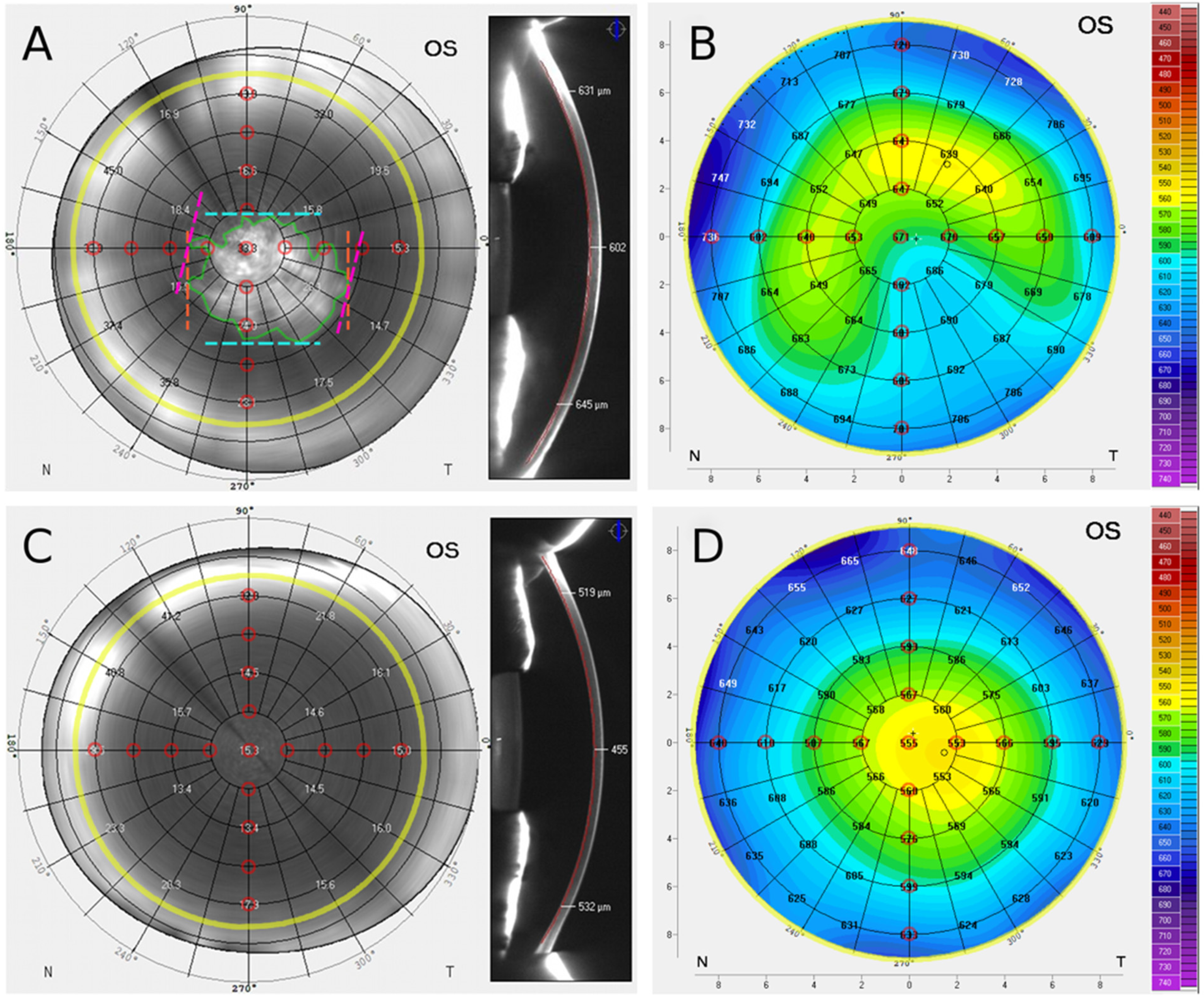
2.2.1. Scheimpflug Imaging

Patients were placed in front of the Pentacam (Oculus GmbH), and the head was fixed to the chin rest and forehead strap. Gaze fixation was performed by an optical fixation object. All images were acquired under unchanged room light conditions. The light source of the Pentacam (Oculus GmbH) is a blue light-emitting diode (LED) with a wavelength of 475 nm. Combined with slit-shaped illumination, a radially oriented rotating measurement procedure generates Scheimpflug cross-sectional images of the anterior segment of the eye. After initialization, 25 images are automatically generated in less than 2 s.
Scheimpflug imaging data were analyzed by a single cornea specialist (OO) following instruction and joint grading of a pilot series with another experienced grader and cornea specialist (MMa). In the Densitometry display (Pentacam software version 1.22r09), the en face Scheimpflug backscatter plane with the highest backscatter gray scale unit (GSU) at the corneal apex within the posterior corneal 100 µm was selected as described before [10]. The image was exported in high-resolution quality. Concentric annuli around the corneal apex superimposed by the Pentacam software served as a metric scale reference (Figure 1). The presence of an FL was defined as a central or paracentral geographic hyperreflectivity with a maximum caliper diameter >1 mm [10]. The boundaries of the FL were manually marked using an image analysis software (ImageJ Version 1.53e, NIH, USA, Figure 1), and the FL area was calculated [13,14]. In addition, manual marking of the horizontal and the vertical caliper diameters of the FL was performed. These caliper diameters are defined as the longest distance between two points on the FL along the x and y axes, respectively (Figure 1). The maximum caliper diameter of the FL was automatically calculated by the image analysis software (Figure 1) [10].
The focal apex corneal backscatter (ACB) value was extracted, and the focal peripheral corneal backscatter (PCB) values were extracted at a radius of 1 mm (PCB1mm), 2 mm (PCB2mm), 3 mm (PCB3mm) and 4 mm (PCB4mm) from the apex along the vertical and horizontal axes (Figure 1). Radius-specific focal PCB values were determined by calculating the mean of the four focal backscatter values located in the respective superior, nasal, inferior, and temporal position (Figure 1).

2.2.2. Scheimpflug Pachymetry Data

The pachymetry map was exported from the Pentacam pachymetry display in high-resolution quality. The central corneal thickness (CCT) and apex corneal thickness (ACT) values were extracted and the peripheral corneal thickness (PCT) values were extracted at a radius of 1 mm (PCT1mm), 2 mm (PCT2mm), 3 mm (PCT3mm) and 4 mm (PCT4mm) from the apex along the vertical and horizontal axes (Figure 1). The radius-specific PCT values were determined by calculating the mean of the four pachymetry values located in the superior, nasal, inferior, and temporal location, respectively (Figure 1).

2.2.3. Comparison of Fibrillar Layer-Positive and -Negative Eyes

Patients were divided into an FL-positive group and an FL-negative group. Scheimpflug backscatter data and pachymetry data were compared between both groups.
In order to analyze the correspondence of the FL localization and of areas of increased corneal thickness, individual focal backscatter values (en face backscatter plane at the level with the highest backscatter gray scale unit (GSU) at the corneal apex within the posterior corneal 100 µm as described above) and pachymetry values were compared between FL-positive and FL-negative corneas at a total of 17 positions within the concentric annuli grid around the corneal apex superimposed by the Pentacam software (corneal apex as well as superior, nasal, inferior, temporal location at 1 mm, 2 mm, 3 mm and 4 mm radial distance from the apex, Figure 1). Comparing FL-positive to FL-negative eyes, significantly elevated focal GSU levels were regarded as indicative of increased presence of an FL, and significantly elevated pachymetry values were regarded as indicative of increased presence of corneal edema in the respective cohort.

2.2.4. Correlation Analysis

Correlation of fibrillar layer dimensions and pachymetry: A correlation analysis was performed in FL-positive eyes to determine correlation between FL dimension parameters (FL area, vertical and horizontal caliper diameters, maximum caliper diameter) and pachymetric parameters (CCT, ACT, PCT1mm, PCT2mm, PCT3mm, PCT4mm).
Correlation of focal backscatter and pachymetry: A correlation analysis of the focal backscatter values (as an indicator for the presence of a fibrillar layer) with the corresponding pachymetry values was performed at the corneal apex and for 1 mm, 2 mm, 3 mm and 4 mm radial distance from the apex.

2.3. Statistics

Continuous variables were summarized by mean ± standard deviation (SD), categorical/nominal variables by counts and percentages. Group comparison of FL-positive vs. FL-negative eyes were performed using a 2-sided t-test or Pearson χ2-test, depending on the distribution. Pearson correlation coefficients were calculated (r = 0 to 1 positive correlation, r = 0 no correlation, r = 0 to −1 negative correlation; r ≥ 0.1 corresponds to a weak correlation, r ≥ 0.3 corresponds to a moderate correlation, r ≥ 0.5 corresponds to a strong correlation.). A p value of < 0.05 was considered statistically significant. No adjustments for multiple testing were made.
Statistical analysis and preparation of graphics were performed using statistical software packages (SPSS 27, IBM Corp., Armonk, NY, USA; creation of statistical charts using Prism 6, GraphPad software, San Diego, CA, USA).

3. Results

A total of n = 96 patients (n = 96 eyes) with advanced FECD were included in the study. The demographic details of all patients are shown in Table 1. Comparing the FL-positive and FL-negative groups, there were no significant differences in age, gender, lens status and time of Scheimpflug imaging. However, there were significant differences with regard to the modified Krachmer grading, with a higher proportion of modified Krachmer grade 6 patients in the FL-positive group and a higher proportion of modified Krachmer grade 5 patients in the FL-negative group (Table 1).

3.1. En Face Scheimpflug Backscatter Data Analysis

An FL was detected in 74% (n = 71) of all patients, whereas 26% (n = 25) were FL-negative. All en face Scheimpflug backscatter images showed a significant increase in hyperreflectivity toward the periphery (>10 mm), and the FL was displayable in higher definition in the corneal center compared to the periphery as previously described (Figure 1) [10,15]. Scheimpflug backscatter analysis showed no values for few individual peripheral corneal measurements (Table 2). The dimensions of the FL (n = 71) were for the area 9.97 ± 5.13 mm², for the vertical caliper diameter 3.50 ± 0.93 mm, for the horizontal caliper diameter FL 4.09 ± 1.10 mm, and for the maximum caliper diameter 4.38 ± 1.07 mm.

3.2. Comparative Analysis of Focal Backscatter in FL-Positive and FL-Negative Eyes

Eyes with FL (n = 71) showed an ACB of 31.7 ± 7.7 GSU and eyes without FL (n = 25) showed an ACB of 23.1 ± 4.8 GSU (p < 0.001). The focal peripheral backscatter values for eyes with FL and without FL, respectively, were for PCB1mm 27.9 ± 6.4 GSU and 20.9 ± 3.5 GSU (p < 0.001), for PCB2mm 23.4 ± 5.7 GSU and 19.2 ± 3.7 GSU (p = 0.001) for PCB3mm 22.8 ± 5.5 GSU and 20.8 ± 4.9 GSU (p = 0.127) and for PCB4mm 27.4 ± 6.6 GSU and 28.6 ± 10.1 GSU (p = 0.579).
The individual focal peripheral corneal backscatter values PCB1mm, PCB2mm, PCB3mm and PCB4mm at superior, nasal, inferior and temporal locations, respectively, are shown in Table 2. In FL-positive eyes, significantly increased GSU values at the corneal apex and at 1 mm radius, as well as inferiorly and temporally at 2 mm and 3 mm radius suggest a primarily central and inferotemporal localization of FL.

3.3. Pachymetry Data Analysis

Analysis of Scheimpflug pachymetry data in all FECD eyes (n = 96) showed a CCT of 604.3 ± 53.6 µm, an ACT of 603.9 ± 52.8 µm, a PCT1mm of 606.5 ± 50.8 µm, a PCT2mm of 618.4 ± 47.2 µm, a PCT3mm of 646.9 ± 45.1 µm, and a PCT4mm of 692.7 ± 49.6 µm.

3.4. Comparative Analysis of Pachymetry in FL-Positive vs. FL-Negative Eyes

Eyes with FL (n = 71) showed a CCT of 614.6 ± 51.8 µm and eyes without FL (n = 25) showed a CCT of 575.1 ± 48.4 µm (p = 0.001). In eyes with FL, the ACT was 614.0 ± 51.6 µm, and in eyes without FL, the ACT was 575.2 ± 46.2 µm (p = 0.001). Peripheral pachymetry values for FECD eyes with FL and without FL, respectively, were for PCT1mm 615.7 ± 50.0 µm and 580.2 ± 44.2 µm (p = 0.002), for PCT2mm 625.2 ± 47.6 µm and 599.1 ± 40.9 µm (p = 0.017) for PCT3mm 651.0 ± 46.3 µm and 635.0 ± 39.9 µm (p = 0.128) and for PCT4mm 695.1 ± 51.8 µm and 686.0 ± 43.2 (p = 0.435).
The individual peripheral corneal thickness values PCT1mm, PCT2mm, PCT3mm and PCT4mm at superior, nasal, inferior and temporal locations, respectively, are shown in Table 3. In FL-positive eyes, significantly increased pachymetry values at the corneal apex and 1 mm radius as well as inferiorly and temporally at 2 mm radius and temporally at 3 mm radius suggest a primarily central and inferotemporal localization of corneal edema.

3.5. Correlation Analysis

Correlation of fibrillar layer dimensions and pachymetry: Weak correlations were found between vertical caliper diameter and ACT (r =0.249, p = 0.036) and between vertical caliper diameter and PCT1mm (r = 0.251, p = 0.035). There were no other significant correlations between the investigated FL dimensions and the pachymetry values.
Correlation of focal backscatter and pachymetry: Correlation analysis showed moderate correlation of ACB values and ACT values (r = 0.309, p = 0.002) and weak correlation of focal backscatter and pachymetric values at 1 mm (r = 0.269, p = 0.008) and 2 mm (r = 0.271, p = 0.007). There were no significant correlations between focal backscatter and pachymetry values at 3 mm (r = 0.146, p = 0.159) and 4 mm (r = 0.004, p = 0.973) radial distance from the apex.

4. Discussion

In the present study, Scheimpflug imaging demonstrates that FL detection is associated with increased corneal thickness and that the FL and increased corneal thickness are primarily located in the central and inferotemporal area of advanced FECD corneas.
Adding to the results from our previous studies showing significantly reduced endothelial cell density [9] and increased light scattering [10] in the FL area, this further supports a potential role of clinical FL detection as a criterion in progression assessment and therapeutic decision making in FECD patients in the future.
Keratoplasty represents the only established definite treatment option for FECD [2,16], and DMEK may be considered the therapeutic gold standard in advanced FECD disease. However, the quest for further minimization of surgical intervention and the limited global supply of donor tissue lead to the continuous development and optimization of the surgical and conservative therapeutic spectrum. New tissue-sparing modalities for the treatment of FECD include hemi- or quarter-DMEK, Descemet stripping only (DSO) or cultured corneal endothelial cell injection, as well as the combined or sole topical drug application including particularly rho-associated protein kinase (ROCK) inhibitors [17,18,19,20,21]. In addition, a customized DMEK based on Scheimpflug images of FL areas with pronounced endothelial loss may be an option to reduce tissue needed for transplantation and thereby potentially also immune reaction rates [16].
With the arrival of new therapeutic options, the optimization of clinical FECD classification systems becomes increasingly important for progression assessment but also for identification of the optimal timepoint, the optimal corneal endothelial area-to-treat and for the selection of the optimal therapeutic approach [22]. The development of such classifications requires a detailed understanding of the underlying pathology and its concomitant characteristic morphological alterations, which may be documented in vivo using a variety of clinical imaging techniques. In this context, previous histology studies demonstrated that subendothelial collagen deposits in the form of an FL are detectable in approximately 80% of patients with advanced FECD (modified Krachmer grades 5 or 6) [8], and that this FL marks areas of sharp and significant reduction in CEC density [9,10]. Moreover, comparing histology and clinical imaging, the FL was identified as a major source of light scattering, enabling FL visualization in vivo by en face Scheimpflug backscatter analysis [10].
Until now, it was unclear whether reduced CEC density in the FL area was accompanied by increased aqueous humor leakage into the corneal tissue and edema formation or whether the FL rather sealed the posterior cornea preventing the entry of aqueous humor in advanced FECD eyes [10]. Moreover, the detailed location of the FL at the posterior corneal surface was unknown.
Results of the present study demonstrate in vivo by Scheimpflug imaging that FL detection in advanced FECD eyes is associated with increased corneal thickness, suggesting edema formation. Moreover, it shows that the FL and increased corneal thickness are located in the central and inferotemporal region. The localization of edema in FL-positive eyes in the central and inferotemporal region replicate and complement previous findings by other groups also showing reduced endothelial cell count and confluent guttae, especially in the inferotemporal corneal area [23,24,25]. This supports the hypothesis that increased aqueous humor entry occurs in the FL area due to significant focal reduction in CEC density or even due to complete defects of significant size within the central and inferotemporal corneal endothelial compound. Furthermore, these results suggest that as a consequence, minimally invasive procedures in (FL-positive) patients with advanced FECD should particularly target the central and inferotemporal endothelium.
A weak correlation of the FL area dimensions (vertical caliper diameter) and corneal thickness was identified in our cohort, pointing toward a potential area growth of the fibrillar layer with FECD progression. However, the correlation was weak and likely limited by the restricted spectrum of FECD stages (exclusively modified Krachmer stages 5 and 6) investigated in the present study. Future studies will examine all stages of FECD in a larger cohort and will provide further insight into the development and area growth of the FL with FECD progression.
Early ex vivo studies of FECD histology specimens showed that FL thickness increases with stromal and epithelial edema (maximum FL thickness in this histologic study 9.3 µm) [26]. It was hypothesized that the increased influx of aqueous humor entering Descemet’s membrane through a rarified endothelium in decompensated corneas contributes to the focal formation of a loose FL continuously growing in thickness [26]. This suggests that the increase in corneal thickness of FL-positive eyes in our study is mainly attributable to corneal edema formation but, to a lesser extent, also to the FL itself. However, based on our experience, Scheimpflug imaging is so far insufficient in resolution to measure the thickness of the FL and thus replicate these studies in vivo. Recent investigations by other groups measuring the thickness of the endothelium/Descemet membrane complex provide evidence that comparable in vivo studies will be possible in the future [27,28]. Using high-definition-optical coherence tomography (HD-OCT) 3D thickness maps, these studies showed a strong correlation between endothelium/Descemet membrane thickness values and FECD severity [27].
Backscatter of the anterior (120 µm), mid and posterior (60 µm) cornea increased with FECD progression and thus with increase in corneal thickness in earlier studies [5,29,30]. In addition, posterior backscatter was among the variables selected by a statistical learning algorithm to predict edema resolution after DMEK in FECD patients based on preoperative assessment [31]. Moreover, posterior backscatter contributed particularly to the development of visual acuity-related disability [4], and there was an early drop in posterior backscatter following DMEK surgery in FECD patients, whereas anterior backscatter regressed over a longer period [15]. Our data confirm the original results from these studies and emphasize the significance of the FL as a source and as an important morphological correlate of posterior backscatter associated with increased edema in FECD. A comparison of patients with and without FL in similar study settings would be interesting to further investigate the role of the FL (and of its removal) as a morphologically relevant target structure in advanced FECD disease. It seems tempting to speculate that DMEK in patients with FL produces a similar backscatter-attenuating effect as a capsulotomy in posterior capsule opacification both through removing the most relevant source of light scattering.
The most important limitation of our study is the restricted range of FECD stages. Moreover, Scheimpflug backscatter images were not adjusted for variations in light intensity in our study. Such adjustments involve the acquisition of a standard scatter source before each examination with subsequent adjustment of the measurements in the human eye. Although these adjustments significantly improve the accuracy, they are not part of the standard automated Scheimpflug imaging procedure [29]. In accordance with previous studies showing reduced repeatability and reproducibility of densitometry in peripherally located corneal areas [32,33], Scheimpflug backscatter analysis showed no values for isolated peripheral corneal measurements in our study. However, the demonstration of a correlation between Scheimpflug FL detection and corneal thickness in our study suggests that the procedure may still be applicable with limitations in clinical practice as described herein.

5. Conclusions

A significant reduction in endothelial cell density, increased backscatter (both demonstrated in previous studies [9,10]), and increased corneal thickness are important features of the FL area, which may primarily be found in a central and inferotemporal location in advanced FECD eyes. This may provide important information for progression assessment and therapeutic decision making in FECD patients in the future. Furthermore, it may open avenues for customized surgical approaches based on preclinical Scheimpflug imaging.

Author Contributions

Conceptualization, O.Ö., M.M. (Mert Mestanoglu), B.B. and M.M. (Mario Matthaei); Data curation, O.Ö., M.M. (Mert Mestanoglu), T.C., P.S., B.B. and M.M. (Mario Matthaei); Formal analysis, O.Ö., M.M. (Mert Mestanoglu), P.S., S.S., N.R., B.B. and M.M. (Mario Matthaei); Funding acquisition, C.C., B.B. and M.M. (Mario Matthaei); Investigation, O.Ö., M.M. (Mert Mestanoglu) and A.H.; Methodology, O.Ö., M.M. (Mert Mestanoglu), B.B. and M.M. (Mario Matthaei); Project administration, C.C., B.B. and M.M. (Mario Matthaei); Supervision, C.C., B.B. and M.M. (Mario Matthaei); Visualization, O.Ö., M.M. (Mert Mestanoglu), A.H., T.C., B.B. and M.M. (Mario Matthaei); Writing—original draft, O.Ö., M.M. (Mert Mestanoglu), A.H., B.B. and M.M. (Mario Matthaei); Writing—review & editing, O.Ö., M.M. (Mert Mestanoglu), A.H., T.C., P.S., S.S., N.R., C.C., B.B. and M.M. (Mario Matthaei). All authors attest that they meet the current ICMJE criteria for authorship. All authors have read and agreed to the published version of the manuscript.

Funding

German Research Foundation (FOR2240 to T.C. and C.C.; CL 751/1-1 to TC; CU 47/6-1, CU 47/9-1, CU 47/12-1 to CC; MA 5110/5-1 to M.M. (Mario Matthaei)), German Research Council Cologne Clinician Scientist Program (Project 413543196 to A.H.), FORTUNE Program University of Cologne (to M.M. (Mario Matthaei)); EU COST BM1302 “Joining Forces in Corneal Regeneration” and EU COST ANIRIDIA (to C.C., B.B., S.S.), EU Arrest Blindness (to C.C.), EU EFRE NRW (to S.S.), Studienstiftung des Deutschen Volkes (to M.M. (Mert Mestanoglu), Forschung für das Sehen e.V. (https://augenklinik.uk-koeln.de/zentrum/foerderverein-forschung-sehen/; accessed on 15 May 2022) (to M.M. (Mario Matthaei)).

Institutional Review Board Statement

Formal approval to conduct this study was obtained from the Ethics Committee of the University of Cologne (16-424). The research adhered to the tenets of the Declaration of Helsinki.

Informed Consent Statement

Written informed consent has been obtained from all participants for the treatment, participation in the research and publication of the results.

Data Availability Statement

The data presented in this study are available on request from the corresponding author. The data are not publicly available due to privacy and ethical considerations.

Conflicts of Interest

The authors declare no conflict of interest. The funders had no role in the design of the study; in the collection, analyses, or interpretation of data; in the writing of the manuscript, or in the decision to publish the results.

References

  1. Gain, P.; Jullienne, R.; He, Z.; Aldossary, M.; Acquart, S.; Cognasse, F.; Thuret, G. Global Survey of Corneal Transplantation and Eye Banking. JAMA Ophthalmol. 2016, 134, 167–173. [Google Scholar] [CrossRef] [PubMed] [Green Version]
  2. Matthaei, M.; Hribek, A.; Clahsen, T.; Bachmann, B.; Cursiefen, C.; Jun, A.S. Fuchs Endothelial corneal dystrophy: Clinical, genetic, pathophysiologic, and therapeutic aspects. Annu. Rev. Vis. Sci. 2019, 5, 151–175. [Google Scholar] [CrossRef] [PubMed]
  3. Loreck, N.; Adler, W.; Siebelmann, S.; Rokohl, A.C.; Heindl, L.M.; Cursiefen, C.; Matthaei, M. Morning myopic shift and glare in advanced fuchs endothelial corneal dystrophy. Am. J. Ophthalmol. 2020, 213, 69–75. [Google Scholar] [CrossRef]
  4. Wacker, K.; Grewing, V.; Fritz, M.; Bohringer, D.; Reinhard, T. Morphological and optical determinants of visual disability in fuchs endothelial corneal dystrophy. Cornea 2020, 39, 726–731. [Google Scholar] [CrossRef]
  5. Wacker, K.; McLaren, J.W.; Amin, S.R.; Baratz, K.H.; Patel, S.V. Corneal High-order aberrations and backscatter in fuchs’ endothelial corneal dystrophy. Ophthalmology 2015, 122, 1645–1652. [Google Scholar] [CrossRef] [PubMed] [Green Version]
  6. Wacker, K.; McLaren, J.W.; Patel, S.V. Optical and anatomic changes in fuchs endothelial dystrophy corneas. In Current Treatment Options for Fuchs Endothelial Dystrophy; Cursiefen, C., Jun, A.S., Eds.; Springer: Cham, Switzerland, 2017; pp. 51–71. [Google Scholar]
  7. Fuchs, E. Dystrophia epithelialis corneae. Albr. Græfe. Arch. Ophthalmol. 1910, 76, 478–508. [Google Scholar] [CrossRef] [Green Version]
  8. Krachmer, J.H.; Purcell, J.J., Jr.; Young, C.W.; Bucher, K.D. Corneal endothelial dystrophy. A study of 64 families. Arch. Ophthalmol. 1978, 96, 2036–2039. [Google Scholar] [CrossRef]
  9. Hribek, A.; Clahsen, T.; Horstmann, J.; Siebelmann, S.; Loreck, N.; Heindl, L.M.; Matthaei, M. Fibrillar layer as a marker for areas of pronounced corneal endothelial cell loss in advanced fuchs endothelial corneal dystrophy. Am. J. Ophthalmol. 2020, 222, 292–301. [Google Scholar] [CrossRef]
  10. Hribek, A.; Mestanoglu, M.; Clahsen, T.; Reinking, N.; Frentzen, F.; Howaldt, A.; Matthaei, M. Scheimpflug backscatter imaging of the fibrillar layer in fuchs endothelial corneal dystrophy. Am. J. Ophthalmol. 2021, 235, 63–70. [Google Scholar] [CrossRef]
  11. Louttit, M.D.; Kopplin, L.J.; Igo, R.P., Jr.; Fondran, J.R.; Tagliaferri, A.; Bardenstein, D.; FECD Genetics Multi-Center Study Group. A multicenter study to map genes for Fuchs endothelial corneal dystrophy: Baseline characteristics and heritability. Cornea 2012, 31, 26–35. [Google Scholar] [CrossRef] [Green Version]
  12. Fritz, M.; Grewing, V.; Maier, P.; Lapp, T.; Böhringer, D.; Reinhard, T.; Wacker, K. Diurnal variation in corneal edema in fuchs endothelial corneal dystrophy. Am. J. Ophthalmol. 2019, 207, 351–355. [Google Scholar] [CrossRef]
  13. Schindelin, J.; Arganda-Carreras, I.; Frise, E.; Kaynig, V.; Longair, M.; Pietzsch, T.; Cardona, A. Fiji: An open-source platform for biological-image analysis. Nat. Methods 2012, 9, 676–682. [Google Scholar] [CrossRef] [Green Version]
  14. Schneider, C.A.; Rasband, W.S.; Eliceiri, K.W. NIH Image to ImageJ: 25 years of image analysis. Nat. Methods 2012, 9, 671–675. [Google Scholar] [CrossRef]
  15. Schaub, F.; Enders, P.; Bluhm, C.; Bachmann, B.O.; Cursiefen, C.; Heindl, L.M. Two-year course of corneal densitometry after descemet membrane endothelial keratoplasty. Am. J. Ophthalmol. 2017, 175, 60–67. [Google Scholar] [CrossRef]
  16. Hos, D.; Matthaei, M.; Bock, F.; Maruyama, K.; Notara, M.; Clahsen, T.; Cursiefen, C. Immune reactions after modern lamellar (DALK, DSAEK, DMEK) versus conventional penetrating corneal transplantation. Prog. Retin. Eye Res. 2019, 73, 100768. [Google Scholar] [CrossRef]
  17. Kinoshita, S.; Koizumi, N.; Ueno, M.; Okumura, N.; Imai, K.; Tanaka, H.; Hamuro, J. Injection of cultured cells with a ROCK inhibitor for bullous keratopathy. N. Engl. J. Med. 2018, 378, 995–1003. [Google Scholar] [CrossRef]
  18. Colby, K. Descemet stripping only for fuchs endothelial corneal dystrophy: Will it become the gold standard? Cornea 2021, 41, 269–271. [Google Scholar] [CrossRef]
  19. Kinoshita, S.; Colby, K.A.; Kruse, F.E. A Close Look at the clinical efficacy of rho-associated protein kinase inhibitor eye drops for fuchs endothelial corneal dystrophy. Cornea 2021, 40, 1225–1228. [Google Scholar] [CrossRef]
  20. Price, M.O.; Price, F.W., Jr. Randomized, double-masked, pilot study of netarsudil 0.02% ophthalmic solution for treatment of corneal edema in fuchs dystrophy. Am. J. Ophthalmol. 2021, 227, 100–105. [Google Scholar] [CrossRef]
  21. Lam, F.C.; Baydoun, L.; Dirisamer, M.; Lie, J.; Dapena, I.; Melles, G.R.J. Hemi-Descemet membrane endothelial keratoplasty transplantation: A potential method for increasing the pool of endothelial graft tissue. JAMA Ophthalmol. 2014, 132, 1469–1473. [Google Scholar] [CrossRef] [Green Version]
  22. Patel, S.V. Imaging fuchs endothelial corneal dystrophy in clinical practice and clinical trials. Cornea 2021, 40, 1505–1511. [Google Scholar] [CrossRef] [PubMed]
  23. Fujimoto, H.; Maeda, N.; Soma, T.; Oie, Y.; Koh, S.; Tsujikawa, M.; Nishida, K. Quantitative regional differences in corneal endothelial abnormalities in the central and peripheral zones in Fuchs’ endothelial corneal dystrophy. Investig. Ophthalmol. Vis. Sci. 2014, 55, 5090–5098. [Google Scholar] [CrossRef] [PubMed]
  24. Soh, Y.Q.; Peh, G.S.L.; Naso, S.L.; Kocaba, V.; Mehta, J.S. Automated clinical assessment of corneal guttae in fuchs endothelial corneal dystrophy. Am. J. Ophthalmol. 2021, 221, 260–272. [Google Scholar] [CrossRef]
  25. Brunette, I.; Sherknies, D.; Terry, M.A.; Chagnon, M.; Bourges, J.L.; Meunier, J. 3-D characterization of the corneal shape in Fuchs dystrophy and pseudophakic keratopathy. Investig. Ophthalmol. Vis. Sci. 2011, 52, 206–214. [Google Scholar] [CrossRef] [Green Version]
  26. Bourne, W.M.; Johnson, D.H.; Campbell, R.J. The ultrastructure of Descemet’s membrane. III. Fuchs’ dystrophy. Arch. Ophthalmol. 1982, 100, 1952–1955. [Google Scholar] [CrossRef]
  27. Eleiwa, T.K.; Cook, J.C.; Elsawy, A.S.; Roongpoovapatr, V.; Volante, V.; Yoo, S.; Abou Shousha, M. Diagnostic performance of three-dimensional endothelium/descemet membrane complex thickness maps in active corneal graft rejection. Am. J. Ophthalmol. 2020, 210, 48–58. [Google Scholar] [CrossRef]
  28. Shousha, M.A.; Perez, V.L.; Wang, J.; Ide, T.; Jiao, S.; Chen, Q.; Yoo, S.H. Use of ultra-high-resolution optical coherence tomography to detect in vivo characteristics of Descemet’s membrane in Fuchs’ dystrophy. Ophthalmology 2010, 117, 1220–1227. [Google Scholar] [CrossRef] [Green Version]
  29. Patel, S.V.; Hodge, D.O.; Treichel, E.J.; Spiegel, M.R.; Baratz, K.H. Predicting the Prognosis of Fuchs Endothelial Corneal Dystrophy by Using Scheimpflug Tomography. Ophthalmology 2020, 127, 315–323. [Google Scholar] [CrossRef] [Green Version]
  30. Repp, D.J.; Hodge, D.O.; Baratz, K.H.; McLaren, J.W.; Patel, S.V. Fuchs’ endothelial corneal dystrophy: Subjective grading versus objective grading based on the central-to-peripheral thickness ratio. Ophthalmology 2013, 120, 687–694. [Google Scholar] [CrossRef] [Green Version]
  31. Zander, D.; Grewing, V.; Glatz, A.; Lapp, T.; Maier, P.C.; Reinhard, T.; Wacker, K. Predicting edema resolution after descemet membrane endothelial keratoplasty for fuchs dystrophy using scheimpflug tomography. JAMA Ophthalmol. 2021, 139, 423–430. [Google Scholar] [CrossRef]
  32. Alnawaiseh, M.; Zumhagen, L.; Wirths, G.; Eveslage, M.; Eter, N.; Rosentreter, A. Corneal densitometry, central corneal thickness, and corneal central-to-peripheral thickness ratio in patients with fuchs endothelial dystrophy. Cornea 2016, 35, 358–362. [Google Scholar] [CrossRef]
  33. Ni Dhubhghaill, S.; Rozema, J.J.; Jongenelen, S.; Ruiz Hidalgo, I.; Zakaria, N.; Tassignon, M.J. Normative values for corneal densitometry analysis by Scheimpflug optical assessment. Investig. Ophthalmol. Vis. Sci. 2014, 55, 162–168. [Google Scholar] [CrossRef] [PubMed] [Green Version]
Figure 1. Scheimpflug backscatter and pachymetry in fibrillar layer (FL)-positive (A,B) and FL-negative (C,D) Fuchs endothelial corneal dystrophy (FECD) eyes: (A) En face Scheimpflug backscatter image of FL-positive FECD cornea: horizontal caliper diameter (orange), vertical caliper diameter (blue), maximum caliper diameter (pink) and FL area (green). FL shows inferotemporal localization. (B) Pachymetry map of FL-positive cornea: Increased corneal thickness inferotemporal. (C) En face Scheimpflug backscatter image of FL-negative cornea, (D) Pachymetry map of FL-negative cornea; superimposed grid marks concentric annuli around the corneal apex at 1 mm radial spacing (black) and at 4.5 mm radius (yellow). The study analyzed backscatter and pachymetry values along the vertical and horizontal axes (red circular marks).
Jcm 11 02815 g001
Table 1. Characteristics of patients and investigated eyes (one eye per patient), fibrillar layer (FL). FECD: Fuchs endothelial corneal dystrophy; AM: Ante Meridiem (before noon).
FL PositiveFL NegativeTotalp-Value
Patients, n (%)71 (74)25 (26)96 (100)-
FECD Krachmer Grade 512 (16.9%)13 (52%)25 (26%)<0.01
FECD Krachmer Grade 659 (83.1%)12 (48%)71 (74%)
Age (years), mean ± standard deviation 67.2 ± 9.569.2 ± 9.267.7 ± 9.40.37
Female, n (%)39 (54.9%)13 (52%)52 (54.2%)0.80
Pseudophakic, n (%)24 (33.8%)5 (20.0%)29 (30.2%)0.20
Time of Scheimpflug imaging: AM, n (%)22 (31%)8 (32%)30 (31.3%)0.93
Data are numbers (percentage) or mean ± standard deviation.
Table 2. Focal backscatter in fibrillar layer (FL)-positive and -negative advanced FECD eyes: The en face Scheimpflug backscatter plane with the highest backscatter gray scale unit (GSU) at the corneal apex within the posterior corneal 100 µm was selected, and focal backscatter values were extracted at the given locations. Apex corneal backscatter (ACB), peripheral corneal backscatter (PCB).
DistancePositionFL PositiveFL Negativep-Value
(n = 71)(n = 25)
ACB (GSU)mean31.7 ± 7.723.1 ± 4.8p < 0.001
PCB1mm (GSU)mean27.9 ± 6.420.9 ± 3.5p < 0.001
superior24.4 ± 8.220.2 ± 4.0p = 0.001
nasal25.4 ± 7.620.6 ± 3.7p < 0.001
inferior30.0 ± 7.920.9 ± 4.4p < 0.001
temporal31.4 ± 9.121.8 ± 3.4p < 0.001
PCB2mm (GSU)mean23.4 ± 5.719.2 ± 3.7p = 0.001
superior22.0 ± 5.820.0 ± 5.1p = 0.124
nasal21.7 ± 6.520.1 ± 5.2p = 0.284
inferior23.9 ± 7.517.8 ± 3.5p < 0.001
temporal25.9 ± 8.019.0 ± 3.3p < 0.001
PCB3mm (GSU)mean 122.8 ± 5.520.8 ± 4.9p = 0.127
superior 228.3 ± 10.725.4 ± 8.4p = 0.235
nasal23.2 ± 6.023.5 ± 9.5p = 0.862
inferior19.4 ± 5.217.6 ± 2.9p = 0.040
temporal20.0 ± 5.218.0 ± 3.9p = 0.048
PCB4mm (GSU)mean 327.4 ± 6.628.6 ± 10.1p = 0.579
superior 440.4 ± 15.138.6 ± 23.6p = 0.672
nasal34.1 ± 10.436.4 ± 12.4p = 0.374
inferior20.6 ± 5.021.7 ± 5.4p = 0.341
temporal19.9 ± 5.119.0 ± 4.2p = 0.430
1,2n = 95 (FL pos. = 71; FL neg. = 24), 3,4 n = 83 (FL pos. = 60; FL neg. = 23).
Table 3. Corneal thickness in fibrillar layer (FL)-positive and -negative advanced FECD eyes. Apex corneal thickness (ACT), peripheral corneal thickness (PCT).
DistancePositionFL PositiveFL Negativep-Value
(n = 71)(n = 25)
ACT (µm)mean614.0 ± 51.6575.2 ± 46.2p = 0.001
PCT1mm (µm)mean615.7 ± 50.0580.2 ± 44.2p = 0.002
superior612.9 ± 51,0583.9 ± 39.1p = 0.011
nasal610.5 ± 49581.8 ± 42.6p = 0.011
inferior619.4 ± 50.6579.0 ± 50.5p = 0.001
temporal618.8 ± 54.0576.2 ± 47.8p = 0.001
PCT2mm (µm)mean625.2 ± 47.6599.1 ± 40.9p = 0.017
superior625.9 ± 48.2609.9 ± 38.1p = 0.135
nasal620.9 ± 46.6602.7 ± 39,5p = 0.084
inferior627.4 ± 49.0594.5 ± 46.5p = 0.004
temporal626.5 ± 54.6589.2 ± 47.7p = 0.003
PCT3mm (µm)mean651.0 ± 46.3635.0 ±39.9p = 0.128
superior659.8 ± 47.5654.7 ± 42.4p = 0.635
nasal654.3 ± 46.6643.2 ± 41.2p = 0.294
inferior645.7 ± 49.8624.1 ± 41.8p = 0.055
temporal644.4 ± 53.5618.2 ± 44.1p = 0.031
PCT4mm (µm)mean695.1 ± 51.8686.0 ± 43.2p = 0.435
superior712.9 ± 56.5713.8 ± 53.8p = 0.939
nasal704.7 ± 53.7696.6 ± 48.8p = 0.507
inferior685.6 ± 61.2672.6 ± 45.7p = 0.333
temporal677.1 ± 56.1661.0 ± 39.2p = 0.189
Publisher’s Note: MDPI stays neutral with regard to jurisdictional claims in published maps and institutional affiliations.

