
Emergency Cervical Cerclage


Abstract
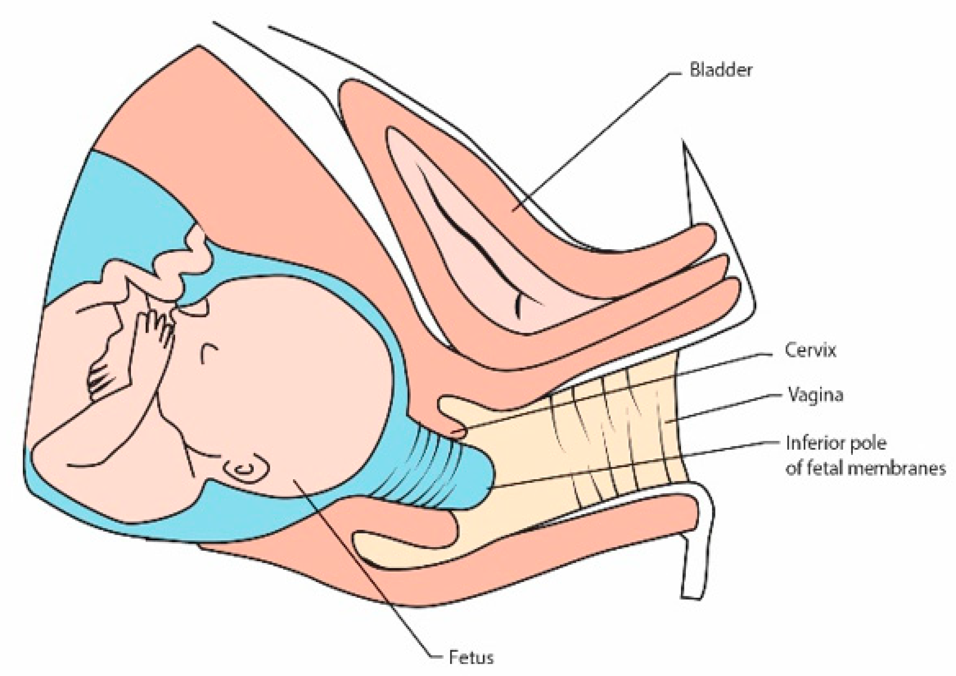
1. Introduction
2. Emergency Suture Procedure
3. Rescue Suture Effectiveness
4. Risk Factors of the Emergency Suture Failure
5. Conclusions
Author Contributions
Funding
Institutional Review Board Statement
Informed Consent Statement
Data Availability Statement
Conflicts of Interest
References
- Rasmark, R.E.; Matthiesen, L.; Rylance, R.; Christiansen, O.B. Is the incidence of recurrent pregnancy loss increasing? A retrospective register-based study in Sweden. Acta Obstet. Gynecol. Scand. 2017, 96, 1365–1372. [Google Scholar] [CrossRef] [PubMed]
- Ticconi, C.; Pietropolli, A.; Specchia, M.; Nicastri, E.; Chiaramonte, C.; Piccione, E.; Scambia, G.; Di Simone, N. Pregnancy-Related Complications in Women with Recurrent Pregnancy Loss: A Prospective Cohort Study. J. Clin. Med. 2020, 9, 2833. [Google Scholar] [CrossRef]
- Berghella, V.; Saccone, G. Fetal fibronectin testing for prevention of preterm birth in singleton pregnancies with threatened preterm labor: A systematic review and metaanalysis of randomized controlled trials. Am. J. Obstet. Gynecol. 2016, 215, 431–438. [Google Scholar] [CrossRef] [PubMed]
- Glass, H.C.; Costarino, A.T.; Stayer, S.A.; Brett, C.M.; Cladis, F.; Davis, P.J. Outcomes for extremely premature infants. Anesth. Analg. 2015, 120, 1337–1351. [Google Scholar] [CrossRef] [PubMed]
- Hafström, M.; Källén, K.; Serenius, F.; Maršál, K.; Rehn, E.; Drake, H.; Ådén, U.; Farooqi, A.; Thorngren-Jerneck, K.; Strömberg, B. Cerebral Palsy in Extremely Preterm Infants. Pediatrics 2018, 141, e20171433. [Google Scholar] [CrossRef]
- Bardin, C.; Piuze, G.; Papageorgiou, A. Outcome at 5 years of age of SGA and AGA infants born less than 28 weeks of gestation. Semin. Perinatol. 2004, 28, 288–294. [Google Scholar] [CrossRef][Green Version]
- Beck, S.; Wojdyla, D.; Say, L.; Betran, A.P.; Merialdi, M.; Requejo, J.H.; Rubens, C.; Menon, R.; van Look, P.F. The worldwide incidence of preterm birth: A systematic review of maternal mortality and morbidity. Bull. World Health Organ. 2010, 88, 31–38. [Google Scholar] [CrossRef]
- Goldenberg, R.L.; Culhane, J.F.; Iams, J.D.; Romero, R. Epidemiology and causes of preterm birth. Lancet 2008, 371, 75–84. [Google Scholar] [CrossRef]
- Davidesko, S.; Wainstock, T.; Sheiner, E.; Pariente, G. Long-Term Infectious Morbidity of Premature Infants: Is There a Critical Threshold? J. Clin. Med. 2020, 9, 3008. [Google Scholar] [CrossRef] [PubMed]
- Platt, M.J. Outcomes in preterm infants. Public Health 2014, 128, 399–403. [Google Scholar] [CrossRef]
- Romero, R.; Dey, S.K.; Fisher, S.J. Preterm Labor: One Syndrome, Many Causes. Science 2014, 15, 760–765. [Google Scholar] [CrossRef] [PubMed]
- Saigal, S.; Doyle, L.W. An overview of mortality and sequelae of preterm birth from infancy to adulthood. Lancet 2008, 371, 261–269. [Google Scholar] [CrossRef]
- Young, B.K. A multidisciplinary approach to pregnancy loss: The pregnancy loss prevention center. J. Perinat. Med. 2018, 47, 41–44. [Google Scholar] [CrossRef] [PubMed]
- Mastrolia, S.A.; Baumfeld, Y.; Hershkovitz, R.; Yohay, D.; Trojano, G.; Weintraub, A.Y. Independent association between uterine malformations and cervical insufficiency: A retrospective population-based cohort study. Arch. Gynecol. Obstet. 2018, 297, 919–926. [Google Scholar] [CrossRef]
- Alfirevic, Z.; Stampalija, T.; Medley, N. Cervical stitch (cerclage) for preventing preterm birth in singleton pregnancy. Cochrane Database Syst. Rev. 2017, 6, CD008991. [Google Scholar] [CrossRef]
- Sneider, K.; Christiansen, O.B.; Sundtoft, I.B.; Langhoff-Roos, J. Recurrence of second trimester miscarriage and extreme preterm delivery at 16–27 weeks of gestation with a focus on cervical insufficiency and prophylactic cerclage. Acta Obstet. Gynecol. Scand. 2016, 95, 1383–1390. [Google Scholar] [CrossRef]
- Lu, C.; Lim, B.; Robson, S.J. Increasing Incidence Rate of Cervical Cerclage in Pregnancy in Australia: A Population-Based Study. Healthcare 2016, 4, 68. [Google Scholar] [CrossRef]
- Tsikouras, P.; Anastasopoulos, G.; Maroulis, V.; Bothou, A.; Chalkidou, A.; Deuteraiou, D.; Anthoulaki, X.; Tsatsaris, G.; Bourazan, A.H.; Iatrakis, G.; et al. Comparative Evaluation of Arabin Pessary and Cervical Cerclage for the Prevention of Preterm Labor in Asymptomatic Women with High Risk Factors. Int. J. Environ. Res. Public Health 2018, 15, 791. [Google Scholar] [CrossRef]
- Vink, J.; Feltovich, H. Cervical etiology of spontaneous preterm birth. Semin. Fetal Neonatal Med. 2016, 21, 106–112. [Google Scholar] [CrossRef]
- Sneider, K.; Christiansen, O.B.; Sundtoft, I.B.; Langhoff-Roos, J. Recurrence rates after abdominal and vaginal cerclages in women with cervical insufficiency: A validated cohort study. Arch. Gynecol. Obstet. 2017, 295, 859–866. [Google Scholar] [CrossRef]
- Koullali, B.; Westervelt, A.R.; Myers, K.M.; House, M.D. Prevention of preterm birth: Novel interventions for the cervix. Semin. Perinatol. 2017, 41, 505–510. [Google Scholar] [CrossRef]
- Danforth, D.N.; Veis, A.; Breen, M.; Weinstein, H.G.; Buckingham, J.C.; Manalo, P. The effect of pregnancy and labor on the human cervix: Changes in collagen, glycoproteins, and glycosaminoglycans. Am. J. Obstet. Gynecol. 1974, 120, 641–651. [Google Scholar] [CrossRef]
- Romero, R.; Espinoza, J.; Erez, O.; Hassan, S. The role of cervical cerclage in obstetric practice: Can the patient who could benefit from this procedure be identified? Am. J. Obstet. Gynecol. 2006, 194, 1–9. [Google Scholar] [CrossRef] [PubMed]
- Barinov, S.V.; Artymuk, N.V.; Novikova, O.N.; Shamina, I.V.; Tirskaya, Y.I.; Belinina, A.A.; Lazareva, O.V.; Kadcyna, T.V.; Borisova, A.V.; Stepanov, S.S.; et al. Analysis of risk factors and predictors of pregnancy loss and strategies for the management of cervical insufficiency in pregnant women at a high risk of preterm birth. J. Matern. Fetal Neonatal Med. 2019, 1–9. [Google Scholar] [CrossRef]
- Sundtoft, I.; Uldbjerg, N.; Steffensen, R.; Sommer, S.; Christiansen, O.B. Polymorphisms in Genes Coding for Cytokines, Mannose-Binding Lectin, Collagen Metabolism and Thrombophilia in Women with Cervical Insufficiency. Gynecol. Obstet. Investig. 2016, 81, 15–22. [Google Scholar] [CrossRef] [PubMed]
- Kimber-Trojnar, Z. Management of concomitant cervical insufficiency and intrauterine adhesions. Ann. Transl. Med. 2020, 8, 526. [Google Scholar] [CrossRef]
- Lotgering, F.K. Clinical aspects of cervical insufficiency. BMC Pregnancy Childbirth 2007, 7, S17. [Google Scholar] [CrossRef] [PubMed]
- Tanner, L.D.; Tucker, L.Y.; Postlethwaite, D.; Greenberg, M. Maternal race/ethnicity as a risk factor for cervical insufficiency. Eur. J. Obstet. Gynecol. Reprod. Biol. 2018, 221, 156–159. [Google Scholar] [CrossRef]
- Christiansen, O.B. Transabdominal cervicoisthmic cerclage in the management of recurrent second trimester miscarriage and preterm delivery. Int. J. Obstet. Gynaecol. 1996, 103, 595–596. [Google Scholar] [CrossRef]
- Dawood, F.; Farquharson, R.G. Transabdominal cerclage: Preconceptual versus first trimester insertion. Eur. J. Obstet. Gynecol. Reprod. Biol. 2016, 199, 27–31. [Google Scholar] [CrossRef]
- To, M.S.; Alfirevic, Z.; Heath, V.C.; Cicero, S.; Cacho, A.M.; Williamson, P.R.; Nicolaides, K.H. Cervical cerclage for prevention of preterm delivery in women with short cervix: Randomised controlled trial. Lancet 2004, 363, 1849–1853. [Google Scholar] [CrossRef]
- Conde-Agudelo, A.; Romero, R.; da Fonseca, E.; O’Brien, J.M.; Cetingoz, E.; Creasy, G.W.; Hassan, S.S.; Erez, O.; Pacora, P.; Nicolaides, K.H. Vaginal progesterone is as effective as cervical cerclage to prevent preterm birth in women with a singleton gestation, previous spontaneous preterm birth, and a short cervix: Updated indirect comparison meta-analysis. Am. J. Obstet. Gynecol. 2018, 219, 10–25. [Google Scholar] [CrossRef]
- Althuisius, S.M.; Dekker, G.A.; Hummel, P.; Bekedam, D.J.; van Geijn, H.P. Final results of the Cervical Incompetence Prevention Randomized Cerclage Trial (CIPRACT): Therapeutic cerclage with bed rest versus bed rest alone. Am. J. Obstet. Gynecol. 2001, 185, 1106–1112. [Google Scholar] [CrossRef] [PubMed]
- Rust, O.A.; Atlas, R.O.; Jones, K.J.; Benham, B.N.; Balducci, J. A randomized trial of cerclage versus no cerclage among patients with ultrasonographically detected second-trimester preterm dilatation of the internal os. Am. J. Obstet. Gynecol. 2000, 183, 830–835. [Google Scholar] [CrossRef]
- Rust, O.A.; Atlas, R.O.; Reed, J.; van Gaalen, J.; Balducci, J. Revisiting the short cervix detected by transvaginal ultrasound in the second trimester: Why cerclage therapy may not help. Am. J. Obstet. Gynecol. 2001, 185, 1098–1105. [Google Scholar] [CrossRef]
- Uzun, C.I.; Sayin, C.; Sutcu, H.; İnan, C.; Erzincan, S.; Yener, C.; Varol, F. Does emergency cerclage really works in patients with advanced cervical dilatation? J. Gynecol. Obstet. Hum. Reprod. 2019, 48, 387–390. [Google Scholar] [CrossRef]
- Liddiard, A.; Bhattacharya, S.; Crichton, L. Elective and emergency cervical cerclage and immediate pregnancy outcomes: A retrospective observational study. JRSM Short Rep. 2011, 2, 91. [Google Scholar] [CrossRef] [PubMed]
- Pang, Q.; Jia, X.; Chen, L. Perinatal Outcomes After Emergency Cervical Cerclage for Cervical Insufficiency with Prolapsed Membranes. Med. Sci. Monit. 2019, 25, 4202–4206. [Google Scholar] [CrossRef] [PubMed]
- Chatzakis, C.; Efthymiou, A.; Sotiriadis, A.; Makrydimas, G. Emergency cerclage in singleton pregnancies with painless cervical dilatation: A meta-analysis. Acta Obstet. Gynecol. Scand. 2020, 99, 1444–1457. [Google Scholar] [CrossRef]
- Royal College of Obstetricians and Gynaecologists. Preterm Prelabour Rupture of Membranes (Green-top Guideline No. 44). Available online: www.rcog.org.uk/en/guidelines-research-services/guidelines/gtg44/ (accessed on 18 November 2020).
- Goodlin, R.C. Cervical incompetence, hourglass membranes, and amniocentesis. Obstet. Gynecol. 1979, 54, 748–750. [Google Scholar]
- Romero, R.; Gonzalez, R.; Sepulveda, W.; Brandt, F.; Ramirez, M.; Sorokin, Y.; Mazor, M.; Treadwell, M.C.; Cotton, D.B. Infection and labor. VIII. Microbial invasion of the amniotic cavity in patients with suspected cervical incompetence: Prevalence and clinical significance. Am. J. Obstet. Gynecol. 1992, 167, 1086–1091. [Google Scholar] [CrossRef]
- Mays, J.K.; Figueroa, R.; Shah, J.; Khakoo, H.; Kaminsky, S.; Tejani, N. Amniocentesis for selection before rescue cerclage. Obstet. Gynecol. 2000, 95, 652–655. [Google Scholar]
- Lee, S.E.; Romero, R.; Park, C.W.; Jun, J.K.; Yoon, B.H. The frequency and significance of intraamniotic inflammation in patients with cervical insufficiency. Am. J. Obstet. Gynecol. 2006, 198, 633.e1–633.e8. [Google Scholar] [CrossRef]
- Bujold, E.; Morency, A.M.; Rallu, F.; Ferland, S.; Tétu, A.; Duperron, L.; Audibert, F.; Laferrière, C. Bacteriology of amniotic fluid in women with suspected cervical insufficiency. J. Obstet. Gynaecol. Can. 2008, 30, 882–887. [Google Scholar] [CrossRef]
- Airoldi, J.; Pereira, L.; Cotter, A.; Gomez, R.; Berghella, V.; Prasertcharoensuk, W.; Rasanen, J.; Chaithongwongwatthana, S.; Mittal, S.; Kearney, E.; et al. Amniocentesis prior to physical exam-indicated cerclage in women with midtrimester cervical dilation: Results from the expectant management compared to Physical Exam-indicated Cerclage international cohort study. Am. J. Perinatol. 2009, 26, 63–68. [Google Scholar] [CrossRef]
- Oh, K.J.; Lee, S.E.; Jung, H.; Kim, G.; Romero, R.; Yoon, B.H. Detection of ureaplasmas by the polymerase chain reaction in the amniotic fluid of patients with cervical insufficiency. J. Perinat. Med. 2010, 38, 261–268. [Google Scholar] [CrossRef] [PubMed]
- Lisonkova, S.; Sabr, Y.; Joseph, K.S. Diagnosis of subclinical amniotic fluid infection prior to rescue cerclage using gram stain and glucose tests: An individual patient meta-analysis. J. Obstet. Gynaecol. Can. 2014, 36, 116–122. [Google Scholar] [CrossRef]
- Oh, K.J.; Romero, R.; Park, J.Y.; Lee, J.; Conde-Agudelo, A.; Hong, J.S.; Yoon, B.H. Evidence that antibiotic administration is effective in the treatment of a subset of patients with intra-amniotic infection/inflammation presenting with cervical insufficiency. Am. J. Obstet. Gynecol. 2019, 221, 140.e1–140.e18. [Google Scholar] [CrossRef]
- Stupin, J.H.; David, M.; Siedentopf, J.P.; Dudenhausen, J.W. Emergency cerclage versus bed rest for amniotic sac prolapse before 27 gestational weeks. A retrospective, comparative study of 161 women. Eur. J. Obstet. Gynecol. Reprod. Biol. 2008, 139, 32–37. [Google Scholar] [CrossRef]
- Pereira, L.; Cotter, A.; Gómez, R.; Berghella, V.; Prasertcharoensuk, W.; Rasanen, J.; Chaithongwongwatthana, S.; Mittal, S.; Daly, S.; Airoldi, J.; et al. Expectant management compared with physical examination-indicated cerclage (EM-PEC) in selected women with a dilated cervix at 140/7–256/7 weeks: Results from the EM-PEC international cohort study. Am. J. Obstet. Gynecol. 2007, 197, 483.e1–483.e8. [Google Scholar] [CrossRef]
- Ko, H.S.; Jo, Y.S.; Kil, K.C.; Chang, H.K.; Park, Y.G.; Park, I.Y.; Lee, G.; Kim, S.; Shin, J.C. The clinical significance of digital examination-indicated cerclage in women with a dilated cervix at 14 0/7–29 6/7 weeks. Int. J. Med. Sci. 2011, 8, 529–536. [Google Scholar] [CrossRef] [PubMed]
- Gimovsky, A.C.; Suhag, A.; Roman, A.; Rochelson, B.L.; Berghella, V. Pessary versus cerclage versus expectant management for cervical dilation with visible membranes in the second trimester. J. Matern. Fetal Neonatal Med. 2016, 29, 1363–1366. [Google Scholar] [CrossRef]
- Ventolini, G.; Genrich, T.J.; Roth, J.; Neiger, R. Pregnancy outcome after placement of ‘rescue’ Shirodkar cerclage. J. Perinatol. 2009, 29, 276–279. [Google Scholar] [CrossRef]
- American College of Obstetricians and Gynecologists. ACOG Practice Bulletin No.142: Cerclage for the management of cervical insufficiency. Obstet. Gynecol. 2014, 123, 372–379. [Google Scholar] [CrossRef]
- Basbug, A.; Bayrak, M.; Doğan, O.; Kaya, A.E.; Goynumer, G. McDonald versus modified Shirodkar rescue cerclage in women with prolapsed fetal membranes. J. Matern. Fetal Neonatal Med. 2018, 27, 1–5. [Google Scholar] [CrossRef]
- Royal College of Obstetricians and Gynaecologists. Preterm Labour and Birth. Green-top Guideline RCOG/NICE. Available online: www.nice.org.uk/guidance/ng25 (accessed on 18 November 2020).
- Barth, W.H., Jr.; Yeomans, E.R.; Hankins, G.D. Emergent cerclage. Surg. Gynecol. Obstet. 1990, 170, 323–326. [Google Scholar]
- Scheerer, L.J.; Lam, F.; Bartolucci, L.; Katz, M. A new technique for reduction of prolapsed fetal membranes for emergency cervical cerclage. Obstet. Gynecol. 1989, 74, 408–410. [Google Scholar]
- Tsatsaris, V.; Senat, M.V.; Gervaise, A.; Fernandez, H. Balloon replacement of fetal membranes to facilitate emergency cervical cerclage. Obstet. Gynecol. 2001, 98, 243–246. [Google Scholar]
- Secher, N.J.; McCormack, C.D.; Weber, T.; Hein, M.; Helmig, R.B. Cervical occlusion in women with cervical insufficiency: Protocol for a randomised, controlled trial with cerclage, with and without cervical occlusion. BJOG Int. J. Obstet. Gynaecol. 2007, 114, 649. [Google Scholar] [CrossRef]
- Debby, A.; Sadan, O.; Glezerman, M.; Golan, A. Favorable outcome following emergency second trimester cerclage. Int. J. Gynaecol. Obstet. 2007, 96, 16–19. [Google Scholar] [CrossRef]
- Son, G.H.; Chang, K.H.; Song, J.E.; Lee, K.Y. Use of a uniconcave balloon in emergency cerclage. Am. J. Obstet. Gynecol. 2015, 212, e1–e4. [Google Scholar] [CrossRef] [PubMed]
- Fuchs, F.; Senat, M.V.; Fernandez, H.; Gervaise, A.; Frydman, R.; Bouyer, J. Predictive score for early preterm birth in decisions about emergency cervical cerclage in singleton pregnancies. Acta Obstet. Gynecol. Scand. 2012, 91, 744–749. [Google Scholar] [CrossRef]
- Lv, M.; Zhao, B.; Chen, Y.; Xi, F.; Zhan, Q.; Wang, Y.; Pu, Y.; Luo, Q. Balloon tamponade for successful emergency cervical cerclage. J. Obstet. Gynaecol. Res. 2020, 46, 418–424. [Google Scholar] [CrossRef]
- Medjedovic, E.; Begic, Z.; Suljevic, A.; Muftic, A.; Dzihic, E.; Kurjak, A. Amnioreduction in Emergency Rescue Cervical Cerclage with Bulging Membranes. Med. Arch. 2020, 74, 151–152. [Google Scholar] [CrossRef] [PubMed]
- Rius, M.; Cobo, T.; García-Posadas, R.; Hernández, S.; Teixidó, I.; Barrau, E.; Abad, C.; Palacio, M. Emergency Cerclage: Improvement of Outcomes by Standardization of Management. Fetal Diagn. Ther. 2016, 39, 134–139. [Google Scholar] [CrossRef] [PubMed]
- Caruso, A.; Trivellini, C.; de Carolis, S.; Paradisi, G.; Mancuso, S.; Ferrazzani, S. Emergency cerclage in the presence of protruding membranes: Is pregnancy outcome predictable? Acta Obstet. Gynecol. Scand. 2000, 79, 265–268. [Google Scholar] [CrossRef]
- Ciancimino, L.; Laganà, A.S.; Imbesi, G.; Chiofalo, B.; Mancuso, A.; Triolo, O. Valuation of Maternal-Fetal Outcomes After Emergency Vaginal Cerclage Performed with Shirodkar-McDonald Combined Modified Technique. J. Clin. Med. Res. 2015, 7, 319–323. [Google Scholar] [CrossRef]
- Çavuş, Y.; Uysal, A.; Balsak, D.; Acar, Z.; İnce, Z.; Uysal, F. Emergency cervical cerclage: Effect on pregnancy outcome and mode of delivery. J. Matern. Fetal Neonatal Med. 2014, 27, 80–83. [Google Scholar] [CrossRef] [PubMed]
- Mitra, A.; Katz, V.; Bowes, W., Jr.; Carmichael, S. Emergency Cerclages: A Review of 40 Consecutive Procedures. Am. J. Perinatol. 1992, 9, 142–145. [Google Scholar] [CrossRef] [PubMed]
- Althuisius, S.M.; Dekker, G.A.; Hummel, P.; van Geijn, H.P. Cervical incompetence prevention randomized cerclage trial: Cervical incompetence prevention randomized cerclage trial: Emergency cerclage with bed rest versus bed rest alone. Am. J. Obstet. Gynecol. 2003, 189, 907–910. [Google Scholar] [CrossRef]
- Aoki, S.; Ohnuma, E.; Kurasawa, K.; Okuda, M.; Takahashi, T.; Hirahara, F. Emergency cerclage versus expectant management for prolapsed fetal membranes: A retrospective, comparative study. J. Obstet. Gynaecol. Res. 2014, 40, 381–386. [Google Scholar] [CrossRef] [PubMed]
- Daskalakis, G.; Papantoniou, N.; Mesogitis, S.; Antsaklis, A. Management of cervical insufficiency and bulging fetal membranes. Obstet. Gynecol. 2006, 107, 221–226. [Google Scholar] [CrossRef]
- Bayrak, M.; Gul, A.; Goynumer, G. Rescue cerclage when foetal membranes prolapse into the vagina. J. Obstet. Gynaecol. 2017, 37, 471–475. [Google Scholar] [CrossRef]
- Ciavattini, A.; Carpini, G.D.; Boscarato, V.; Febi, T.; Di Giuseppe, J.; Landi, B. Effectiveness of emergency cerclage in cervical insufficiency. J. Matern. Fetal Neonatal Med. 2016, 29, 2088–2092. [Google Scholar] [CrossRef]
- Costa, M.M.F.; Amorim Filho, A.G.; Barros, M.F.; Rodrigues, A.S.; Zugaib, M.; Francisco, R.P.V.; Carvalho, M.H.B. Emergency cerclage: Gestational and neonatal outcomes. Rev. Assoc. Med. Braz. 2019, 65, 598–602. [Google Scholar] [CrossRef] [PubMed]
- Cockwell, H.A.; Smith, G.N. Cervical incompetence and the role of emergency cerclage. J. Obstet. Gynaecol. Can. 2005, 27, 123–129. [Google Scholar] [CrossRef]
- Ito, A.; Maseki, Y.; Ikeda, S.; Tezuka, A.; Kuribayashi, M.; Furuhashi, M. Factors associated with delivery at or after 28 weeks gestation in women with bulging fetal membranes before 26 weeks gestation. J. Matern. Fetal Neonatal Med. 2017, 30, 2046–2050. [Google Scholar] [CrossRef]
- Tita, A.T.; Andrews, W.W. Diagnosis and management of clinical chorioamnionitis. Clin. Perinatol. 2010, 37, 339–354. [Google Scholar] [CrossRef] [PubMed]
- Govia, R.N.M.; Birse, K.D.; Sepehri, S.; Khafipour, E.; Menticoglou, S.M.; Burgener, A.D.; Poliquin, V. Amniotic fluid proteomic signatures of cervical insufficiency and their association with length of latency. Am. J. Reprod. Immunol. 2018, 80, e13030. [Google Scholar] [CrossRef] [PubMed]
- Lee, J.; Lee, J.E.; Choi, J.W.; Han, M.H.; Seong, S.Y.; Park, K.H.; Park, J.W. Proteomic Analysis of Amniotic Fluid Proteins for Predicting the Outcome of Emergency Cerclage in Women with Cervical Insufficiency. Reprod. Sci. 2020, 27, 1318–1329. [Google Scholar] [CrossRef]
- Schneider, K.; Fimmers, R.; Jörgens, M.; Peter, S.; Pelzer, V.; Redlich, T. Emergency cerclage following a standardized protocol offers an effective and safe therapeutic option for women with high risk for prematurity—A retrospective monocentric cohort study on 130 pregnancies and 155 neonates. J. Matern. Fetal Neonatal Med. 2019, 18, 1–7. [Google Scholar] [CrossRef] [PubMed]
- Chun, S.H.; Chun, J.; Lee, K.Y.; Sung, T.J. Effects of emergency cerclage on the neonatal outcomes of preterm twin pregnancies compared to preterm singleton pregnancies: A neonatal focus. PLoS ONE 2018, 13, e0208136. [Google Scholar] [CrossRef] [PubMed]
- Cilingir, I.U.; Sayin, C.; Sutcu, H.; İnan, C.; Erzincan, S.; Yener, C.; Varol, F. Emergency cerclage in twins during mid gestation may have favorable outcomes: Results of a retrospective cohort. J. Gynecol. Obstet. Hum. Reprod. 2018, 47, 451–453. [Google Scholar] [CrossRef]
- Suff, N.; Hall, M.; Shennan, A.; Chandiramani, M. The use of quantitative fetal fibronectin for the prediction of preterm birth in women with exposed fetal membranes undergoing emergency cervical cerclage. Eur. J. Obstet. Gynecol. Reprod. Biol. 2020, 246, 19–22. [Google Scholar] [CrossRef] [PubMed]


| Prolongation of Pregnancy | Gestational Age at Delivery | Delivery Before 32 Weeks 34 Weeks | Mean Birth Weight | Neonatal Survival | ||
|---|---|---|---|---|---|---|
| Althuisius et al. [72] | Cerclage group (n = 13) | 54 days | 29.9 weeks | 53.8% | N/A | 56.2% |
| Bed rest group (n = 10) | 20 days * | 25.9 weeks NS | 100% * | 28.6% NS | ||
| Aoki et al. [73] | Cerclage group (n = 15) | 44 days | 32.4 weeks | 33.3% | N/A | N/A |
| Bed rest group (n = 20) | 12.5 days ** | 26.0 weeks * | 90% ** | |||
| Daskalakis et al. [74] | Cerclage group (n = 29) | 8.8 weeks | N/A | 31% | 2101 g | 96% |
| Bed rest group (n = 17) | 3.1 weeks *** | 94.1% *** | 739 g *** | 57.1% * | ||
| Stupin et al. [50] | Cerclage group (n = 89) | 41 days | 28.0 weeks | N/A | 1340 g | 72% |
| Bed rest group (n = 72) | 3 days *** | 23.0 weeks *** | 750 g *** | 25% *** | ||
| Bayrak et al. [75] | Cerclage group (n = 27) | 64 days | 31.5 weeks | 51.9% | N/A | 63% |
| Bed rest group (n = 8) | 13.5 days ** | 24.1 weeks ** | 100% ** | 0% ** | ||
| Ciavattini et al. [76] | Cerclage group (n = 18) | 16.8 weeks | 34.8 weeks | 16.7% | 2814 g | 100% |
| Bed rest group (n = 19) | 7.2 weeks *** | 26.7 weeks ** | 21.1% NS | 1482 g *** | 100% # | |
| Costa et al. [77] | Cerclage group (n = 19) | 48.6 days | 28.6 weeks | N/A | 1468.3 g | 47.4% |
| 861.2 g NS | 36.3% NS | |||||
| Bed rest group (n = 11) | 16 days * | 23.3 weeks * |
| Risk Factor | |
|---|---|
| 1 | Primigravidas |
| 2 | Multigravidas with a history of second-trimester pregnancy loss |
| 3 | Cervical dilatation ≥ 4 cm |
| 4 | Bulging membranes into the vagina |
| 5 | Infection |
| 6 | The presence of myeloperoxidase, lactoferrin, glucose-6-phosphate isomerase, lipocalin-2, and lymphocyte cytosolic protein 1 in amniotic fluid |
| 7 | Multiple gestation |
| 8 | Level of fetal fibronectin over 500 ng/mL |
| Variable Adjusted 95% Confidence Score Odds Ratio Interval Points | |||
|---|---|---|---|
| Obstetric History | |||
| Multigravidas without history of STPL | 1 | 0 | |
| Primigravidas | 4.8 | 1.1–23.6 | 4 |
| Multigravidas with history of STPL | 7.5 | 1.3–43.9 | 5 |
| Cervical Dilatation | |||
| 1 cm | 1 | 0 | |
| 2 cm | 1.4 | 1.1–2.3 | 1 |
| 3 cm | 2.0 | 1.2–5.5 | 2 |
| ≥4 cm | 4.1 | 1.9–30.0 | 4 |
| Membranes | |||
| Visible at external os | 1 | 0 | |
| Bulging into the vagina | 4.2 | 1.1–16.8 | 4 |
| Infection WBC ≥ 13,600/ mm3 or C-Reactive Protein > 15 mg/L | |||
| No | 1 | 0 | |
| Yes | 2.3 | 1.5–7.8 | 2 |
Publisher’s Note: MDPI stays neutral with regard to jurisdictional claims in published maps and institutional affiliations. |
© 2021 by the authors. Licensee MDPI, Basel, Switzerland. This article is an open access article distributed under the terms and conditions of the Creative Commons Attribution (CC BY) license (http://creativecommons.org/licenses/by/4.0/).
Share and Cite
Wierzchowska-Opoka, M.; Kimber-Trojnar, Ż.; Leszczyńska-Gorzelak, B. Emergency Cervical Cerclage. J. Clin. Med. 2021, 10, 1270. https://doi.org/10.3390/jcm10061270
Wierzchowska-Opoka M, Kimber-Trojnar Ż, Leszczyńska-Gorzelak B. Emergency Cervical Cerclage. Journal of Clinical Medicine. 2021; 10(6):1270. https://doi.org/10.3390/jcm10061270
Chicago/Turabian StyleWierzchowska-Opoka, Magdalena, Żaneta Kimber-Trojnar, and Bożena Leszczyńska-Gorzelak. 2021. "Emergency Cervical Cerclage" Journal of Clinical Medicine 10, no. 6: 1270. https://doi.org/10.3390/jcm10061270
APA StyleWierzchowska-Opoka, M., Kimber-Trojnar, Ż., & Leszczyńska-Gorzelak, B. (2021). Emergency Cervical Cerclage. Journal of Clinical Medicine, 10(6), 1270. https://doi.org/10.3390/jcm10061270