Postmortem Micro-CT of Human Fetal Heart—A Systematic Literature Review

Abstract
1. Introduction
2. Materials and Methods
2.1. Eligibility Criteria
2.2. Data Extraction and Tabulation
2.3. Data Analysis
3. Results
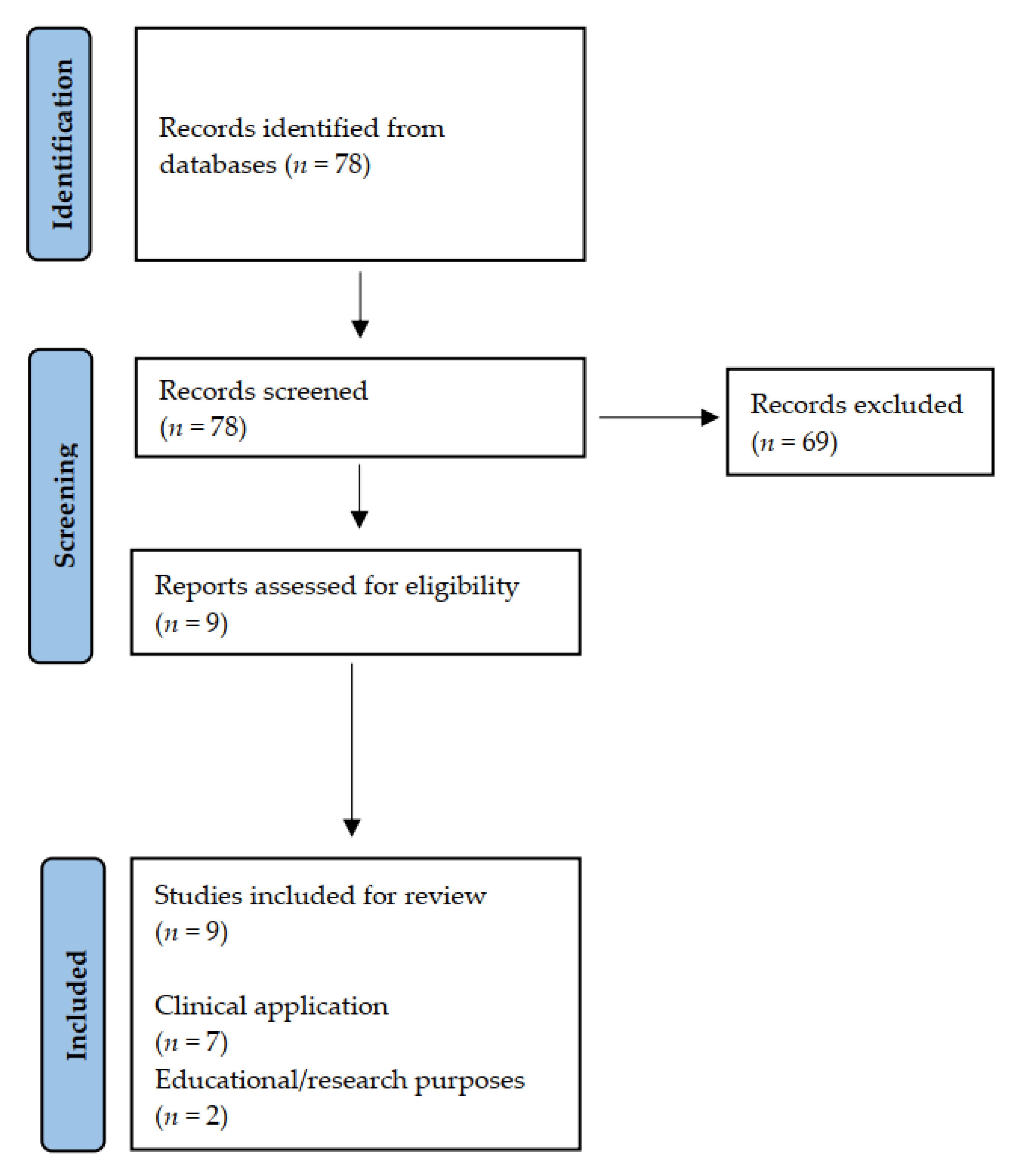
3.1. Selected Studies
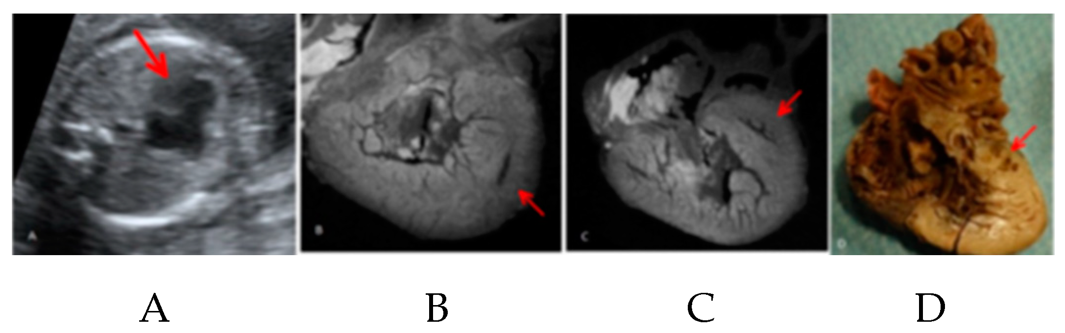
3.2. Micro-CT in Clinical Scenario
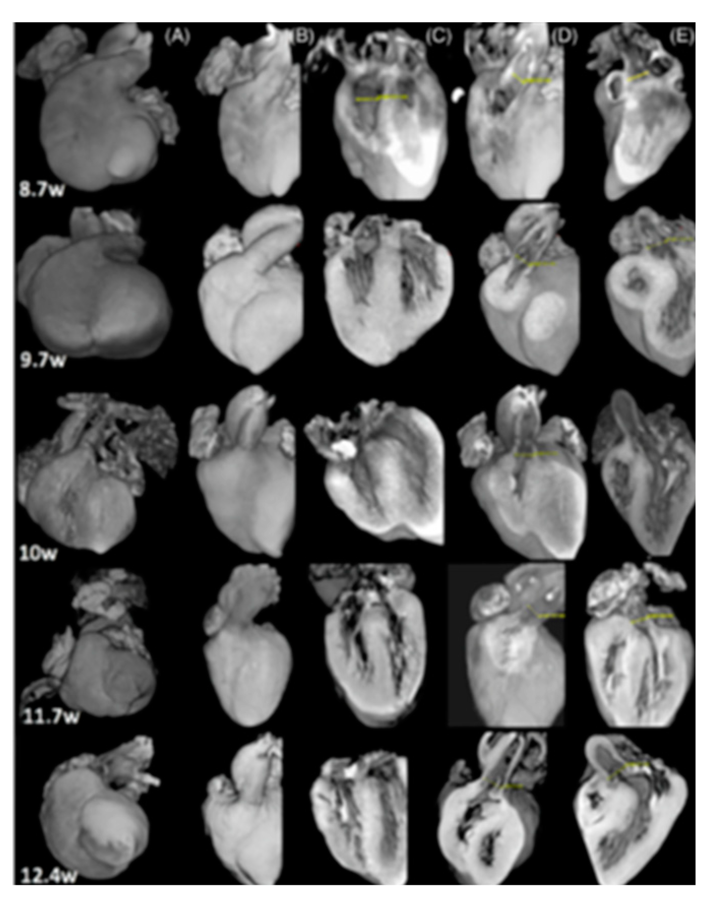
3.3. Research and Educational Purposes
4. Discussion and Conclusions
Author Contributions
Funding
Data Availability Statement
Conflicts of Interest
References
- Hoffman, J.I.E. Incidence of congenital heart disease: II. Prenatal incidence. Pediatr. Cardiol. 1995, 16, 155–165. [Google Scholar] [CrossRef] [Scilit] [PubMed]
- Hoffman, J.I.E.; Kaplan, S. The incidence of congenital heart disease. J. Am. Coll. Cardiol. 2002, 39, 1890–1900. [Google Scholar] [CrossRef] [Scilit]
- Evans, W.; Castillo, W.; Rollins, R.; Luna, C.; Kip, K.; Ludwick, J.; Madan, N.; Ciccolo, M.; Galindo, A.; Rothman, A.; et al. Moving towards universal prenatal detection of critical congenital heart disease in southern Nevada: A community-wide program. Pediatr. Cardiol. 2015, 36, 281–288. [Google Scholar] [CrossRef] [Scilit]
- Sekar, P.; Heydarian, H.; Cnota, J.; Hornberger, L.; Michelfelder, E. Diagnosis of congenital heart disease in an era of universal prenatal ultrasound screening in southwest Ohio. Cardiol. Young 2015, 25, 35–41. [Google Scholar] [CrossRef] [Scilit]
- Van Velzen, C.; Clur, S.; Rijlaarsdam, M.; Bax, C.; Pajkrt, E.; Heymans, M.; Bekker, M.N.; Hruda, J.; de Groot, C.J.M.; Blom, N.A.; et al. Prenatal detection of congenital heart disease—Results of a national screening programme. BJOG 2016, 123, 400–407. [Google Scholar] [CrossRef] [Scilit] [PubMed]
- Hartge, D.R.; Weichert, J.; Krapp, M.; Germer, U.; Gembruch, U.; Axt-Fliedner, R. Results of early foetal echocardiography and cumulative detection rate of congenital heart disease. Cardiol. Young 2011, 21, 505–517. [Google Scholar] [CrossRef] [Scilit] [PubMed]
- Jegatheeswaran, A.; Oliveira, C.; Batsos, C.; Moon-Grady, A.J.; Silverman, N.H.; Hornberger, L.K.; Coyte, P.; Friedberg, M.K. Costs of prenatal detection of congenital heart disease. Am. J. Cardiol. 2011, 108, 1808–1814. [Google Scholar] [CrossRef] [Scilit] [PubMed]
- EUROCAT. Available online: www.eurocat-network.eu (accessed on 16 August 2021).
- Carvalho, J.S.; Moscoso, G.; Tekay, A.; Campbell, S.; Thilaganathan, B.; Shinebourne, E.A. Clinical impact of first and early second trimester fetal echocardiography on high risk pregnancies. Heart 2004, 90, 921–926. [Google Scholar] [CrossRef] [Scilit]
- Smrcek, J.M.; Berg, C.; Geipel, A.; Fimmers, R.; Diedrich, K.; Gembruch, U. Early fetal echocardiography: Heart biometry and visualization of cardiac structures between 10 and 15 week’ gestation. J. Ultrasound Med. 2006, 25, 173–182. [Google Scholar] [CrossRef] [Scilit] [PubMed]
- Persico, N.; Moratalla, J.; Lombardi, C.M.; Zidere, V.; Allan, L.; Nicolaides, K.H. Fetal echocardiography at 11–13 weeks by transabdominal high-frequency ultrasound. Ultrasound Obstet. Gynecol. 2011, 37, 296–301. [Google Scholar] [CrossRef] [Scilit]
- Hutchinson, D.; McBrien, A.; Howley, L.; Yamamoto, Y.; Sekar, P.; Motan, T.; Jain, V.; Savard, W.; Hornberger, L.K. First-Trimester Fetal Echocardiography: Identification of Cardiac Structures for Screening from 6 to 13 Weeks’ Gestational Age. J. Am. Soc. Echocardiogr. 2017, 30, 763–772. [Google Scholar] [CrossRef] [Scilit]
- McBrien, A.; Hornberger, L.K. Early fetal echocardiography. Birth Defects Res. 2019, 111, 370–379. [Google Scholar] [CrossRef] [Scilit] [PubMed]
- Rizzo, G.; Calì, G.; on behalf of the “Società Italiana di Ecografia Ostetrico Ginecologica e Metodologie Biofisiche”. Linee Guida SIEOG; EDITEAM Gruppo Editoriale: Ferrara, Italy, 2015. [Google Scholar]
- Sonek, J. First Trimester Ultrasonography in Screening and Detection of Fetal Anomalies. Am. J. Med. Genet. C Semin. Med. Genet. 2007, 45, 45–61. [Google Scholar] [CrossRef] [Scilit]
- Nicolaides, K.H.; Azar, G.; Byrne, D.; Mansur, C.; Marks, K. Fetal nuchal translucency: Ultrasound screening for chromosomal defects in first trimester of pregnancy. BMJ Clin. Res. Ed. 1992, 304, 867–869. [Google Scholar] [CrossRef] [Scilit]
- Hyett, J.; Moscoso, G.; Papapanagiotou, G.; Perdu, M.; Nicolaides, K.H. Abnormalities of the heart and great arteries in chromosomally normal fetuses with increased nuchal translucency thickness at 11–13 weeks of gestation. Ultrasound Obstet. Gynecol. 1996, 7, 245–250. [Google Scholar] [CrossRef] [Scilit] [PubMed]
- Hyett, J.; Perdu, M.; Sharland, G.; Snijders, R.; Nicolaides, K.H. Using fetal nuchal translucency to screen for major congenital cardiac defects at 1014 weeks of gestation: Population based cohort study. BMJ 1999, 318, 81–85. [Google Scholar] [CrossRef] [Scilit] [PubMed]
- Ghi, T.; Huggon, I.C.; Zosmer, N.; Nicolaides, K.H. Incidence of major structural cardiac defects associated with increased nuchal translucency but normal karyotype. Ultrasound Obstet. Gynecol. 2001, 18, 610–614. [Google Scholar] [CrossRef] [Scilit]
- Sotiriadis, A.; Papatheodorou, S.; Eleftheriades, M.; Makrydimas, G. Nuchal translucency and major congenital heart defects in fetuses with normal karyotype: A meta-analysis. Ultrasound Obstet. Gynecol. 2013, 42, 383–389. [Google Scholar] [CrossRef] [Scilit] [PubMed]
- Nicolaides, K.H. Screening for fetal aneuploidies at 11 to 13 weeks. Prenat. Diagn. 2011, 31, 7–15. [Google Scholar] [CrossRef] [Scilit]
- Jakobsen, T.R.; Søgaard, K.; Tabor, A. Implications of a first trimester Down syndrome screening program on timing of malformation detection. Acta Obstet. Gynecol. Scand. 2011, 90, 728–736. [Google Scholar] [CrossRef] [Scilit] [PubMed]
- Dickinson, J.E.; Prime, D.K.; Charles, A.K. The role of autopsy following pregnancy termination for fetal abnormality. Aust. N. Z. J. Obstet. Gynaecol. 2007, 47, 445–449. [Google Scholar] [CrossRef] [Scilit]
- Rossi, A.C.; Prefumo, F. Correlation between fetal autopsy and prenatal diagnosis by ultrasound: A systematic review. Eur. J. Obstet. Gynecol. Reprod. Biol. 2016, 210, 201–206. [Google Scholar] [CrossRef] [Scilit] [PubMed]
- Sinard, J.H. Factors affecting autopsy rate, autopsy requested rates, and autopsy findings at a large academic medical center. Exp. Mol. Pathol. 2001, 70, 333–343. [Google Scholar] [CrossRef] [Scilit]
- Cannie, M.; Votino, C.; Moerman, P.H.; Vanheste, R.; Segers, V.; Van Berkel, K.; Hanssens, M.; Kang, X.; Cos, T.; Kir, M.; et al. Acceptance, reliability and confidence of diagnosis of fetal and neonatal virtuopsy compared with conventional autopsy: A prospective study. Ultrasound Obstet. Gynecol. 2012, 39, 659–665. [Google Scholar] [CrossRef] [Scilit] [PubMed]
- Lombardi, C.M.; Zambelli, V.; Botta, G.; Moltrasio, F.; Cattoretti, G.; Lucchini, V.; Fesslova, V.; Cuttin, S. Postmortem microcomputed tomography (micro-CT) of small fetuses and hearts. Ultrasound Obstet. Gynecol. 2014, 44, 600–609. [Google Scholar] [CrossRef] [Scilit] [PubMed]
- Hutchinson, J.C.; Arthurs, O.J.; Ashworth, M.T.; Ramsey, A.T.; Mifsud, W.; Lombardi, C.M.; Sebire, N.J. Clinical utility of postmortem microcomputed tomography of the fetal heart: Diagnostic imaging vs macroscopic dissection. Ultrasound Obstet. Gynecol. 2016, 47, 58–64. [Google Scholar] [CrossRef] [Scilit]
- Sandrini, C.; Rossetti, L.; Zambelli, V.; Zanarotti, R.; Bettinazzi, F.; Soldá, R.; Di Pace, C.; Hoxha, S.; Ribichini, F.L.; Faggian, G.; et al. Accuracy of Micro-Computed Tomography in Post-mortem Evaluation of Fetal Congenital Heart Disease. Comparison Between Post-mortem Micro-CT and Conventional Autopsy. Front. Pediatr. 2019, 7, 92. [Google Scholar] [CrossRef] [Scilit]
- Sandaite, I.; Lombardi, C.; Cook, A.C.; Fabietti, I.; Deprest, J.; Boito, S. Micro-computed tomography of isolated fetal hearts following termination of pregnancy: A feasibility study at 8 to 12 weeks’ gestation. Prenat. Diagn. 2020, 40, 984–990. [Google Scholar] [CrossRef] [Scilit] [PubMed]
- Hutchinson, J.C.; Kang, X.; Shelmerdine, S.C.; Segers, V.; Lombardi, C.L.; Cannie, M.M.; Sebire, N.J.; Jani, J.C.; Arthurs, O.J. Postmortem microfocus computed tomography for early gestation fetuses: A validation study against conventional autopsy. Am. J. Obstet. Gynecol. 2018, 218, 445.e1–445.e12. [Google Scholar] [CrossRef] [Scilit]
- Shelmerdine, S.C.; Simcock, I.C.; Hutchinson, J.C.; Guy, A.; Ashworth, M.T.; Sebire, N.J.; Arthurs, O.J. Postmortem microfocus computed tomography for noninvasive autopsies: Experience in >250 human fetuses. Am. J. Obstet. Gynecol. 2021, 224, 103.e1–103.e15. [Google Scholar] [CrossRef] [Scilit]
- Lombardi, S.; Scola, E.; Ippolito, D.; Zambelli, V.; Botta, G.; Cuttin, S.; Triulzi, F.; Lombardi, C.M. Micro-computed tomography: A new diagnostic tool in postmortem assessment of brain anatomy in small fetuses. Neuroradiology 2019, 61, 737–746. [Google Scholar] [CrossRef] [Scilit]
- Hutchinson, J.C.; Ashworth, M.T.; Sebire, N.J.; Arthurs, O.J. Multiple Cardiac Rhabdomyomas Visualised Using Micro-CT in a Case of Tuberous Sclerosis. Fetal Diagn. Ther. 2017, 41, 157–160. [Google Scholar] [CrossRef] [Scilit]
- Dawood, Y.; Strijkers, G.J.; Limpens, J.; Oostra, R.J.; De Bakker, B.S. Novel imaging techniques to study postmortem human fetal anatomy: A systematic review on microfocus-CT and ultra-high-field MRI. Eur. Radiol. 2020, 30, 2280–2292. [Google Scholar] [CrossRef] [Scilit]
- Shelmerdine, S.C.; Simcock, I.C.; Hutchinson, J.C.; Aughwane, R.; Melbourne, A.; Nikitichev, D.I.; Ong, J.L.; Borghi, A.; Cole, G.; Kingham, E.; et al. 3D printing from microfocus computed tomography (micro-CT) in human specimens: Education and future implications. Br. J. Radiol. 2018, 91, 1088. [Google Scholar] [CrossRef] [Scilit]
- Sandrini, C.; Lombardi, C.; Shearn, A.I.U.; Ordonez, M.V.; Caputo, M.; Presti, F.; Luciani, G.B.; Rossetti, L.; Biglino, G. Three-Dimensional Printing of Fetal Models of Congenital Heart Disease Derived From Microfocus Computed Tomography: A Case Series. Front. Pediatr. 2020, 7, 567. [Google Scholar] [CrossRef] [Scilit] [PubMed]
- Vukicevic, M.; Mosadegh, B.; Min, J.K.; Little, S.H. Cardiac 3D Printing and its Future Directions. JACC Cardiovasc. Imaging 2017, 10, 171–184. [Google Scholar] [CrossRef] [Scilit]
- Valverde, I.; Gomez-Ciriza, G.; Hussain, T.; Suarez-Mejias, C.; Velasco-Forte, M.N.; Byrne, N.; Ordoñez, A.; Gonzalez-Calle, A.; Anderson, D.; Hazekamp, M.G.; et al. Three-dimensional printed models for surgical planning of complex congenital heart defects: An international multicentre study. Eur. J. Cardiothorac. Surg. 2017, 52, 1139–1148. [Google Scholar] [CrossRef] [Scilit]
- Milano, E.G.; Capelli, C.; Wray, J.; Biffi, B.; Layton, S.; Lee, M.; Caputo, M.; Taylor, A.M.; Schievano, S.; Biglino, G. Current and future applications of 3D printing in congenital cardiology and cardiac surgery. Br. J. Radiol. 2019, 92, 20180389. [Google Scholar] [CrossRef] [Scilit] [PubMed]
- Taylor, A.M.; Arthurs, O.J.; Sebire, N.J. Postmortem cardiac imaging in fetuses and children. Pediatr. Radiol. 2015, 45, 549–555. [Google Scholar] [CrossRef] [Scilit] [PubMed]
- Arthurs, O.J.; Guy, A.; Thayyil, S.; Wade, A.; Jones, R.; Norman, W.; Scott, R.; Robertson, N.J.; Jacques, T.S.; Kling’Chong, W.K.; et al. Comparison of diagnostic performance for perinatal and paediatric post-mortem imaging: CT versus MRI. Eur. Radiol. 2016, 26, 2327–2336. [Google Scholar] [CrossRef] [Scilit]
- Arthurs, O.J.; Hutchinson, J.C.; Sebire, N.J. Current issues in postmortem imaging of perinatal and forensic childhood deaths. Forensic Sci. Med. Pathol. 2017, 13, 58–66. [Google Scholar] [CrossRef] [Scilit] [PubMed]
- Kang, X.; Carlin, A.; Cannie, M.; Sanchez, T.C.; Jani, J.C. Fetal postmortem imaging: An overview of current techniques and future perspectives. Am. J. Obstet. Gynecol. 2020, 223, 493–515. [Google Scholar] [CrossRef] [Scilit] [PubMed]
- Lupariello, F.; Genova, T.; Mussano, F.D.C.; Di Vella, G.; Botta, G. Micro-CT processing’s effects on microscopic appearance of human fetal cardiac samples. Leg. Med. 2021, 53, 101934. [Google Scholar] [CrossRef] [Scilit] [PubMed]


| Ref. | Sample (n.) | CHD (n.) | Weight (g) | Dimension (cm) | GA (w) | Fixation Protocol (Agent; Duration | Staining Protocol (Agent; Duration; %w/v) | Voxel Size (µm) | Current (µA) | Voltage (Kv) | Objective of the Study | Method | Comparison | Result |
|---|---|---|---|---|---|---|---|---|---|---|---|---|---|---|
| Lombardi CM 2014 [27] | 21; 7 whole bodies, 14 isolated hearts | 6 | Whole bodies: 0.1–90 Isolate hearts: 0.1–5.2 | NP | Whole bodies: 7–17 Isolate heart 11–22 | 4% PFA; 4–7 days | Lugol; 2–7 d; 3.75, 7.50 | 9, 18, 35 | 300, 313 | 80 | Asses feasibility and utility of micro-CT in identifying structural anomalies | Analysis of four chamber view, crux cordis and av valves; three vessels cross sectional view of PA, Ao and SVC; no gold standard technique; descriptive analysis | Micro-CT vs. conventional dissection (available only for samples > 13 w) | Feasibility of micro-CT for identification of structural anomalies |
| Hutchinson JC 2016 [28] | 6; isolated hearts | 5 | 1.1–5.3 | NP | 17–23 | 10% formalin; 48 h | Lugol; 48 h; 3.75 | 19–31 | 50–135 | 85–125 | Describe initial experience with micro-CT for the examination of complex CHD | Analysis of 21 indices of cardiac anatomy; autopsy = gold standard technique; statistical analysis | Micro-CT vs. autopsy | High accuracy of micro-CT for CHD |
| Hutchinson JC 2017 [34] | 1; isolated heart | 1 | NP | NP | 35 | NP; NP | Iodine agent; NP; NP | NP | NP | NP | Case report | Descriptive analysis | NP | 1st description of cardiac tumor identification using micro-CT |
| Hutchinson JC 2018 [31] | 20; whole bodies | 2 | NP | NP | 11–21 | 10% formalin; 72 h | Lugol; 72 h; 3.75 | 7.4–51 | 87–180 | 80–110 | Evaluate the diagnostic accuracy of micro-CT for non-invasive human fetal autopsy for early gestation fetuses | Analysis of 9 indices of cardiac anatomy; autopsy = gold standard technique; statistical analysis | Micro-CT vs. autopsy | High accuracy of micro-CT in early gestation fetuses; better performance of micro-CT in first trimester fetuses Heart: sensitivity 90.5%, specificity 100%, PPV 100%, NPV 98.6%, concordance 98.7% |
| Sandrini C 2019 [29] | 10; isolated hearts or heart-lungs block | 10 | 0.39–4 | 0.5–1.8 | 12–22 | 10% formalin; 16–260 d | Lugol; 72 h; 3–3.75 | 9–18 | 264–500 | 50–89 | Evaluate the diagnostic accuracy of micro-CT for diagnosis of CHD | Segmental approach; analysis of 25 indices of cardiac anatomy; autopsy = gold standard technique; statistical analysis | Micro-CT vs. autopsy | Equal diagnostic power of micro-CT as compared to autopsy for CHD; more diagnostic accuracy of micro-CT in small specimens |
| Shelmerdine S.C 2020 [32] | 268; whole bodies | 6 | 3–350 | 6–26 | 11–24 | 10% formalin; NP | Lugol; ≥96; NP | 18.6–121.7 | 78–350 | 60–160 | Describe the range of anomalies detectable on fetal micro-CT; estimate the invasive autopsy avoidance rate | Definition of normal, abnormal or non-diagnostic for different body areas (including cardiovascular system); autopsy/MIA/limited autopsy/NIA performed only if needed; autopsy = gold standard technique (when performed); statistical analysis (when possible) | Micro-CT vs. autopsy/limited autopsy (when performed) | Postmortem micro-CT as part of a NIA examination is feasible |
| Sandaite I 2020 [30] | 49; isolated hearts | 6 | NP | 0.5 | 8–12 | 10% formalin; NP | Lugol; 24 h; NP | 9 | NP | NP | Asses the feasibility of retrieval of heart after 1st trimester TOP; asses the feasibility of micro-CT for anatomical evaluation | Segmental approach; analysis of cardiac chambers, av connection, outflow tracts, va connection, av and semilunar valves, ventricular septum; no gold standard technique; descriptive analysis | No | Fetal heart can be retrieved after 1st trimester TOP; micro-CT can be used from as early as 8 w to describe cardiac structures |
Publisher’s Note: MDPI stays neutral with regard to jurisdictional claims in published maps and institutional affiliations. |
© 2021 by the authors. Licensee MDPI, Basel, Switzerland. This article is an open access article distributed under the terms and conditions of the Creative Commons Attribution (CC BY) license (https://creativecommons.org/licenses/by/4.0/).
Share and Cite
Sandrini, C.; Boito, S.; Lombardi, C.M.; Lombardi, S. Postmortem Micro-CT of Human Fetal Heart—A Systematic Literature Review. J. Clin. Med. 2021, 10, 4726. https://doi.org/10.3390/jcm10204726
Sandrini C, Boito S, Lombardi CM, Lombardi S. Postmortem Micro-CT of Human Fetal Heart—A Systematic Literature Review. Journal of Clinical Medicine. 2021; 10(20):4726. https://doi.org/10.3390/jcm10204726
Chicago/Turabian StyleSandrini, Camilla, Simona Boito, Claudio M. Lombardi, and Sophie Lombardi. 2021. "Postmortem Micro-CT of Human Fetal Heart—A Systematic Literature Review" Journal of Clinical Medicine 10, no. 20: 4726. https://doi.org/10.3390/jcm10204726
APA StyleSandrini, C., Boito, S., Lombardi, C. M., & Lombardi, S. (2021). Postmortem Micro-CT of Human Fetal Heart—A Systematic Literature Review. Journal of Clinical Medicine, 10(20), 4726. https://doi.org/10.3390/jcm10204726