Article

The Relative Role of Toxins A and B in the Virulence of Clotridioides difficile

1
Research Service, Edward Hines Jr., Veterans Affairs Hospital, Infectious Disease Section, Hines, IL 60141, USA
2
Department of Medicine, Loyola University Medical Center, Chicago, IL 60153, USA
3
Department of Medicine, Northwestern University Medical School, Chicago, IL 60611, USA
4
Pritzker School of Medicine, University of Chicago, Chicago, IL 60637, USA
*
Author to whom correspondence should be addressed.
J. Clin. Med. 2021, 10(1), 96; https://doi.org/10.3390/jcm10010096
Submission received: 14 December 2020 / Revised: 24 December 2020 / Accepted: 26 December 2020 / Published: 30 December 2020
(This article belongs to the Special Issue Clostridium difficile Infection: Unmet Needs and Unsolved Questions)

Abstract

:
Most pathogenic strains of C. difficile possess two large molecular weight single unit toxins with four similar functional domains. The toxins disrupt the actin cytoskeleton of intestinal epithelial cells leading to loss of tight junctions, which ultimately manifests as diarrhea in the host. While initial studies of purified toxins in animal models pointed to toxin A (TcdA) as the main virulence factor, animal studies using isogenic mutants demonstrated that toxin B (TcdB) alone was sufficient to cause disease. In addition, the natural occurrence of TcdA−/TcdB+ (TcdA−/B+)mutant strains was shown to be responsible for cases of C. difficile infection (CDI) with symptoms identical to CDI caused by fully toxigenic (A+/B+) strains. Identification of these cases was delayed during the period when clinical laboratories were using immunoassays that only detected TcdA (toxA EIA). Our hospital laboratory at the time performed culture as well as toxA EIA on patient stool samples. A total of 1.6% (23/1436) of all clinical isolates recovered over a 2.5-year period were TcdA−/B+ variants, the majority of which belonged to the restriction endonuclease analysis (REA) group CF and toxinotype VIII. Despite reports of serious disease due to TcdA−/B+ CF strains, these infections were typically mild, often not requiring specific treatment. While TcdB alone may be sufficient to cause disease, clinical evidence suggests that both toxins have a role in disease.

1. Introduction

Most virulent strains of Clostridioides difficile produce two large single unit toxins which act via glycosylation of small GTP-binding proteins involved in cell cytoskeleton organization resulting in the disruption of epithelial tight junctions [1,2,3]. These two primary toxins, identified as toxin A (TcdA) and toxin B (TcdB), are encoded by tcdA and tcdB located with the pathogenicity locus [4]. As such, the detection of toxins A and B in patient stool specimens has been utilized by clinical laboratories to diagnose C. difficile infection (CDI) for decades [5]. The first toxin assay available was a cell culture cytotoxin assay, which primarily reflects the cytopathic effects of TcdB on eukaryotic cell culture [6]. In the late 1990s, many clinical laboratories implemented TcdA enzyme immunoassays (EIA) for C. difficile testing in place of cytotoxin assays because of the rapidity and simplicity of the test, as well as initial data showing comparable results with cell culture assays [7,8]. These EIAs employed a monoclonal antibody (PCG-4) that recognizes epitopes encoded by a region of highly repetitive DNA sequences in the 3′ end of tcdA [9]. Previous studies of purified toxins in animal models indicated that TcdA was the primary virulence determinant; therefore, TcdA was included as the sole target antigen in these early immunoassays [10]. Subsequent studies identified the importance of TcdB, including animal studies with challenges by isogenic tcdA and tcdB mutant strains, which demonstrated that TcdB alone could result in disease [11,12].
C. difficile toxin variants have been described that do not react with TcdA EIA but are cytotoxic in cell culture assays [13]. These strains share common genetic alterations including truncation at the 3′ end of tcdA and a point mutation at the 5′ end of tcdA that introduces a premature stop codon at amino acid position 47 [13,14]. CDI due to these toxin A−/B+ (TcdA−/B+) variants has been increasingly common across the world, including multiple nosocomial outbreaks, highlighting the limitations of TcdA EIA as a diagnostic tool [15,16,17,18,19,20,21,22,23]. These limitations were further highlighted by a fatal case of pseudomembranous colitis due to a TcdA−/B+ variant strain (REA Type CF4) for which the diagnosis of CDI was delayed by reliance on the TcdA EIA test which was repeatedly negative [24].
C. difficile testing has evolved over the past 20 years due to detection limitations and the inability of TcdA EIAs to detect C. difficile TcdA−/B+ variant strains and TcdA EIA has been replaced by EIAs that detect both toxins. However, still unanswered is the clinical and epidemiologic significance of TcdA−/B+ variant C. difficile isolates. In this study, we investigated the incidence, outcomes, and laboratory characterization of TcdA−/B+ variant CDI.

2. Experimental Section

We conducted a single-center retrospective study from September 1999 to January 2002 at Northwestern Memorial Hospital located in Chicago, Illinois to determine the incidence, clinical impact, and the molecular epidemiology of CDI related to TcdA−/B+ variant isolates.

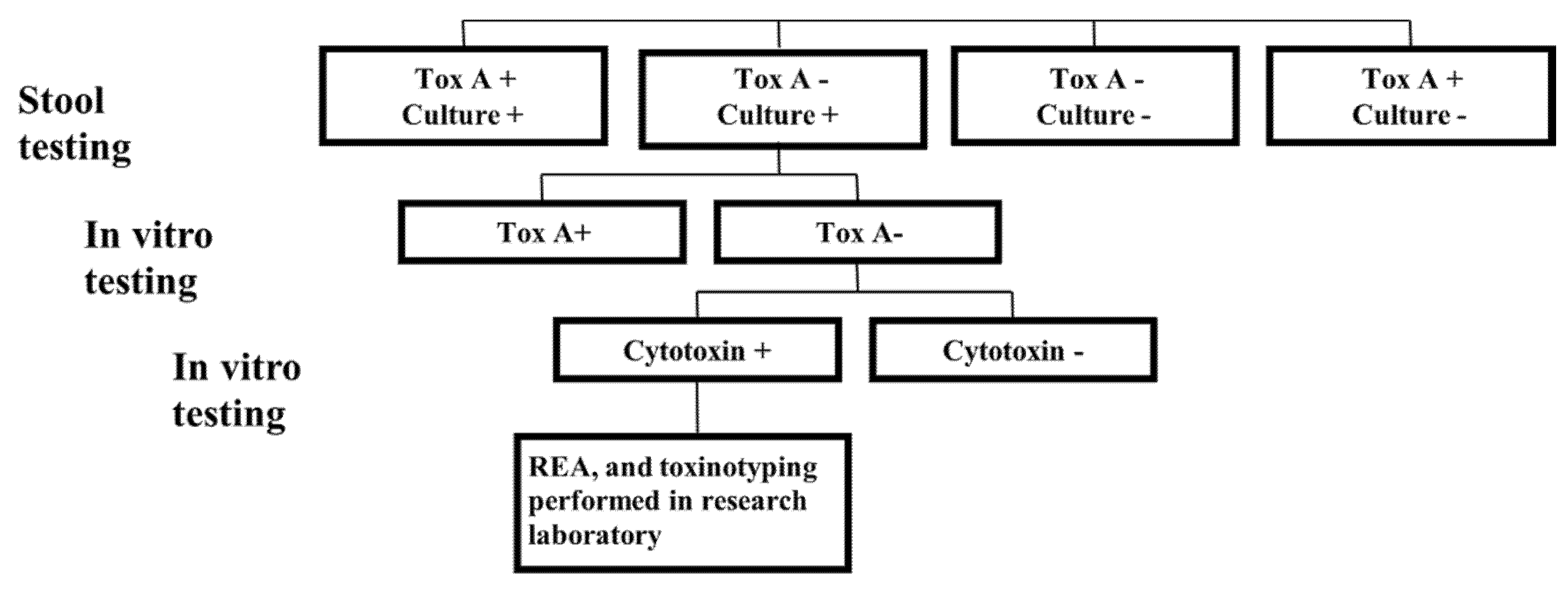
2.1. Clinical Microbiology Laboratory Testing

From September 1999 to January 2002, patient stool samples were submitted to the clinical microbiology laboratory for C. difficile testing. Specimens were tested for the presence of TcdA by immunoassay (Clearview C. difficile Toxin A EIA, Wampole Laboratories, Cranbury, NJ, USA) and for the presence of C. difficile by culture.

2.1.1. Culture

All stool samples were cultured anaerobically for C. difficile on selective cycloserine cefoxitin fructose agar (CCFA) media as previously described [25]. Colonies of C. difficile identified on culture were inoculated into Chopped Meat Carbohydrate Medium (CMCM) and incubated at 35 degrees C for 48 h.

2.1.2. In Vitro Testing of Recovered C. difficile Isolates for TcdA and Cytotoxicity

C. difficile isolates that were recovered from stool samples but were negative for TcdA by EIA in the clinical laboratory were subjected to in vitro testing for TcdA. Supernatant of the CMCM was diluted 1:10 and tested by TcdA EIA as per the manufacturer’s directions (C. difficile TOX-A, TechLab, Blacksburg, VA, USA). Isolates that were negative for TcdA underwent a qualitative cytotoxin analysis. C. difficile-inoculated CMCM was diluted 1:10 and aliquots were pre-incubated with either diluent or C. difficile antitoxin. Each separate aliquot was inoculated onto cell cultures of human foreskin fibroblasts (final dilution 1:200) (Toxi-Titer cytotoxicity assay, Trinity Biotech, Carlsbad, CA, USA). Cells were screened for cytopathic changes at 24 and 48 h. Supernatant aliquots that demonstrated cytotoxicity at the final titer (1:200) and which were neutralized by the antitoxin were considered positive. Supernatants from known toxigenic and non-toxigenic strains were included as the positive and negative controls, respectively. C. difficile isolates that were negative by TcdA EIA but had a positive cytotoxicity assay underwent REA typing and toxinotyping in our research laboratory (Figure 1).

2.2. Research Laboratory Testing

2.2.1. Restriction Endonuclease Analysis (REA) Typing

C. difficile isolates were cultured in brain heart infusion or tryptic soy broth (TSB). Using methods previously described by Clabots et al., DNA was isolated, purified, and subjected to HindIII restriction enzyme digestion and electrophoresis on 0.7% agarose gel [25]. The pattern of each isolate was visually compared with a known database [26,27]. Patterns that had a 90% similarity index were placed within the same REA group (letter designations) and identical patterns were classified by a subgroup (number designation).

2.2.2. Toxinotyping

C. difficile strains were inoculated into TSB broth from a blood agar plate 48-h culture and incubated anaerobically overnight. Total cellular DNA was isolated as previously described [25]. Polymerase chain reaction (PCR) was performed to create fragments A3 and B1, which were subsequently restricted by HincII or AccI (B1) and EcoRI (A3) and compared to reference strains as described by Rupnik et al. [7].

2.3. Clinical Data

A chart review of patients with isolates determined to be TcdA−/B+ was conducted to obtain demographics, medications, CDI treatment, and outcomes. The determination of initial versus recurrent CDI, healthcare associated CDI (HA-CDI), and community associated CDI (CA-CDI) were classified per the Infectious Diseases Society of America and Society for Healthcare Epidemiology of America (IDSA/SHEA) guidelines [5]. The institutional review board of the hospital approved the study protocol.

3. Results

3.1. Surveillance by TcdA Immunoassay

From 1 September, 1999 to 1 January, 2002, 10,668 stools were submitted for C. difficile testing. In total, 1661 of the stool specimens were positive by Toxin A EIA or culture (15.6%, 95% Confidence Interval (CI): 14.9–16.3%). Of these 1661 C. difficile-positive stool specimens, C. difficile culture and TcdA EIA were positive in 1436 (86.5%, 95% CI: 84.8–88.1%) and 586 (35.3%, 95% CI: 32.9–37.6%), respectively (Table 1). Among the C. difficile 1436 stools that were culture positive, 1075 were TcdA EIA negative in the clinical laboratory (74.9%, 95% CI: 72.6–77.1%).
Our reference laboratory documented in vitro TcdA production in 786 of the 1075 isolates (73.1%, 95% CI: 70.4–75.8%). Of the remaining isolates, 289 (26.9%, CI:24.3–29.6%) were negative for TcdA by in vitro analysis. Further analysis of the 289 in vitro TcdA−negative isolates revealed that 23 isolates were positive by in vitro cytotoxicity assay indicating the presence of TcdB. Therefore, 1.6% (23/1436) of the isolates that were recovered during this 2.5-year surveillance period were TcdA−/B+ variants (Table 1).

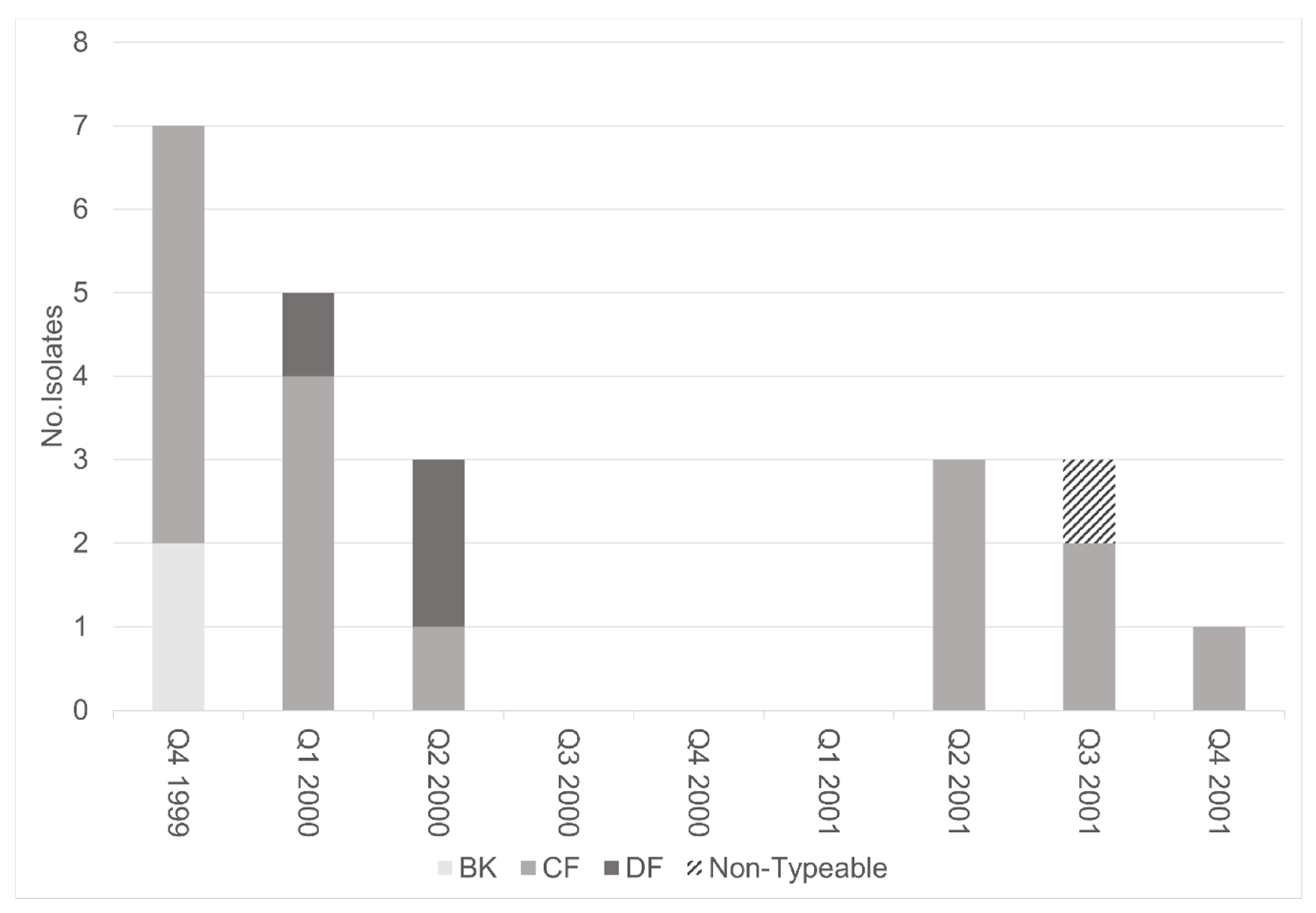
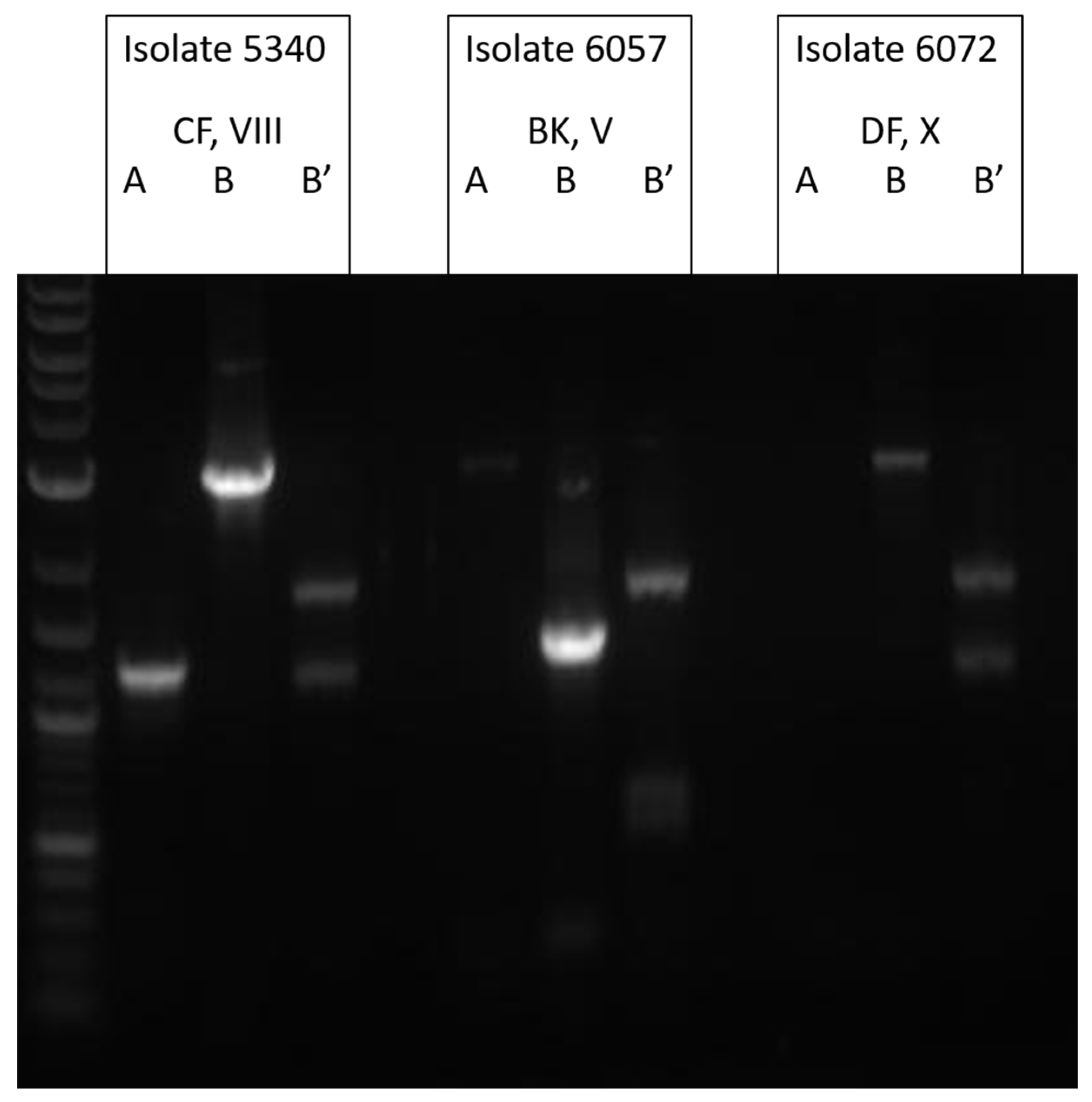
3.2. Molecular Analysis of TcdA−Negative, B-Positive Variant Isolates

Twenty-two TcdA−/B+ variant isolates were characterized by REA and representative isolates were analyzed by toxinotyping (one isolate was unrecoverable after initial testing). These variants were found throughout the study time period with no obvious clustering (Figure 2). The majority of toxin variants examined belonged to REA group CF (73%, 16/22). Eight of these group CF isolates were the specific REA type, CF4. The other 8 CF isolates were identified as eight different REA subgroups (Figure 2). This strain was previously identified as the cause of a fatal case of CDI in our institution [24]. Toxinotyping of a representative CF isolate confirmed this strain to be toxinotype VIII (Figure 3), consistent with our previous analyses of REA group CF strains [28].
In addition to the REA group CF strains, we identified two new TcdA-/B+ variants. The restriction pattern of three isolates had never been seen before at the time of diagnosis and was designated as a new REA group (DF) and REA type, DF 1. The toxinotype pattern of this isolate was consistent with toxinotype X (Figure 3). Two isolates were identified as REA group BK and toxinotype V (Figure 3).

3.3. Clinical Outcomes

The 23 TcdA−/B+ variant isolates were recovered from 21 patients whose clinical charts were reviewed for clinical management and outcome (Table 2). Among these patients, 48% (10/21) were female and the median age was 60 years (range: 37–86). The initial CDI episode was healthcare facility-associated in 71% (15/21) of the cases. Recent antibiotic exposure was documented in 84% (16/19) and diarrhea was noted in 95% (19/20). Less than half of the patients (37%, 7/19) received specific therapy for CDI and the symptoms in patients who did not receive specific therapy resolved spontaneously. There were no deaths and no other identified complications.

4. Discussion

From September 1999 to January 2002, TcdA−/B+ variants accounted for 1.6% of all the recovered C. difficile isolates within a single healthcare system in United States. The majority of these isolates were identified as REA group CF, a well-characterized TcdA−/B+ C. difficile variant [13,14]. We also found two additional REA groups, BK and DF, which were also identified as TcdA−/B+ isolates.
REA group CF commonly correlates with PCR-Ribotype 017 (RT017), toxinotype VIII, NAP9, and ST37 [28,29,30,31] and has been well established as a TcdA−/B+ variant that results in a symptomatic CDI through the actions of toxin B [32]. CF/RT017 has been found worldwide but is endemic to East Asia and historically accounted for 15–40% of CDI in South Korea, Taiwan, and China [33,34,35]. Initial studies indicated that CF/RT017 was capable of causing severe disease and even mortality [21,22,23,24,28,32,33,34,35,36]. Within our own healthcare system, we reported a death due to REA group CF related CDI [24]. The outcome of this case was likely attributed to a lack of specific antibiotic treatment due to an overreliance on clinical laboratory testing which at the time only included toxin identification by TcdA EIA, despite overwhelming clinical evidence of CDI [24]. Severe outcomes due to TcdA−/B+ CDI are well documented, particularly in outbreak settings, however, the full spectrum of infection with these variants is not clear, particularly in certain regions of Asia where RT 017 is common and where severe disease and mortality are uncommon [32]. Data provided by our study would further support that CDI caused by TcdA−/B+ variants more commonly results in less severe CDI. There were no reported fatal events within our study and the diagnoses and treatments were often delayed in subjects with TcdA−/B+ related CDI due to the delay in final test results which involved culture and in vitro toxin testing of the recovered isolate. In fact, the final test results were not reported in some cases until after the patient had improved and was discharged from the hospital.
Over the past three decades, the understanding of TcdA and TcdB has expanded greatly. Initial animal models using purified TcdA and TcdB indicated that TcdA was the primary virulence determinant [10,37]. As such, emphasis was placed on TcdA for the detection of C. difficile within clinical laboratories [8]. However, subsequent animal experiments using isogenic TcdA and TcdB C. difficile mutants demonstrated that TcdB is not only sufficient in causing disease, but also causes CDI through multiple mechanisms [11,12,38]. Moreover, Carter, et al. used isogenic C. difficile mutants in three different animal models, showing that the TcdA−/B+ mutants were attenuated in virulence [39]. Furthermore, TcdB alone caused severe localized intestinal damage as well as systemic organ damage and TcdB was primarily responsible for inducing host innate and inflammatory immune responses [39].
These data supporting the importance of TcdB are further bolstered by the clinical observations of TcdA−/B+ isolates resulting in CDI. Additionally, the importance of TcdB is also supported by a randomized clinical trial using the monoclonal bezlotoxumab [40,41]. Bezlotoxumab, a monoclonal antibody directed towards TcdB, has demonstrated the capacity to decrease recurrent CDI through binding TcdB [40,41]. However, no such effect was noted with the monoclonal antibody with activity towards TcdA [40]. As such, the primary focus in both clinical and molecular epidemiology has focused more so on TcdB.
Recent data have demonstrated that TcdA causes cellular apoptosis in a manner distinctly different than TcdB [38]. TcdA, a potent enterotoxin, has been shown to induce epithelial apoptosis through glucosyltransferase-dependent caspase-3 activation [38]. This action by TcdA in turn results in epithelial tight junction disruption and likely results in mucosal damage and fluid loss [42]. TcdB, a cytotoxin, causes cellular necrosis and apoptosis via a glucosyltransferase-independent reactive oxygen species production [38,43]. Despite these differences, toxin A+/B- mutants have demonstrated virulence in vitro, albeit with somewhat attenuated virulence [12,39]. Further data supporting the importance of TcdA can be seen in animal models. Hamster models have demonstrated that while TcdA−/B+ REA CF strains are virulent, they also demonstrate delayed colonization and incomplete mortality when compared with fully toxigenic (A+/B+) strains [44]. These findings support roles for both toxin A and B in inducing a CDI as well as colonization.
The studies surrounding REA group CF (RT017) reveal that TcdA−/B+ variants are clearly pathogenic [21,22,23,24,28,32,33,34,35,36]. However, a detailed clinical study to determine the clinical outcomes and the virulence of this group strain is lacking. Our data, while limited to one institution, indicate that these variant isolates may not carry the same risk of severe CDI as fully toxigenic A+/B+ isolates. Our study was limited by the lack of a contemporary control group comparing clinical outcomes in patients infected with toxin A+/B+ strains. However, we included all patients identified with TcdA−/TcdB+ over a 2.5-year period in our institution and did not identify any severe or complicated infections.
Moreover, while CF/RT017 is more common in Asia, our study reveals that REA group BK (RT078/128), a strain more common in Europe, can lack TcdA [45]. Current data surrounding REA group BK (RT078/128) support the hypothesis that the group is a fully toxigenic and harbors both TcdA and TcdB [46]. Our findings indicate that group strains which we presume to be fully toxigenic may harbor mutations which render them TcdA−/B+ variants, yet remain virulent [30]. To fully understand the implications of C. difficile TcdA−/B+ variant isolates, robust genomic and molecular epidemiologic studies will be required to answer these still yet unanswered questions regarding the importance of C. difficile toxins A and B.

Author Contributions

Conceptualization, S.J., L.R.P., and D.N.G.; Methodology, S.J., L.R.P.; Investigation, K.J.O.; Data Curation, M.M.M., S.P.S., and F.S.; Writing-Original Draft Preparation, A.M.S.; Writing-Review & Editing, A.M.S., S.T.P., L.R.P., D.N.G., and S.J.; Supervision, S.J.; Funding Acquisition, S.J. All authors have read and agreed to the published version of the manuscript.

Funding

Stuart Johnson and Dale Gerding are supported in part by the Veterans Administration Merit Review Research Program.

Institutional Review Board Statement

The study was approved by the Institutional Review Board of Northwestern University (Project#:659-003; Approved 9/8/2000).

Informed Consent Statement

Informed consent was waived.

Data Availability Statement

The data presented in this study are available on request from the corresponding author. The data are not publicly available due to privacy and ethical concerns.

Acknowledgments

We thank Iris Figueroa for her help with the REA and toxinotyping analyses and John Warren and Pam Kelly for their support and the resources utilized in the clinical microbiology laboratory at Northwestern Memorial Hospital.

Conflicts of Interest

The authors declare no significant conflict of interest.

References

  1. Just, I.; Fritz, G.; Aktories, K.; Giry, M.; Popoff, M.R.; Boquet, P.; Hegenbarth, S.; von Eichel-Streiber, C. Clostridium Difficile Toxin B Acts on the GTP-Binding Protein Rho. J. Biol. Chem. 1994, 269, 10706–10712. [Google Scholar] [PubMed]
  2. Hundsberger, T.; Braun, V.; Weidmann, M.; Leukel, P.; Sauerborn, M.; von Eichel-Streiber, C. Transcription Analysis of the Genes TcdA-E of the Pathogenicity Locus of Clostridium Difficile. Eur. J. Biochem. 1997, 244, 735–742. [Google Scholar] [CrossRef] [PubMed]
  3. Hecht, G.; Pothoulakis, C.; LaMont, J.T.; Madara, J.L. Clostridium Difficile Toxin A Perturbs Cytoskeletal Structure and Tight Junction Permeability of Cultured Human Intestinal Epithelial Monolayers. J. Clin. Investig. 1988, 82, 1516–1524. [Google Scholar] [CrossRef] [PubMed]
  4. Hammond, G.A.; Johnson, J.L. The Toxigenic Element of Clostridium Difficile Strain VPI 10463. Microb. Pathog. 1995, 19, 203–213. [Google Scholar] [CrossRef]
  5. McDonald, L.C.; Gerding, D.N.; Johnson, S.; Bakken, J.S.; Carroll, K.C.; Coffin, S.E.; Dubberke, E.R.; Garey, K.W.; Gould, C.V.; Kelly, C.; et al. Clinical Practice Guidelines for Clostridium Difficile Infection in Adults and Children: 2017 Update by the Infectious Diseases Society of America (IDSA) and Society for Healthcare Epidemiology of America (SHEA). Clin. Infect. Dis. 2018, 66, e1–e48. [Google Scholar] [CrossRef]
  6. Musher, D.M.; Manhas, A.; Jain, P.; Nuila, F.; Waqar, A.; Logan, N.; Marino, B.; Graviss, E.A. Detection of Clostridium Difficile Toxin: Comparison of Enzyme Immunoassay Results with Results Obtained by Cytotoxicity Assay. J. Clin. Microbiol. 2007, 45, 2737–2739. [Google Scholar] [CrossRef] [Green Version]
  7. Rupnik, M.; Avesani, V.; Janc, M.; von Eichel-Streiber, C.; Delmée, M. A Novel Toxinotyping Scheme and Correlation of Toxinotypes with Serogroups of Clostridium DifficileIsolates. J. Clin. Microbiol. 1998, 36, 2240–2247. [Google Scholar] [CrossRef] [Green Version]
  8. Borek, A.; Kini, A.; Kelly, P.; Wunderlickh-Weiorka, L.; Peterson, L. Prospective Comparison of Clearview C. DIFF A (CDA) with Meridian Immunocard (IMM), Stool Culture (StC) and Cytotoxin Detection (CyD) for Diagnosis of Clostridium Difficile Associated Diarrhea (CDAD). In Proceedings of the Ninety-Eighth Annual Meeting of the American Society for Microbiology, Atlanta, GA, USA, 17–21 May 1998. [Google Scholar]
  9. Lyerly, D.M.; Neville, L.M.; Evans, D.T.; Fill, J.; Allen, S.; Greene, W.; Sautter, R.; Hnatuck, P.; Torpey, D.J.; Schwalbe, R. Multicenter Evaluation of the Clostridium DifficileTOX A/B TEST. J. Clin. Microbiol. 1998, 36, 184–190. [Google Scholar] [CrossRef] [Green Version]
  10. Lyerly, D.M.; Lockwood, D.E.; Richardson, S.H.; Wilkins, T.D. Biological Activities of Toxins A and B of Clostridium Difficile. Infect. Immun. 1982. [Google Scholar] [CrossRef] [Green Version]
  11. Lyras, D.; O’Connor, J.R.; Howarth, P.M.; Sambol, S.P.; Carter, G.P.; Phumoonna, T.; Poon, R.; Adams, V.; Vedantam, G.; Johnson, S.; et al. Toxin B Is Essential for Virulence of Clostridium Difficile. Nature 2009, 458, 1176–1179. [Google Scholar] [CrossRef]
  12. Kuehne, S.A.; Cartman, S.T.; Heap, J.T.; Kelly, M.L.; Cockayne, A.; Minton, N.P. The Role of Toxin A and Toxin B in Clostridium Difficile Infection. Nature 2010, 467, 711–713. [Google Scholar] [CrossRef] [PubMed] [Green Version]
  13. Sambol, S.P.; Merrigan, M.M.; Lyerly, D.; Gerding, D.N.; Johnson, S. Toxin Gene Analysis of a Variant Strain of Clostridium Difficile That Causes Human Clinical Disease. Infect. Immun. 2000. [Google Scholar] [CrossRef] [PubMed] [Green Version]
  14. Von Eichel-Streiber, C. A Nonsense Mutation Abrogates Production of a Functional Enterotoxin A in Clostridium Difficile Toxinotype VIII Strains of Serogroups F and X. FEMS Microbiol. Lett. 1999, 178, 163–168. [Google Scholar] [CrossRef]
  15. Kato, H.; Kato, N.; Watanabe, K.; Iwai, N.; Nakamura, H.; Yamamoto, T.; Suzuki, K.; Kim, S.-M.; Chong, Y.; Wasito, E.B. Identification of Toxin A-Negative, Toxin B-Positive Clostridium Difficile by PCR. J. Clin. Microbiol. 1998, 36, 2178–2182. [Google Scholar] [CrossRef] [Green Version]
  16. Barbut, F.; Lalande, V.; Burghoffer, B.; Thien, H.V.; Grimprel, E.; Petit, J.-C. Prevalence and Genetic Characterization of Toxin A Variant Strains of Clostridium Difficile among Adults and Children with Diarrhea in France. J. Clin. Microbiol. 2002, 40, 2079–2083. [Google Scholar] [CrossRef] [PubMed] [Green Version]
  17. Limaye, A.P.; Turgeon, D.K.; Cookson, B.T.; Fritsche, T.R. Pseudomembranous Colitis Caused by a Toxin A(-) B(+) Strain of Clostridium Difficile. J. Clin. Microbiol. 2000, 38, 1696–1697. [Google Scholar] [CrossRef] [Green Version]
  18. Rupnik, M.; Kato, N.; Grabnar, M.; Kato, H. New Types of Toxin A-Negative, Toxin B-Positive Strains among Clostridium Difficile Isolates from Asia. J. Clin. Microbiol. 2003, 41, 1118–1125. [Google Scholar] [CrossRef] [Green Version]
  19. Goorhuis, A.; Legaria, M.C.; van den Berg, R.J.; Harmanus, C.; Klaassen, C.H.W.; Brazier, J.S.; Lumelsky, G.; Kuijper, E.J. Application of Multiple-Locus Variable-Number Tandem-Repeat Analysis to Determine Clonal Spread of Toxin A-Negative Clostridium Difficile in a General Hospital in Buenos Aires, Argentina. Clin. Microbiol. Infect. 2009, 15, 1080–1086. [Google Scholar] [CrossRef] [Green Version]
  20. Kim, H.; Riley, T.V.; Kim, M.; Kim, C.K.; Yong, D.; Lee, K.; Chong, Y.; Park, J.-W. Increasing Prevalence of Toxin A-Negative, Toxin B-Positive Isolates of Clostridium Difficile in Korea: Impact on Laboratory Diagnosis. J. Clin. Microbiol. 2008, 46, 1116–1117. [Google Scholar] [CrossRef] [Green Version]
  21. Alfa, M.J.; Kabani, A.; Lyerly, D.; Moncrief, S.; Neville, L.M.; Al-Barrak, A.; Harding, G.K.H.; Dyck, B.; Olekson, K.; Embil, J.M. Characterization of a Toxin A-Negative, Toxin B-Positive Strain of Clostridium Difficile Responsible for a Nosocomial Outbreak of Clostridium Difficile-Associated Diarrhea. J. Clin. Microbiol. 2000, 38, 2706–2714. [Google Scholar] [CrossRef] [Green Version]
  22. Kuijper, E.; Weerdt, J.; Kato, H.; Kato, N.; Dam, A.; Vorm, E.; Weel, J.; Rheenen, C.; Dankert, J. Nosocomial Outbreak of Clostridium Difficile-Associated Diarrhoea Due to a Clindamycin-Resistant Enterotoxin A-Negative Strain. Eur. J. Clin. Microbiol. Infect. Dis. 2001, 20, 528–534. [Google Scholar] [CrossRef] [PubMed]
  23. Drudy, D.; Harnedy, N.; Fanning, S.; Hannan, M.; Kyne, L. Emergence and Control of Fluoroquinolone-Resistant, Toxin A–Negative, Toxin B–Positive Clostridium Difficile. Infect. Control Hosp. Epidemiol. 2007, 28, 932–940. [Google Scholar] [CrossRef] [PubMed] [Green Version]
  24. Johnson, S.; Kent, S.A.; O’Leary, K.J.; Merrigan, M.M.; Sambol, S.P.; Peterson, L.R.; Gerding, D.N. Fatal Pseudomembranous Colitis Associated with a Variant Clostridium Difficile Strain Not Detected by Toxin A Immunoassay. Ann. Intern. Med. 2001, 135, 434. [Google Scholar] [CrossRef] [PubMed]
  25. Clabots, C.R.; Johnson, S.; Bettin, K.M.; Mathie, P.A.; Mulligan, M.E.; Schaberg, D.R.; Peterson, L.R.; Gerding, D.N. Development of a Rapid and Efficient Restriction Endonuclease Analysis Typing System for Clostridium Difficile and Correlation with Other Typing Systems. J. Clin. Microbiol. 1993, 31, 1870–1875. [Google Scholar] [CrossRef] [PubMed] [Green Version]
  26. Manzo, C.E.; Merrigan, M.M.; Johnson, S.; Gerding, D.N.; Riley, T.V.; Silva, J.; Brazier, J.S. International Typing Study of Clostridium Difficile. Anaerobe 2014, 28, 4–7. [Google Scholar] [CrossRef] [PubMed]
  27. Kociolek, L.K.; Perdue, E.R.; Fawley, W.N.; Wilcox, M.H.; Gerding, D.N.; Johnson, S. Correlation between Restriction Endonuclease Analysis and PCR Ribotyping for the Identification of Clostridioides (Clostridium) Difficile Clinical Strains. Anaerobe 2018, 54, 1–7. [Google Scholar] [CrossRef] [Green Version]
  28. Johnson, S.; Sambol, S.P.; Brazier, J.S.; Delmee, M.; Avesani, V.; Merrigan, M.M.; Gerding, D.N. International Typing Study of Toxin A-Negative, Toxin B-Positive Clostridium Difficile Variants. J. Clin. Microbiol. 2003, 41, 1543–1547. [Google Scholar] [CrossRef] [Green Version]
  29. Rupnik, M.; Brazier, J.S.; Duerden, B.I.; Grabnar, M.; Stubbs, S.L.J. Comparison of Toxinotyping and PCR Ribotyping of Clostridium Difficile Strains and Description of Novel Toxinotypes. Microbiology 2001, 147, 439–447. [Google Scholar] [CrossRef] [Green Version]
  30. Tenover, F.C.; Åkerlund, T.; Gerding, D.N.; Goering, R.V.; Boström, T.; Jonsson, A.M.; Wong, E.; Wortman, A.T.; Persing, D.H. Comparison of Strain Typing Results for Clostridium Difficile Isolates from North America. J. Clin. Microbiol. 2011, 49, 1831–1837. [Google Scholar] [CrossRef] [Green Version]
  31. Griffiths, D.; Fawley, W.; Kachrimanidou, M.; Bowden, R.; Crook, D.W.; Fung, R.; Golubchik, T.; Harding, R.M.; Jeffery, K.J.M.; Jolley, K.A.; et al. Multilocus Sequence Typing of Clostridium Difficile. J. Clin. Microbiol. 2010, 48, 770–778. [Google Scholar] [CrossRef] [Green Version]
  32. Imwattana, K.; Knight, D.R.; Kullin, B.; Collins, D.A.; Putsathit, P.; Kiratisin, P.; Riley, T.V. Clostridium Difficile Ribotype 017—Characterization, Evolution and Epidemiology of the Dominant Strain in Asia. Emerg. Microbes Infect. 2019, 8, 796–807. [Google Scholar] [CrossRef] [PubMed] [Green Version]
  33. Kim, J.; Kim, Y.; Pai, H. Clinical Characteristics and Treatment Outcomes of Clostridium Difficile Infections by PCR Ribotype 017 and 018 Strains. PLoS ONE 2016, 11, e0168849. [Google Scholar] [CrossRef] [PubMed] [Green Version]
  34. Huang, H.; Fang, H.; Weintraub, A.; Nord, C.E. Distinct Ribotypes and Rates of Antimicrobial Drug Resistance in Clostridium Difficile from Shanghai and Stockholm. Clin. Microbiol. Infect. 2009, 15, 1170–1173. [Google Scholar] [CrossRef] [PubMed] [Green Version]
  35. Kim, H.; Jeong, S.H.; Roh, K.H.; Hong, S.G.; Kim, J.W.; Shin, M.-G.; Kim, M.-N.; Shin, H.B.; Uh, Y.; Lee, H.; et al. Investigation of Toxin Gene Diversity, Molecular Epidemiology, and Antimicrobial Resistance of Clostridium Difficile Isolated from 12 Hospitals in South Korea. Ann. Lab. Med. 2010, 30, 491–497. [Google Scholar] [CrossRef] [PubMed]
  36. Goorhuis, A.; Debast, S.B.; Dutilh, J.C.; van Kinschot, C.M.; Harmanus, C.; Cannegieter, S.C.; Hagen, E.C.; Kuijper, E.J. Type-Specific Risk Factors and Outcome in an Outbreak With 2 Different Clostridium Difficile Types Simultaneously in 1 Hospital. Clin. Infect. Dis. 2011, 53, 860–869. [Google Scholar] [CrossRef] [PubMed] [Green Version]
  37. Mitchell, T.J.; Ketley, J.M.; Haslam, S.C.; Stephen, J.; Burdon, D.W.; Candy, D.C.; Daniel, R. Effect of Toxin A and B of Clostridium Difficile on Rabbit Ileum and Colon. Gut 1986, 27, 78–85. [Google Scholar] [CrossRef] [PubMed]
  38. Chumbler, N.M.; Farrow, M.A.; Lapierre, L.A.; Franklin, J.L.; Lacy, D.B. Clostridium Difficile Toxins TcdA and TcdB Cause Colonic Tissue Damage by Distinct Mechanisms. Infect. Immun. 2016, 84, 2871–2877. [Google Scholar] [CrossRef] [Green Version]
  39. Carter, G.P.; Chakravorty, A.; Pham Nguyen, T.A.; Mileto, S.; Schreiber, F.; Li, L.; Howarth, P.; Clare, S.; Cunningham, B.; Sambol, S.P.; et al. Defining the Roles of TcdA and TcdB in Localized Gastrointestinal Disease, Systemic Organ Damage, and the Host Response during Clostridium Difficile Infections. MBio 2015, 6. [Google Scholar] [CrossRef] [Green Version]
  40. Wilcox, M.H.; Gerding, D.N.; Poxton, I.R.; Kelly, C.; Nathan, R.; Birch, T.; Cornely, O.A.; Rahav, G.; Bouza, E.; Lee, C.; et al. Bezlotoxumab for Prevention of Recurrent Clostridium Difficile Infection. N. Engl. J. Med. 2017, 376, 305–317. [Google Scholar] [CrossRef]
  41. Gerding, D.N.; Kelly, C.P.; Rahav, G.; Lee, C.; Dubberke, E.R.; Kumar, P.N.; Yacyshyn, B.; Kao, D.; Eves, K.; Ellison, M.C.; et al. Bezlotoxumab for Prevention of Recurrent Clostridium Difficile Infection in Patients at Increased Risk for Recurrence. Clin. Infect. Dis. 2018, 67, 649–656. [Google Scholar] [CrossRef]
  42. Moncrief, J.S.; Lyerly, D.; Wilkins, T. Molecular biology of the clostridium difficile toxins. In The Clostridia: Molecular Biology and Pathogenesis; Rood, J.I., McClane, B.A., Songer, J.G., Titball, R.W., Eds.; Academic Press: London, UK, 1997; pp. 369–392. [Google Scholar]
  43. Voth, D.E.; Ballard, J.D. Clostridium Difficile Toxins: Mechanism of Action and Role in Disease. Clin. Microbiol. Rev. 2005, 18, 247–263. [Google Scholar] [CrossRef] [PubMed] [Green Version]
  44. Sambol, S.P.; Tang, J.K.; Merrigan, M.M.; Johnson, S.; Gerding, D.N. Infection of Hamsters with Epidemiologically Important Strains of Clostridium Difficile. J. Infect. Dis. 2001, 183, 1760–1766. [Google Scholar] [CrossRef] [PubMed] [Green Version]
  45. Goorhuis, A.; Bakker, D.; Corver, J.; Debast, S.B.; Harmanus, C.; Notermans, D.W.; Bergwerff, A.A.; Dekker, F.W.; Kuijper, E.J. Emergence of Clostridium Difficile Infection Due to a New Hypervirulent Strain, Polymerase Chain Reaction Ribotype 078. Clin. Infect. Dis. 2008, 47, 1162–1170. [Google Scholar] [CrossRef] [PubMed] [Green Version]
  46. Álvarez-Pérez, S.; Blanco, J.L.; Harmanus, C.; Kuijper, E.; García, M.E. Subtyping and Antimicrobial Susceptibility of Clostridium Difficile PCR Ribotype 078/126 Isolates of Human and Animal Origin. Vet. Microbiol. 2017, 199, 15–22. [Google Scholar] [CrossRef]
Figure 1. Stool C. difficile testing protocol using TcdA (Tox A) immunoassay and culture from 1 September 1999 to 1 February 2002.
Jcm 10 00096 g001
Figure 2. Epidemiologic curve of all TcdA−/B+ variant isolates and (B) restriction endonuclease analysis (REA) group CF isolates per yearly quarter (3 month time interval: Q1, Q2, Q3, Q4). Among REA group CF, 50% (8/16) were the specific REA type CF4. REA CF4 was found throughout the study period. Other CF types included: CF2, CF6, CF7, CF8, CF9, CF10, CF18, CF19 (1 isolate each).
Jcm 10 00096 g002
Figure 3. Toxinotyping of toxin variant strains of C. difficile. Listed by isolate number, REA type and toxinotype. “A” represents the A3-PCR fragment digested with EcoRI, “B” is the B1-PCR fragment digested with HincII and “B’” is the B1-PCR fragment digested with AccI. Shown here is the previously unreported REA type DF strain, identified as possibly similar to toxinotype X, but we were unable to amplify the A3 fragment.
Jcm 10 00096 g003
Table 1. C. difficile testing results on submitted stool specimens and recovered C. difficile isolates.
Testing ModalityAll Stools (n = 10,668)95% Confidence Interval
TcdA EIA positive
OR
Culture positive
1661 (15.6%)14.9–16.3%
Culture- or toxin A EIA-positive stools
(n = 1661)
Culture positive1436 (86.5%)84.8–88.1%
 TcdA EIA positive 361 (25.1%) 22.9–27.4%
 TcdA EIA negative 1075 (74.9%) 72.6–77.1%
TcdA EIA positive586 (35.3%)32.9–37.6%
 Culture negative 225 (38.4%) 34.5–42.3%
Recovered C. difficile isolates from culture-positive/EIA-negative stools
(n = 1075)
In vitro TcdA positive786 (73.1%)70.4–75.8%
In vitro TcdA negative289 (26.9%)24.3–29.6%
 In vitro cytotoxicity assay positive 23 (8.0%) * 5.1–11.7%
EIA: Enzyme immunoassay.* 8.0% of isolates that were negative for TcdA in vitro (23/289) or 1.6% of all culture-positive stools were TcdA−/B+ (23/1436).
Table 2. Clinical characteristics * of patients with TcdA−/B+ variant C. difficile infection.
Median Age (Range)60 (37–86)
Female (n)47.6% (10/21)
HA-CDI (n)71.4% (15/21)
Prior Antibiotics (n)84.2% (16/19)
Diarrhea (n)95% (19/20)
Treated for CDI (n)36.8% (7/19)
* Clinical data were not available for all 21 patients.
Publisher’s Note: MDPI stays neutral with regard to jurisdictional claims in published maps and institutional affiliations.

