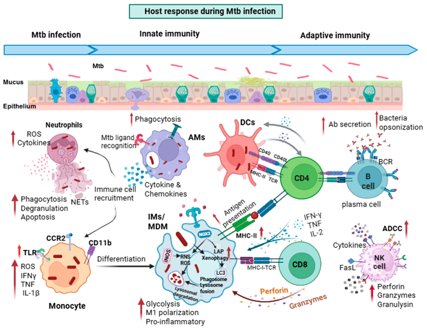
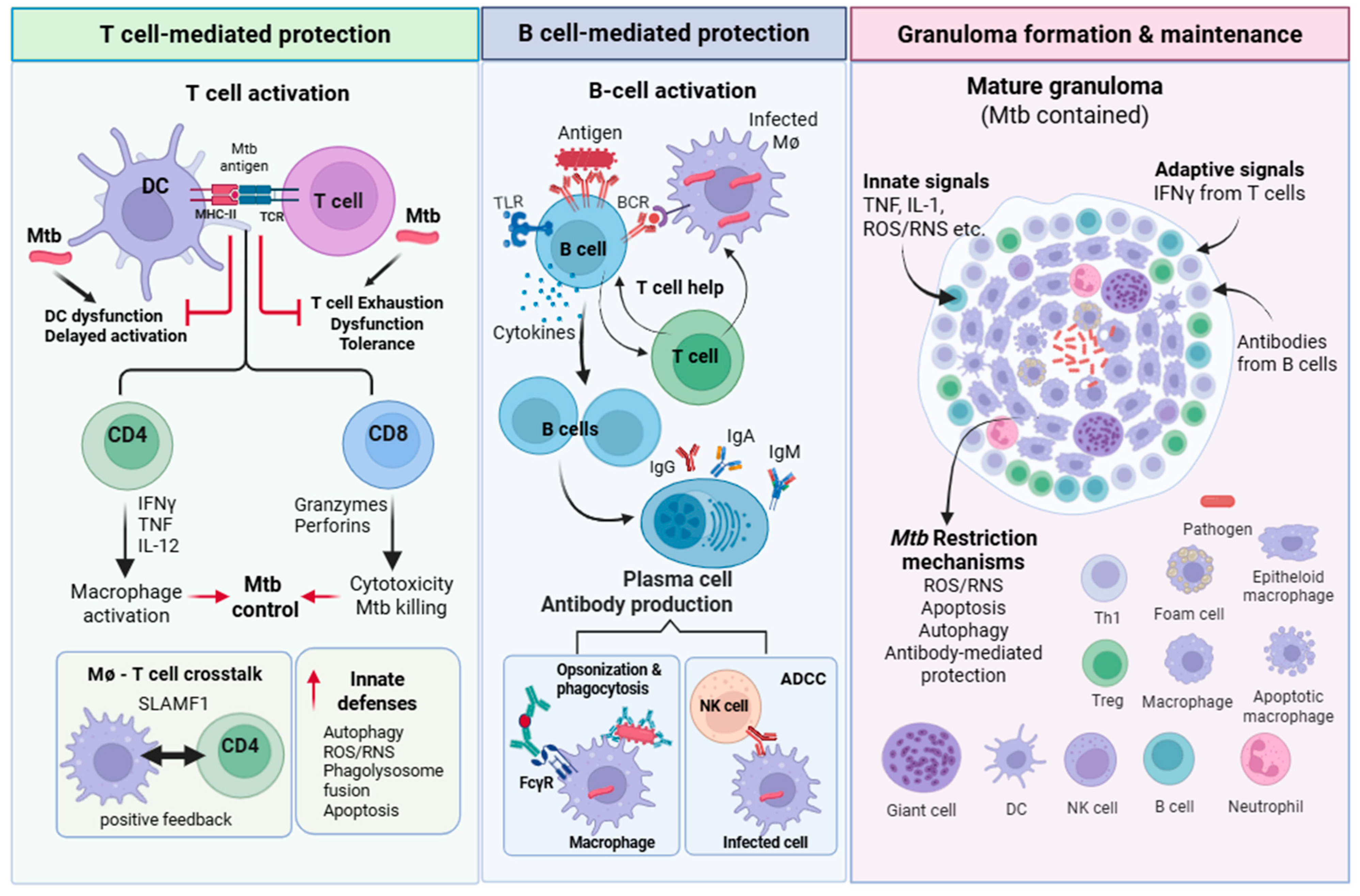
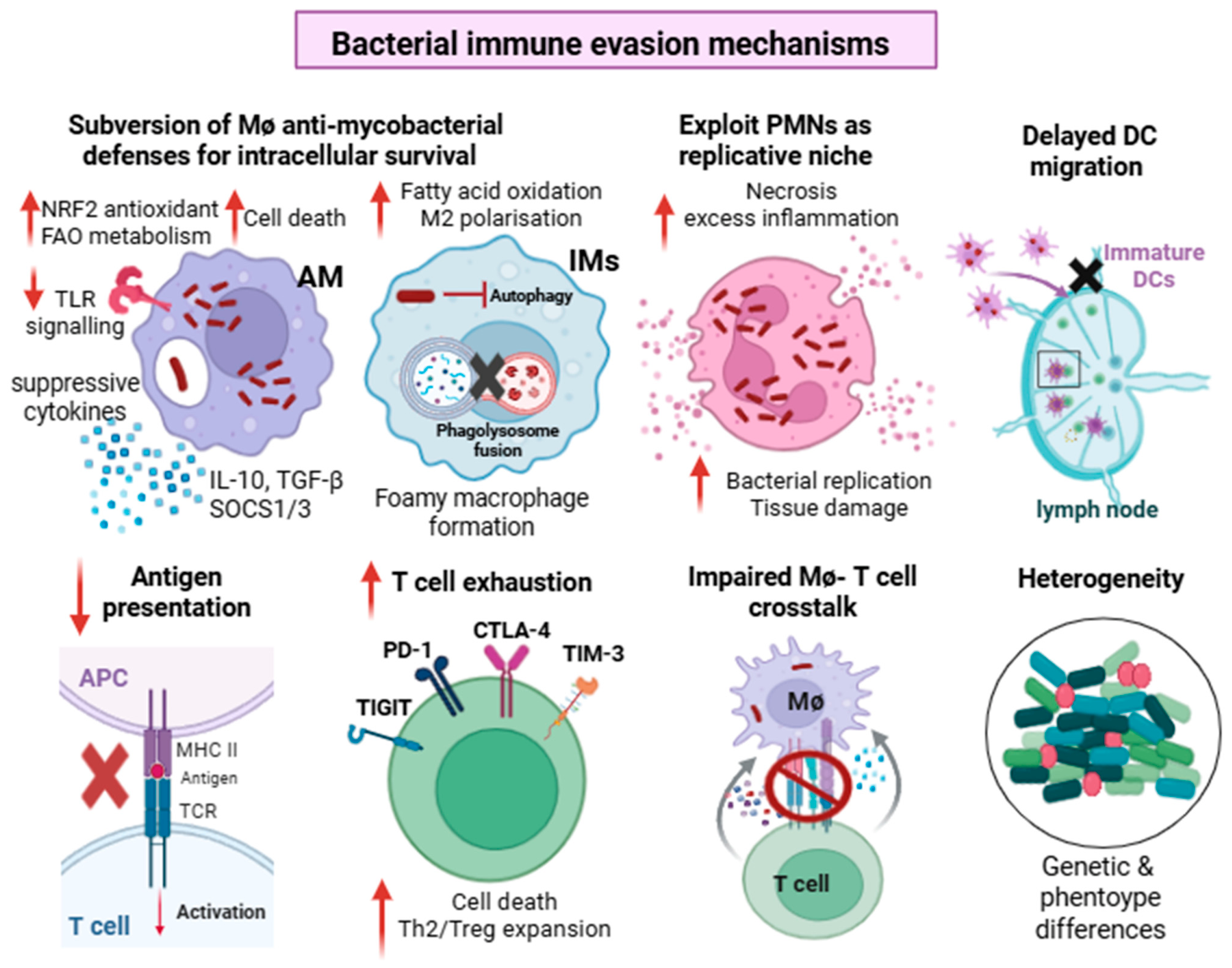
How Mycobacterium tuberculosis Subverts Innate and Adaptive Immunity and Their Crosstalk: Implications for Vaccine Design

Abstract
1. Introduction
2. Overview of Innate Immune Response
2.1. The Mucosal Barrier
2.2. Pathogen Sensing
2.3. Cell Death
2.4. Reactive Oxygen and Nitrogen Species
2.5. Autophagy
3. Overview of Adaptive Immune Response to Mtb
3.1. T Cell-Mediated Protection
3.2. B-Cell-Mediated Protection
4. Bacterial Evasion Mechanisms
4.1. Type VII Secretion Systems
4.2. Evading and Disrupting Immune Recognition
4.3. Phagosomal Damage and Intracellular Survival
5. Challenges in Vaccine Design and Efficacy
5.1. Dendritic Cell Dysfunction and Delayed Activation of the Adaptive Immune Response
5.2. T Cell Exhaustion, Dysfunction, and Establishment of Immune Tolerance
5.3. Disruption of Macrophage–T Cell Crosstalk
5.4. Bacterial Heterogeneity
5.5. Rational TB Vaccine Design: Engaging Protective Innate and Adaptive Immunity
6. Conclusions

| Vaccine Type | Vaccine Candidate
(Composition) | Mode of Action/Key Immunological Mechanisms | References |
|---|---|---|---|
| Live attenuated whole-cell vaccines | VPM1002 (recombinant BCG (ΔureC::hly) urease C-deficient strain expressing listeriolysin O) |
| [210,211,212,213,214] |
| MTBVAC (M. tuberculosis with deletion mutations in two virulence genes phoP, fadD26. It contains the RD1 locus) |
| [215,216,217] | |
| BCG (revaccination, travel vaccine) (M. bovis BCG Tokyo-172 strain) |
| [218,219,220,221] | |
| Inactivated whole-cell vaccines | DAR-901 heat-inactivated Mycobacterium obuense |
| [222,223,224] |
| ImmuVac heat-killed Mycobacterium indicus pranii |
| [225,226,227] | |
| RUTI (polyantigenic liposomal suspension of detoxified M. tuberculosis cell wall components |
| [228,229,230] | |
| Subunit vaccine | M72/AS01E recombinant fusion protein M72 composed of Mtb proteins Mtb32A, Mtb39A and AS01E adjuvant |
| [103,207,231,232,233] |
| GamTBMtbvac (Ag85A and ESAT6-CFP10 fusion proteins fused with dextran-binding domain from Leuconostoc mesenteroides and DEAE-dextran/CpG adjuvant) |
| [233,234,235,236] | |
| ID93+GLA-SE (recombinant fusion protein ID93 made of four tandem-linked Mtb antigens Rv3619, Rv1813, Rv3620, Rv2608 and TLR4 agonist adjuvant GLA-SE) |
| [233,237,238,239] | |
| H107e/CAF10b (eight Mtb antigens formulated with a novel liposomal adjuvant CAF10b containing MINCLE and TLR9 agonists) |
| [205,233,240,241,242] | |
| AEC/BC02 (Ag85b, ESAT6-CFP10 fusion proteins and BC02 adjuvant composed of BCG-CpG DNA and aluminum hydroxide) |
| [233,243,244,245,246] | |
| Viral vector-based vaccine | AdHu5Ag85A (human adenovirus serotype-5 vector-expressing Ag85A) |
| [247,248,249,250] |
| Ad5-105K (adenovirus type 5 vector-expressing Ag85A, Mtb32A and Mtb39A) |
| [207,247,248,251] | |
| ChAdOx1.85A+MVA85A (simian adenoviral vector expressing Ag85A administered by aerosol route followed by intramuscular administration of modified vaccinia Ankara virus expressing Ag85A) |
| [252,253,254] | |
| TB/FLU-05E (recombinant attenuated influenza vector Flu/THSP co-expressing truncated NS1 protein and full-length Mtb proteins TB10.4 and HspX) |
| [202,255,256,257,258,259] | |
| DNA/RNA vaccines | BNT164a1 and BNT164b1 (mRNA vaccines encoding eight Mtb antigens: Ag85A, Hrp1, ESAT-6, RpfD, RpfA, HbhA, M72, VapB47). BNT164a1 uses unmodified mRNA and BNT164b1 uses N1-methylpsuedouridine mRNA |
| [260,261,262] |
Author Contributions
Funding
Data Availability Statement
Acknowledgments
Conflicts of Interest
Abbreviations
| CCR | C-C chemokine receptor |
| CD80/86 | Cluster of differentiation 80/86 |
| cGAS | Cyclic GMP-AMP synthase |
| CIITA | Class II major histocompatibility complex transactivator |
| CMV | Cytomegalovirus |
| CTLA-4 | Cytotoxic T-lymphocyte-associated protein 4 |
| CVID | Common variable immunodeficiency |
| DO | Diversity Outbred |
| ESX-1 | Early secretory antigenic target 6 kDa system 1 |
| FcγR | Fc gamma receptor |
| FAO | Fatty acid oxidation |
| HAUSP | Herpesvirus-associated ubiquitin-specific protease |
| IFN | Interferon |
| IL | Interleukin |
| KLRG1 | Killer cell lectin-like receptor G1 |
| LAG-3 | Lymphocyte-activation gene 3 |
| LAM | Lipoarabinomannan |
| LC3 | Microtubule-associated protein 1A/1B-Light Chain 3 |
| LTBI | Latent TB infection |
| MAPK | Mitogen-activated protein kinase |
| MHCII | Major Histocompatibility Complex (MHC) II |
| MOI | Multiplicity of infection |
| Mtb | Mycobacterium tuberculosis |
| MTBC | Mycobacterium tuberculosis complex |
| NADPH | Nicotinamide adenine dinucleotide phosphate |
| NDP52 | Nuclear dot protein 52 kDa |
| NF-κB | Nuclear factor kappa-light chain enhancer of activated B cells |
| NHP | Non-human primate |
| NO | Nitric oxide |
| OXPHOS | Oxidative phosphorylation |
| PD-1 | Programmed cell death protein 1 |
| PDIM | Phthiocerol dimycocerosate |
| PE protein | Proline–glutamic acid |
| PPE | Proline–proline–glutamic acid protein |
| PstS1 | Phosphate-binding protein S1 |
| Rab5/7 | Ras-related protein Rab-5a/7a |
| SLAMF1 | Signaling lymphocyte activation molecule family member 1 |
| SNPs | Single-nucleotide polymorphisms |
| STING | Stimulator of interferon genes |
| TAX1BP1 | Tax1 (human T cell leukemia virus type 1)-binding protein 1 |
| TCR | T cell receptor |
| TGF-β | Transforming growth factor-beta |
| Tim-3 | T cell immunoglobulin and mucin domain-containing protein-3 |
| TNF | Tumor necrosis factor |
| TRAF6 | TNF receptor-associated factor 6 |
References
- Global Tuberculosis Report 2024; WHO: Geneva, Switzerland, 2024.
- Emery, J.C.; Richards, A.S.; Dale, K.D.; McQuaid, C.F.; White, R.G.; Denholm, J.T.; Rein, M.G.J.H. Self-clearance of Mycobacterium tuberculosis infection: Implications for lifetime risk and population at-risk of tuberculosis disease. Proc. Biol. Sci. 2021, 288, 20201635. [Google Scholar] [CrossRef]
- Behr, M.A.; Edelstein, P.H.; Ramakrishnan, L. Revisiting the timetable of tuberculosis. BMJ 2018, 362, k2738. [Google Scholar] [CrossRef] [PubMed]
- Roy, A.; Eisenhut, M.; Harris, R.J.; Rodrigues, L.C.; Sridhar, S.; Habermann, S.; Snell, L.; Mangtani, P.; Adetifa, I.; Lalvani, A.; et al. Effect of BCG vaccination against Mycobacterium tuberculosis infection in children: Systematic review and meta-analysis. BMJ 2014, 349, g4643. [Google Scholar] [CrossRef] [PubMed]
- Martinez, L.; Cords, O.; Liu, Q.; Acuna-Villaorduna, C.; Bonnet, M.; Fox, G.J.; Carvalho, A.C.C.; Chan, P.C.; Croda, J.; Hill, P.C.; et al. Infant BCG vaccination and risk of pulmonary and extrapulmonary tuberculosis throughout the life course: A systematic review and individual participant data meta-analysis. Lancet Glob. Health 2022, 10, e1307–e1316. [Google Scholar] [CrossRef]
- Comas, I.; Coscolla, M.; Luo, T.; Borrell, S.; Holt, K.E.; Kato-Maeda, M.; Parkhill, J.; Malla, B.; Berg, S.; Thwaites, G.; et al. Out-of-Africa migration and Neolithic coexpansion of Mycobacterium tuberculosis with modern humans. Nat. Genet. 2013, 45, 1176–1182. [Google Scholar] [CrossRef] [PubMed]
- Rahlwes, K.C.; Dias, B.R.S.; Campos, P.C.; Alvarez-Arguedas, S.; Shiloh, M.U. Pathogenicity and virulence of Mycobacterium tuberculosis. Virulence 2023, 14, 2150449. [Google Scholar] [CrossRef]
- Scordo, J.M.; Knoell, D.L.; Torrelles, J.B. Alveolar Epithelial Cells in Mycobacterium tuberculosis Infection: Active Players or Innocent Bystanders? J. Innate Immun. 2016, 8, 3–14. [Google Scholar] [CrossRef]
- de Waal, A.M.; Hiemstra, P.S.; Ottenhoff, T.H.; Joosten, S.A.; van der Does, A.M. Lung epithelial cells interact with immune cells and bacteria to shape the microenvironment in tuberculosis. Thorax 2022, 77, 408–416. [Google Scholar] [CrossRef]
- Cohen, S.B.; Gern, B.H.; Delahaye, J.L.; Adams, K.N.; Plumlee, C.R.; Winkler, J.K.; Sherman, D.R.; Gerner, M.Y.; Urdahl, K.B. Alveolar Macrophages Provide an Early Mycobacterium tuberculosis Niche and Initiate Dissemination. Cell Host Microbe 2018, 24, 439–446.e4. [Google Scholar] [CrossRef]
- Yang, S.; Ouyang, J.; Lu, Y.; Harypursat, V.; Chen, Y. A Dual Role of Heme Oxygenase-1 in Tuberculosis. Front. Immunol. 2022, 13, 842858. [Google Scholar] [CrossRef]
- Rothchild, A.C.; Olson, G.S.; Nemeth, J.; Amon, L.M.; Mai, D.; Gold, E.S.; Diercks, A.H.; Aderem, A. Alveolar macrophages generate a noncanonical NRF2-driven transcriptional response. Sci. Immunol. 2019, 4, eaaw6693. [Google Scholar] [CrossRef]
- Silva-Gomes, S.; Appelberg, R.; Larsen, R.; Soares, M.P.; Gomes, M.S. Heme catabolism by heme oxygenase-1 confers host resistance to Mycobacterium infection. Infect. Immun. 2013, 81, 2536–2545. [Google Scholar] [CrossRef]
- Pisu, D.; Huang, L.; Grenier, J.K.; Russell, D.G. Dual RNA-Seq of Mtb-Infected Macrophages In Vivo Reveals Ontologically Distinct Host-Pathogen Interactions. Cell Rep. 2020, 30, 335–350.e4. [Google Scholar] [CrossRef] [PubMed]
- Huang, L.; Nazarova, E.V.; Tan, S.; Liu, Y.; Russell, D.G. Growth of Mycobacterium tuberculosis in vivo segregates with host macrophage metabolism and ontogeny. J. Exp. Med. 2018, 215, 1135–1152. [Google Scholar] [CrossRef]
- Alcantara, C.A.; Glassman, I.; Nguyen, K.H.; Parthasarathy, A.; Venketaraman, V. Neutrophils in Mycobacterium tuberculosis. Vaccines 2023, 11, 631. [Google Scholar] [CrossRef]
- Repasy, T.; Martinez, N.; Lee, J.; West, K.; Li, W.; Kornfeld, H. Bacillary replication and macrophage necrosis are determinants of neutrophil recruitment in tuberculosis. Microbes Infect. 2015, 17, 564–574. [Google Scholar] [CrossRef]
- Hult, C.; Mattila, J.T.; Gideon, H.P.; Linderman, J.J.; Kirschner, D.E. Neutrophil Dynamics Affect Mycobacterium tuberculosis granuloma outcomes and dissemination. Front. Immunol. 2021, 12, 712457. [Google Scholar] [CrossRef] [PubMed]
- Ravesloot-Chávez, M.M.; Van Dis, E.; Stanley, S.A. The Innate Immune Response to Mycobacterium tuberculosis infection. Annu. Rev. Immunol. 2021, 39, 611–637. [Google Scholar] [CrossRef]
- Srivastava, S.; Ernst, J.D.; Desvignes, L. Beyond macrophages: The diversity of mononuclear cells in tuberculosis. Immunol. Rev. 2014, 262, 179–192. [Google Scholar] [CrossRef] [PubMed]
- Wolf, A.J.; Linas, B.; Trevejo-Nuñez, G.J.; Kincaid, E.; Tamura, T.; Takatsu, K.; Ernst, J.D. Mycobacterium tuberculosis infects dendritic cells with high frequency and impairs their function in vivo. J. Immunol. 2007, 179, 2509–2519. [Google Scholar] [CrossRef]
- Srivastava, S.; Ernst, J.D. Cell-to-cell transfer of M. tuberculosis antigens optimizes CD4 T cell priming. Cell Host Microbe 2014, 15, 741–752. [Google Scholar] [CrossRef] [PubMed]
- Guilliams, M.; Lambrecht, B.N.; Hammad, H. Division of labor between lung dendritic cells and macrophages in the defense against pulmonary infections. Mucosal Immunol. 2013, 6, 464–473. [Google Scholar] [CrossRef]
- Wolf, A.J.; Desvignes, L.; Linas, B.; Banaiee, N.; Tamura, T.; Takatsu, K.; Ernst, J.D. Initiation of the adaptive immune response to Mycobacterium tuberculosis depends on antigen production in the local lymph node, not the lungs. J. Exp. Med. 2008, 205, 105–115. [Google Scholar] [CrossRef]
- Middleton, A.M.; Chadwick, M.V.; Nicholson, A.G.; Wilson, R.; Thornton, D.J.; Kirkham, S.; Sheehan, J.K. Interaction between mycobacteria and mucus on a human respiratory tissue organ culture model with an air interface. Exp. Lung Res. 2004, 30, 17–29. [Google Scholar] [CrossRef]
- Gao, X.; Feng, J.; Wei, L.; Dong, P.; Chen, J.; Zhang, L.; Yang, Y.; Xu, L.; Wang, H.; Luo, J.; et al. Defensins: A novel weapon against Mycobacterium tuberculosis? Int. Immunopharmacol. 2024, 127, 111383. [Google Scholar] [CrossRef] [PubMed]
- Dong, H.; Lv, Y.; Zhao, D.; Barrow, P.; Zhou, X. Defensins: The Case for Their Use against Mycobacterial Infections. J. Immunol. Res. 2016, 2016, 7515687. [Google Scholar] [CrossRef] [PubMed]
- Tjärnlund, A.; Rodríguez, A.; Cardona, P.J.; Guirado, E.; Ivanyi, J.; Singh, M.; Troye-Blomberg, M.; Fernández, C. Polymeric IgR knockout mice are more susceptible to mycobacterial infections in the respiratory tract than wild-type mice. Int. Immunol. 2006, 18, 807–816. [Google Scholar] [CrossRef]
- Gaynor, C.D.; McCormack, F.X.; Voelker, D.R.; McGowan, S.E.; Schlesinger, L.S. Pulmonary surfactant protein A mediates enhanced phagocytosis of Mycobacterium tuberculosis by a direct interaction with human macrophages. J. Immunol. 1995, 155, 5343–5351. [Google Scholar] [CrossRef]
- Dodd, C.E.; Pyle, C.J.; Glowinski, R.; Rajaram, M.V.; Schlesinger, L.S. CD36-Mediated Uptake of Surfactant Lipids by Human Macrophages Promotes Intracellular Growth of Mycobacterium tuberculosis. J. Immunol. 2016, 197, 4727–4735. [Google Scholar] [CrossRef]
- Shukla, S.; Richardson, E.T.; Drage, M.G.; Boom, W.H.; Harding, C.V. Mycobacterium tuberculosis Lipoprotein and Lipoglycan Binding to Toll-Like Receptor 2 Correlates with Agonist Activity and Functional Outcomes. Infect. Immun. 2018, 86, e00450-18. [Google Scholar] [CrossRef]
- Bowdish, D.M.; Sakamoto, K.; Kim, M.J.; Kroos, M.; Mukhopadhyay, S.; Leifer, C.A.; Tryggvason, K.; Gordon, S.; Russell, D.G. MARCO, TLR2, and CD14 are required for macrophage cytokine responses to mycobacterial trehalose dimycolate and Mycobacterium tuberculosis. PLoS Pathog. 2009, 5, e1000474. [Google Scholar] [CrossRef] [PubMed]
- Bafica, A.; Scanga, C.A.; Feng, C.G.; Leifer, C.; Cheever, A.; Sher, A. TLR9 regulates Th1 responses and cooperates with TLR2 in mediating optimal resistance to Mycobacterium tuberculosis. J. Exp. Med. 2005, 202, 1715–1724. [Google Scholar] [CrossRef] [PubMed]
- Bulut, Y.; Michelsen, K.S.; Hayrapetian, L.; Naiki, Y.; Spallek, R.; Singh, M.; Arditi, M. Mycobacterium tuberculosis heat shock proteins use diverse Toll-like receptor pathways to activate pro-inflammatory signals. J. Biol. Chem. 2005, 280, 20961–20967. [Google Scholar] [CrossRef]
- Goyal, S.; Klassert, T.E.; Slevogt, H. C-type lectin receptors in tuberculosis: What we know. Med. Microbiol. Immunol. 2016, 205, 513–535. [Google Scholar] [CrossRef]
- Ishikawa, E.; Ishikawa, T.; Morita, Y.S.; Toyonaga, K.; Yamada, H.; Takeuchi, O.; Kinoshita, T.; Akira, S.; Yoshikai, Y.; Yamasaki, S. Direct recognition of the mycobacterial glycolipid, trehalose dimycolate, by C-type lectin Mincle. J. Exp. Med. 2009, 206, 2879–2888. [Google Scholar] [CrossRef] [PubMed]
- Coulombe, F.; Divangahi, M.; Veyrier, F.; de Léséleuc, L.; Gleason, J.L.; Yang, Y.; Kelliher, M.A.; Pandey, A.K.; Sassetti, C.M.; Reed, M.B.; et al. Increased NOD2-mediated recognition of N-glycolyl muramyl dipeptide. J. Exp. Med. 2009, 206, 1709–1716. [Google Scholar] [CrossRef]
- Yang, Y.; Yin, C.; Pandey, A.; Abbott, D.; Sassetti, C.; Kelliher, M.A. NOD2 pathway activation by MDP or Mycobacterium tuberculosis infection involves the stable polyubiquitination of Rip2. J. Biol. Chem. 2007, 282, 36223–36229. [Google Scholar] [CrossRef]
- Watson, R.O.; Bell, S.L.; MacDuff, D.A.; Kimmey, J.M.; Diner, E.J.; Olivas, J.; Vance, R.E.; Stallings, C.L.; Virgin, H.W.; Cox, J.S. The Cytosolic Sensor cGAS Detects Mycobacterium tuberculosis DNA to Induce Type I Interferons and Activate Autophagy. Cell Host Microbe 2015, 17, 811–819. [Google Scholar] [CrossRef]
- Mvubu, N.E. Innate immune recognition of Mycobacterium tuberculosis: Receptor engagement and inflammatory outcomes at the site of infection. Cell Surf. 2025, 14, 100150. [Google Scholar] [CrossRef]
- Kleinnijenhuis, J.; Oosting, M.; Joosten, L.A.; Netea, M.G.; Van Crevel, R. Innate immune recognition of Mycobacterium tuberculosis. Clin. Dev. Immunol. 2011, 2011, 405310. [Google Scholar] [CrossRef]
- Vu, A.; Glassman, I.; Campbell, G.; Yeganyan, S.; Nguyen, J.; Shin, A.; Venketaraman, V. Host Cell Death and Modulation of Immune Response against Mycobacterium tuberculosis infection. Int. J. Mol. Sci. 2024, 25, 6255. [Google Scholar] [CrossRef]
- Briken, V. “With a little help from my friends”: Efferocytosis as an antimicrobial mechanism. Cell Host Microbe 2012, 12, 261–263. [Google Scholar] [CrossRef]
- Stutz, M.D.; Allison, C.C.; Ojaimi, S.; Preston, S.P.; Doerflinger, M.; Arandjelovic, P.; Whitehead, L.; Bader, S.M.; Batey, D.; Asselin-Labat, M.L.; et al. Macrophage and neutrophil death programs differentially confer resistance to tuberculosis. Immunity 2021, 54, 1758–1771.e7. [Google Scholar] [CrossRef] [PubMed]
- Bustamante, J.; Arias, A.A.; Vogt, G.; Picard, C.; Galicia, L.B.; Prando, C.; Grant, A.V.; Marchal, C.C.; Hubeau, M.; Chapgier, A. Germline CYBB mutations that selectively affect macrophages in kindreds with X-linked predisposition to tuberculous mycobacterial disease. Nat. Immunol. 2011, 12, 213–221. [Google Scholar] [CrossRef]
- Olive, A.J.; Smith, C.M.; Kiritsy, M.C.; Sassetti, C.M. The Phagocyte Oxidase Controls Tolerance to Mycobacterium tuberculosis infection. J. Immunol. 2018, 201, 1705–1716. [Google Scholar] [CrossRef]
- Deffert, C.; Schäppi, M.G.; Pache, J.C.; Cachat, J.; Vesin, D.; Bisig, R.; Ma Mulone, X.; Kelkka, T.; Holmdahl, R.; Garcia, I.; et al. Bacillus calmette-guerin infection in NADPH oxidase deficiency: Defective mycobacterial sequestration and granuloma formation. PLoS Pathog. 2014, 10, e1004325. [Google Scholar] [CrossRef]
- Deffert, C.; Cachat, J.; Krause, K.H. Phagocyte NADPH oxidase, chronic granulomatous disease and mycobacterial infections. Cell. Microbiol. 2014, 16, 1168–1178. [Google Scholar] [CrossRef] [PubMed]
- Scheuermann, L.; Pei, G.; Domaszewska, T.; Zyla, J.; Oberbeck-Müller, D.; Bandermann, S.; Feng, Y.; Ruiz Moreno, J.S.; Opitz, B.; Mollenkopf, H.J.; et al. Platelets Restrict the Oxidative Burst in Phagocytes and Facilitate Primary Progressive Tuberculosis. Am. J. Respir. Crit. Care Med. 2020, 202, 730–744. [Google Scholar] [CrossRef]
- Chandra, P.; He, L.; Zimmerman, M.; Yang, G.; Köster, S.; Ouimet, M.; Wang, H.; Moore, K.J.; Dartois, V.; Schilling, J.D.; et al. Inhibition of Fatty Acid Oxidation Promotes Macrophage Control of Mycobacterium tuberculosis. mBio 2020, 11, e01139-20. [Google Scholar] [CrossRef] [PubMed]
- Choi, H.S.; Rai, P.R.; Chu, H.W.; Cool, C.; Chan, E.D. Analysis of nitric oxide synthase and nitrotyrosine expression in human pulmonary tuberculosis. Am. J. Respir. Crit. Care Med. 2002, 166, 178–186. [Google Scholar] [CrossRef]
- MacMicking, J.D.; North, R.J.; LaCourse, R.; Mudgett, J.S.; Shah, S.K.; Nathan, C.F. Identification of nitric oxide synthase as a protective locus against tuberculosis. Proc. Natl. Acad. Sci. USA 1997, 94, 5243–5248. [Google Scholar] [CrossRef]
- Chan, J.; Tanaka, K.; Carroll, D.; Flynn, J.; Bloom, B.R. Effects of nitric oxide synthase inhibitors on murine infection with Mycobacterium tuberculosis. Infect. Immun. 1995, 63, 736–740. [Google Scholar] [CrossRef]
- Stei, F.; McCaffrey, E.F.; Zessin, B.; Pioch, J.; Simm, S.; Stubbe, B.; Ewert, R.; Verreck, F.; Schneider, B.E.; Dorhoi, A.; et al. Species-specific iNOS expression distinguishes epithelial and myeloid IFNγ responses in tuberculosis. bioRxiv 2026. [Google Scholar] [CrossRef]
- Shiloh, M.U.; MacMicking, J.D.; Nicholson, S.; Brause, J.E.; Potter, S.; Marino, M.; Fang, F.; Dinauer, M.; Nathan, C. Phenotype of mice and macrophages deficient in both phagocyte oxidase and inducible nitric oxide synthase. Immunity 1999, 10, 29–38. [Google Scholar] [CrossRef] [PubMed]
- Silwal, P.; Paik, S.; Kim, J.K.; Yoshimori, T.; Jo, E.K. Regulatory Mechanisms of Autophagy-Targeted Antimicrobial Therapeutics Against Mycobacterial Infection. Front. Cell. Infect. Microbiol. 2021, 11, 633360. [Google Scholar] [CrossRef] [PubMed]
- Strong, E.J.; Lee, S. Targeting Autophagy as a Strategy for Developing New Vaccines and Host-Directed Therapeutics Against Mycobacteria. Front. Microbiol. 2020, 11, 614313. [Google Scholar] [CrossRef]
- Schiebler, M.; Brown, K.; Hegyi, K.; Newton, S.M.; Renna, M.; Hepburn, L.; Klapholz, C.; Coulter, S.; Obregón-Henao, A.; Henao Tamayo, M.; et al. Functional drug screening reveals anticonvulsants as enhancers of mTOR-independent autophagic killing of Mycobacterium tuberculosis through inositol depletion. EMBO Mol. Med. 2015, 7, 127–139. [Google Scholar] [CrossRef]
- Kinsella, R.L.; Kimmey, J.M.; Smirnov, A.; Woodson, R.; Gaggioli, M.R.; Chavez, S.M.; Kreamalmeyer, D.; Stallings, C.L. Autophagy prevents early proinflammatory responses and neutrophil recruitment during Mycobacterium tuberculosis infection without affecting pathogen burden in macrophages. PLoS Biol. 2023, 21, e3002159. [Google Scholar] [CrossRef]
- Feng, S.; McNehlan, M.E.; Kinsella, R.L.; Sur Chowdhury, C.; Chavez, S.M.; Naik, S.K.; McKee, S.R.; Van Winkle, J.A.; Dubey, N.; Samuels, A.; et al. Autophagy promotes efficient T cell responses to restrict high-dose Mycobacterium tuberculosis infection in mice. Nat. Microbiol. 2024, 9, 684–697. [Google Scholar] [CrossRef]
- Aylan, B.; Bernard, E.M.; Pellegrino, E.; Botella, L.; Fearns, A.; Athanasiadi, N.; Bussi, C.; Santucci, P.; Gutierrez, M.G. ATG7 and ATG14 restrict cytosolic and phagosomal Mycobacterium tuberculosis replication in human macrophages. Nat. Microbiol. 2023, 8, 803–818. [Google Scholar] [CrossRef]
- Augenstreich, J.; Phan, A.T.; Allen, C.N.S.; Poddar, A.; Chen, H.; Srinivasan, L.; Briken, V. Dynamic interplay of autophagy and membrane repair during Mycobacterium tuberculosis Infection. PLoS Pathog. 2025, 21, e1012830. [Google Scholar] [CrossRef]
- Li, J.; Feng, H.; Chen, D.; Zhang, H.; Liao, Y. Autophagy in mycobacterial infections: Molecular mechanisms, host-pathogen interactions, and therapeutic opportunities. Front. Cell. Infect. Microbiol. 2025, 15, 1640647. [Google Scholar] [CrossRef] [PubMed]
- Golovkine, G.R.; Roberts, A.W.; Morrison, H.M.; Rivera-Lugo, R.; McCall, R.M.; Nilsson, H.; Garelis, N.E.; Repasy, T.; Cronce, M.; Budzik, J.; et al. Autophagy restricts Mycobacterium tuberculosis during acute infection in mice. Nat. Microbiol. 2023, 8, 819–832. [Google Scholar] [CrossRef]
- Kwan, C.K.; Ernst, J.D. HIV and tuberculosis: A deadly human syndemic. Clin. Microbiol. Rev. 2011, 24, 351–376. [Google Scholar] [CrossRef] [PubMed]
- Mogues, T.; Goodrich, M.E.; Ryan, L.; LaCourse, R.; North, R.J. The relative importance of T cell subsets in immunity and immunopathology of airborne Mycobacterium tuberculosis infection in mice. J. Exp. Med. 2001, 193, 271–280. [Google Scholar] [CrossRef]
- Caruso, A.M.; Serbina, N.; Klein, E.; Triebold, K.; Bloom, B.R.; Flynn, J.L. Mice deficient in CD4 T cells have only transiently diminished levels of IFN-gamma, yet succumb to tuberculosis. J. Immunol. 1999, 162, 5407–5416. [Google Scholar] [CrossRef] [PubMed]
- Diedrich, C.R.; Mattila, J.T.; Klein, E.; Janssen, C.; Phuah, J.; Sturgeon, T.J.; Montelaro, R.C.; Lin, P.L.; Flynn, J.L. Reactivation of latent tuberculosis in cynomolgus macaques infected with SIV is associated with early peripheral T cell depletion and not virus load. PLoS ONE 2010, 5, e9611. [Google Scholar] [CrossRef]
- Lin, P.L.; Rutledge, T.; Green, A.M.; Bigbee, M.; Fuhrman, C.; Klein, E.; Flynn, J.L. CD4 T cell depletion exacerbates acute Mycobacterium tuberculosis while reactivation of latent infection is dependent on severity of tissue depletion in cynomolgus macaques. AIDS Res. Hum. Retroviruses 2012, 28, 1693–1702. [Google Scholar] [CrossRef]
- Krishna Prasad, G.V.R.; Grigsby, S.J.; Erkenswick, G.A.; Portal-Celhay, C.; Mittal, E.; Yang, G.; Fallon, S.M.; Chen, F.; Klevorn, T.; Jain, N.; et al. Macrophage-T cell interactions promote SLAMF1 expression for enhanced TB defense. Nat. Commun. 2025, 16, 6794. [Google Scholar] [CrossRef]
- Srivastava, S.; Ernst, J.D. Cutting edge: Direct recognition of infected cells by CD4 T cells is required for control of intracellular Mycobacterium tuberculosis in vivo. J. Immunol. 2013, 191, 1016–1020. [Google Scholar] [CrossRef]
- Ankley, L.; Thomas, S.; Olive, A.J. Fighting Persistence: How Chronic Infections with Mycobacterium tuberculosis Evade T Cell-Mediated Clearance and New Strategies To Defeat Them. Infect. Immun. 2020, 88, e00916-19. [Google Scholar] [CrossRef] [PubMed]
- Kireev, F.D.; Lopatnikova, J.A.; Alshevskaya, A.A.; Sennikov, S.V. Role of Tumor Necrosis Factor in Tuberculosis. Biomolecules 2025, 15, 709. [Google Scholar] [CrossRef]
- Khelghati, F.; Rahmanian, M.; Eghbal, E.; Seghatoleslami, Z.S.; Goudarzi, M.; Keramatinia, A.; Ong, C.W.; Goletti, D.; D’Ambrosio, L.; Centis, R.; et al. Risk of tuberculosis disease in patients receiving TNF-α antagonist therapy: A meta-analysis of randomized controlled trials. New Microbes New Infect. 2024, 62, 101533. [Google Scholar] [CrossRef]
- Imran, M.; Alshrari, A.S.; Khan, A.; Alzahrani, A.R. IFN-γ-Driven macrophage responses in the immunity to Mycobacterium tuberculosis and Mycobacterium leprae. Front. Cell. Infect. Microbiol. 2025, 15, 1679691. [Google Scholar] [CrossRef]
- Mortensen, R.; Arlehamn, C.S.L.; Coler, R.N.; Gerner, M.Y.; Goletti, D.; Lewinsohn, D.A.; Modlin, R.L.; Musvosvi, M.; Rengarajan, J.; Urdahl, K.B.; et al. T cell-macrophage interactions in tuberculosis: What we’ve got here is failure to communicate. J. Intern. Med. 2026, 299, 44–65. [Google Scholar] [CrossRef] [PubMed]
- Bustamante, J.; Boisson-Dupuis, S.; Abel, L.; Casanova, J.L. Mendelian susceptibility to mycobacterial disease: Genetic, immunological, and clinical features of inborn errors of IFN-γ immunity. Semin. Immunol. 2014, 26, 454–470. [Google Scholar] [CrossRef] [PubMed]
- Maciag, K.; Plumlee, C.R.; Cohen, S.B.; Gern, B.H.; Urdahl, K.B. Reappraising the Role of T Cell-Derived IFN-γ in Restriction of Mycobacterium tuberculosis in the Murine Lung. J. Immunol. 2024, 213, 339–346. [Google Scholar] [CrossRef]
- Sakai, S.; Kauffman, K.D.; Sallin, M.A.; Sharpe, A.H.; Young, H.A.; Ganusov, V.V.; Barber, D.L. CD4 T Cell-Derived IFN-γ Plays a Minimal Role in Control of Pulmonary Mycobacterium tuberculosis Infection and Must Be Actively Repressed by PD-1 to Prevent Lethal Disease. PLoS Pathog. 2016, 12, e1005667. [Google Scholar] [CrossRef]
- Lu, L.L.; Smith, M.T.; Yu, K.K.Q.; Luedemann, C.; Suscovich, T.J.; Grace, P.S.; Grace, P.S.; Cain, A.; Yu, W.H.; McKitrick, T.R.; et al. IFN-γ-independent immune markers of Mycobacterium tuberculosis exposure. Nat. Med. 2019, 25, 977–987. [Google Scholar] [CrossRef]
- Gallegos, A.M.; van Heijst, J.W.; Samstein, M.; Su, X.; Pamer, E.G.; Glickman, M.S. A gamma interferon independent mechanism of CD4 T cell mediated control of M. tuberculosis infection in vivo. PLoS Pathog. 2011, 7, e1002052. [Google Scholar] [CrossRef]
- Ahrends, T.; Spanjaard, A.; Pilzecker, B.; Bąbała, N.; Bovens, A.; Xiao, Y.; Jacobs, H.; Borst, J. CD4 T Cell Help Confers a Cytotoxic T Cell Effector Program Including Coinhibitory Receptor Downregulation and Increased Tissue Invasiveness. Immunity 2017, 47, 848–861.e5. [Google Scholar] [CrossRef]
- Lu, Y.J.; Barreira-Silva, P.; Boyce, S.; Powers, J.; Cavallo, K.; Behar, S.M. CD4 T cell help prevents CD8 T cell exhaustion and promotes control of Mycobacterium tuberculosis infection. Cell Rep. 2021, 36, 109696. [Google Scholar] [CrossRef]
- Sakai, S.; Mayer-Barber, K.D.; Barber, D.L. Defining features of protective CD4 T cell responses to Mycobacterium tuberculosis. Curr. Opin. Immunol. 2014, 29, 137–142. [Google Scholar] [CrossRef] [PubMed]
- Lin, P.L.; Flynn, J.L. CD8 T cells and Mycobacterium tuberculosis infection. Semin. Immunopathol. 2015, 37, 239–249. [Google Scholar] [CrossRef]
- Noschka, R.; Wondany, F.; Kizilsavas, G.; Weil, T.; Weidinger, G.; Walther, P.; Michaelis, J.; Stenger, S. Gran1: A Granulysin-Derived Peptide with Potent Activity against Intracellular Mycobacterium tuberculosis. Int. J. Mol. Sci. 2021, 22, 8392. [Google Scholar] [CrossRef] [PubMed]
- Rijnink, W.F.; Ottenhoff, T.H.M.; Joosten, S.A. B-Cells and Antibodies as Contributors to Effector Immune Responses in Tuberculosis. Front. Immunol. 2021, 12, 640168. [Google Scholar] [CrossRef] [PubMed]
- Chen, X.F.; Wang, W.F.; Zhang, Y.D.; Zhao, W.; Wu, J.; Chen, T.X. Clinical characteristics and genetic profiles of 174 patients with X-linked agammaglobulinemia: Report from Shanghai, China (2000–2015). Medicine 2016, 95, e4544. [Google Scholar] [CrossRef]
- Hunter, L.; Hingley-Wilson, S.; Stewart, G.R.; Sharpe, S.A.; Salguero, F.J. Dynamics of Macrophage, T and B Cell Infiltration Within Pulmonary Granulomas Induced by Mycobacterium tuberculosis in two Non Human Primate models of aerosol infection. Front. Immunol. 2021, 12, 776913. [Google Scholar] [CrossRef]
- Phuah, J.Y.; Mattila, J.T.; Lin, P.L.; Flynn, J.L. Activated B cells in the granulomas of nonhuman primates infected with Mycobacterium tuberculosis. Am. J. Pathol. 2012, 181, 508–514. [Google Scholar] [CrossRef]
- van Meijgaarden, K.E.; Grace, P.S.; Freund, N.T.; Achkar, J.M.; Lindenstrøm, T.; Tan, J.; Chan, J.; King, C.G.; Ottenhoff, T.H.M.; Joosten, S.A.; et al. Antibodies in tuberculosis: Functional capacity as key determinant. Infect. Immun. 2026, 94, e0039425. [Google Scholar] [CrossRef]
- Lu, L.L.; Chung, A.W.; Rosebrock, T.R.; Ghebremichael, M.; Yu, W.H.; Grace, P.S.; Schoen, M.K.; Tafesse, F.; Martin, C.; Leung, V.; et al. A Functional Role for Antibodies in Tuberculosis. Cell 2016, 167, 433–443.e14. [Google Scholar] [CrossRef]
- Chen, T.; Blanc, C.; Eder, A.Z.; Prados-Rosales, R.; Souza, A.C.; Kim, R.S.; Glatman-Freedman, A.; Joe, M.; Bai, Y.; Lowary, T.L.; et al. Association of Human Antibodies to Arabinomannan With Enhanced Mycobacterial Opsonophagocytosis and Intracellular Growth Reduction. J. Infect. Dis. 2016, 214, 300–310. [Google Scholar] [CrossRef] [PubMed]
- Aguilo, N.; Uranga, S.; Mata, E.; Tarancon, R.; Gómez, A.B.; Marinova, D.; Otal, I.; Monzón, M.; Badiola, J.; Montenegro, D.; et al. Respiratory Immunization With a Whole Cell Inactivated Vaccine Induces Functional Mucosal Immunoglobulins Against Tuberculosis in Mice and Non-human Primates. Front. Microbiol. 2020, 11, 1339. [Google Scholar] [CrossRef] [PubMed]
- Fletcher, H.A.; Snowden, M.A.; Landry, B.; Rida, W.; Satti, I.; Harris, S.A.; Matsumiya, M.; Tanner, R.; O’Shea, M.K.; Dheenadhayalan, V.; et al. T-cell activation is an immune correlate of risk in BCG vaccinated infants. Nat. Commun. 2016, 7, 11290. [Google Scholar] [CrossRef] [PubMed]
- Carpenter, S.M.; Lu, L.L. Leveraging Antibody, B Cell and Fc Receptor Interactions to Understand Heterogeneous Immune Responses in Tuberculosis. Front. Immunol. 2022, 13, 830482. [Google Scholar] [CrossRef]
- Kumar, S.K.; Singh, P.; Sinha, S. Naturally produced opsonizing antibodies restrict the survival of Mycobacterium tuberculosis in human macrophages by augmenting phagosome maturation. Open Biol. 2015, 5, 150171. [Google Scholar] [CrossRef]
- Prados-Rosales, R.; Carreño, L.; Cheng, T.; Blanc, C.; Weinrick, B.; Malek, A.; Lowary, T.L.; Baena, A.; Joe, M.; Bai, Y.; et al. Enhanced control of Mycobacterium tuberculosis extrapulmonary dissemination in mice by an arabinomannan-protein conjugate vaccine. PLoS Pathog. 2017, 13, e1006250. [Google Scholar] [CrossRef]
- Bouzeyen, R.; Sithole, N.; Watson, A.; Abramovitz, L.; Rakayev, K.; Fillion, A.; Dickson, T.; MacAry, P.A.; Darrah, P.A.; Seder, R.A.; et al. Early Antibody-Mediated Immunity to Tuberculosis in Mice Requires NLRP3. bioRxiv 2025. [Google Scholar] [CrossRef]
- Darrah, P.A.; Zeppa, J.J.; Maiello, P.; Hackney, J.A.; Wadsworth, M.H.; Hughes, T.K.; Pokkali, S.; Swanson, P.A.; Grant, N.L.; Rodgers, M.A.; et al. Prevention of tuberculosis in macaques after intravenous BCG immunization. Nature 2020, 577, 95–102. [Google Scholar] [CrossRef]
- Hansen, S.G.; Zak, D.E.; Xu, G.; Ford, J.C.; Marshall, E.E.; Malouli, D.; Gilbride, R.M.; Hughes, C.M.; Ventura, A.B.; Ainslie, E.; et al. Prevention of tuberculosis in rhesus macaques by a cytomegalovirus-based vaccine. Nat. Med. 2018, 24, 130–143. [Google Scholar] [CrossRef]
- Vierboom, M.P.M.; Dijkman, K.; Sombroek, C.C.; Hofman, S.O.; Boot, C.; Vervenne, R.A.W.; Haanstra, K.G.; van der Sande, M.; van Emst, L.; Domínguez-Andrés, J.; et al. Stronger induction of trained immunity by mucosal BCG or MTBVAC vaccination compared to standard intradermal vaccination. Cell Rep. Med. 2021, 2, 100185. [Google Scholar] [CrossRef] [PubMed]
- Tait, D.R.; Hatherill, M.; Van Der Meeren, O.; Ginsberg, A.M.; Van Brakel, E.; Salaun, B.; Scriba, T.J.; Akite, E.J.; Ayles, H.M.; Bollaerts, A.; et al. Final Analysis of a Trial of M72/AS01E vaccine to prevent tuberculosis. N. Engl. J. Med. 2019, 381, 2429–2439. [Google Scholar] [CrossRef]
- Roy, S.; Ghatak, D.; Das, P.; BoseDasgupta, S. ESX secretion system: The gatekeepers of mycobacterial survivability and pathogenesis. Eur. J. Microbiol. Immunol. 2020, 10, 202–209. [Google Scholar] [CrossRef]
- Clemmensen, H.S.; Knudsen, N.P.H.; Rasmussen, E.M.; Winkler, J.; Rosenkrands, I.; Ahmad, A.; Lillebaek, T.; Sherman, D.R.; Andersen, P.L.; Aagaard, C.; et al. An attenuated Mycobacterium tuberculosis clinical strain with a defect in ESX-1 secretion induces minimal host immune responses and pathology. Sci. Rep. 2017, 7, 46666. [Google Scholar] [CrossRef] [PubMed]
- Peng, X.; Jiang, G.; Liu, W.; Zhang, Q.; Qian, W.; Sun, J. Characterization of differential pore-forming activities of ESAT-6 proteins from Mycobacterium tuberculosis and Mycobacterium smegmatis. FEBS Lett. 2016, 590, 509–519. [Google Scholar] [CrossRef]
- Tiwari, S.; Casey, R.; Goulding, C.W.; Hingley-Wilson, S.; Jacobs, W.R. Infect and Inject: How Mycobacterium tuberculosis Exploits Its Major Virulence-Associated Type VII Secretion System, ESX-1. Microbiol. Spectr. 2019, 7. [Google Scholar] [CrossRef]
- Gröschel, M.I.; Sayes, F.; Simeone, R.; Majlessi, L.; Brosch, R. ESX secretion systems: Mycobacterial evolution to counter host immunity. Nat. Rev. Microbiol. 2016, 14, 677–691. [Google Scholar] [CrossRef]
- Conrad, W.H.; Osman, M.M.; Shanahan, J.K.; Chu, F.; Takaki, K.K.; Cameron, J.; Hopkinson-Woolley, D.; Brosch, R.; Ramakrishnan, L. Mycobacterial ESX-1 secretion system mediates host cell lysis through bacterium contact-dependent gross membrane disruptions. Proc. Natl. Acad. Sci. USA 2017, 114, 1371–1376. [Google Scholar] [CrossRef]
- Serafini, A.; Pisu, D.; Palù, G.; Rodriguez, G.M.; Manganelli, R. The ESX-3 secretion system is necessary for iron and zinc homeostasis in Mycobacterium tuberculosis. PLoS ONE 2013, 8, e78351. [Google Scholar] [CrossRef]
- Ravindran Nair, R.; Meikle, V.; Dubey, S.; Pavlenok, M.; Tischler, A.D.; Niederweis, M. Control of Mycobacterium tuberculosis protein secretion by ESX-4 and the outer membrane EsxUT-EsxEF complex. Nat. Commun. 2025, 16, 10228. [Google Scholar] [CrossRef] [PubMed]
- Pajuelo, D.; Tak, U.; Zhang, L.; Danilchanka, O.; Tischler, A.D.; Niederweis, M. Toxin secretion and trafficking by Mycobacterium tuberculosis. Nat. Commun. 2021, 12, 6592. [Google Scholar] [CrossRef]
- Mittal, E.; Roth, A.T.; Seth, A.; Singamaneni, S.; Beatty, W.; Philips, J.A. Single cell preparations of Mycobacterium tuberculosis damage the mycobacterial envelope and disrupt macrophage interactions. eLife 2023, 12, e85416. [Google Scholar] [CrossRef]
- Oldenburg, R.; Mayau, V.; Prandi, J.; Arbues, A.; Astarie-Dequeker, C.; Guilhot, C.; Werts, C.; Winter, N.; Demangel, C. Mycobacterial Phenolic Glycolipids Selectively Disable TRIF-Dependent TLR4 Signaling in Macrophages. Front. Immunol. 2018, 9, 2. [Google Scholar] [CrossRef]
- Cambier, C.J.; O’Leary, S.M.; O’Sullivan, M.P.; Keane, J.; Ramakrishnan, L. Phenolic Glycolipid Facilitates Mycobacterial Escape from Microbicidal Tissue-Resident Macrophages. Immunity 2017, 47, 552–565.e4. [Google Scholar] [CrossRef] [PubMed]
- Cambier, C.J.; Banik, S.M.; Buonomo, J.A.; Bertozzi, C.R. Spreading of a mycobacterial cell-surface lipid into host epithelial membranes promotes infectivity. eLife 2020, 9, e60648. [Google Scholar] [CrossRef] [PubMed]
- Kim, J.S.; Kim, H.K.; Cho, E.; Mun, S.J.; Jang, S.; Jang, J.; Jang, J.; Yang, C.S. PE_PGRS38 Interaction With HAUSP Downregulates Antimycobacterial Host Defense via TRAF6. Front. Immunol. 2022, 13, 862628. [Google Scholar] [CrossRef] [PubMed]
- Madan-Lala, R.; Peixoto, K.V.; Re, F.; Rengarajan, J. Mycobacterium tuberculosis Hip1 dampens macrophage proinflammatory responses by limiting toll-like receptor 2 activation. Infect. Immun. 2011, 79, 4828–4838. [Google Scholar] [CrossRef]
- Rooyakkers, A.W.; Stokes, R.W. Absence of complement receptor 3 results in reduced binding and ingestion of Mycobacterium tuberculosis but has no significant effect on the induction of reactive oxygen and nitrogen intermediates or on the survival of the bacteria in resident and interferon-gamma activated macrophages. Microb. Pathog. 2005, 39, 57–67. [Google Scholar] [CrossRef]
- Velasco-Velázquez, M.A.; Barrera, D.; González-Arenas, A.; Rosales, C.; Agramonte-Hevia, J. Macrophage--Mycobacterium tuberculosis interactions: Role of complement receptor 3. Microb. Pathog. 2003, 35, 125–131. [Google Scholar] [CrossRef]
- Kang, P.B.; Azad, A.K.; Torrelles, J.B.; Kaufman, T.M.; Beharka, A.; Tibesar, E.; DesJardin, L.E.; Schlesinger, L.S. The human macrophage mannose receptor directs Mycobacterium tuberculosis lipoarabinomannan-mediated phagosome biogenesis. J. Exp. Med. 2005, 202, 987–999. [Google Scholar] [CrossRef]
- Astarie-Dequeker, C.; N’Diaye, E.N.; Le Cabec, V.; Rittig, M.G.; Prandi, J.; Maridonneau-Parini, I. The mannose receptor mediates uptake of pathogenic and nonpathogenic mycobacteria and bypasses bactericidal responses in human macrophages. Infect. Immun. 1999, 67, 469–477. [Google Scholar] [CrossRef] [PubMed]
- Sun, J.; Singh, V.; Lau, A.; Stokes, R.W.; Obregón-Henao, A.; Orme, I.M.; Wong, D.; Av-Gay, Y.; Hmama, Z. Mycobacterium tuberculosis nucleoside diphosphate kinase inactivates small GTPases leading to evasion of innate immunity. PLoS Pathog. 2013, 9, e1003499. [Google Scholar] [CrossRef]
- Zulauf, K.E.; Sullivan, J.T.; Braunstein, M. The SecA2 pathway of Mycobacterium tuberculosis exports effectors that work in concert to arrest phagosome and autophagosome maturation. PLoS Pathog. 2018, 14, e1007011. [Google Scholar] [CrossRef] [PubMed]
- Wong, D.; Bach, H.; Sun, J.; Hmama, Z.; Av-Gay, Y. Mycobacterium tuberculosis protein tyrosine phosphatase (PtpA) excludes host vacuolar-H+-ATPase to inhibit phagosome acidification. Proc. Natl. Acad. Sci. USA 2011, 108, 19371–19376. [Google Scholar] [CrossRef] [PubMed]
- Levitte, S.; Adams, K.N.; Berg, R.D.; Cosma, C.L.; Urdahl, K.B.; Ramakrishnan, L. Mycobacterial Acid Tolerance Enables Phagolysosomal Survival and Establishment of Tuberculous Infection In Vivo. Cell Host Microbe 2016, 20, 250–258. [Google Scholar] [CrossRef]
- Botella, H.; Vaubourgeix, J.; Lee, M.H.; Song, N.; Xu, W.; Makinoshima, H.; Glickman, M.S.; Ehrt, S. Mycobacterium tuberculosis protease MarP activates a peptidoglycan hydrolase during acid stress. EMBO J. 2017, 36, 536–548. [Google Scholar] [CrossRef]
- Mittal, E.; Prasad, G.V.R.K.; Upadhyay, S.; Sadadiwala, J.; Olive, A.J.; Yang, G.; Sassetti, C.M.; Philips, J.A. Mycobacterium tuberculosis virulence lipid PDIM inhibits autophagy in mice. Nat. Microbiol. 2024, 9, 2970–2984. [Google Scholar] [CrossRef]
- Köster, S.; Upadhyay, S.; Chandra, P.; Papavinasasundaram, K.; Yang, G.; Hassan, A.; Grigsby, S.J.; Mittal, E.; Park, H.S.; Jones, V.; et al. Mycobacterium tuberculosis is protected from NADPH oxidase and LC3-associated phagocytosis by the LCP protein CpsA. Proc. Natl. Acad. Sci. USA 2017, 114, E8711–E8720. [Google Scholar] [CrossRef]
- Quigley, J.; Hughitt, V.K.; Velikovsky, C.A.; Mariuzza, R.A.; El-Sayed, N.M.; Briken, V. The Cell Wall Lipid PDIM Contributes to Phagosomal Escape and Host Cell Exit of Mycobacterium tuberculosis. mBio 2017, 8, e00148-17. [Google Scholar] [CrossRef]
- Russell, D.G. The ins and outs of the Mycobacterium tuberculosis-containing vacuole. Cell. Microbiol. 2016, 18, 1065–1069. [Google Scholar] [CrossRef]
- Simeone, R.; Sayes, F.; Song, O.; Gröschel, M.I.; Brodin, P.; Brosch, R.; Majlessi, L. Cytosolic access of Mycobacterium tuberculosis: Critical impact of phagosomal acidification control and demonstration of occurrence in vivo. PLoS Pathog. 2015, 11, e1004650. [Google Scholar] [CrossRef] [PubMed]
- Augenstreich, J.; Briken, V. Host Cell Targets of Released Lipid and Secreted Protein Effectors of Mycobacterium tuberculosis. Front. Cell. Infect. Microbiol. 2020, 10, 595029. [Google Scholar] [CrossRef] [PubMed]
- Urdahl, K.B. Understanding and overcoming the barriers to T cell-mediated immunity against tuberculosis. Semin. Immunol. 2014, 26, 578–587. [Google Scholar] [CrossRef]
- Rahman, F. Characterizing the immune response to Mycobacterium tuberculosis: A comprehensive narrative review and implications in disease relapse. Front. Immunol. 2024, 15, 1437901. [Google Scholar] [CrossRef]
- Madan-Lala, R.; Sia, J.K.; King, R.; Adekambi, T.; Monin, L.; Khader, S.A.; Pulendran, B.; Rengarajan, J. Mycobacterium tuberculosis impairs dendritic cell functions through the serine hydrolase Hip1. J. Immunol. 2014, 192, 4263–4272. [Google Scholar] [CrossRef]
- Roberts, L.L.; Robinson, C.M. Mycobacterium tuberculosis infection of human dendritic cells decreases integrin expression, adhesion and migration to chemokines. Immunology 2014, 141, 39–51. [Google Scholar] [CrossRef]
- Jeyanathan, M.; McCormick, S.; Lai, R.; Afkhami, S.; Shaler, C.R.; Horvath, C.N.; Damjanovic, D.; Zganiacz, A.; Barra, N.; Ashkar, A.; et al. Pulmonary M. tuberculosis infection delays Th1 immunity via immunoadaptor DAP12-regulated IRAK-M and IL-10 expression in antigen-presenting cells. Mucosal Immunol. 2014, 7, 670–683. [Google Scholar] [CrossRef] [PubMed]
- Su, H.; Peng, B.; Zhang, Z.; Liu, Z. The Mycobacterium tuberculosis glycoprotein Rv1016c protein inhibits dendritic cell maturation, and impairs Th1 /Th17 responses during mycobacteria infection. Mol. Immunol. 2019, 109, 58–70. [Google Scholar] [CrossRef]
- Strong, E.J.; Wang, J.; Ng, T.W.; Porcelli, S.A.; Lee, S. Mycobacterium tuberculosis PPE51 Inhibits Autophagy by Suppressing Toll-Like Receptor 2-Dependent Signaling. mBio 2022, 13, e0297421. [Google Scholar] [CrossRef]
- Saini, N.K.; Baena, A.; Ng, T.W.; Venkataswamy, M.M.; Kennedy, S.C.; Kunnath-Velayudhan, S.; Carreño, L.J.; Xu, J.; Chan, J.; Larsen, M.H.; et al. Suppression of autophagy and antigen presentation by Mycobacterium tuberculosis PE_PGRS47. Nat. Microbiol. 2016, 1, 16133. [Google Scholar] [CrossRef]
- Blomgran, R.; Desvignes, L.; Briken, V.; Ernst, J.D. Mycobacterium tuberculosis inhibits neutrophil apoptosis, leading to delayed activation of naive CD4 T cells. Cell Host Microbe 2012, 11, 81–90. [Google Scholar] [CrossRef]
- Kim, H.; Shin, S.J. Pathological and protective roles of dendritic cells in Mycobacterium tuberculosis infection. Front. Cell. Infect. Microbiol. 2022, 12, 891878. [Google Scholar] [CrossRef] [PubMed]
- Geijtenbeek, T.B.; Van Vliet, S.J.; Koppel, E.A.; Sanchez-Hernandez, M.; Vandenbroucke-Grauls, C.M.; Appelmelk, B.; Van Kooyk, Y. Mycobacteria target DC-SIGN to suppress dendritic cell function. J. Exp. Med. 2003, 197, 7–17. [Google Scholar] [CrossRef]
- Lugo-Villarino, G.; Troegeler, A.; Balboa, L.; Lastrucci, C.; Duval, C.; Mercier, I.; Bénard, A.; Capilla, F.; Al Saati, T.; Poincloux, R.; et al. The C-Type Lectin Receptor DC-SIGN Has an Anti-Inflammatory Role in Human M(IL-4) Macrophages in Response to Mycobacterium tuberculosis. Front. Immunol. 2018, 9, 1123. [Google Scholar] [CrossRef] [PubMed]
- Griffiths, K.L.; Ahmed, M.; Das, S.; Gopal, R.; Horne, W.; Connell, T.D.; Moynihan, K.D.; Kolls, J.K.; Irvine, D.J.; Artyomov, M.N.; et al. Targeting dendritic cells to accelerate T-cell activation overcomes a bottleneck in tuberculosis vaccine efficacy. Nat. Commun. 2016, 7, 13894. [Google Scholar] [CrossRef]
- Maerz, M.D.; Makatsa, M.S.; Bucsan, A.N.; Sutton, M.S.; Bishop, E.; Tian, Z.; Layton, E.D.; Roederer, M.; Shalek, A.K.; Seder, R.A.; et al. BCG vaccination induces antibacterial effector functions among Vδ1/3 T cells that are associated with protection against tuberculosis. Cell Rep. Med. 2026, 7, 102536. [Google Scholar] [CrossRef]
- Makatsa, M.S.; Bishop, E.; Bucsan, A.N.; Sutton, M.S.; Lehman, C.C.; Robertson, M.; Yu, K.K.Q.; Peters, J.M.; Bryson, B.D.; Roederer, M.; et al. IFN-γ- GranzymeB + Natural killer cells are induced by IV BCG vaccination and associated with protection against tuberculosis in rhesus macaques. Mucosal Immunol. 2026, 19, 1550–1560. [Google Scholar] [CrossRef] [PubMed]
- Simonson, A.W.; Zeppa, J.J.; Bucsan, A.N.; Chao, M.C.; Pokkali, S.; Hopkins, F.; Chase, M.R.; Vickers, A.J.; Sutton, M.S.; Winchell, C.G.; et al. Intravenous BCG-mediated protection against tuberculosis requires CD4+ T cells and CD8α+ lymphocytes. J. Exp. Med. 2025, 222, e20241571. [Google Scholar] [CrossRef]
- Chang, E.; Cavallo, K.; Behar, S.M. CD4 T cell dysfunction is associated with bacterial recrudescence during chronic tuberculosis. Nat. Commun. 2025, 16, 2636. [Google Scholar] [CrossRef]
- Lombardi, A.; Villa, S.; Castelli, V.; Bandera, A.; Gori, A. T-Cell Exhaustion in Mycobacterium tuberculosis and Nontuberculous Mycobacteria Infection: Pathophysiology and Therapeutic Perspectives. Microorganisms 2021, 9, 2460. [Google Scholar] [CrossRef]
- Zilinskas, A.; Balakhmet, A.; Fox, D.; Ni, H.M.; Agudelo, C.; Samani, H.; Stanley, S.A. Mycobacterium tuberculosis suppresses protective Th17 responses during infection. bioRxiv 2025. [Google Scholar] [CrossRef]
- Liu, Q.; Que, S.; Qiu, Y.; Tang, M.; Liu, S.; Yang, G.; Wang, Y.; Deng, A.; Hu, X.; Lian, X.; et al. Host Immune Response to Mycobacterium tuberculosis Infection: Implications for Vaccine Development. J. Inflamm. Res. 2025, 18, 8429–8445. [Google Scholar] [CrossRef]
- Cardona, P.; Cardona, P.J. Regulatory T Cells in Mycobacterium tuberculosis infection. Front. Immunol. 2019, 10, 2139. [Google Scholar] [CrossRef]
- Sutiwisesak, R.; Hicks, N.D.; Boyce, S.; Murphy, K.C.; Papavinasasundaram, K.; Carpenter, S.M.; Boucau, J.; Joshi, N.; Le Gall, S.; Fortune, S.M.; et al. A natural polymorphism of Mycobacterium tuberculosis in the esxH gene disrupts immunodomination by the TB10.4-specific CD8 T cell response. PLoS Pathog. 2020, 16, e1009000. [Google Scholar] [CrossRef]
- Culviner, P.H.; Frey, A.M.; Liu, Q.; Ha, D.T.M.; Thai, P.V.K.; Thu, D.D.A.; Quang, N.L.; Calderon, R.; Lecca, L.; Caws, M.; et al. Evolution of Mycobacterium tuberculosis transcription regulation is associated with increased transmission and drug resistance. Cell 2025, 188, 6620–6635.e14. [Google Scholar] [CrossRef] [PubMed]
- Chen, X.; Zhang, M.; Liao, M.; Graner, M.W.; Wu, C.; Yang, Q.; Liu, H.; Zhou, B. Reduced Th17 response in patients with tuberculosis correlates with IL-6R expression on CD4+ T Cells. Am. J. Respir. Crit. Care Med. 2010, 181, 734–742. [Google Scholar] [CrossRef] [PubMed]
- Qin, Y.; Xu, J.; Wang, Q.; Shi, J. Cytotoxic granules and effector molecules from immune cells in tuberculosis: Mechanisms of host defense and therapeutic potential. Virulence 2025, 16, 2542466. [Google Scholar] [CrossRef] [PubMed]
- Pan, J.; Zhang, X.; Xu, J.; Chang, Z.; Xin, Z.; Wang, G. Landscape of Exhausted T Cells in Tuberculosis Revealed by Single-Cell Sequencing. Microbiol. Spectr. 2023, 11, e0283922. [Google Scholar] [CrossRef]
- Russell, S.L.; Lamprecht, D.A.; Mandizvo, T.; Jones, T.T.; Naidoo, V.; Addicott, K.W.; Moodley, C.; Ngcobo, B.; Crossman, D.K.; Wells, G.; et al. Compromised Metabolic Reprogramming Is an Early Indicator of CD8+ T Cell Dysfunction during Chronic Mycobacterium tuberculosis Infection. Cell Rep. 2019, 29, 3564–3579.e5. [Google Scholar] [CrossRef]
- Mahon, R.N.; Sande, O.J.; Rojas, R.E.; Levine, A.D.; Harding, C.V.; Boom, W.H. Mycobacterium tuberculosis ManLAM inhibits T-cell-receptor signaling by interference with ZAP-70, Lck and LAT phosphorylation. Cell Immunol. 2012, 275, 98–105. [Google Scholar] [CrossRef]
- Kumar, N.P.; Gopinath, V.; Sridhar, R.; Hanna, L.E.; Banurekha, V.V.; Jawahar, M.S.; Nutman, T.B.; Babu, S. IL-10 dependent suppression of type 1, type 2 and type 17 cytokines in active pulmonary tuberculosis. PLoS ONE 2013, 8, e59572. [Google Scholar] [CrossRef]
- Moguche, A.O.; Musvosvi, M.; Penn-Nicholson, A.; Plumlee, C.R.; Mearns, H.; Geldenhuys, H.; Smit, E.; Abrahams, D.; Rozot, V.; Dintwe, O.; et al. Antigen Availability Shapes T Cell Differentiation and Function during Tuberculosis. Cell Host Microbe 2017, 21, 695–706.e5. [Google Scholar] [CrossRef]
- Sakai, S.; Kauffman, K.D.; Schenkel, J.M.; McBerry, C.C.; Mayer-Barber, K.D.; Masopust, D.; Barber, D.L. Cutting edge: Control of Mycobacterium tuberculosis infection by a subset of lung parenchyma-homing CD4 T cells. J. Immunol. 2014, 192, 2965–2969. [Google Scholar] [CrossRef]
- Becker, S.H.; Ronayne, C.E.; Bold, T.D.; Jenkins, M.K. Antigen-specific CD4+ T cells promote monocyte recruitment and differentiation into glycolytic lung macrophages to control Mycobacterium tuberculosis. PLoS Pathog. 2025, 21, e1013208. [Google Scholar] [CrossRef]
- Yang, J.D.; Mott, D.; Sutiwisesak, R.; Lu, Y.J.; Raso, F.; Stowell, B.; Babunovic, G.H.; Lee, J.; Carpenter, S.M.; Way, S.S.; et al. Mycobacterium tuberculosis-specific CD4+ and CD8+ T cells differ in their capacity to recognize infected macrophages. PLoS Pathog. 2018, 14, e1007060. [Google Scholar] [CrossRef] [PubMed]
- Patankar, Y.R.; Sutiwisesak, R.; Boyce, S.; Lai, R.; Lindestam Arlehamn, C.S.; Sette, A.; Sette, A.; Behar, S.M. Limited recognition of Mycobacterium tuberculosis-infected macrophages by polyclonal CD4 and CD8 T cells from the lungs of infected mice. Mucosal Immunol. 2020, 13, 140–148. [Google Scholar] [CrossRef] [PubMed]
- Mott, D.; Yang, J.; Baer, C.; Papavinasasundaram, K.; Sassetti, C.M.; Behar, S.M. High Bacillary Burden and the ESX-1 Type VII Secretion System Promote MHC Class I Presentation by Mycobacterium tuberculosis-Infected Macrophages to CD8 T Cells. J. Immunol. 2023, 210, 1531–1542. [Google Scholar] [CrossRef]
- Srivastava, S.; Grace, P.S.; Ernst, J.D. Antigen Export Reduces Antigen Presentation and Limits T Cell Control of M. tuberculosis. Cell Host Microbe 2016, 19, 44–54. [Google Scholar] [CrossRef] [PubMed]
- Branchett, W.J.; Stavropoulos, E.; Shields, J.; Al-Dibouni, A.; Cardoso, M.; Fernandes, A.I.; Moreira-Teixeira, L.; Slawinski, H.; Mikolajczak, A.; Rodgers, A.; et al. Type I IFN drives neutrophil swarming, impeding lung T cell-macrophage interactions and TB control. J. Exp. Med. 2025, 222, e20250466. [Google Scholar] [CrossRef]
- Gern, B.H.; Klas, J.M.; Foster, K.A.; Kanagy, M.E.; Cohen, S.B.; Plumlee, C.R.; Duffy, F.J.; Neal, M.L.; Halima, M.; Gustin, A.T.; et al. Early and opposing neutrophil and CD4 T cell responses shape pulmonary tuberculosis pathology. J. Exp. Med. 2025, 222, 20250161. [Google Scholar] [CrossRef]
- Pham, L.K.; Cervantes, M.M.; Lim, P.N.; Dubey, D.; Tufts, A.; Shinkawa, T.; Behar, S.M.; Rothchild, A.C. NRF2 inhibition of alveolar macrophage MHC II expression during Mycobacterium tuberculosis infection. bioRxiv 2025. [Google Scholar] [CrossRef]
- Lai, R.; Williams, T.; Rakib, T.; Lee, J.; Behar, S.M. Heterogeneity in lung macrophage control of Mycobacterium tuberculosis is modulated by T cells. Nat. Commun. 2024, 15, 5710. [Google Scholar] [CrossRef]
- Bold, T.D.; Banaei, N.; Wolf, A.J.; Ernst, J.D. Suboptimal activation of antigen-specific CD4+ effector cells enables persistence of M. tuberculosis in vivo. PLoS Pathog. 2011, 7, e1002063. [Google Scholar] [CrossRef] [PubMed]
- Gail, D.P.; Suzart, V.G.; Du, W.; Kaur Sandhu, A.; Jarvela, J.; Nantongo, M.; Mwebaza, I.; Panigrahi, S.; Freeman, M.L.; Canaday, D.H.; et al. Mycobacterium tuberculosis impairs human memory CD4 + T cell recognition of M2 but not M1-like macrophages. iScience 2023, 26, 107706. [Google Scholar] [CrossRef]
- Krueger, G.; Faisal, S.; Dorhoi, A. Microenvironments of tuberculous granuloma: Advances and opportunities for therapy. Front. Immunol. 2025, 16, 1575133. [Google Scholar] [CrossRef]
- Gengenbacher, M.; Kaufmann, S.H. Mycobacterium tuberculosis: Success through dormancy. FEMS Microbiol. Rev. 2012, 36, 514–532. [Google Scholar] [CrossRef]
- McCaffrey, E.F.; Delmastro, A.C.; Fitzhugh, I.; Ranek, J.S.; Douglas, S.; Peters, J.M.; Fullaway, C.C.; Bosse, M.; Liu, C.C.; Gillen, C.; et al. The immunometabolic topography of cellular organization and bacterial control in tuberculosis granulomas. Nat. Immunol. 2026, 27, 867–880. [Google Scholar] [CrossRef] [PubMed]
- Liu, X.; Yang, M.; Xu, P.; Du, M.; Li, S.; Shi, J.; Li, Q.; Yuan, J.; Pang, Y. Kynurenine-AhR reduces T-cell infiltration and induces a delayed T-cell immune response by suppressing the STAT1-CXCL9/CXCL10 axis in tuberculosis. Cell Mol. Immunol. 2024, 21, 1426–1440. [Google Scholar] [CrossRef]
- Singh, B.; Moodley, C.; Singh, D.K.; Escobedo, R.A.; Sharan, R.; Arora, G.; Ganatra, S.R.; Shivanna, V.; Gonzalez, O.; Hall-Ursone, S.; et al. Inhibition of indoleamine dioxygenase leads to better control of tuberculosis adjunctive to chemotherapy. JCI Insight. 2023, 8, 163101. [Google Scholar] [CrossRef]
- Niemann, S.; Merker, M.; Kohl, T.; Supply, P. Impact of Genetic Diversity on the Biology of Mycobacterium tuberculosis Complex Strains. Microbiol. Spectr. 2016, 4. [Google Scholar] [CrossRef] [PubMed]
- Behruznia, M.; Marin, M.; Whiley, D.J.; Farhat, M.R.; Thomas, J.C.; Domingo-Sananes, M.R.; Domingo-Sananes, M.R.; Meehan, C.J. The Mycobacterium tuberculosis complex pangenome is smalll and shaped by sub-lineage-specific regions of pangenome is small and shaped by sub-lineage-specific regions of difference. eLife 2025, 13, RP97870. [Google Scholar] [CrossRef]
- Luo, Y.; Huang, C.C.; Howard, N.C.; Wang, X.; Liu, Q.; Li, X.; Zhu, J.; Amariuta, T.; Asgari, S.; Ishigaki, K.; et al. Paired analysis of host and pathogen genomes identifies determinants of human tuberculosis. Nat. Commun. 2024, 15, 10393. [Google Scholar] [CrossRef]
- Carey, A.F.; Wang, X.; Cicchetti, N.; Spaulding, C.N.; Liu, Q.; Hopkins, F.; Brown, J.; Sixsmith, J.; Sutiwisesak, R.; Behar, S.M.; et al. Multiplexed Strain Phenotyping Defines Consequences of Genetic Diversity in Mycobacterium tuberculosis for Infection and Vaccination Outcomes. mSystems 2022, 7, e0011022. [Google Scholar] [CrossRef]
- Ahmed, M.; Das, S.; Rosa, B.A.; Rangel Moreno, J.; Kaushal, D.; Mitreva, M.; Khader, S.A. Targeting the lung innate pathways during tuberculosis can improve vaccine-induced protection via Th17 responses in diversity outbred mice. mBio 2026, 17, e0323225. [Google Scholar] [CrossRef] [PubMed]
- Kurtz, S.L.; Rossi, A.P.; Beamer, G.L.; Gatti, D.M.; Kramnik, I.; Elkins, K.L. The Diversity Outbred Mouse Population Is an Improved Animal Model of Vaccination against Tuberculosis That Reflects Heterogeneity of Protection. mSphere 2020, 5, e00097-20. [Google Scholar] [CrossRef] [PubMed]
- Chung, E.S.; Johnson, W.C.; Aldridge, B.B. Types and functions of heterogeneity in mycobacteria. Nat. Rev. Microbiol. 2022, 20, 529–541. [Google Scholar] [CrossRef]
- Dhar, N.; McKinney, J.; Manina, G. Phenotypic Heterogeneity in Mycobacterium tuberculosis. Microbiol. Spectr. 2016, 4. [Google Scholar] [CrossRef] [PubMed]
- Yadav, V.; Sahoo, S.; Malhotra, N.; Mishra, R.; Sreedharan, S.; Rajmani, R.S.; Shanmugam, S.; Shandil, R.K.; Narayanan, S.; Thacker, V.V.; et al. Bioenergetic reprogramming of macrophages reduces drug tolerance in Mycobacterium tuberculosis. Nat. Commun. 2025, 16, 9370. [Google Scholar] [CrossRef]
- Lin, P.L.; Ford, C.B.; Coleman, M.T.; Myers, A.J.; Gawande, R.; Ioerger, T.; Sacchettini, J.; Fortune, S.M.; Flynn, J.L. Sterilization of granulomas is common in active and latent tuberculosis despite within-host variability in bacterial killing. Nat. Med. 2014, 20, 75–79. [Google Scholar] [CrossRef]
- Martin, C.J.; Cadena, A.M.; Leung, V.W.; Lin, P.L.; Maiello, P.; Hicks, N.; Chase, M.R.; Flynn, J.L.; Fortune, S.M. Digitally Barcoding Mycobacterium tuberculosis reveals in vivo infection dynamics in the macaque model of tuberculosis. mBio 2017, 8, e00312-17. [Google Scholar] [CrossRef]
- Cooper, S.K.; Ackart, D.F.; Lanni, F.; Henao-Tamayo, M.; Anderson, G.B.; Podell, B.K. Heterogeneity in immune cell composition is associated with Mycobacterium tuberculosis replication at the granuloma level. Front. Immunol. 2024, 15, 1427472. [Google Scholar] [CrossRef]
- Lavin, R.C.; Tan, S. Spatial relationships of intra-lesion heterogeneity in Mycobacterium tuberculosis microenvironment, replication status, and drug efficacy. PLoS Pathog. 2022, 18, e1010459. [Google Scholar] [CrossRef]
- Liu, Q.; Via, L.E.; Luo, T.; Liang, L.; Liu, X.; Wu, S.; Shen, Q.; Wei, W.; Ruan, X.; Yuan, X.; et al. Within patient microevolution of Mycobacterium tuberculosis correlates with heterogeneous responses to treatment. Sci. Rep. 2015, 5, 17507. [Google Scholar] [CrossRef]
- Ernst, J.D.; Cornelius, A.; Bolz, M. Dynamics of Mycobacterium tuberculosis Ag85B Revealed by a Sensitive Enzyme-Linked Immunosorbent Assay. mBio 2019, 10, e00611-19. [Google Scholar] [CrossRef]
- Ramli, S.; Karimah, N.; Nurhasanah, A.; Hidayah, N.; Massi, M.N.; Setyo Utomo, D.I.; Sjahril, R.; Syamsuri, F.; Handayani, I. Mutation analysis of the latency-associated gene Rv2660c in Mycobacterium tuberculosis clinical isolates from South Sulawesi, Indonesia: In-silico prediction of implications for antigenicity. Indian. J. Tuberc. 2025, 72, S60–S71. [Google Scholar] [CrossRef]
- Liang, Y.; Zhang, X.; Bai, X.; Yang, Y.; Gong, W.; Wang, T.; Li, G.; Chen, Y.; Chen, X.; Wu, X. Immunogenicity and Therapeutic Effects of Latency-Associated Genes in a Mycobacterium Tuberculosis Reactivation Mouse Model. Hum. Gene Ther. Methods 2019, 30, 60–69. [Google Scholar] [CrossRef]
- Niu, H.; Peng, J.; Bai, C.; Liu, X.; Hu, L.; Luo, Y.; Wang, B.; Zhang, Y.; Chen, J.; Yu, H.; et al. Multi-Stage Tuberculosis Subunit Vaccine Candidate LT69 Provides High Protection against Mycobacterium tuberculosis Infection in Mice. PLoS ONE 2015, 10, e0130641. [Google Scholar] [CrossRef]
- Aagaard, C.; Hoang, T.; Dietrich, J.; Cardona, P.J.; Izzo, A.; Dolganov, G.; Schoolnik, G.K.; Cassidy, J.P.; Billeskov, R.; Andersen, P. A multistage tuberculosis vaccine that confers efficient protection before and after exposure. Nat. Med. 2011, 17, 189–194. [Google Scholar] [CrossRef] [PubMed]
- Vidal, S.J.; Lasrado, N.; Tostanoski, L.H.; Chaudhari, J.; Mbiwan, E.R.; Neka, G.D.; Strutton, E.A.; Espinosa Perez, A.A.; Sellers, D.; Barrett, J.; et al. Mining the CD4 antigen repertoire for next-generation tuberculosis vaccines. Cell 2025, 188, 6791–6803.e13. [Google Scholar] [CrossRef] [PubMed]
- Shin, E.; Kim, S.M.; Yun, J.S.; Park, S.H.; Jeong, H.S.; Kim, Y.J. Status of Tuberculosis Vaccines Development in Clinical Trials. Public Health Wkly. Rep. 2025, 18, 88–101. [Google Scholar]
- Zhou, F.; Zhang, D. Recent advance in the development of tuberculosis vaccines in clinical trials and virus-like particle-based vaccine candidates. Front. Immunol. 2023, 14, 1238649. [Google Scholar] [CrossRef]
- Sable, S.B.; Posey, J.E.; Scriba, T.J. Tuberculosis Vaccine Development: Progress in Clinical Evaluation. Clin. Microbiol. Rev. 2019, 33, e00100-19. [Google Scholar] [CrossRef]
- Counoupas, C.; Triccas, J.A.; Britton, W.J. Deciphering protective immunity against tuberculosis: Implications for vaccine development. Expert. Rev. Vaccines 2019, 18, 353–364. [Google Scholar] [CrossRef]
- An, Y.; Ni, R.; Zhuang, L.; Yang, L.; Ye, Z.; Li, L.; Parkkila, S.; Aspatwar, A.; Gong, W. Tuberculosis vaccines and therapeutic drug: Challenges and future directions. Mol. Biomed. 2025, 6, 4. [Google Scholar] [CrossRef]
- Sachdeva, K.S.; Chadha, V.K. TB-vaccines: Current status & challenges. Indian. J. Med. Res. 2024, 160, 338–345. [Google Scholar]
- Ji, Z.; Jian, M.; Chen, T.; Luo, L.; Li, L.; Dai, X.; Bai, R.; Ding, Z.; Bi, Y.; Wen, S.; et al. Immunogenicity and Safety of the M72/AS01E candidate vaccine against tuberculosis: A meta-analysis. Front. Immunol. 2019, 10, 2089. [Google Scholar] [CrossRef] [PubMed]
- Pisu, D.; Johnston, L.; Mattila, J.T.; Russell, D.G. The frequency of CD38+ alveolar macrophages correlates with early control of M. tuberculosis in the murine lung. Nat. Commun. 2024, 15, 8522. [Google Scholar] [CrossRef] [PubMed]
- Ndiaye, B.P.; Thienemann, F.; Ota, M.; Landry, B.S.; Camara, M.; Dièye, S.; Dieye, T.N.; Esmail, H.; Goliath, R.; Huygen, K.; et al. Safety, immunogenicity, and efficacy of the candidate tuberculosis vaccine MVA85A in healthy adults infected with HIV-1: A randomised, placebo-controlled, phase 2 trial. Lancet Respir. Med. 2015, 3, 190–200. [Google Scholar] [CrossRef]
- Nieuwenhuizen, N.E.; Kulkarni, P.S.; Shaligram, U.; Cotton, M.F.; Rentsch, C.A.; Eisele, B.; Grode, L.; Kaufmann, S.H.E. The Recombinant Bacille Calmette-Guérin Vaccine VPM1002: Ready for Clinical Efficacy Testing. Front. Immunol. 2017, 8, 1147. [Google Scholar] [CrossRef] [PubMed]
- Gengenbacher, M.; Nieuwenhuizen, N.; Vogelzang, A.; Liu, H.; Kaiser, P.; Schuerer, S.; Lazar, D.; Wagner, I.; Mollenkopf, H.J.; Kaufmann, S.H. Deletion of nuoG from the Vaccine Candidate Mycobacterium bovis BCG ΔureC::hly Improves Protection against Tuberculosis. mBio 2016, 7, e00679-16. [Google Scholar] [CrossRef]
- Vogelzang, A.; Perdomo, C.; Zedler, U.; Kuhlmann, S.; Hurwitz, R.; Gengenbacher, M.; Kaufmann, S.H. Central memory CD4+ T cells are responsible for the recombinant Bacillus Calmette-Guérin ΔureC::hly vaccine’s superior protection against tuberculosis. J. Infect. Dis. 2014, 210, 1928–1937. [Google Scholar] [CrossRef] [PubMed]
- Desel, C.; Dorhoi, A.; Bandermann, S.; Grode, L.; Eisele, B.; Kaufmann, S.H. Recombinant BCG ΔureC hly+ induces superior protection over parental BCG by stimulating a balanced combination of type 1 and type 17 cytokine responses. J. Infect. Dis. 2011, 204, 1573–1584. [Google Scholar] [CrossRef] [PubMed]
- Cotton, M.F.; Madhi, S.A.; Luabeya, A.K.; Tameris, M.; Hesseling, A.C.; Shenje, J.; Schoeman, E.; Hatherill, M.; Desai, S.; Kapse, D.; et al. Safety and immunogenicity of VPM1002 versus BCG in South African newborn babies: A randomised, phase 2 non-inferiority double-blind controlled trial. Lancet Infect. Dis. 2022, 22, 1472–1483. [Google Scholar] [CrossRef]
- Gonzalo-Asensio, J.; Marinova, D.; Martin, C.; Aguilo, N. MTBVAC: Attenuating the Human Pathogen of Tuberculosis (TB) Toward a Promising Vaccine against the TB Epidemic. Front. Immunol. 2017, 8, 1803. [Google Scholar] [CrossRef]
- Tameris, M.; Mearns, H.; Penn-Nicholson, A.; Gregg, Y.; Bilek, N.; Mabwe, S.; Geldenhuys, H.; Shenje, J.; Luabeya, A.K.K.; Murillo, I.; et al. Live-attenuated Mycobacterium tuberculosis vaccine MTBVAC versus BCG in adults and neonates: A randomised controlled, double-blind dose-escalation trial. Lancet Respir. Med. 2019, 7, 757–770. [Google Scholar] [CrossRef]
- Tarancón, R.; Domínguez-Andrés, J.; Uranga, S.; Ferreira, A.V.; Groh, L.A.; Domenech, M.; González-Camacho, F.; Riksen, N.P.; Aguilo, N.; Yuste, J.; et al. New live attenuated tuberculosis vaccine MTBVAC induces trained immunity and confers protection against experimental lethal pneumonia. PLoS Pathog. 2020, 16, e1008404. [Google Scholar] [CrossRef]
- Ahmed, A.; Tripathi, H.; van Meijgaarden, K.E.; Kumar, N.C.; Adiga, V.; Rakshit, S.; Parthiban, C.; Eveline, J.S.; D’Souza, G.; Dias, M.; et al. BCG revaccination in adults enhances pro-inflammatory markers of trained immunity along with anti-inflammatory pathways. iScience 2023, 26, 107889. [Google Scholar] [CrossRef]
- Du, J.; Su, Y.; Wang, R.; Dong, E.; Cao, Y.; Zhao, W.; Gong, W. Research progress on specific and non-specific immune effects of BCG and the possibility of BCG protection against COVID-19. Front. Immunol. 2023, 14, 1118378. [Google Scholar] [CrossRef] [PubMed]
- Debisarun, P.A.; Kilic, G.; de Bree, L.C.J.; Pennings, L.J.; van Ingen, J.; Benn, C.S.; Aaby, P.; Dijkstra, H.; Lemmers, H.; Domínguez-Andrés, J.; et al. The impact of BCG dose and revaccination on trained immunity. Clin. Immunol. 2023, 246, 109208. [Google Scholar] [CrossRef]
- Villanueva, P.; Wadia, U.; Crawford, N.; Messina, N.L.; Kollmann, T.R.; Lucas, M.; Manning, L.; Richmond, P.; Pittet, L.F.; Curtis, N. Revaccination with Bacille Calmette-Guérin (BCG) is associated with an increased risk of abscess and lymphadenopathy. npj Vaccines 2022, 7, 6. [Google Scholar] [CrossRef]
- Masonou, T.; Hokey, D.A.; Lahey, T.; Halliday, A.; Berrocal-Almanza, L.C.; Wieland-Alter, W.F.; Arbeit, R.D.; Lalvani, A.; von Reyn, C.F. CD4+ T cell cytokine responses to the DAR-901 booster vaccine in BCG-primed adults: A randomized, placebo-controlled trial. PLoS ONE 2019, 14, e0217091. [Google Scholar] [CrossRef]
- von Reyn, C.F.; Lahey, T.; Arbeit, R.D.; Landry, B.; Kailani, L.; Adams, L.V.; Haynes, B.C.; Mackenzie, T.; Wieland-Alter, W.; Connor, R.I.; et al. Safety and immunogenicity of an inactivated whole cell tuberculosis vaccine booster in adults primed with BCG: A randomized, controlled trial of DAR-901. PLoS ONE 2017, 12, e0175215. [Google Scholar] [CrossRef]
- Lahey, T.; Laddy, D.; Hill, K.; Schaeffer, J.; Hogg, A.; Keeble, J.; Dagg, B.; Ho, M.M.; Arbeit, R.D.; von Reyn, C.F. Immunogenicity and Protective Efficacy of the DAR-901 Booster Vaccine in a Murine Model of Tuberculosis. PLoS ONE 2016, 11, e0168521. [Google Scholar] [CrossRef] [PubMed]
- Singh, M.; Joshi, S.; Vohra, V.; Sarin, R.; Kamble, S.V.; Velayutham, B.; Mohan, A.; Singh, U.B.; Kumar, R.; Pati, S.; et al. Efficacy and safety of VPM1002 and Immuvac in preventing tuberculosis: Phase 3 randomised clinical trial (PreVenTB trial). BMJ 2026, 393, e085716. [Google Scholar] [CrossRef]
- Faisal, S.; Vats, D.; Panda, S.; Sharma, V.; Luthra, K.; Mohan, A.; Kulkarni, S.; Gupta, P.K.; Singh, A. Synergistic role of Mycobacterium indicus pranii and human beta Defensin-2 as adjunctive therapy against Mycobacterium tuberculosis. Tuberculosis 2024, 149, 102571. [Google Scholar] [CrossRef] [PubMed]
- Dogra, S.; Jain, S.; Sharma, A.; Chhabra, S.; Narang, T. (MIP) Vaccine: Pharmacology, Indication, Dosing Schedules, Administration, and Side Effects in Clinical Practice. Indian. Dermatol. Online J. 2023, 14, 753–761. [Google Scholar] [CrossRef] [PubMed]
- Prabowo, S.A.; Painter, H.; Zelmer, A.; Smith, S.G.; Seifert, K.; Amat, M.; Cardona, P.J.; Fletcher, H.A. RUTI Vaccination Enhances Inhibition of Mycobacterial Growth. Front. Immunol. 2019, 10, 894. [Google Scholar] [CrossRef]
- Cardona, P.J. The Progress of Therapeutic Vaccination with Regard to Tuberculosis. Front. Microbiol. 2016, 7, 1536. [Google Scholar] [CrossRef]
- Pennisi, M.; Russo, G.; Sgroi, G.; Bonaccorso, A.; Parasiliti Palumbo, G.A.; Fichera, E.; Mitra, D.K.; Walker, K.B.; Cardona, P.J.; Amat, M.; et al. Predicting the artificial immunity induced by RUTI® vaccine against tuberculosis using universal immune system simulator (UISS). BMC Bioinform. 2019, 20, 504. [Google Scholar] [CrossRef]
- Ullah, I.; Bibi, S.; Ul Haq, I.; Safia; Ullah, K.; Ge, L.; Shi, X.; Bin, M.; Niu, H.; Tian, J. The Systematic Review and Meta-Analysis on the Immunogenicity and Safety of the Tuberculosis Subunit Vaccines M72/AS01. Front. Immunol. 2020, 11, 1806. [Google Scholar] [CrossRef]
- Gillard, P.; Yang, P.C.; Danilovits, M.; Su, W.J.; Cheng, S.L.; Pehme, L.; Bollaerts, A.; Jongert, E.; Moris, P.; Ofori-Anyinam, O.; et al. Safety and immunogenicity of the M72/AS01E candidate tuberculosis vaccine in adults with tuberculosis: A phase II randomised study. Tuberculosis 2016, 100, 118–127. [Google Scholar] [CrossRef]
- Zhang, Y.; Xu, J.C.; Hu, Z.D.; Fan, X.Y. Advances in protein subunit vaccines against tuberculosis. Front. Immunol. 2023, 14, 1238586. [Google Scholar] [CrossRef]
- Vasina, D.V.; Kleymenov, D.A.; Manuylov, V.A.; Mazunina, E.P.; Koptev, E.Y.; Tukhovskaya, E.A.; Murashev, A.N.; Gintsburg, A.L.; Gushchin, V.A.; Tkachuk, A.P. First-In-Human Trials of GamTBvac, a Recombinant Subunit Tuberculosis Vaccine Candidate: Safety and Immunogenicity Assessment. Vaccines 2019, 7, 166. [Google Scholar] [CrossRef]
- Tkachuk, A.P.; Gushchin, V.A.; Potapov, V.D.; Demidenko, A.V.; Lunin, V.G.; Gintsburg, A.L. Multi-subunit BCG booster vaccine GamTBvac: Assessment of immunogenicity and protective efficacy in murine and guinea pig TB models. PLoS ONE 2017, 12, e0176784. [Google Scholar] [CrossRef] [PubMed]
- Tkachuk, A.P.; Bykonia, E.N.; Popova, L.I.; Kleymenov, D.A.; Semashko, M.A.; Chulanov, V.P.; Fitilev, S.B.; Maksimov, S.L.; Smolyarchuk, E.A.; Manuylov, V.A.; et al. Safety and Immunogenicity of the GamTBvac, the Recombinant Subunit Tuberculosis Vaccine Candidate: A Phase II, Multi-Center, Double-Blind, Randomized, Placebo-Controlled Study. Vaccines 2020, 8, 652. [Google Scholar] [CrossRef]
- Baldwin, S.L.; Reese, V.A.; Huang, P.W.; Beebe, E.A.; Podell, B.K.; Reed, S.G.; Coler, R.N. Protection and Long-Lived Immunity Induced by the ID93/GLA-SE Vaccine Candidate against a Clinical Mycobacterium tuberculosis Isolate. Clin. Vaccine Immunol. 2016, 23, 137–147. [Google Scholar] [CrossRef] [PubMed]
- Sagawa, Z.K.; Goman, C.; Frevol, A.; Blazevic, A.; Tennant, J.; Fisher, B.; Day, T.; Jackson, S.; Lemiale, F.; Toussaint, L.; et al. Safety and immunogenicity of a thermostable ID93 + GLA-SE tuberculosis vaccine candidate in healthy adults. Nat. Commun. 2023, 14, 1138. [Google Scholar] [CrossRef]
- Day, T.A.; Penn-Nicholson, A.; Luabeya, A.K.K.; Fiore-Gartland, A.; Du Plessis, N.; Loxton, A.G.; Vergara, J.; Rolf, T.A.; Reid, T.D.; Toefy, A.; et al. Safety and immunogenicity of the adjunct therapeutic vaccine ID93 + GLA-SE in adults who have completed treatment for tuberculosis: A randomised, double-blind, placebo-controlled, phase 2a trial. Lancet Respir. Med. 2021, 9, 373–386. [Google Scholar] [CrossRef]
- Woodworth, J.S.; Contreras, V.; Christensen, D.; Naninck, T.; Kahlaoui, N.; Gallouët, A.S.; Langlois, S.; Burban, E.; Joly, C.; Gros, W.; et al. MINCLE and TLR9 agonists synergize to induce Th1/Th17 vaccine memory and mucosal recall in mice and non-human primates. Nat. Commun. 2024, 15, 8959. [Google Scholar] [CrossRef]
- Woodworth, J.S.; Clemmensen, H.S.; Battey, H.; Dijkman, K.; Lindenstrøm, T.; Laureano, R.S.; Taplitz, R.; Morgan, J.; Aagaard, C.; Rosenkrands, I.; et al. A Mycobacterium tuberculosis-specific subunit vaccine that provides synergistic immunity upon co-administration with Bacillus Calmette-Guérin. Nat. Commun. 2021, 12, 6658. [Google Scholar] [CrossRef] [PubMed]
- Dijkman, K.; Lindenstrøm, T.; Rosenkrands, I.; Søe, R.; Woodworth, J.S.; Lindestam Arlehamn, C.S.; Mortensen, R. A protective, single-visit TB vaccination regimen by co-administration of a subunit vaccine with BCG. npj Vaccines 2023, 8, 66. [Google Scholar] [CrossRef]
- Chen, L.; Xu, M.; Wang, Z.Y.; Chen, B.W.; Du, W.X.; Su, C.; Shen, X.B.; Zhao, A.H.; Dong, N.; Wang, Y.J.; et al. The development and preliminary evaluation of a new Mycobacterium tuberculosis vaccine comprising Ag85b, HspX and CFP-10:ESAT-6 fusion protein with CpG DNA and aluminum hydroxide adjuvants. FEMS Immunol. Med. Microbiol. 2010, 59, 42–52. [Google Scholar] [CrossRef]
- Guo, X.; Lu, J.; Li, J.; Du, W.; Shen, X.; Su, C.; Wu, Y.; Zhao, A.; Xu, M. The Subunit AEC/BC02 Vaccine Combined with Antibiotics Provides Protection in Mycobacterium tuberculosis-Infected Guinea Pigs. Vaccines 2022, 10, 2164. [Google Scholar] [CrossRef]
- Lu, J.; Guo, X.; Wang, C.; Du, W.; Shen, X.; Su, C.; Wu, Y.; Xu, M. Therapeutic Effect of Subunit Vaccine AEC/BC02 on. Vaccines 2022, 10, 825. [Google Scholar] [CrossRef] [PubMed]
- Li, J.; Fu, L.; Wang, G.; Subbian, S.; Qin, C.; Zhao, A. Unmethylated CpG motif-containing genomic DNA fragment of. Innate Immun. 2020, 26, 183–203. [Google Scholar] [CrossRef] [PubMed]
- Jeyanathan, M.; Fritz, D.K.; Afkhami, S.; Aguirre, E.; Howie, K.J.; Zganiacz, A.; Dvorkin-Gheva, A.; Thompson, M.R.; Silver, R.F.; Cusack, R.P.; et al. Aerosol delivery, but not intramuscular injection, of adenovirus-vectored tuberculosis vaccine induces respiratory-mucosal immunity in humans. J. Clin. Investig. 2022, 7, 155655. [Google Scholar] [CrossRef]
- Smaill, F.; Xing, Z. Human type 5 adenovirus-based tuberculosis vaccine: Is the respiratory route of delivery the future? Expert. Rev. Vaccines 2014, 13, 927–930. [Google Scholar] [CrossRef]
- Xing, Z.; McFarland, C.T.; Sallenave, J.M.; Izzo, A.; Wang, J.; McMurray, D.N. Intranasal mucosal boosting with an adenovirus-vectored vaccine markedly enhances the protection of BCG-primed guinea pigs against pulmonary tuberculosis. PLoS ONE 2009, 4, e5856. [Google Scholar] [CrossRef]
- Santosuosso, M.; McCormick, S.; Zhang, X.; Zganiacz, A.; Xing, Z. Intranasal boosting with an adenovirus-vectored vaccine markedly enhances protection by parenteral Mycobacterium bovis BCG immunization against pulmonary tuberculosis. Infect. Immun. 2006, 74, 4634–4643. [Google Scholar] [CrossRef]
- Ding, Y.; Li, Y.; Wu, Z.; Zhou, Y.; Guo, Y.; Tian, S.; Yu, R.; Deng, C.; Wei, R.; Chen, H.; et al. A novel nanoparticle vaccine displaying multistage tuberculosis antigens confers protection in mice infected with H37Rv. npj Vaccines 2025, 10, 173. [Google Scholar] [CrossRef] [PubMed]
- Audran, R.; Karoui, O.; Donnet, L.; Soumas, V.; Fares, F.; Lovis, A.; Noirez, L.; Cavassini, M.; Fayet-Mello, A.; Satti, I.; et al. Randomised, double-blind, controlled phase 1 trial of the candidate tuberculosis vaccine ChAdOx1-85A delivered by aerosol versus intramuscular route. J. Infect. 2024, 89, 106205. [Google Scholar] [CrossRef] [PubMed]
- Wajja, A.; Nassanga, B.; Natukunda, A.; Serubanja, J.; Tumusiime, J.; Akurut, H.; Oduru, G.; Nassuuna, J.; Kabagenyi, J.; Morrison, H.; et al. Safety and immunogenicity of ChAdOx1 85A prime followed by MVA85A boost compared with BCG revaccination among Ugandan adolescents who received BCG at birth: A randomised, open-label trial. Lancet Infect. Dis. 2024, 24, 285–296. [Google Scholar] [CrossRef] [PubMed]
- Wilkie, M.; Satti, I.; Minhinnick, A.; Harris, S.; Riste, M.; Ramon, R.L.; Sheehan, S.; Thomas, Z.M.; Wright, D.; Stockdale, L.; et al. A phase I trial evaluating the safety and immunogenicity of a candidate tuberculosis vaccination regimen, ChAdOx1 85A prime—MVA85A boost in healthy UK adults. Vaccine 2020, 38, 779–789. [Google Scholar] [CrossRef]
- Stosman, K.I.; Aleksandrov, A.G.; Sivak, K.V.; Buzitskaya, Z.V.; Stukova, M.A. Evaluation of the immunotoxicity and allergenicity of a new intranasal influenza vector vaccine against tuberculosis carrying TB10.4 and HspX antigens. Iran. J. Basic Med. Sci. 2023, 26, 558–563. [Google Scholar]
- Vasilyev, K.; Shurygina, A.P.; Zabolotnykh, N.; Sergeeva, M.; Romanovskaya-Romanko, E.; Pulkina, A.; Buzitskaya, J.; Dogonadze, M.Z.; Vinogradova, T.I.; Stukova, M.A. Enhancement of the Local CD8+ T cellular immune response to Mycobacterium tuberculosis in BCG-primed mice after intranasal administration of Influenza vector vaccine carrying TB10.4 and HspX antigens. Vaccines 2021, 9, 1273. [Google Scholar] [CrossRef]
- Sergeeva, M.; Romanovskaya-Romanko, E.; Zabolotnyh, N.; Pulkina, A.; Vasilyev, K.; Shurigina, A.P.; Buzitskaya, J.; Zabrodskaya, Y.; Fadeev, A.; Vasin, A.; et al. Mucosal Influenza Vector Vaccine Carrying TB10.4 and HspX Antigens Provides Protection against. Vaccines 2021, 9, 394. [Google Scholar] [CrossRef]
- Stylianou, E.; Satti, I. Inhaled aerosol viral-vectored vaccines against tuberculosis. Curr. Opin. Virol. 2024, 66, 101408. [Google Scholar] [CrossRef]
- Shurygina, A.P.; Zabolotnykh, N.; Vinogradova, T.; Khairullin, B.; Kassenov, M.; Nurpeisova, A.; Sarsenbayeva, G.; Sansyzbay, A.; Vasilyev, K.; Buzitskaya, J.; et al. Preclinical Evaluation of TB/FLU-04L-An Intranasal Influenza Vector-Based Boost Vaccine against Tuberculosis. Int. J. Mol. Sci. 2023, 24, 7439. [Google Scholar] [CrossRef]
- Li, J.; Liu, D.; Li, X.; Wei, J.; Du, W.; Zhao, A.; Xu, M. RNA vaccines: The dawn of a new age for tuberculosis? Hum. Vaccines Immunother. 2025, 21, 2469333. [Google Scholar] [CrossRef] [PubMed]
- Agrawal, N.; Ates, L.S.; Schille, S.; Chaturvedi, A.; Vogt, J.; Vukovic, N.; Vogel, A.B.; Diekmann, J.; Diken, M.; Şahin, U. mRNA-based tuberculosis vaccines BNT164a1 and BNT164b1 are immunogenic, well-tolerated and efficacious in rodent models. BioRxiv 2025. [Google Scholar] [CrossRef]
- Larsen, S.E.; Erasmus, J.H.; Reese, V.A.; Pecor, T.; Archer, J.; Kandahar, A.; Hsu, F.C.; Nicholes, K.; Reed, S.G.; Baldwin, S.L.; et al. An RNA-Based Vaccine Platform for Use against. Vaccines 2023, 11, 130. [Google Scholar] [CrossRef]


Disclaimer/Publisher’s Note: The statements, opinions and data contained in all publications are solely those of the individual author(s) and contributor(s) and not of MDPI and/or the editor(s). MDPI and/or the editor(s) disclaim responsibility for any injury to people or property resulting from any ideas, methods, instructions or products referred to in the content. |
© 2026 by the authors. Licensee MDPI, Basel, Switzerland. This article is an open access article distributed under the terms and conditions of the Creative Commons Attribution (CC BY) license.
Share and Cite
Prasad, G.V.R.K.; Philips, J.A. How Mycobacterium tuberculosis Subverts Innate and Adaptive Immunity and Their Crosstalk: Implications for Vaccine Design. Vaccines 2026, 14, 414. https://doi.org/10.3390/vaccines14050414
Prasad GVRK, Philips JA. How Mycobacterium tuberculosis Subverts Innate and Adaptive Immunity and Their Crosstalk: Implications for Vaccine Design. Vaccines. 2026; 14(5):414. https://doi.org/10.3390/vaccines14050414
Chicago/Turabian StylePrasad, G V R Krishna, and Jennifer A. Philips. 2026. "How Mycobacterium tuberculosis Subverts Innate and Adaptive Immunity and Their Crosstalk: Implications for Vaccine Design" Vaccines 14, no. 5: 414. https://doi.org/10.3390/vaccines14050414
APA StylePrasad, G. V. R. K., & Philips, J. A. (2026). How Mycobacterium tuberculosis Subverts Innate and Adaptive Immunity and Their Crosstalk: Implications for Vaccine Design. Vaccines, 14(5), 414. https://doi.org/10.3390/vaccines14050414