Article

Restricting O-Linked Glycosylation of the Mucin-like Domains Enhances Immunogenicity and Protective Efficacy of a Respiratory Syncytial Virus G Glycoprotein Vaccine Antigen

by
Sara M. O’Rourke
1,†,
Jackelyn Murray
2,†,
Maria G. Juarez
3,
Ralph A. Tripp
2,* and
Rebecca M. DuBois
1,*
1
Department of Biomolecular Engineering, University of California Santa Cruz, Santa Cruz, CA 95064, USA
2
Department of Infectious Diseases, University of Georgia, Athens, GA 30605, USA
3
Department of Molecular, Cell, and Developmental Biology, University of California Santa Cruz, Santa Cruz, CA 95064, USA
*
Authors to whom correspondence should be addressed.
Equal contribution and first authorship.
Vaccines 2025, 13(10), 1004; https://doi.org/10.3390/vaccines13101004
Submission received: 26 August 2025 / Revised: 20 September 2025 / Accepted: 23 September 2025 / Published: 25 September 2025
(This article belongs to the Special Issue Respiratory Syncytial Virus (RSV) Vaccine)

Abstract

Background: As of 2024, three approved respiratory syncytial virus (RSV) vaccines are licensed for use in adults in the United States: Arexvy™, Abrysvo™, and mRESVIA™. These vaccines are specifically designed to prevent lower respiratory tract disease caused by RSV in adults aged 60 and older. All licensed vaccines rely on stabilized RSV pre-fusion F (pre-F) as the sole antigen. RSV vaccines targeted to the other key RSV surface protein, the G glycoprotein, have been slow to advance because of sequence diversity and a historical association with vaccine-enhanced disease in animal models of infection. The recent development of structure-guided subunit immunogens and immune-modulating adjuvants has renewed interest in RSV G, as the combination of both F and G glycoproteins appears to improve vaccine efficacy over either one individually. RSV G is extensively O-glycosylated, with two mucin-like regions. Methods: This study investigated the effects of manipulation of O-linked glycosylation on a recombinant RSV G vaccine antigen in an RSV/A2 challenge study in BALB/c mice. Conclusions: We found that restricting the O-linked glycosylation on a recombinant RSV G vaccine antigen enhances its immunogenicity and protective efficacy in BALB/c mice.

1. Introduction

Respiratory syncytial virus (RSV) infection is the major cause of lower respiratory tract disease in infants and young children and the elderly. In 2019, 33 million RSV-associated acute lower respiratory infections and 3 to 6 million RSV-associated hospital admissions were recorded worldwide [1]. Recently, three adult-use vaccines have been approved in the United States: Abrysvo (Pfizer), Arexvy (GSK), and mRESVIA (Moderna). All licensed vaccines are focused on the F glycoprotein primarily because it is more conserved than the G glycoprotein [2]. The Abrysvo vaccine offers up to 70% protection from severe RSV disease in infants of immunized mothers up to 180 days after birth, but this response falls to ~40% efficacy after the infant reaches six months of age [3]. Additionally, all three vaccines offer protection against severe RSV disease in the elderly population, with efficacies ranging from approximately 62% to 84% [4,5,6].
The RSV G attachment glycoprotein mediates viral attachment to cells and modulates host immunity [7,8,9]. The G glycoprotein consists of an extracellular region that consists of two highly variable mucin-like domains, and a central conserved domain (CCD) of approximately 40 amino acids [2]. RSV has two major subgroups, A and B, defined by the G glycoprotein sequence. RSV/A tends to be more prevalent globally and may be linked to more severe disease. RSV/B can co-circulate simultaneously and may dominate in different regions or seasons. Genetic differences between subgroups are significant, with numerous distinct genotypes within each group, which influence their prevalence and spread [10]. The RSV G mucin-like domains 1 and 2 display strain-specific epitopes [11,12], and their mucin type O-glycosylation, which is highly diverse in nature and function, may impact both protein folding and immunogenicity [13,14]. Within the central conserved domain is a CX3C motif (aa182–186) that mimics the cysteine signature found in the chemokine CX3CL1 [15]. Soluble CX3CL1 is an immune-cell-recruiting chemokine that binds to the CX3CR1 receptor, while a membrane-bound version of CX3CL1 promotes cell adhesion [16]. Engagement of CX3CR1 on ciliated airway epithelia by the RSV G glycoprotein facilitates viral attachment and subsequent infection [17,18,19,20].
Human infant neonatal B regulatory cells are also infected via the CX3CR1 receptor, and upregulated expression of CX3CR1 correlates with disease severity [21]. Although the biological function of the RSV G protein mucin-like domains and the benefit of antibodies targeting these regions is still unclear, over the last 10 years, sequence duplications have been observed in the second mucin domain of circulating RSV A and B strains [22,23].
There has been a historical hesitance to explore the use of the RSV G glycoprotein as a vaccine antigen, primarily due to its extensive mucin-like domain sequence variability, low immunogenicity, and the potential, for dysregulation of CX3CL1-mediated CX3CR1 host immune interactions [15,24,25,26,27,28,29,30,31]. Nevertheless, structure-guided immunogen design and novel adjuvants have revived interest in G as a vaccine antigen. Monoclonal antibodies targeting the CCD block the interaction of RSV G glycoprotein with CX3CR1 [32,33,34,35,36,37] and are protective against inflammatory disease in mice [38,39,40,41,42,43,44,45] and cotton rat models of RSV infection [46]. Several studies have shown that antibodies to both F and G surface glycoproteins are associated with protection against RSV disease and may act synergistically [35,47,48,49,50,51,52]. A combination of both F and G glycoproteins appears to improve vaccine efficacy over either one individually [38,39,41,46,48,51,52,53], and the complementary use of Toll-like receptor TLR-4 and TLR9 agonist adjuvants, or T-cell-blocking molecules such as Cyclosporine A, has improved vaccine safety [54,55,56,57,58].
RSV infection of different mammalian epithelial-like cell lines produces RSV G glycoprotein with differing glycosylation and antigenic properties [59]. Recombinant mammalian-cell-produced RSV G ectodomain (Gecto) comprises greater than 60% by mass of oligosaccharide, the majority in the form of O-linked glycans that densely mask the protein backbone of the mucin-like domains. In contrast, the immunodominant conserved central domain is non-glycosylated. RSV G glycoprotein derived from virus-infected human airway epithelial cells likely comprises substantially more oligosaccharide [60] than that produced in commonly used mammalian cell lines. Glycosylation can facilitate protein folding, protein solubility, protease resistance, pattern recognition, epitope masking, immune evasion, and, in some cases, viral entry and egress, but the biological function of the mucin-like domains on the RSV G glycoprotein is yet to be fully elucidated [61,62]. Non-glycosylated E. coli-expressed RSV Gecto was shown to be more immunogenic in mice than mammalian cell-produced RSV Gecto containing glycosylation. While the latter induced significant lung pathology, non-glycosylated RSV Gecto did not. Non-glycosylated E. coli-expressed RSV Gecto also demonstrated a robust protective response in mice vaccinated with recombinant RSV Gecto in combination with a CpG TLR9 agonist [57]. In these studies, no vaccine-induced lung pathology was observed. Furthermore, a recombinant non-glycosylated RSV G protein-based vaccine (BARS13) has just completed first-in-human randomized, double-blind, placebo-controlled dose-escalation phase II clinical trials, evaluating safety and immunogenicity in healthy adults aged 18–45 [55,63]. This protein-based vaccine is an E. coli-expressed recombinant RSV Gecto administered in combination with Cyclosporine A. While the E. coli expression strategy was to eliminate glycosylation modifications that may function as a “glycan shield” for immune evasion, there are examples where glycosylation modifications play a nuanced but beneficial role in immunogenicity and vaccine design. For example, antibody recognition of specific high-mannose moieties provided an unexpected site of vulnerability in the HIV envelope glycoprotein [64]. This allowed the development of a new generation of HIV vaccine antigens [65] and potent neutralizing antibodies [66]. O-linked glycosylation is less well studied, but the recent characterization of the O-glycopeptidome [67], and identification of viral O-GalNAc peptide epitopes [13,61], is enlightening the field. For example, O-linked N-Acetyl galactosamine (GalNAc) is immunogenic and, due to its small size, may constitute a proteoglycan B-cell epitope absent on a non-glycosylated protein [68].
To explore the potential to enhance immunogenicity by restricting, but not completely excluding, O-linked glycosylation on an RSV Gecto vaccine antigen, we performed a preclinical study using three Chinese Hamster Ovary (CHO) cell lines to express and purify RSV Gecto with an identical amino acid sequence, but differing O-linked glycosylation. We selected the wild-type cell line, CHO-S, which generates simple mono- or di-sialylated O-glycosylation modifications, and two engineered CHO cell lines: (1) SimpleCell [69,70,71], which restricts O-linked glycosylation to the addition of the innermost N-acetylgalactosamine (GalNAc) on permissive serine and threonine amino acids, and (2) SimpleCell GALNT3, a SimpleCell line that expresses polypeptide N-acetylgalactosaminyltransferase 3 (GALNT3), which is otherwise absent in CHO cells in [72,73]. We chose to use the GALNT3 knock-in CHO line as polypeptide N-acetylgalactosaminyltransferase isoenzymes have partially overlapping but different peptide specificities [74], and down-regulation of GALNT3 is known to severely inhibit O-glycosylation of some mucins, such as the Muc10 protein [75]. Proteins were analyzed for appropriate folding by size exclusion chromatography and reactivity to conformation-dependent antibodies.
BALB/c mice were vaccinated with the three RSV Gecto protein antigens, along with monophosphoryl-lipid A (MPLA) adjuvant, and then challenged with autologous RSV. We selected the MPLA as an adjuvant as it has been reported to engage the TLR4 receptor and promote a Th1-type response [76]. Vaccinated groups were tested for markers indicative of a protective response, such as specific antibody titer, neutralizing antibody induction, and lung viral load at peak infection. Additionally, we examined the IgG1 and IgG2a levels in serum and the cellular composition of bronchoalveolar lavage fluid [77].

2. Materials and Methods

2.1. Cell Lines

FreeStyle CHO-S cells were purchased (R80007, Thermo Fisher Scientific, Waltham, MA, USA). SimpleCell CHO SimpleCell GALNT3 CHO cell lines kindly provided by Dr. H. Claussen and Dr. Y. Narimatsu from the University of Copenhagen, Denmark. SimpleCells were generated by Zinc Finger Nuclease modification of an original CHO line deposited at the European Collection of Authenticated Cell Cultures as product number 85,051,005 via Sigma Aldrich (Burlington, MA, USA), as described in detail in Yang et al. 2014 [71]. The resulting CHO glycoproteome derived from analysis of these cells is described in the O-Glycoproteome DB: http://glycoproteomics.somee.com. The GALNT3 “knock in” protocol is described in [73,78]. Freestyle HEK293F cells were purchased (R79007, Thermo Fisher Scientific, Waltham, MA, USA). Hep2 cells (CCL-023), A549 cells (CCL-185) and Vero cells (CCL-81) were purchased from the American Type Culture Collection (ATCC, Manassas, Virginia, USA). Hep2 cells were used to propagate viruses, and Vero cells for plaque assay. Neither line is susceptible to anti-RSV G-mediated neutralization [79]; therefore, the human lung adenocarcinoma epithelial cell line (A549) was used for microneutralization assay as described below. All cell lines were immediately frozen on receipt, used at low passage number and tested negative for mycoplasma contamination. Murine mAbs 143-6C and clone 131-2A were isolated from hybridomas described in [80], kindly provided to the Tripp Lab by Dr. L. Anderson of Emory University (Atlanta, GA, USA).

2.2. Immunogens

Recombinant RSV Gecto proteins were produced in serum-free suspension cultures of CHO-S, SimpleCell and SimpleCell GALNT3 CHO cells. A codon-optimized synthetic gene encoding RSV strain A2 (RSV/A2) G glycoprotein amino acids 64–298 (UnitProtKB entry P03423) from Genscript (Piscataway, NJ, USA) was cloned into a pcDNA3.1 vector optimized for expression, with an N-terminal CD5 signal peptide, and tandem C-terminal 6× His-tag and Twin-StrepTactin purification tags. For expression, endotoxin-free plasmid was introduced into each cell line via electroporation, and stable pools were selected based on G418 resistance conferred by the Neomycin resistance gene from Tn5 encoding an aminoglycoside 3′-phosphotransferase, APH 3′ II. Media were harvested and processed for protein purification as previously described [65,80,81,82]. Secreted RSV Gecto protein was affinity purified on a StrepTactin XT column (Cytiva, Marlborough, MA, USA), followed by size-exclusion chromatography on a Superose-6 increase 10-300 column (Cytiva). All proteins were concentrated to 1 mg/mL, dialyzed in PBS, flash-frozen in liquid nitrogen, and stored at −80 °C until use. The protein purity was evaluated via sodium dodecyl sulfate-polyacrylamide gel electrophoresis (SDS-PAGE), size-exclusion chromatography, and anti-His-tag Western blot analysis. Freestyle HEK293F were cultured according to the manufacturer’s protocol, in Freestyle media (Thermo Fisher Scientific, Waltham, MA, USA).

2.3. Monoclonal Antibodies (mAbs) and Recombinant CCD-Fc Protein

The anti-RSV G glycoprotein monoclonal antibody (mAb) 2D10, was expressed by transient transfection of CHO-S cells and purified from CHO-S conditioned media as previously described [82]. MAb 3D3 was from Trellis Bioscience (Redwood City, CA, USA). Murine mAbs 143-6C and clone 131-2A [80] were isolated from cultured hybridomas. To measure serum antibodies against the central conserved domain (CCD) alone, amino acids a synthetic gene 157–198 (Genscript) was cloned in-frame with a human Fc domain in a plasmid previously used for expression of CX3CL1 chemokine domain fused to Fc [83] using Gibson Assembly. The CCD-Fc fusion has an N-terminal murine kappa-light chain secretory signal and a C-terminal 6× His-Tag. Recombinant CCD-Fc was produced from G418-resistant HEK293F transfected cells, purified using His-Trap Excel column chromatography (Cytiva), and dialyzed in PBS before flash freezing for storage at 1 mg/mL at −80 °C. Accessibility of the CCD was assessed by measuring the affinity of the CCD-Fc protein to conformation-dependent Fab fragments of two anti-CCD antibodies, mAbs 2D10 and 3D3 [36].

2.4. Binding-Affinity Analysis

The binding affinities of O-glycan-restricted RSV Gecto to mAbs 2D10 and 3D3 were determined using biolayer interferometry (Octet RED384, Sartorius, Freemont, CA, USA). Anti-human-Fc Capture (AHC) biosensors (Sartorius, 18-5060) were immersed in binding buffer (PBS pH 7.4, 1% bovine serum albumin, 0.05% Tween-20) for 60 s and then loaded sequentially with 1 µg/mL of antibody for 120 s, followed by 120 s in binding buffer. Association was measured with a 2-fold serial dilution series (20–5 nM) of Gecto over 300 s and dissociation was measured over 600 s. A global 1:1 fit model was used to fit at least three concentrations of RSV Gecto to calculate the equilibrium dissociation constant (KD). Average KD values are reported as the average of the three replicates calculated from the dilution series.

2.5. Mice

Specific-pathogen-free, 6–8-week-old female BALB/c mice (Jackson Labs, Bar Harbor, ME, USA) were used in all studies. The mice were housed in microisolator cages (5 animals per cage), 5 animals per experimental group, 30 animals in total, and given ad libitum access to food and water. All experiments were performed according to the guidelines of the University of Georgia’s Institutional Animal Care and Use Committee (IACUC), following protocols approved by the University of Georgia IACUC (approval date: 5 May 2018; approval: A2018 04-018-A2). Mice were randomly assigned to treatment groups.

2.6. Virus

RSV/A2 was propagated in HEp-2 cells (American Tissue Culture Collection ATCC, CCL-023), as previously described [84]. The HEp-2 cell line contains HeLa marker chromosomes and were derived via HeLa contamination. Mice were anesthetized via intraperitoneal administration of 2,2,2-tribromoethanol (Avertin; 200 mg/kg) and intranasally challenged with 106 plaque-forming units (PFUs) of RSV/A2 in 30 μL PBS (Hyclone, South Logan, UT, USA).

2.7. Vaccination

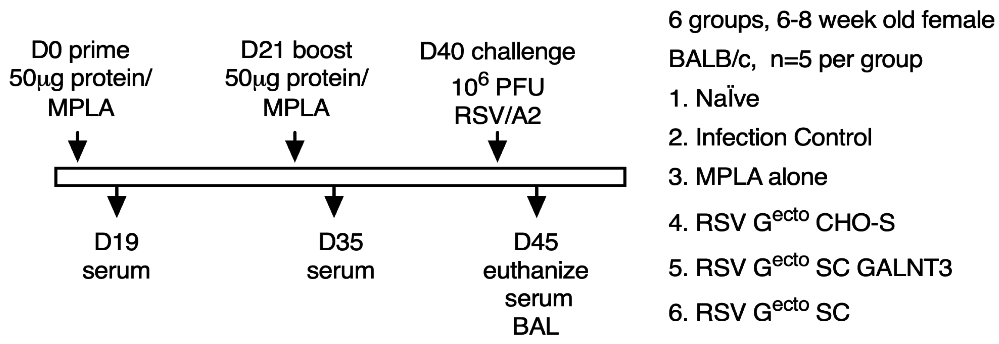
Mice were vaccinated intramuscularly with 50 μg of immunogen or PBS + 50 μg synthetic monophosphoryl lipid A (MPLA, InVivogen, San Diego, CA, USA), diluted in PBS. Mice (n = 5 per group) were primed on day 0 and boosted on day 21 via homogenous vaccination. On day 40 (19 days post-boost), the mice were intranasally challenged with 106 PFUs RSV/A2 and subsequently euthanized on day 5 post-challenge.

2.8. Detection of Anti-RSV Gecto and Anti-CCD Specific IgG by ELISA

Flat-bottom, high-binding 96-well ELISA plates (Corning, 3590) were coated overnight at 4 °C with 5 µg/mL of recombinant RSV Gecto or CCD-Fc protein, diluted in PBS. The plates were washed three times with wash-buffer, PBS, pH7.4, 0.1% Tween (Accuwash, Thermo Fisher Scientific, Waltham, MA, USA), and blocked for two hours at 25 °C with Blotto (PBS pH7.4, 5% non-fat dry milk). Pre-challenge sera samples collected at Day 19 and Day 35 of the experiment were serially diluted 1:3 in Blotto (starting at a 1/40 dilution) and then incubated for two hours at 25 °C. After washing three times with wash buffer, the plates were incubated for one hour at 25 °C with a goat anti-mouse Fc-specific horseradish peroxidase (HRP)-conjugated secondary antibody (115-035-071, Jackson-ImmunoResearch, West Grove, PA, USA), diluted 1/5000 in Blotto. The plates were washed 3 times and developed for 10 min in the dark with TMB Substrate (Thermo Fisher Scientific, Waltham MA, USA), before stopping the reaction with 1 N H2SO4. The optical density at 450 nm, OD450, was read on a plate reader (Molecular Devices, San Jose, CA, USA). The resulting graphs are representative of two independent experiments. The background cut-off was calculated as the mean OD450 signal in the absence of primary antibody, but the presence of secondary antibody, plus three times the standard deviation, plus 10% of the calculated number.

2.9. Determining RSV Lung Titers by Plaque Assays

RSV lung titers in vaccinated and control mice were determined as previously described [76]. Briefly, lungs were aseptically removed from exsanguinated mice on day 5 after challenge, and individual lung specimens were placed in 1 mL Dulbecco’s modified Eagle’s medium and homogenized using a gentleMACS dissociator (Miltenyi Biotec, San Jose, CA, USA). After centrifugation of the extract at 400× g for 10 min at 4 °C, 200 µL/well, serial 10-fold dilutions of the soluble homogenate fraction were added to 90% confluent Vero cell (ATCC CCL-81) monolayers in 24-well tissue culture plates, allowing 2 h at 37 °C for viral absorption. The cell monolayers were overlaid with medium containing 2% methylcellulose and further incubated at 37 °C for 6 days. To enumerate plaques, the monolayers were fixed with 60% acetone/40% methanol, then incubated overnight at 4 °C with a 1 μg/mL of monoclonal antibody against the RSV F protein (clone 131-2A), followed by a further 1 h incubation at 25 °C with a 1/1,000 dilution of goat anti-mouse HRP-conjugated IgG antibody (Jackson ImmunoResearch West Grove, PA, USA) before visualizing with KPL TrueBlue peroxidase substrate (SeraCare, Milford, MA, USA).

2.10. Detection of Anti-RSV/A2 Antibodies by ELISA

An indirect ELISA specific to RSV/2 was examined using flat-bottom high-binding ELISA plates (Corning, 3590) that were coated with 106 PFU/mL RSV/A2 was diluted in PBS (pH 7.4) and added to the wells for 1 h at 37 °C. The plates were washed three times with 1× KPL Wash Buffer (SeraCare Milford, MA, USA) and blocked for 2 h at 37 °C with 5% non-fat dry milk + 1% BSA (Sigma Aldrich, Burlington, MA, USA) diluted in 1× KPL wash buffer. The blocking solution was decanted, and the plates were incubated for 1h at 37 °C with sera diluted in 5% non-fat dry milk + 1% BSA. The solution was decanted, and the wells were washed 3x with KPL wash buffer. Subsequently, the wells were incubated for 1 h at 37 °C with a 1/5,000 dilution of an HRP-conjugated secondary goat anti-mouse antibody against murine Fc (Thermo Fisher Scientific, Waltham, MA, USA) or an HRP-conjugated secondary IgG1 or IgG2a goat anti-mouse antibody (Southern Biotech, Birmingham, AL, USA), diluted per the manufacturer’s instructions. The plates were washed in PBS (pH 7.4) and developed with 1-Step Ultra TMB Substrate (Thermo Fisher Scientific, Waltham, MA, USA). The reactions were stopped with Stop-Solution (Invitrogen). OD450 and values read using a plate reader (BioTek, Winooski, VT, USA). The data were plotted using GraphPad Prism software, version 10.4 (GraphPad, Boston, MA, USA), and error bars represent standard deviation.

2.11. Bronchoalveolar Lavage (BAL) and Flow Cytometry

To investigate the composition of BAL cells in vaccinated mice at day 5 post-challenge, lungs were washed 3x with 1 mL ice-cold PBS. BAL was centrifuged at 500× g for 5 min at 4 °C. BAL cells were washed and resuspended in FACS buffer (PBS with 1% BSA and 1 mM EDTA). Cell-surface marker expression patterns were used to identify CD3+ T cells, eosinophils (CD45+/SiglecF+/CD11clow), alveolar macrophages (CD45+/SiglecF+/CD11chigh) and activated B cells (CD45+, CD220+). Cell suspensions were blocked with an anti-FcγII/III receptor antibody (BD Bioscience, Franklin Lakes, NJ, USA) and subsequently stained with fluorescent antibodies from eBiosciences (San Diego, CA, USA), BioLegend (San Diego, CA, USA), or BD Bioscience, i.e., PerCP-conjugated anti-CD45 (30-F11), APC-conjugated anti-CD11c (HL3 or N418), PE-conjugated anti-SiglecF (E50-2440), CD45+ CD220+ (clone RA3-6B2), FITC-conjugated anti-CD3e (145-2C11), per the suppliers’ instructions. The cells were analyzed on a Guava easyCyte 8 flow cytometers (Millipore, Burlington, MA, USA), and the data were analyzed using Guavasoft software (version 2.7 or 3.1.1). The cells were gated based on forward scatter and side scatter, and then by CD45+ staining. Eosinophils were identified as FSChigh/SSChigh/CD45+/SiglecF+/CD11clow cells.

2.12. Microneutralization Assay

A microneutralization assay was used to assess anti-RSV-mediated neutralization in mouse serum samples [85]. Briefly, the sera were pooled and heat-inactivated at 55 °C for 30 min. The sera were diluted 1:40 in DMEM containing 2% fetal bovine serum and incubated for 1 h at 37 °C with 200 fluorescent focus units (FFUs) RSV/A2-GFP with or without 10% guinea pig complement (C’) (Sigma Aldrich, Burlington, MA, USA). Following pre-incubation, the virus/serum mixture was added to 95% confluent A549 (purchased from the ATCC, A549-CCL-185) and used at low passage number) and the cells were incubated for 48 h. Anti-RSV F protein neutralizing mAb 143.6C was included as a technical control. FFUs were visualized using a Cellomics ArrayScan (Thermo Fisher Scientific, Waltham, MA, USA), and the mean FFU value of replicate wells was determined using HCS Studio™ Cell Analysis Software (Thermo Fisher Scientific, Waltham, MA, USA, Scotts Valley, CA, USA). Neutralization was defined as the percent reduction in the mean FFU when compared with infection in the absence of serum.

2.13. Statistical Analysis

The challenge study was designed as a preliminary pilot experiment to investigate the experimental variation and attrition, as a guide to future larger experiments. No power calculation was performed. No animals were excluded from analysis. All statistical analyses were performed using GraphPad Prism software, version 10.4 (GraphPad Software). A single challenge study was performed. The data are presented as the mean ± standard deviation. After applying the Shapiro–Wilk normality test, differences between two groups were analyzed with one-way or two-way analysis of variance (ANOVA), where appropriate, followed by Dunnett’s multiple comparison test for group to control analysis, or Tukey HSD test for comparison among groups. p < 0.05 was considered statistically significant. Non-parametric tests were performed where indicated, using the Mann–Whitney U test (for two independent groups), the Kruskal–Wallis test (for more than two independent groups). Results of our statistical tests are indicated in the figure legends. For statistical analysis of flow cytometry, the data were analyzed by one-way ANOVA with Bonferroni’s correction or Student’s t-test for pairwise analysis.

3. Results

3.1. Expression and Purification of O-Glycan-Restricted RSV Gecto Glycoproteins

RSV G glycoprotein from RSV/A2 is a 298 amino acid polypeptide, with three predicted N-linked glycosylation sites (PNGS) and between 30–40 predicted O-linked glycosylation sites, a transmembrane domain, and a short cytoplasmic tail. For the murine RSV challenge study, we produced protein immunogens corresponding to the mature ectodomain of RSV G (Gecto) that incorporates amino acids 64 to 298 (Figure 1A), and C-terminal tandem 6× histidine and Twin-StrepTactin purification tags, for detection and purification. RSV Gecto protein produced in CHO cells has a slightly lower motility on an SDS-PAGE gel than RSV Gecto protein produced in HEK 293 cells (Figure 1B). To generate O-glycan-restricted RSV Gecto proteins, we used SimpleCell (SC) and SC GALNT3 CHO lines [69,70,71]. CHO-S, SC GALNT3, and SC lines produced RSV Gecto proteins exhibiting apparent molecular masses of, 90 kDa, 75 kDa, and 55 kDa, respectively, by SDS-PAGE (Figure 1C), and PNGase F treatment resulted in a molecular mass reduction concomitant with stripping of the predicted 3 N-linked glycans (Figure 1C, panel 2) but having no effect on O-linked glycans. Interestingly, SC GALNT3 RSV Gecto is ~20 kDa larger than SC RSV Gecto, which is due to increased occupancy by GalNAc, catalyzed by GALNT3 (Figure 1A). Western blot analysis with an anti-His-tag antibody confirmed that the peaks contained the recombinant RSV Gecto proteins (Figure 1D).
Analytical-scale size exclusion chromatography of purified RSV Gecto proteins resulted in a major peak eluting between the globular gel-filtration standards representing molecular weights of 670 and 158 kDa, with the CHO-S RSV Gecto eluting at a single peak at around 15.5 mL. In contrast, SC GALNT3 and SC RSV Gecto were retained longer, eluting at 16 and 16.5 mL, respectively (Figure 1E). Previous cryo-electron microscopy studies of the SC GALNT3 RSV Gecto protein showed it to be monomeric, flexible, and having the capacity to simultaneously bind two conformation-dependent antibodies with high affinity [81].

3.2. Antigenicity of O-Glycan-Restricted RSV Gecto Glycoproteins

To evaluate the effect of O-glycosylation restriction on RSV Gecto protein folding, we determined the affinities of the RSV Gecto proteins for two anti-RSV G CCD conformation-dependent monoclonal antibodies (mAbs) that bind to neighboring epitopes spanning the cysteine loop [36,81], by biolayer interferometry (BLI). In line with previous studies [36,81,82], mAbs 2D10 and 3D3 had low picomolar binding affinity for CHO-S RSV Gecto (Table 1). Similar results were observed for SC-produced RSV Gecto. These studies reveal that the O-glycan-restricted recombinant RSV Gecto retains conformational epitopes in the CCD.

3.3. Immunogenicity of O-Glycan-Restricted RSV Gecto Protein Immunogens

A 50 µg RSV Gecto was combined with MPLA, an established TLR4 agonist. Six groups of 6–8-week-old female BALB/c mice were immunized intramuscularly in a prime-boost regimen and then challenged with RSV/A2, following the schedule shown (Figure 2).
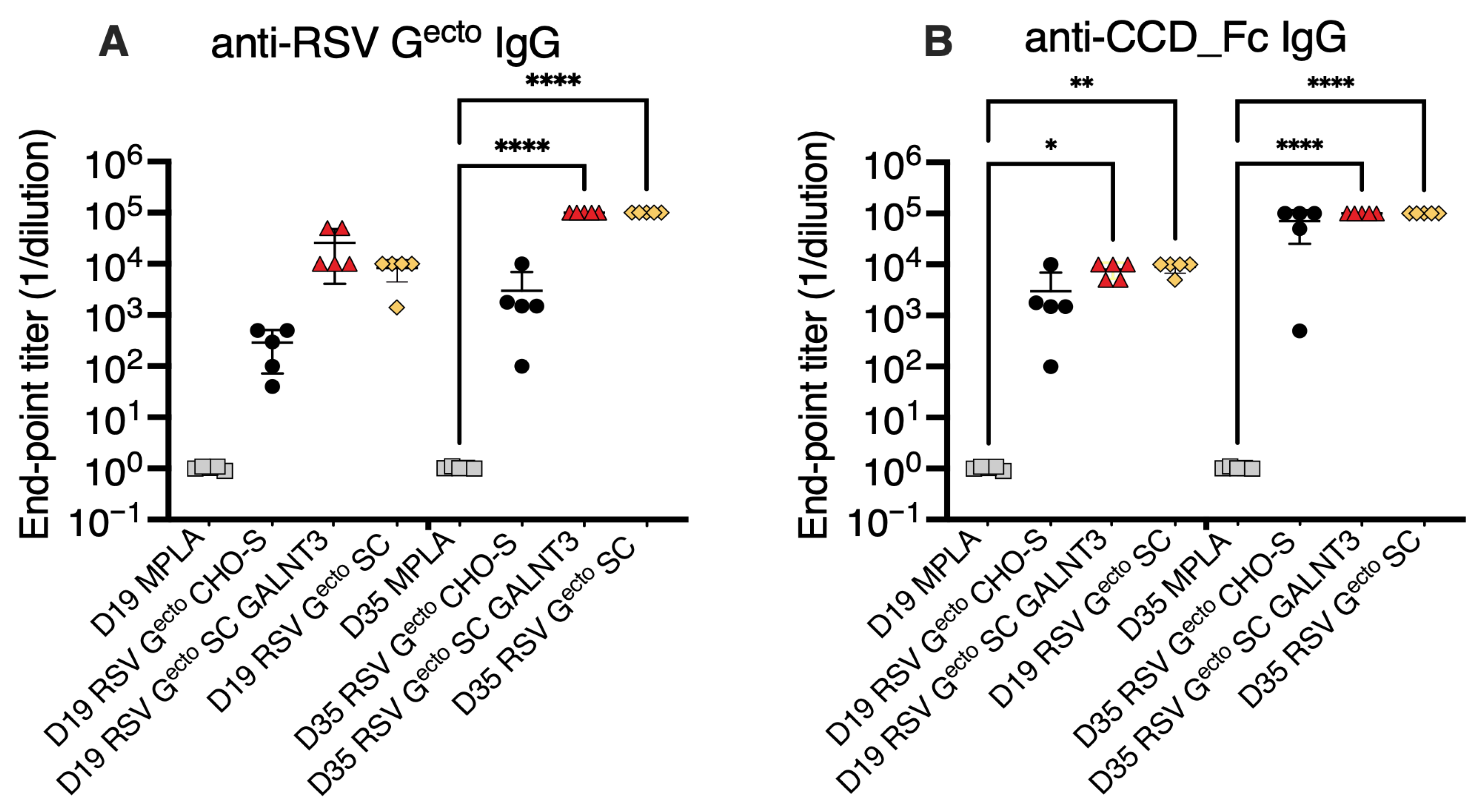
To compare vaccine antigen immunogenicity, we calculated the endpoint titer of anti-RSV Gecto and anti-CCD-specific IgG circulating after a single priming (D19), or the complete prime/boost regime (D35) by ELISA, as summarized in Figure 3. Neither anti-RSV Gecto nor anti-CCD IgG was detected in the MPLA-alone group. Post-boost (D35), groups of mice vaccinated with recombinant O-glycan-restricted RSV Gecto (SC GALNT3 and SC) antigens had significantly (p = 0.0001) higher anti-Gecto endpoint titers (1/dilution) both at ~105, compared to MPLA control group. Similarly, statistically significant anti-CCD-specific IgG endpoint titers of ~105 (p > 0.0001), were only achieved post-boost in the O-glycosylation restricted RSV Gecto vaccinated groups (Figure 3B).
Interestingly, although only approaching statistical significance for the RSV Gecto SC GALNT3 vaccination (p = 0.0643), a single immunization (D19) elicited 10-fold more total anti-RSV-Gecto IgG than the non-restricted O-glycosylated protein (Figure 3A). Measuring the anti-CCD specific IgG response after a single vaccination (D19), all mice vaccinated with O-glycosylation restricted RSV Gecto adjuvanted with MPLA achieved a titer of 104 (p = 0.0069 and p = 0.0226) compared to the baseline MPLA response (Figure 3B).

3.4. Evaluation of Post-RSV/A2 Challenge Responses

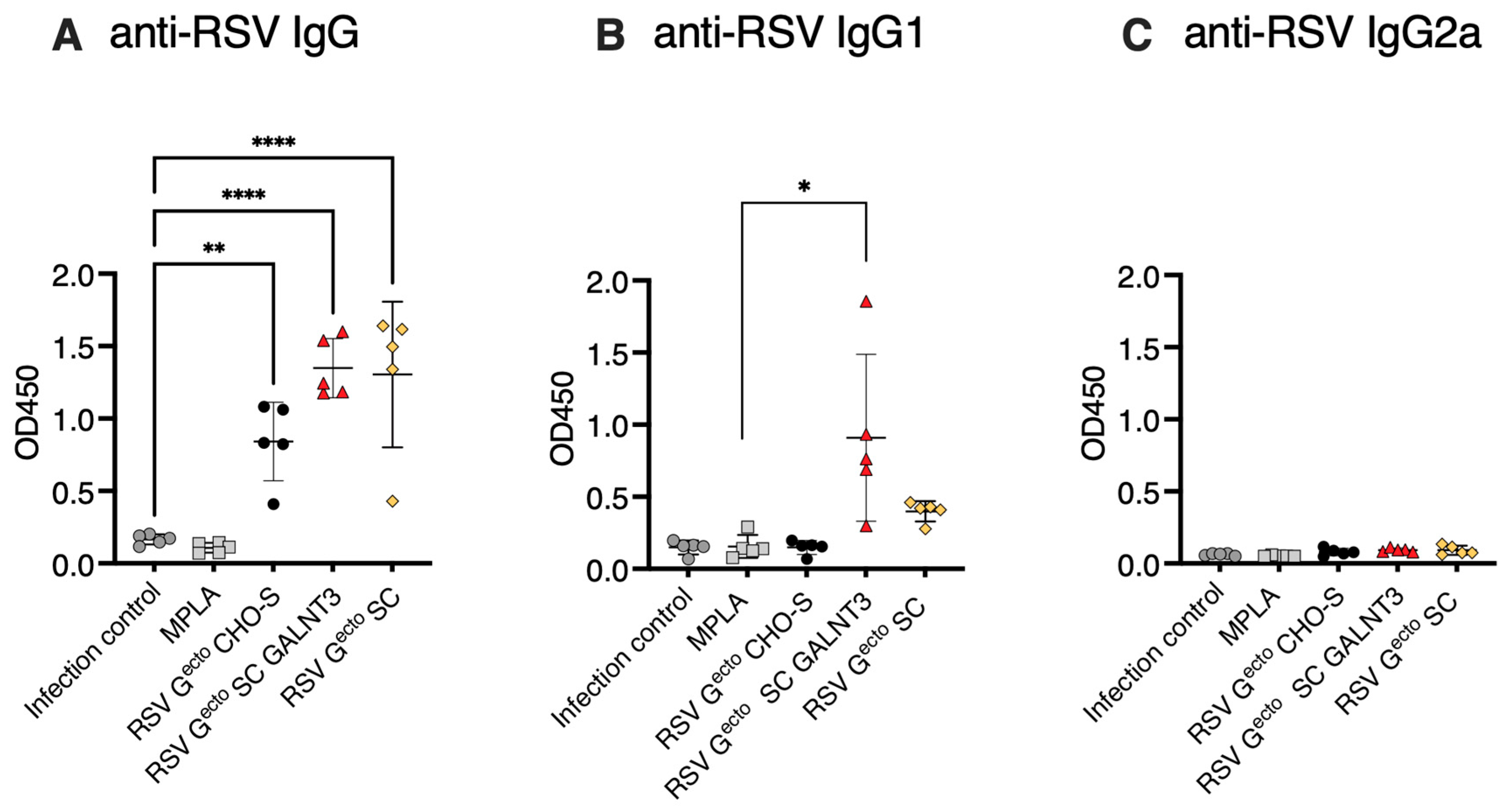
RSV/A2-specific serum IgG antibody recall, serum neutralizing activity, and viral lung titer at the peak of infection were evaluated five days after intranasal challenge with 106 PFU RSV/A2. All RSV Gecto-vaccinated groups had robust anti-RSV/A2 IgG recall responses, consistent with pre-challenge immunogen-specific titer (Figure 4A), further analyzed as IgG1 and IgG2a components in Figure 4B,C and discussed below. The group immunized with RSV Gecto SC GALNT3 had a higher IgG1 response than the infection control (p < 0.05).
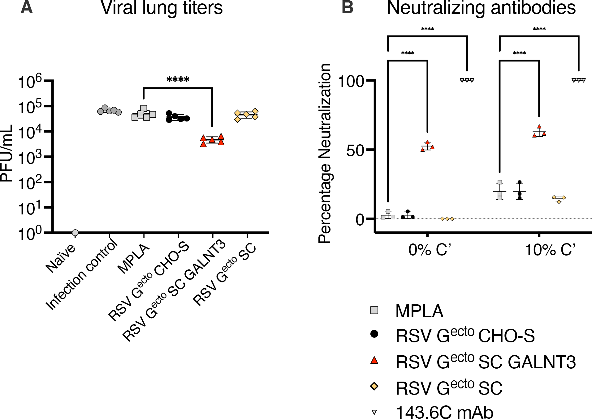
Lung viral load was quantified by plaque assay, and the data (Figure 5A) were summarized as PFU/mL of lung homogenate. A single vaccine immunogen, the O-glycan-restricted RSV Gecto GALNT3 protein, conferred a significant reduction of mean viral load from 4.9 × 10⁴ PFU/mL in the mock vaccinated MPLA control group, to 4.8 × 10³ PFU/mL (p < 0.001). Finally, a significant (p = 0.0001) complement-mediated and classical (non-complement-dependent) neutralization of autologous RSV A2 virus was detected in the assay of sera from mice vaccinated with the O-glycan-restricted RSV Gecto SC GALNT3 (Figure 5B) compared to MPLA controls.

3.5. Evaluation of Lung Biomarkers Associated with Vaccine-Enhanced Disease

The RSV G protein modulates host immunity by interfering with the CX3CL1/CX3CR1 interaction, raising questions about the safety of RSV G-based vaccines [7]. Host-mediated pathogenesis correlates with a Th2-type T-cell-mediated imbalance and cytokine cascade. Although the BALB/c murine infection model of infection is imperfect with respect to accurately reflecting human RSV infection [9,86,87,88,89], previous studies vaccinating BALB/c with fully glycosylated recombinant RSV G-derived proteins [28,57,90] have caused measurable increases in surrogate markers of vaccine enhanced disease such as inflammatory infiltrate, lung eosinophilia, cytokine profile and an imbalance in the ratio of RSV G protein specific IgG antibody subtypes.
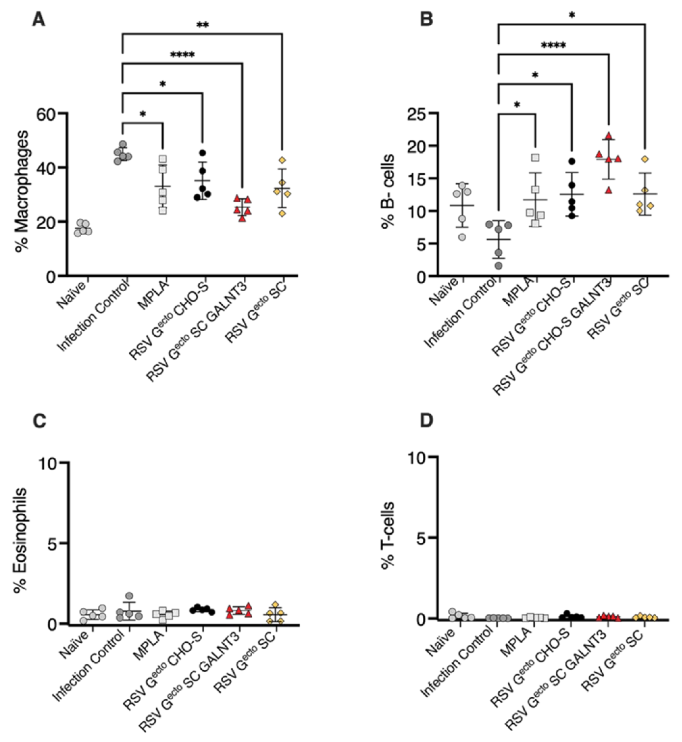
We evaluated the levels of serum anti-RSV/A2 IgG1 and IgG2a as a surrogate for a potential vaccine-induced Th2:Th1 imbalance. BALB/c mice vaccinated with O-glycan-restricted RSV Gecto. SC GALNT3 had more RSV/A2-specific IgG1 than the adjuvant alone vaccinated mice (p = 0.05), and no vaccine antigen promoted the induction of an IgG2a response (Figure 4B,C). Examination of the BAL shown in Figure 6 did not indicate the presence of a pathological post-challenge vaccine-enhanced inflammatory infiltrate in the lungs of any of the groups of mice vaccinated with MPLA adjuvanted RSV Gecto. It is known that airway macrophages line the bronchiole membranes, promoting protection and homeostasis [91]. Still, the recruitment of activated macrophages following RSV challenge occurred in approximately 45% of the BAL leukocytes analyzed in the infection control. Typical BAL leukocytes were Siglecf+ macrophages (CD45+/Siglecf+CD11chigh). Pro-inflammatory macrophage recruitment is dampened by MPLA alone (~30% total BAL cells), with the most significant dampening observed after vaccination with the O-glycan-restricted RSV Gecto SC GALNT3 protein (p = 0.0001) (Figure 6A). The study indicated a localized enhanced presence of mature antibody-secreting B220+ B cells in all mice administered MPLA (p = 0.05), with a notable increase in lung-resident mature B cells in mice vaccinated with O-glycan-restricted RSV Gecto SC GALNT3 (p = 0.0001) (Figure 6B). There was no substantive Siglecf+ eosinophilic infiltrate (CD45+/Siglecf+CD11clow) (Figure 6C), and unusually few infiltrating T lymphocytes were detected (Figure 6D).

4. Discussion

To determine if the RSV vaccine response was modified by restricting O-linked glycosylation on an RSV Gecto glycoprotein vaccine, we examined the outcome in a preliminary BALB/c mouse challenge study, challenging with a virus with an identical RSV G sequence that was used for immunization. RSV Gecto immunogens with identical amino acid sequence but differing O-linked glycosylation were expressed and purified from three CHO cell lines. Comparing O-glycosylation-restricted RSV Gecto proteins by electrophoretic mobility by SDS-PAGE, we observed that only approximately half of the predicted O-linked glycosylation sites on RSV Gecto are occupied by GalNAc in the absence of GALNT3. This finding emphasizes the reason to carefully examine O-glycosylation when producing recombinant viral glycoproteins with mucin-like domains as vaccine antigens.
We subsequently vaccinated six groups of BALB/c mice (Figure 2) with one of each of the three RSV Gecto proteins (50 µg) adjuvanted with MPLA, followed by an identical boost three weeks later. The vaccinated mice were challenged with 106 PFU RSV/A2 a week after the vaccination regime was completed. Control groups included naïve, MPLA-immunized, and infection-only controls. O-glycosylation-restricted RSV Gecto proteins were significantly more immunogenic than non-restricted O-glycosylated RSV Gecto. Even a single dose of O-glycosylation-restricted RSV Gecto vaccine antigen raised a moderate anti-RSV G CCD response. Post boost, a circulating anti-RSV Gecto IgG endpoint titer of 105 was attained compared to a mean titer of 5 × 10³ by mice vaccinated with a non-restricted RSV Gecto protein. Only the O-glycosylation-restricted RSV Gecto protein immunized groups achieved statistical significance (p = 0.0001) and despite the immunogenicity of both RSV Gecto SC and SC GALNT3 proteins, only vaccination with RSV Gecto SC GALNT3 protein induced autologous-virus-specific neutralizing antibodies. This could be due to the requirement for a very particular configuration of GalNAc loaded residues to expose neutralization epitopes, and/or due to differences in proteolysis due to the more extensive glycan occupancy of RSV Gecto SC GALNT3.
In concordance with the detection of neutralizing antibodies, the RSV Gecto SC GALNT3 protein-vaccinated group of mice had significantly reduced lung viral load, five days after challenge with autologous virus. RSV Gecto proteins, when administered in combination with the TLR4 agonist MPLA, had no significant effect on post-vaccination pulmonary infiltrate in our BALB/c experiment.
Precedent for the manipulation of O-linked glycans in vaccine antigen design. Recently, the improved efficacy of a currently available CHO-produced recombinant Herpes Zoster glycoprotein gE subunit vaccine was determined to be due to altered exposure of a B cell epitope that is shielded by O-linked glycosylation when the vaccine antigen is produced in a different cell line [92]. There is also precedent for the elimination of N- and O-linked glycans in the generation of RSV Gecto vaccine antigens via protein expression in E. coli [90]. However, this protein was produced in an insoluble form and then refolded in the presence of a redox buffer, so it is unclear if this antigen displays conformational epitopes like those for mAbs 3D3 and 2D10. Even so, this non-glycosylated RSV Gecto vaccine antigen was found to be significantly more immunogenic than the fully glycosylated protein, eliciting neutralizing antibodies in both BALB/c mice and cotton rats [90,93]. Furthermore, the non-glycosylated RSV Gecto has antigenic epitopes outside of the central conserved domain, which may contribute towards a protective anti-RSV response [94].
Here, by employing the SimpleCell strategy to produce soluble RSV Gecto antigens, the conformational epitopes in the CCD are retained, sites previously masked by O-linked core polysaccharide extension may be de-shielded, and new epitopes added. This is a new use of the SimpleCell technology, which was originally developed to produce O-linked glycoproteins with homogeneous O-glycosylation to map the O-glycoproteome by mass spectrometry. We found that O-glycan-restricted RSV Gecto glycoproteins appear to be more effective in raising antibodies to full-length RSV G than a non-restricted protein when used in combination with MPLA to vaccinate BALB/c mice. All RSV Gecto proteins were effective in raising a response to the non-glycosylated and highly conserved CCD, but again, only mice immunized with the O-glycan-restricted RSV Gecto glycoproteins demonstrated a titer with a statistical confidence higher than MPLA vaccination alone. Vaccination with RSV Gecto SC GALNT3 protein raised antibodies capable of neutralizing autologous virus in vitro and reducing lung viral load in vivo. Preliminary examination of inflammatory lung infiltrate post challenge suggests that vaccine antigens were well-tolerated in this experiment. Future work should include examining the theoretical mechanisms of vaccine-enhanced disease (VED), including antibody-enhanced disease and Th2-mediated pathology. It should be noted that this is a preliminary pilot study and the first use of a GalNAc-enriched viral glycoprotein as a vaccine antigen in vivo. The cotton rat model may be better to mimic RSV pathology and potential VED, and examination of cross-reactivity to other RSV A or B strains might be justified. GalNAc proteoglycan B cell epitopes have been identified on other viruses [61,67,68]. They are known to be immunogenic, but these antigens are thought to be rapidly cleared from serum due to their high affinity for liver-expressed asialoglycoprotein receptor [95].
The production of viral glycoproteins in differing cell types with differing capacity to initiate and extend core O-linked glycans is an underexplored field. Our study supports the importance in further exploring the manipulation of O-linked glycosylation to enhance the efficacy of RSV Gecto as a vaccine antigen.

Author Contributions

R.M.D., R.A.T., S.M.O., and J.M. contributed to study design. J.M. and S.M.O. contributed equally to data collection and analysis, S.M.O., R.M.D., R.A.T., and J.M. wrote the manuscript. M.G.J. supported experimental antigen design. All authors have read and agreed to the published version of the manuscript.

Funding

This research was funded by an NIH grant R01 AI166066 to R.M.D. and R.A.T. Funding for the purchase of the Octet RED384 instrument was supported by the NIH S10 shared instrumentation grant 1S10OD027012-01.

Data Availability Statement

Experimental data is deposited at https://datadryad.org.

Acknowledgments

The authors acknowledge H. Clausen and Y. Narimatsu from the University of Copenhagen, Denmark, for the CHO SimpleCell lines, and Trellis Biosciences for the mAb 3D3. We would also like to thank Anand Verma for technical assistance with the Octet BLI studies.

Conflicts of Interest

The authors declare that the research was conducted in the absence of any commercial or financial relationships that could be construed as a potential conflict of interest.

References

  1. Li, Y.; Wang, X.; Blau, D.M.; Caballero, M.T.; Feikin, D.R.; Gill, C.J.; Madhi, S.A.; Omer, S.B.; Simoes, E.A.F.; Campbell, H.; et al. Global, regional, and national disease burden estimates of acute lower respiratory infections due to respiratory syncytial virus in children younger than 5 years in 2019: A systematic analysis. Lancet 2022, 399, 2047–2064. [Google Scholar] [CrossRef]
  2. McLellan, J.S.; Ray, W.C.; Peeples, M.E. Structure and function of respiratory syncytial virus surface glycoproteins. Curr. Top. Microbiol. Immunol. 2013, 372, 83–104. [Google Scholar] [PubMed]
  3. Kampmann, B.; Madhi, S.A.; Munjal, I.; Simoes, E.A.F.; Pahud, B.A.; Llapur, C.; Baker, J.; Perez Marc, G.; Radley, D.; Shittu, E.; et al. Bivalent prefusion F vaccine in pregnancy to prevent RSV illness in infants. N. Engl. J. Med. 2023, 388, 1451–1464. [Google Scholar] [CrossRef] [PubMed]
  4. Papi, A.; Ison, M.G.; Langley, J.M.; Lee, D.G.; Leroux-Roels, I.; Martinon-Torres, F.; Schwarz, T.F.; van Zyl-Smit, R.N.; Campora, L.; Dezutter, N.; et al. Respiratory syncytial virus prefusion F protein vaccine in older adults. N. Engl. J. Med. 2023, 388, 595–608. [Google Scholar] [CrossRef]
  5. Walsh, E.E.; Eiras, D.; Woodside, J.; Jiang, Q.; Patton, M.; Marc, G.P.; Llapur, C.; Ramet, M.; Fukushima, Y.; Hussen, N.; et al. Efficacy, immunogenicity, and safety of the bivalent RSV prefusion F (RSVpreF) vaccine in older adults over 2 RSV seasons. Clin. Infect. Dis. Off. Publ. Infect. Dis. Soc. Am. 2025, 388, 1465–1477 . [Google Scholar] [CrossRef]
  6. Wilson, E.; Goswami, J.; Baqui, A.H.; Doreski, P.A.; Perez-Marc, G.; Zaman, K.; Monroy, J.; Duncan, C.J.A.; Ujiie, M.; Ramet, M.; et al. Efficacy and safety of an mRNA-based RSV preF vaccine in older adults. N. Engl. J. Med. 2023, 389, 2233–2244. [Google Scholar] [CrossRef]
  7. Anderson, J.; Do, L.A.H.; van Kasteren, P.B.; Licciardi, P.V. The role of respiratory syncytial virus G protein in immune cell infection and pathogenesis. EBioMedicine 2024, 107, 105318. [Google Scholar] [CrossRef]
  8. Anderson, L.J.; Jadhao, S.J.; Paden, C.R.; Tong, S. Functional features of the respiratory syncytial virus G protein. Viruses 2021, 13, 1214. [Google Scholar] [CrossRef]
  9. Castilow, E.M.; Olson, M.R.; Varga, S.M. Understanding respiratory syncytial virus (RSV) vaccine-enhanced disease. Immunol. Res. 2007, 39, 225–239. [Google Scholar] [CrossRef]
  10. Shishir, T.A.; Saha, O.; Rajia, S.; Mondol, S.M.; Masum, M.H.U.; Rahaman, M.M.; Hossen, F.; Bahadur, N.M.; Ahmed, F.; Naser, I.B.; et al. Genome-wide study of globally distributed respiratory syncytial virus (RSV) strains implicates diversification utilizing phylodynamics and mutational analysis. Sci. Rep. 2023, 13, 13531. [Google Scholar] [CrossRef]
  11. Anderson, L.J.; Hierholzer, J.C.; Tsou, C.; Hendry, R.M.; Fernie, B.F.; Stone, Y.; McIntosh, K. Antigenic characterization of respiratory syncytial virus strains with monoclonal antibodies. J. Infect. Dis. 1985, 151, 626–633. [Google Scholar] [CrossRef]
  12. Martinez, I.; Dopazo, J.; Melero, J.A. Antigenic structure of the human respiratory syncytial virus G glycoprotein and relevance of hypermutation events for the generation of antigenic variants. J. Gen. Virol. 1997, 78 Pt 10, 2419–2429. [Google Scholar] [CrossRef] [PubMed]
  13. Olofsson, S.; Bally, M.; Trybala, E.; Bergstrom, T. Structure and role of O-linked glycans in viral envelope proteins. Annu. Rev. Virol. 2023, 10, 283–304. [Google Scholar] [CrossRef] [PubMed]
  14. Wandall, H.H.; Nielsen, M.A.I.; King-Smith, S.; de Haan, N.; Bagdonaite, I. Global functions of O-glycosylation: Promises and challenges in O-glycobiology. FEBS J. 2021, 288, 7183–7212. [Google Scholar] [CrossRef] [PubMed]
  15. Tripp, R.A.; Jones, L.P.; Haynes, L.M.; Zheng, H.; Murphy, P.M.; Anderson, L.J. CX3C chemokine mimicry by respiratory syncytial virus G glycoprotein. Nat. Immunol. 2001, 2, 732–738. [Google Scholar] [CrossRef]
  16. Imai, T.; Hieshima, K.; Haskell, C.; Baba, M.; Nagira, M.; Nishimura, M.; Kakizaki, M.; Takagi, S.; Nomiyama, H.; Schall, T.J.; et al. Identification and molecular characterization of fractalkine receptor CX3CR1, which mediates both leukocyte migration and adhesion. Cell 1997, 91, 521–530. [Google Scholar] [CrossRef]
  17. Anderson, C.S.; Chirkova, T.; Slaunwhite, C.G.; Qiu, X.; Walsh, E.E.; Anderson, L.J.; Mariani, T.J. CX3CR1 engagement by respiratory syncytial virus leads to induction of nucleolin and dysregulation of cilia-related genes. J. Virol. 2021, 95, A1171. [Google Scholar] [CrossRef]
  18. Chirkova, T.; Lin, S.; Oomens, A.G.; Gaston, K.A.; Boyoglu-Barnum, S.; Meng, J.; Stobart, C.C.; Cotton, C.U.; Hartert, T.V.; Moore, M.L.; et al. CX3CR1 is an important surface molecule for respiratory syncytial virus infection in human airway epithelial cells. J. Gen. Virol. 2015, 96, 2543–2556. [Google Scholar] [CrossRef]
  19. Jeong, K.I.; Piepenhagen, P.A.; Kishko, M.; DiNapoli, J.M.; Groppo, R.P.; Zhang, L.; Almond, J.; Kleanthous, H.; Delagrave, S.; Parrington, M. CX3CR1 is expressed in differentiated human ciliated airway cells and co-localizes with respiratory syncytial virus on cilia in a G protein-dependent manner. PLoS ONE 2015, 10, e0130517. [Google Scholar] [CrossRef]
  20. Johnson, S.M.; McNally, B.A.; Ioannidis, I.; Flano, E.; Teng, M.N.; Oomens, A.G.; Walsh, E.E.; Peeples, M.E. Respiratory syncytial virus uses CX3CR1 as a receptor on primary human airway epithelial cultures. PLoS Pathog. 2015, 11, e1005318. [Google Scholar] [CrossRef]
  21. Zhivaki, D.; Lemoine, S.; Lim, A.; Morva, A.; Vidalain, P.O.; Schandene, L.; Casartelli, N.; Rameix-Welti, M.A.; Herve, P.L.; Deriaud, E.; et al. Respiratory syncytial virus infects regulatory B cells in human neonates via chemokine receptor CX3CR1 and promotes lung disease severity. Immunity 2017, 46, 301–314. [Google Scholar] [CrossRef] [PubMed]
  22. Cui, G.; Liu, H.; Li, X.; Ming, L. Preliminary functional and phylogeographic analyses of the 72 nucleotide duplication region in the emerging human respiratory syncytial virus ON1 strain attachment glycoprotein gene. Biomed. Pharmacother. 2020, 123, 109800. [Google Scholar] [CrossRef] [PubMed]
  23. Hotard, A.L.; Laikhter, E.; Brooks, K.; Hartert, T.V.; Moore, M.L. Functional analysis of the 60-nucleotide duplication in the respiratory syncytial virus buenos aires strain attachment glycoprotein. J. Virol. 2015, 89, 8258–8266. [Google Scholar] [CrossRef] [PubMed]
  24. Chirkova, T.; Boyoglu-Barnum, S.; Gaston, K.A.; Malik, F.M.; Trau, S.P.; Oomens, A.G.; Anderson, L.J. Respiratory syncytial virus G protein CX3C motif impairs human airway epithelial and immune cell responses. J. Virol. 2013, 87, 13466–13479. [Google Scholar] [CrossRef]
  25. Hancock, G.E.; Speelman, D.J.; Heers, K.; Bortell, E.; Smith, J.; Cosco, C. Generation of atypical pulmonary inflammatory responses in BALB/c mice after immunization with the native attachment (G) glycoprotein of respiratory syncytial virus. J. Virol. 1996, 70, 7783–7791. [Google Scholar] [CrossRef]
  26. Johnson, T.R.; Parker, R.A.; Johnson, J.E.; Graham, B.S. IL-13 is sufficient for respiratory syncytial virus G glycoprotein-induced eosinophilia after respiratory syncytial virus challenge. J. Immunol. 2003, 170, 2037–2045. [Google Scholar] [CrossRef]
  27. Johnson, T.R.; Varga, S.M.; Braciale, T.J.; Graham, B.S. Vβ14+ T cells mediate the vaccine-enhanced disease induced by immunization with respiratory syncytial virus (RSV) G glycoprotein but not with formalin-inactivated RSV. J. Virol. 2004, 78, 8753–8760. [Google Scholar] [CrossRef]
  28. Kawahara, E.; Senpuku, K.; Kawaguchi, Y.; Yamamoto, S.; Yasuda, K.; Kuroda, E.; Ouji-Sageshima, N.; Ito, T.; Hirai, T.; Shibata, T.; et al. Recombinant RSV G protein vaccine induces enhanced respiratory disease via IL-13 and mucin overproduction. npj Vaccines 2024, 9, 187. [Google Scholar] [CrossRef]
  29. Martinez, M.E.; Capella Gonzalez, C.; Huey, D.; Peeples, M.E.; McCarty, D.; Niewiesk, S. Immunogenicity and inflammatory properties of respiratory syncytial virus attachment G protein in cotton rats. PLoS ONE 2021, 16, e0246770. [Google Scholar] [CrossRef]
  30. Polack, F.P.; Teng, M.N.; Collins, P.L.; Prince, G.A.; Exner, M.; Regele, H.; Lirman, D.D.; Rabold, R.; Hoffman, S.J.; Karp, C.L.; et al. A role for immune complexes in enhanced respiratory syncytial virus disease. J. Exp. Med. 2002, 196, 859–865. [Google Scholar] [CrossRef]
  31. Stott, E.J.; Taylor, G.; Ball, L.A.; Anderson, K.; Young, K.K.; King, A.M.; Wertz, G.W. Immune and histopathological responses in animals vaccinated with recombinant vaccinia viruses that express individual genes of human respiratory syncytial virus. J. Virol. 1987, 61, 3855–3861. [Google Scholar] [CrossRef]
  32. Bergeron, H.C.; Tripp, R.A. Immunopathology of RSV: An updated review. Viruses 2021, 13, 2478. [Google Scholar] [CrossRef] [PubMed]
  33. Choi, Y.; Mason, C.S.; Jones, L.P.; Crabtree, J.; Jorquera, P.A.; Tripp, R.A. Antibodies to the central conserved region of respiratory syncytial virus (RSV) G protein block RSV G protein CX3C-CX3CR1 binding and cross-neutralize RSV A and B strains. Viral Immunol. 2012, 25, 193–203. [Google Scholar] [CrossRef] [PubMed]
  34. Collarini, E.J.; Lee, F.E.; Foord, O.; Park, M.; Sperinde, G.; Wu, H.; Harriman, W.D.; Carroll, S.F.; Ellsworth, S.L.; Anderson, L.J.; et al. Potent high-affinity antibodies for treatment and prophylaxis of respiratory syncytial virus derived from B cells of infected patients. J. Immunol. 2009, 183, 6338–6345. [Google Scholar] [CrossRef] [PubMed]
  35. Cortjens, B.; Yasuda, E.; Yu, X.; Wagner, K.; Claassen, Y.B.; Bakker, A.Q.; van Woensel, J.B.M.; Beaumont, T. Broadly reactive anti-respiratory syncytial virus G antibodies from exposed individuals effectively inhibit infection of primary airway epithelial cells. J. Virol. 2017, 91, e02357-16. [Google Scholar] [CrossRef]
  36. Fedechkin, S.O.; George, N.L.; Wolff, J.T.; Kauvar, L.M.; DuBois, R.M. Structures of respiratory syncytial virus G antigen bound to broadly neutralizing antibodies. Sci. Immunol. 2018, 3, eaar3534. [Google Scholar] [CrossRef]
  37. Harcourt, J.L.; Karron, R.A.; Tripp, R.A. Anti-G protein antibody responses to respiratory syncytial virus infection or vaccination are associated with inhibition of G protein CX3C-CX3CR1 binding and leukocyte chemotaxis. J. Infect. Dis. 2004, 190, 1936–1940. [Google Scholar] [CrossRef]
  38. Boyoglu-Barnum, S.; Gaston, K.A.; Todd, S.O.; Boyoglu, C.; Chirkova, T.; Barnum, T.R.; Jorquera, P.; Haynes, L.M.; Tripp, R.A.; Moore, M.L.; et al. A respiratory syncytial virus (RSV) anti-G protein F(ab′)2 monoclonal antibody suppresses mucous production and breathing effort in RSV rA2-line19F-infected BALB/c mice. J. Virol. 2013, 87, 10955–10967. [Google Scholar] [CrossRef]
  39. Boyoglu-Barnum, S.; Todd, S.O.; Chirkova, T.; Barnum, T.R.; Gaston, K.A.; Haynes, L.M.; Tripp, R.A.; Moore, M.L.; Anderson, L.J. An anti-G protein monoclonal antibody treats RSV disease more effectively than an anti-F monoclonal antibody in BALB/c mice. Virology 2015, 483, 117–125. [Google Scholar] [CrossRef]
  40. Caidi, H.; Harcourt, J.L.; Tripp, R.A.; Anderson, L.J.; Haynes, L.M. Combination therapy using monoclonal antibodies against respiratory syncytial virus (RSV) G glycoprotein protects from RSV disease in BALB/c mice. PLoS ONE 2012, 7, e51485. [Google Scholar] [CrossRef]
  41. Caidi, H.; Miao, C.; Thornburg, N.J.; Tripp, R.A.; Anderson, L.J.; Haynes, L.M. Anti-respiratory syncytial virus (RSV) G monoclonal antibodies reduce lung inflammation and viral lung titers when delivered therapeutically in a BALB/c mouse model. Antivir. Res. 2018, 154, 149–157. [Google Scholar] [CrossRef]
  42. Haynes, L.M.; Caidi, H.; Radu, G.U.; Miao, C.; Harcourt, J.L.; Tripp, R.A.; Anderson, L.J. Therapeutic monoclonal antibody treatment targeting respiratory syncytial virus (RSV) G protein mediates viral clearance and reduces the pathogenesis of RSV infection in BALB/c mice. J. Infect. Dis. 2009, 200, 439–447. [Google Scholar] [CrossRef]
  43. Lee, H.J.; Lee, J.Y.; Park, M.H.; Kim, J.Y.; Chang, J. Monoclonal antibody against G glycoprotein increases respiratory syncytial virus clearance in vivo and prevents vaccine-enhanced diseases. PLoS ONE 2017, 12, e0169139. [Google Scholar] [CrossRef] [PubMed]
  44. Miao, C.; Radu, G.U.; Caidi, H.; Tripp, R.A.; Anderson, L.J.; Haynes, L.M. Treatment with respiratory syncytial virus G glycoprotein monoclonal antibody or F(ab′)2 components mediates reduced pulmonary inflammation in mice. J. Gen. Virol. 2009, 90, 1119–1123. [Google Scholar] [CrossRef] [PubMed]
  45. Zhang, W.; Choi, Y.; Haynes, L.M.; Harcourt, J.L.; Anderson, L.J.; Jones, L.P.; Tripp, R.A. Vaccination to induce antibodies blocking the CX3C-CX3CR1 interaction of respiratory syncytial virus G protein reduces pulmonary inflammation and virus replication in mice. J. Virol. 2010, 84, 1148–1157. [Google Scholar] [CrossRef] [PubMed]
  46. Boukhvalova, M.S.; Yim, K.C.; Blanco, J. Cotton rat model for testing vaccines and antivirals against respiratory syncytial virus. Antivir. Chem. Chemother. 2018, 26, 2040206618770518. [Google Scholar] [CrossRef]
  47. Capella, C.; Chaiwatpongsakorn, S.; Gorrell, E.; Risch, Z.A.; Ye, F.; Mertz, S.E.; Johnson, S.M.; Moore-Clingenpeel, M.; Ramilo, O.; Mejias, A.; et al. Prefusion F, postfusion F, G antibodies and disease severity in infants and young children with acute respiratory syncytial virus infection. J. Infect. Dis. 2017, 216, 1398–1406. [Google Scholar] [CrossRef]
  48. Fuentes, S.; Coyle, E.M.; Beeler, J.; Golding, H.; Khurana, S. Antigenic fingerprinting following primary RSV infection in young children identifies novel antigenic sites and reveals unlinked evolution of human antibody repertoires to fusion and attachment glycoproteins. PLoS Pathog. 2016, 12, e1005554. [Google Scholar] [CrossRef]
  49. Kishko, M.; Catalan, J.; Swanson, K.; DiNapoli, J.; Wei, C.J.; Delagrave, S.; Chivukula, S.; Zhang, L. Evaluation of the respiratory syncytial virus G-directed neutralizing antibody response in the human airway epithelial cell model. Virology 2020, 550, 21–26. [Google Scholar] [CrossRef]
  50. Nziza, N.; Jung, W.; Mendu, M.; Chen, T.; Julg, B.; Graham, B.; Ramilo, O.; Mejias, A.; Alter, G. Longitudinal humoral analysis in RSV-infected infants identifies pre-existing RSV strain-specific G and evolving cross-reactive F antibodies. Immunity 2024, 57, 1681–1695.e4. [Google Scholar] [CrossRef]
  51. Shinoff, J.J.; O’Brien, K.L.; Thumar, B.; Shaw, J.B.; Reid, R.; Hua, W.; Santosham, M.; Karron, R.A. Young infants can develop protective levels of neutralizing antibody after infection with respiratory syncytial virus. J. Infect. Dis. 2008, 198, 1007–1015. [Google Scholar] [CrossRef] [PubMed]
  52. Walsh, E.E.; Falsey, A.R. Humoral and mucosal immunity in protection from natural respiratory syncytial virus infection in adults. J. Infect. Dis. 2004, 190, 373–378. [Google Scholar] [CrossRef] [PubMed]
  53. Lee, Y.; Klenow, L.; Coyle, E.M.; Grubbs, G.; Golding, H.; Khurana, S. Monoclonal antibodies targeting sites in respiratory syncytial virus attachment G protein provide protection against RSV-A and RSV-B in mice. Nat. Commun. 2024, 15, 2900. [Google Scholar] [CrossRef] [PubMed]
  54. Boukhvalova, M.S.; Prince, G.A.; Soroush, L.; Harrigan, D.C.; Vogel, S.N.; Blanco, J.C. The TLR4 agonist, monophosphoryl lipid A, attenuates the cytokine storm associated with respiratory syncytial virus vaccine-enhanced disease. Vaccine 2006, 24, 5027–5035. [Google Scholar] [CrossRef]
  55. Cheng, X.; Zhao, G.; Dong, A.; He, Z.; Wang, J.; Jiang, B.; Wang, B.; Wang, M.; Huai, X.; Zhang, S.; et al. A first-in-human trial to evaluate the safety and immunogenicity of a G protein-based recombinant respiratory syncytial virus vaccine in healthy adults 18–45 years of age. Vaccines 2023, 11, 999. [Google Scholar] [CrossRef]
  56. Hancock, G.E.; Heers, K.M.; Pryharski, K.S.; Smith, J.D.; Tiberio, L. Adjuvants recognized by toll-like receptors inhibit the induction of polarized type 2 T cell responses by natural attachment (G) protein of respiratory syncytial virus. Vaccine 2003, 21, 4348–4358. [Google Scholar] [CrossRef]
  57. Kawahara, E.; Shibata, T.; Hirai, T.; Yoshioka, Y. Non-glycosylated G protein with CpG ODN provides robust protection against respiratory syncytial virus without inducing eosinophilia. Front. Immunol. 2023, 14, 1282016. [Google Scholar] [CrossRef]
  58. Li, C.; Zhou, X.; Zhong, Y.; Li, C.; Dong, A.; He, Z.; Zhang, S.; Wang, B. A recombinant G protein plus cyclosporine A-based respiratory syncytial virus vaccine elicits humoral and regulatory T cell responses against infection without vaccine-enhanced disease. J. Immunol. 2016, 196, 1721–1731. [Google Scholar] [CrossRef]
  59. Garcia-Beato, R.; Martinez, I.; Franci, C.; Real, F.X.; Garcia-Barreno, B.; Melero, J.A. Host cell effect upon glycosylation and antigenicity of human respiratory syncytial virus G glycoprotein. Virology 1996, 221, 301–309. [Google Scholar] [CrossRef]
  60. Kwilas, S.; Liesman, R.M.; Zhang, L.; Walsh, E.; Pickles, R.J.; Peeples, M.E. Respiratory syncytial virus grown in vero cells contains a truncated attachment protein that alters its infectivity and dependence on glycosaminoglycans. J. Virol. 2009, 83, 10710–10718. [Google Scholar] [CrossRef]
  61. Olofsson, S.; Blixt, O.; Bergstrom, T.; Frank, M.; Wandall, H.H. Viral O-GalNAc peptide epitopes: A novel potential target in viral envelope glycoproteins. Rev. Med. Virol. 2016, 26, 34–48. [Google Scholar] [CrossRef]
  62. Palomo, C.; Cane, P.A.; Melero, J.A. Evaluation of the antibody specificities of human convalescent-phase sera against the attachment (G) protein of human respiratory syncytial virus: Influence of strain variation and carbohydrate side chains. J. Med. Virol. 2000, 60, 468–474. [Google Scholar] [CrossRef]
  63. Zhang, L.; Zhao, G.; Cheng, X.; Wang, S.; Wang, J.; Huai, X.; Xia, Y.; Xiao, Y.; Ren, S.; Zhang, S.; et al. A phase ii random, double-blind, placebo-controlled study of the safety and immunogenicity of a recombinant G protein-based respiratory syncytial virus vaccine in healthy older adults. Vaccines 2025, 13, 885. [Google Scholar] [CrossRef] [PubMed]
  64. Kong, L.; Lee, J.H.; Doores, K.J.; Murin, C.D.; Julien, J.P.; McBride, R.; Liu, Y.; Marozsan, A.; Cupo, A.; Klasse, P.J.; et al. Supersite of immune vulnerability on the glycosylated face of HIV-1 envelope glycoprotein gp120. Nat. Struct. Mol. Biol. 2013, 20, 796–803. [Google Scholar] [CrossRef] [PubMed]
  65. Byrne, G.; O’Rourke, S.M.; Alexander, D.L.; Yu, B.; Doran, R.C.; Wright, M.; Chen, Q.; Azadi, P.; Berman, P.W. CRISPR/Cas9 gene editing for the creation of an MGAT1-deficient CHO cell line to control HIV-1 vaccine glycosylation. PLoS Biol. 2018, 16, e2005817. [Google Scholar] [CrossRef] [PubMed]
  66. Walker, L.M.; Huber, M.; Doores, K.J.; Falkowska, E.; Pejchal, R.; Julien, J.P.; Wang, S.K.; Ramos, A.; Chan-Hui, P.Y.; Moyle, M.; et al. Broad neutralization coverage of HIV by multiple highly potent antibodies. Nature 2011, 477, 466–470. [Google Scholar] [CrossRef]
  67. Clo, E.; Kracun, S.K.; Nudelman, A.S.; Jensen, K.J.; Liljeqvist, J.A.; Olofsson, S.; Bergstrom, T.; Blixt, O. Characterization of the viral O-glycopeptidome: A novel tool of relevance for vaccine design and serodiagnosis. J. Virol. 2012, 86, 6268–6278. [Google Scholar] [CrossRef]
  68. D’Arrigo, I.; Clo, E.; Bergstrom, T.; Olofsson, S.; Blixt, O. Diverse IgG serum response to novel glycopeptide epitopes detected within immunodominant stretches of Epstein-Barr virus glycoprotein 350/220: Diagnostic potential of O-glycopeptide microarrays. Glycoconj. J. 2013, 30, 633–640. [Google Scholar] [CrossRef]
  69. Steentoft, C.; Vakhrushev, S.Y.; Joshi, H.J.; Kong, Y.; Vester-Christensen, M.B.; Schjoldager, K.T.; Lavrsen, K.; Dabelsteen, S.; Pedersen, N.B.; Marcos-Silva, L.; et al. Precision mapping of the human O-GalNAc glycoproteome through SimpleCell technology. EMBO J. 2013, 32, 1478–1488. [Google Scholar] [CrossRef]
  70. Steentoft, C.; Vakhrushev, S.Y.; Vester-Christensen, M.B.; Schjoldager, K.T.; Kong, Y.; Bennett, E.P.; Mandel, U.; Wandall, H.; Levery, S.B.; Clausen, H. Mining the O-glycoproteome using zinc-finger nuclease-glycoengineered SimpleCell lines. Nat. Methods 2011, 8, 977–982. [Google Scholar] [CrossRef]
  71. Yang, Z.; Halim, A.; Narimatsu, Y.; Jitendra Joshi, H.; Steentoft, C.; Schjoldager, K.T.; Alder Schulz, M.; Sealover, N.R.; Kayser, K.J.; Paul Bennett, E.; et al. The GalNAc-type O-glycoproteome of CHO cells characterized by the SimpleCell strategy. Mol. Cell. Proteom. MCP 2014, 13, 3224–3235. [Google Scholar] [CrossRef]
  72. Clausen, H.; Wandall, H.H.; DeLisa, M.P.; Stanley, P.; Schnaar, R.L. Glycosylation engineering. In Essentials of Glycobiology, 4th ed.; Varki, A., Cummings, R.D., Esko, J.D., Stanley, P., Hart, G.W., Aebi, M., Mohnen, D., Kinoshita, T., Packer, N.H., Prestegard, J.H., et al., Eds.; Cold Spring Harbor Laboratory Press: Cold Spring Harbor, NY, USA, 2022. [Google Scholar]
  73. Kato, K.; Jeanneau, C.; Tarp, M.A.; Benet-Pages, A.; Lorenz-Depiereux, B.; Bennett, E.P.; Mandel, U.; Strom, T.M.; Clausen, H. Polypeptide GalNAc-transferase T3 and familial tumoral calcinosis. Secretion of fibroblast growth factor 23 requires O-glycosylation. J. Biol. Chem. 2006, 281, 18370–18377. [Google Scholar] [CrossRef]
  74. Gill, D.J.; Clausen, H.; Bard, F. Location, location, location: New insights into O-GalNAc protein glycosylation. Trends Cell Biol. 2011, 21, 149–158. [Google Scholar] [CrossRef]
  75. Peluso, G.; Tian, E.; Abusleme, L.; Munemasa, T.; Mukaibo, T.; Ten Hagen, K.G. Loss of the disease-associated glycosyltransferase Galnt3 alters Muc10 glycosylation and the composition of the oral microbiome. J. Biol. Chem. 2020, 295, 1411–1425. [Google Scholar] [CrossRef] [PubMed]
  76. Sarkar, I.; Garg, R.; van Drunen Littel-van den Hurk, S. Selection of adjuvants for vaccines targeting specific pathogens. Expert Rev. Vaccines 2019, 18, 505–521. [Google Scholar] [CrossRef] [PubMed]
  77. Mountford, A.P.; Fisher, A.; Wilson, R.A. The profile of IgG1 and IgG2a antibody responses in mice exposed to Schistosoma mansoni. Parasite Immunol. 1994, 16, 521–527. [Google Scholar] [CrossRef] [PubMed]
  78. Schjoldager, K.T.; Joshi, H.J.; Kong, Y.; Goth, C.K.; King, S.L.; Wandall, H.H.; Bennett, E.P.; Vakhrushev, S.Y.; Clausen, H. Deconstruction of O-glycosylation—GalNAc-T isoforms direct distinct subsets of the O-glycoproteome. EMBO Rep. 2015, 16, 1713–1722. [Google Scholar] [CrossRef]
  79. Boukhvalova, M.S.; Mbaye, A.; Kovtun, S.; Yim, K.C.; Konstantinova, T.; Getachew, T.; Khurana, S.; Falsey, A.R.; Blanco, J.C.G. Improving ability of RSV microneutralization assay to detect G-specific and cross-reactive neutralizing antibodies through immortalized cell line selection. Vaccine 2018, 36, 4657–4662. [Google Scholar] [CrossRef]
  80. Anderson, L.J.; Bingham, P.; Hierholzer, J.C. Neutralization of respiratory syncytial virus by individual and mixtures of F and G protein monoclonal antibodies. J. Virol. 1988, 62, 4232–4238. [Google Scholar] [CrossRef]
  81. Juarez, M.G.; O’Rourke, S.M.; Dzimianski, J.V.; Gagnon, D.; Penunuri, G.; Serrao, V.H.B.; Corbett-Detig, R.B.; Kauvar, L.M.; DuBois, R.M. Structures of respiratory syncytial virus G bound to broadly reactive antibodies provide insights into vaccine design. Sci. Rep. 2025, 15, 8666. [Google Scholar] [CrossRef]
  82. Nuñez Castrejon, A.M.; O’Rourke, S.M.; Kauvar, L.M.; DuBois, R.M. Structure-based design and antigenic validation of respiratory syncytial virus G immunogens. J. Virol. 2022, 96, e0220121. [Google Scholar] [CrossRef] [PubMed]
  83. Burg, J.S.; Ingram, J.R.; Venkatakrishnan, A.J.; Jude, K.M.; Dukkipati, A.; Feinberg, E.N.; Angelini, A.; Waghray, D.; Dror, R.O.; Ploegh, H.L.; et al. Structural biology. Structural basis for chemokine recognition and activation of a viral G protein-coupled receptor. Science 2015, 347, 1113–1117. [Google Scholar] [CrossRef]
  84. Tripp, R.A.; Moore, D.; Jones, L.; Sullender, W.; Winter, J.; Anderson, L.J. Respiratory syncytial virus G and/or SH protein alters Th1 cytokines, natural killer cells, and neutrophils responding to pulmonary infection in BALB/c mice. J. Virol. 1999, 73, 7099–7107. [Google Scholar] [CrossRef]
  85. Shambaugh, C.; Azshirvani, S.; Yu, L.; Pache, J.; Lambert, S.L.; Zuo, F.; Esser, M.T. Development of a high-throughput respiratory syncytial virus fluorescent focus-based microneutralization assay. Clin. Vaccine Immunol. CVI 2017, 24, e00225-17. [Google Scholar] [CrossRef]
  86. Altamirano-Lagos, M.J.; Diaz, F.E.; Mansilla, M.A.; Rivera-Perez, D.; Soto, D.; McGill, J.L.; Vasquez, A.E.; Kalergis, A.M. Current animal models for understanding the pathology caused by the respiratory syncytial virus. Front. Microbiol. 2019, 10, 873. [Google Scholar] [CrossRef]
  87. Graham, B.S.; Perkins, M.D.; Wright, P.F.; Karzon, D.T. Primary respiratory syncytial virus infection in mice. J. Med. Virol. 1988, 26, 153–162. [Google Scholar] [CrossRef]
  88. Openshaw, P.J.; Tregoning, J.S. Immune responses and disease enhancement during respiratory syncytial virus infection. Clin. Microbiol. Rev. 2005, 18, 541–555. [Google Scholar] [CrossRef]
  89. Schulte, S.; Sukhova, G.K.; Libby, P. Genetically programmed biases in Th1 and Th2 immune responses modulate atherogenesis. Am. J. Pathol. 2008, 172, 1500–1508. [Google Scholar] [CrossRef]
  90. Fuentes, S.; Coyle, E.M.; Golding, H.; Khurana, S. Nonglycosylated G-protein vaccine protects against homologous and heterologous respiratory syncytial virus (RSV) challenge, while glycosylated G enhances RSV lung pathology and cytokine levels. J. Virol. 2015, 89, 8193–8205. [Google Scholar] [CrossRef] [PubMed]
  91. Aegerter, H.; Lambrecht, B.N.; Jakubzick, C.V. Biology of lung macrophages in health and disease. Immunity 2022, 55, 1564–1580. [Google Scholar] [CrossRef] [PubMed]
  92. Norden, R.; Nilsson, J.; Samuelsson, E.; Risinger, C.; Sihlbom, C.; Blixt, O.; Larson, G.; Olofsson, S.; Bergstrom, T. Recombinant glycoprotein e of varicella zoster virus contains glycan-peptide motifs that modulate B cell epitopes into discrete immunological signatures. Int. J. Mol. Sci. 2019, 20, 954. [Google Scholar] [CrossRef]
  93. Fuentes, S.; Klenow, L.; Golding, H.; Khurana, S. Preclinical evaluation of bacterially produced RSV-G protein vaccine: Strong protection against RSV challenge in cotton rat model. Sci. Rep. 2017, 7, 42428. [Google Scholar] [CrossRef]
  94. Lee, J.; Klenow, L.; Coyle, E.M.; Golding, H.; Khurana, S. Protective antigenic sites in respiratory syncytial virus G attachment protein outside the central conserved and cysteine noose domains. PLoS Pathog. 2018, 14, e1007262. [Google Scholar] [CrossRef]
  95. Spiess, M. The asialoglycoprotein receptor: A model for endocytic transport receptors. Biochemistry 1990, 29, 10009–10018. [Google Scholar] [CrossRef]
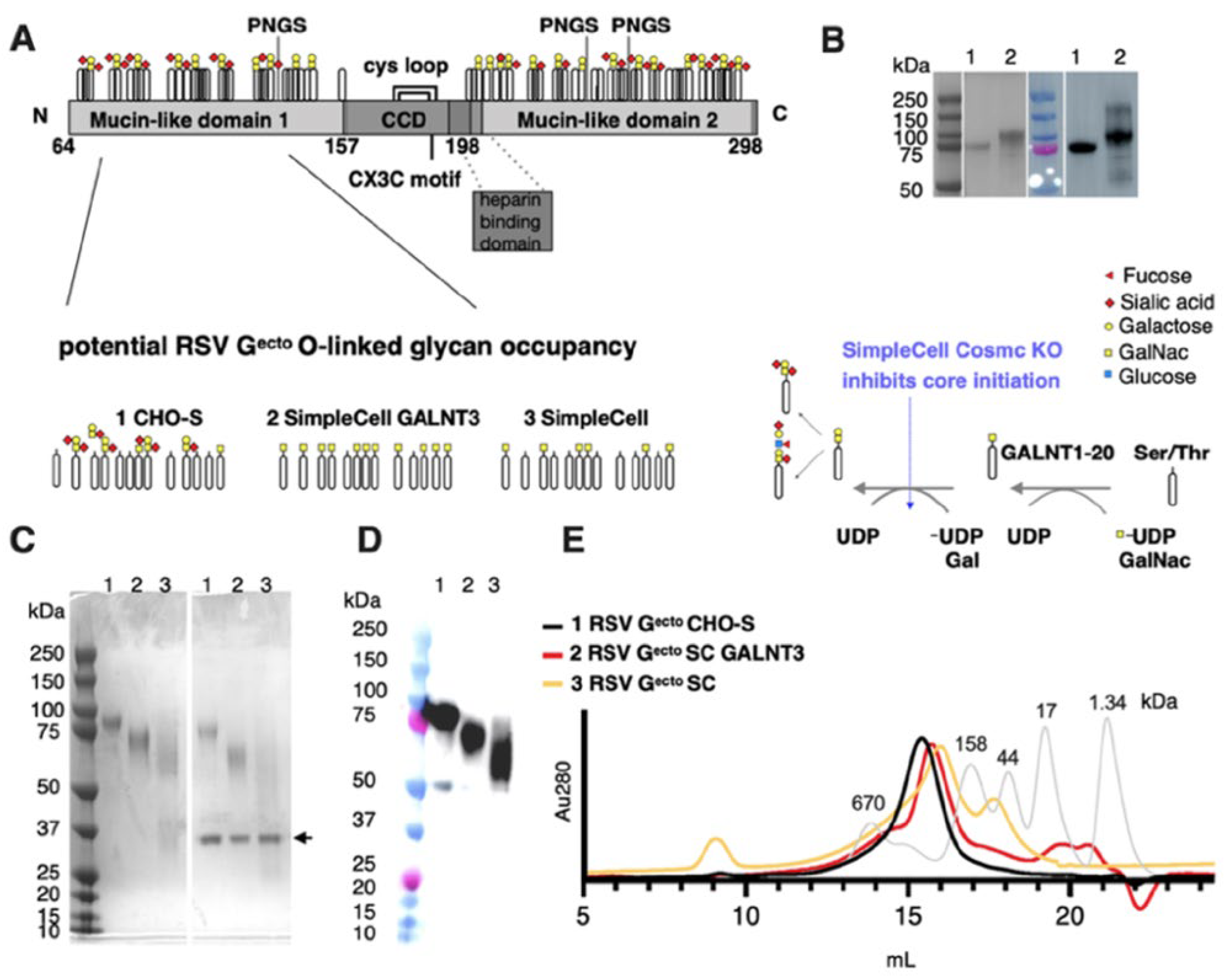
Figure 1. Evaluation of recombinant RSV Gecto proteins produced in SC cell lines. (A) Schematic of recombinant RSV Gecto comprising the conserved central domain (CCD), including its CX3C motif and cysteine loop, heparin-binding domain, and mucin-like domains 1 and 2, with a simplified depiction of potential O-linked glycosylation patterns and potential N-linked glycosylation sites (PNGS). O-glycosylation addition is initiated via polypeptide N-acetylgalactosaminyltransferases GALNT1-20. GALNT3 is not expressed in wild-type CHO cells. These transferases catalyze the addition of GalNac to Ser/Thr residues, permitting subsequent core synthesis, extension, and capping. In SimpleCell lines, core-1 synthesis is inhibited by the knockout of an essential chaperone, Cosmc, restricting O-glycosylation to the initial GalNAc on Ser/Thr residues. In SC GALNT3, GalNac is added to additional permissive Ser/Thr residues by GALNT3. (B) SDS PAGE followed by Coomassie stain and Western blot of RSV Gecto purified from (1) CHO-S or (2) HEK 293 cells, with apparent molecular weights of ~90 and ~100 kDa, respectively. (C) SDS PAGE followed by Coomassie stain of RSV Gecto purified from (1) CHO-S, (2) SC GALNT3, and (3) SC cells, with apparent molecular weights of ~90, 75, and 55 kDa, respectively. In the presence of PNGaseF (enzyme indicated by arrow), the bands downshift, indicative of the removal of N-linked glycans. (D) Western blot of recombinant RSV Gecto with an anti-His-tag antibody. (E) Analytical size exclusion chromatography on a Superose 6 10-300 column of recombinant RSV Gecto proteins (black, red, and yellow lines) compared to gel filtration standards (grey line).
Vaccines 13 01004 g001
Figure 2. Outline of vaccination and challenge schedule. Six groups of BALB/c mice (n = 5) were primed with 50 µg of RSV Gecto combined with 50 µg of MPLA, MPLA alone or PBS, and boosted at day 21 (D21) with the same vaccine antigen and adjuvant combination. Serum samples were assayed 19 days after priming at D19 and 14 days after homologous boosting at D35. Animals were subjected to intranasal challenge at D40, five days after boost, with 106 PFU RSV/A2 virus, then euthanized five days later. Serum, BAL, and lung tissue were collected for analysis.
Vaccines 13 01004 g002
Figure 3. Comparison of non-restricted and O-glycan-restricted RSV Gecto immunogenicity in mice. Circulating (A) Anti-RSV Gecto-specific IgG (B) and anti-CCD-Fc specific IgG were detected by an indirect-binding ELISA two weeks after prime (D19), and two weeks after boost (D35), with the three vaccine antigens (n = 5 mice per group) adjuvanted with MPLA or MPLA alone. Endpoint titer was calculated from a sigmoidal 5PL, where the X-axis is a log (serum dilution) plot with a cutoff value of baseline +3 times SD plus 10%. Groups were compared for statistical significance using a one-way ANOVA with Tukey’s post hoc correction, * p = 0.0226, ** p = 0.0069, **** p < 0.0001, 95% confidence.
Vaccines 13 01004 g003
Figure 4. The IgG recall response of RSV/A2 challenged mice vaccinated with O-glycan restricted RSV Gecto immunogens. Vaccinated mice (n = 5 per group) were intranasally challenged with 106 PFU of RSV/A2. (A) Five days post-challenge, circulating anti-RSV-specific IgG was assayed by direct binding of antibodies in serially diluted serum samples to RSV/A2-coated ELISA plates. All antigens induced a significant anti-RSV/A2/specific IgG response when compared to the infection control (** p < 0.0031, and **** p < 0.001). OD450 (Y-axis) at single point dilution (1/1280) on the linear segment of the titration curve where OD values are proportional to amount of antibody, permitting comparison. Bars represent mean values and SD. (B) Relative amounts of circulating anti-RSV A2-specific IgG1, (C) IgG2a, were assessed at a single (1/40) serum dilution for mice in each challenge group. IgG1 was elevated in mice immunized with RSV Gecto SC GALNT3 protein compared to the infection control (* p < 0.05). No group raised an IgG2a response above that observed in the infection control. Statistical testing was performed by a one-way ANOVA test using Tukey’s post hoc correction for (A) (as the dilution is on the linear part of the curve) and a Kruskal–Wallis test for (B,C).
Vaccines 13 01004 g004
Figure 5. The lung viral load and neutralizing antibody response in BALB/c mice following vaccination with O-glycan restricted RSV Gecto vaccines. (A) Viral lung titer/mL of lung homogenate as determined by plaque assay. Mice were immunized with O-glycan-restricted RSV Gecto SC GALNT3 + MPLA. They had a significant (**** p = 0.0001) reduction in lung virus load (D45) at day 5 post-challenge compared to mice vaccinated with MPLA alone. (B) Pooled antisera and control anti-F glycoprotein neutralizing murine mAb 143.6C were heat-inactivated and diluted to 1/40 for microneutralization assay in A549 cells in the presence or absence of 10% guinea-pig complement. Fluorescent focus units (FFU) were enumerated on a Cellomics ArrayScan using HTS software. Bars represent mean ± SEM. Sera derived from RSV Gecto SC GALNT3 protein vaccinated mice had a greater capacity to neutralize RSV A2 infection of A549 cells (**** p = 0.0001) than MPLA-treated controls. Bars represent mean ± SD. Group titers were compared by one-way ANOVA with Dunnett’s multiple comparison.
Vaccines 13 01004 g005
Figure 6. BAL leukocyte composition at day 5 in BALB/c mice with O-glycosylation restricted RSV Gecto vaccines and challenged with 106 PFU of RSV/A2. BAL composition by flow cytometry: charts represent the percentage of the total number of cells isolated with values for each vaccine control group compared to the infection control, applying a one-way ANOVA analysis with Tukey’s post hoc correction to compare groups. Bars represent mean ± SD. (A) Siglecf+ staining macrophages (CD45+/Siglecf+CD11clow) (* p = <0.05, ** p < 0.0056, **** p < 0.0001). (B) Percentage B220-positive B-cells (* p < 0.05, **** p < 0.0001). (C) Percentage Siglecf+ (CD45+/Siglecf+CD11clow) eosinophils. (D) Percentage positive CD3+ T-cells.
Vaccines 13 01004 g006
Table 1. Binding affinity constant (KD), standard deviation (KD SD), on-rates (ka), off-rates (kd), R2, and x2 of RSV Gecto immunogens to conformation-dependent mAbs 3D3 and 2D10.
mAbKD (pM) *KD SD (pM)ka (105 M−1s−1)kd (10−7s−1)R2x2
CHO-S Gecto3D3<1.03.01.00<10.99270.0562
CHO-S Gecto2D10<1.01.75.00<10.99470.1022
SC GALNT3 Gecto3D31.0<1.06.00<10.99060.0667
SC GALNT3 Gecto2D10<1.0<1.02.00<10.99430.0468
SC Gecto3D3<1.0<1.08.00<10.97380.1689
SC Gecto2D102.01.65.00<10.98880.0300
* Average KD values are reported as the average of the three replicate binding studies.
Disclaimer/Publisher’s Note: The statements, opinions and data contained in all publications are solely those of the individual author(s) and contributor(s) and not of MDPI and/or the editor(s). MDPI and/or the editor(s) disclaim responsibility for any injury to people or property resulting from any ideas, methods, instructions or products referred to in the content.