


Pilot Findings on SARS-CoV-2 Vaccine-Induced Pituitary Diseases: A Mini Review from Diagnosis to Pathophysiology


Abstract
1. Introduction
2. Materials and Methods
3. Results
3.1. Hypophysitis
3.1.1. Clinical Presentations
3.1.2. MRI Findings
3.1.3. Treatment
3.2. Pituitary Apoplexy
3.2.1. Clinical Presentations
3.2.2. MRI Findings
3.2.3. Treatment
4. Discussion
4.1. Hypophysitis
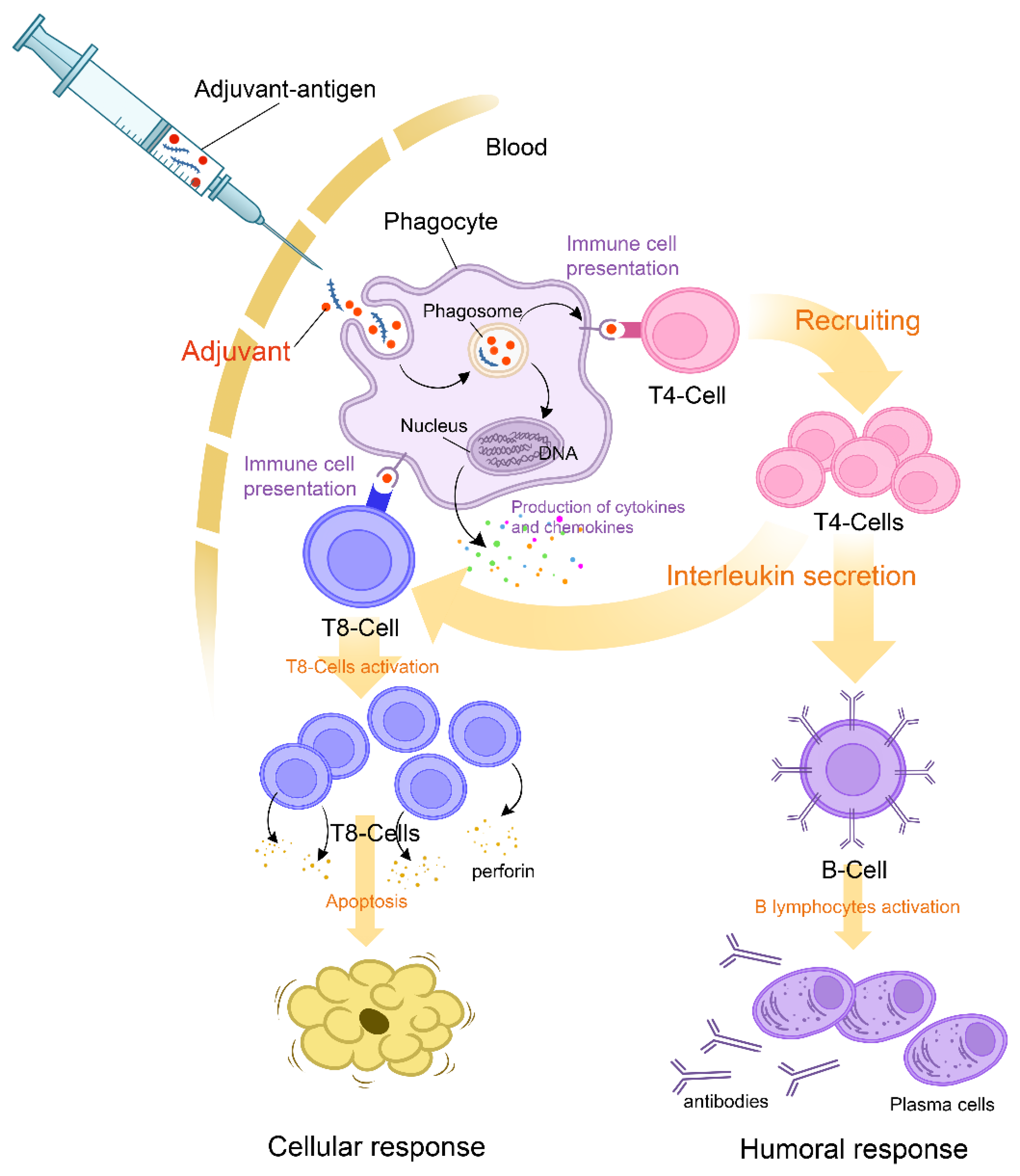
4.1.1. Pathogenesis
4.1.2. Clinical Presentations
4.1.3. Treatment
4.2. Pituitary Apoplexy
4.2.1. Pathogenesis
4.2.2. Clinical Presentations
4.2.3. Treatment
5. Conclusions
Author Contributions
Funding
Institutional Review Board Statement
Informed Consent Statement
Acknowledgments
Conflicts of Interest
References
- Yu, J.; Chai, P.; Ge, S.; Fan, X. Recent Understandings Toward Coronavirus Disease 2019 (COVID-19): From Bench to Bedside. Front. Cell Dev. Biol. 2020, 8, 476. [Google Scholar] [CrossRef] [PubMed]
- Wong, R.S.Y. The SARS-CoV-2 Outbreak: An Epidemiological and Clinical Perspective. SN Compr. Clin. Med. 2020, 2, 1983–1991. [Google Scholar] [CrossRef] [PubMed]
- Johnson, K.D.; Harris, C.; Cain, J.K.; Hummer, C.; Goyal, H.; Perisetti, A. Pulmonary and Extra-Pulmonary Clinical Manifestations of COVID-19. Front. Med. 2020, 7, 526. [Google Scholar] [CrossRef] [PubMed]
- Rabail, R.; Ahmed, W.; Ilyas, M.; Rajoka, M.S.R.; Hassoun, A.; Khalid, A.R.; Khan, M.R.; Aadil, R.M. The Side Effects and Adverse Clinical Cases Reported after COVID-19 Immunization. Vaccines 2022, 10, 488. [Google Scholar] [CrossRef] [PubMed]
- Zhao, Y.; Wu, X. Influence of COVID-19 vaccines on endocrine system. Endocrine 2022, 78, 241–246. [Google Scholar] [CrossRef]
- Ku, C.R.; Jung, K.Y.; Ahn, C.H.; Moon, J.S.; Lee, J.H.; Kim, E.H.; Kwon, H.; Kim, H.K.; Suh, S.; Hong, S.; et al. COVID-19 Vaccination for Endocrine Patients: A Position Statement from the Korean Endocrine Society. Endocrinol. Metab. 2021, 36, 757–765. [Google Scholar] [CrossRef]
- Soldevila, B.; Puig-Domingo, M.; Marazuela, M. Basic mechanisms of SARS-CoV-2 infection. What endocrine systems could be implicated? Rev. Endocr. Metab. Disord. 2022, 23, 137–150. [Google Scholar] [CrossRef]
- Young, M.J.; Clyne, C.D.; Chapman, K.E. Endocrine aspects of ACE2 regulation: RAAS, steroid hormones and SARS-CoV-2. J. Endocrinol. 2020, 247, R45–R62. [Google Scholar] [CrossRef]
- Garg, M.K.; Gopalakrishnan, M.; Yadav, P.; Misra, S. Endocrine Involvement in COVID-19: Mechanisms, Clinical Features, and Implications for Care. Indian J. Endocrinol. Metab. 2020, 24, 381–386. [Google Scholar] [CrossRef]
- Lisco, G.; De Tullio, A.; Stragapede, A.; Solimando, A.; Albanese, F.; Capobianco, M.; Giagulli, V.; Guastamacchia, E.; De Pergola, G.; Vacca, A.; et al. COVID-19 and the Endocrine System: A Comprehensive Review on the Theme. J. Clin. Med. 2021, 10, 2920. [Google Scholar] [CrossRef]
- Mirza, S.A.; Sheikh, A.A.E.; Barbera, M.; Ijaz, Z.; Javaid, M.A.; Shekhar, R.; Pal, S.; Sheikh, A.B. COVID-19 and the Endocrine System: A Review of the Current Information and Misinformation. Infect. Dis. Rep. 2022, 14, 184–197. [Google Scholar] [CrossRef]
- Zhang, C.; Zhou, C.; Shi, L.; Liu, G. Perspectives on development of vaccines against severe acute respiratory syndrome coronavirus 2 (SARS-CoV-2). Hum. Vaccines Immunother. 2020, 16, 2366–2369. [Google Scholar] [CrossRef] [PubMed]
- Zhou, H.; Møhlenberg, M.; Thakor, J.C.; Tuli, H.S.; Wang, P.; Assaraf, Y.G.; Dhama, K.; Jiang, S. Sensitivity to Vaccines, Therapeutic Antibodies, and Viral Entry Inhibitors and Advances To Counter the SARS-CoV-2 Omicron Variant. Clin. Microbiol. Rev. 2022, 35, e0001422. [Google Scholar] [CrossRef] [PubMed]
- Caironi, V.; Pitoia, F.; Trimboli, P. Thyroid Inconveniences With Vaccination Against SARS-CoV-2: The Size of the Matter. A Systematic Review. Front. Endocrinol. 2022, 1206, 13. [Google Scholar] [CrossRef] [PubMed]
- Patrizio, A.; Ferrari, S.M.; Elia, G.; Ragusa, F.; Paparo, S.R.; Mazzi, V.; Antonelli, A.; Fallahi, P. Graves’ Disease Following SARS-CoV-2 Vaccination: A Systematic Review. Vaccines 2022, 10, 1445. [Google Scholar] [CrossRef]
- Banerjee, M.; Pal, R.; Dutta, S. Risk of incident diabetes post-COVID-19: A systematic review and meta-analysis. Prim. Care Diabetes 2022, 16, 591–593. [Google Scholar] [CrossRef]
- Yano, M.; Morioka, T.; Natsuki, Y.; Sasaki, K.; Kakutani, Y.; Ochi, A.; Yamazaki, Y.; Shoji, T.; Emoto, M. New-onset Type 1 Diabetes after COVID-19 mRNA Vaccination. Intern. Med. 2022, 61, 1197–1200. [Google Scholar] [CrossRef]
- Markovic, N.; Faizan, A.; Boradia, C.; Nambi, S. Adrenal Crisis Secondary to COVID-19 Vaccination in a Patient With Hypopituitarism. AACE Clin. Case Rep. 2022, 8, 171–173. [Google Scholar] [CrossRef]
- Alexandraki, K.I.; Grossman, A. Management of Hypopituitarism. J. Clin. Med. 2019, 8, 2153. [Google Scholar] [CrossRef]
- Fleseriu, M.; Hashim, I.A.; Karavitaki, N.; Melmed, S.; Murad, M.H.; Salvatori, R.; Samuels, M.H. Hormonal Replacement in Hypopituitarism in Adults: An Endocrine Society Clinical Practice Guideline. J. Clin. Endocrinol. Metab. 2016, 101, 3888–3921. [Google Scholar] [CrossRef]
- Jonklaas, J.; Bianco, A.C.; Bauer, A.J.; Burman, K.D.; Cappola, A.R.; Celi, F.S.; Cooper, D.S.; Kim, B.W.; Peeters, R.B.; Rosenthal, M.S.; et al. Guidelines for the treatment of hypothyroidism: Prepared by the american thyroid association task force on thyroid hormone replacement. Thyroid. Off. J. Am. Thyroid. Assoc. 2014, 24, 1670–1751. [Google Scholar] [CrossRef] [PubMed]
- Ach, T.; Yosra, H.; Jihen, M.; Asma, B.A.; Maha, K.; Molka, C.; Rouatbi, S.; Monia, Z.; Ach, K. Cortisol cut-points for the glucagon stimulation test in the evaluation of hypothalamic pituitary adrenal axis. Endocr. J. 2018, 65, 935–942. [Google Scholar] [CrossRef] [PubMed]
- Hoffman, A.R.; Mathison, T.; Andrews, D.; Murray, K.; Kelepouris, N.; Fleseriu, M. Adult Growth Hormone Deficiency: Diagnostic and Treatment Journeys From the Patients’ Perspective. J. Endocr. Soc. 2022, 6, bvac077. [Google Scholar] [CrossRef] [PubMed]
- Mutter, C.M.; Smith, T.; Menze, O.; Zakharia, M.; Nguyen, H. Diabetes Insipidus: Pathogenesis, Diagnosis, and Clinical Management. Cureus 2021, 13, e13523. [Google Scholar] [CrossRef] [PubMed]
- Kalra, S.; Zargar, A.H.; Jain, S.M.; Sethi, B.; Chowdhury, S.; Singh, A.K.; Thomas, N.; Unnikrishnan, A.G.; Thakkar, P.B.; Malve, H. Diabetes insipidus: The other diabetes. Indian J. Endocrinol. Metab. 2016, 20, 9. [Google Scholar] [PubMed]
- Shin, J.H.; Lee, H.K.; Choi, C.G.; Suh, D.C.; Kim, C.J.; Hong, S.K.; Na, D.G. MR imaging of central diabetes insipidus: A pictorial essay. Korean J. Radiol. 2001, 2, 222–230. [Google Scholar] [CrossRef]
- Majumdar, A.; Mangal, N.S. Hyperprolactinemia. J. Hum. Reprod. Sci. 2013, 6, 168–175. [Google Scholar] [CrossRef]
- Zainordin, N.A.; Hatta, S.; Ab Mumin, N.; Shah, F.Z.M.; Ghani, R.A. Pituitary apoplexy after COVID-19 vaccination: A case report. J. Clin. Transl. Endocrinol. Case Rep. 2022, 25, 100123. [Google Scholar] [CrossRef]
- Roncati, L.; Manenti, A. Pituitary apoplexy following adenoviral vector-based COVID-19 vaccination. Brain Hemorrhages 2022, 4, 27–29. [Google Scholar] [CrossRef]
- Piñar-Gutiérrez, A.; Remón-Ruiz, P.; Soto-Moreno, A. Case report: Pituitary apoplexy after COVID-19 vaccination. Med. Clin. (Engl. Ed.) 2022, 158, 498–499. [Google Scholar]
- Morita, S.; Tsuji, T.; Kishimoto, S.; Uraki, S.; Takeshima, K.; Iwakura, H.; Furuta, H.; Nishi, M.; Inaba, H.; Matsuoka, T.A. Isolated ACTH deficiency following immunization with the BNT162b2 SARS-CoV-2 vaccine: A case report. BMC Endocr. Disord. 2022, 22, 185. [Google Scholar] [CrossRef] [PubMed]
- Bouça, B.; Roldão, M.; Bogalho, P.; Cerqueira, L.; Silva-Nunes, J. Central Diabetes Insipidus Following Immunization With BNT162b2 mRNA COVID-19 Vaccine: A Case Report. Front. Endocrinol. 2022, 13, 889074. [Google Scholar] [CrossRef] [PubMed]
- Ankireddypalli, A.R.; Chow, L.S.; Radulescu, A.; Kawakami, Y.; Araki, T. A Case of Hypophysitis Associated With SARS-CoV-2 Vaccination. AACE Clin. Case Rep. 2022, 8, 204–209. [Google Scholar] [CrossRef]
- Ach, T.; Kammoun, F.; El Fekih, H.; Slama, N.B.H.; Kahloun, S.; Fredj, F.B. Central diabetes insipidus revealing a hypophysitis induced by SARS-CoV-2 vaccine. Therapie 2022. [Google Scholar] [CrossRef] [PubMed]
- Murvelashvili, N.; Tessnow, A. A Case of Hypophysitis Following Immunization With the mRNA-1273 SARS-CoV-2 Vaccine. J. Investig. Med. High Impact Case Rep. 2021, 9, 23247096211043386. [Google Scholar] [CrossRef] [PubMed]
- Trougakos, I.P.; Terpos, E.; Alexopoulos, H.; Politou, M.; Paraskevis, D.; Scorilas, A.; Kastritis, E.; Anndreakos, E.; Dimopoulos, M.A. Adverse effects of COVID-19 mRNA vaccines: The spike hypothesis. Trends Mol. Med. 2022, 28, 542–554. [Google Scholar] [CrossRef] [PubMed]
- Cosentino, M.; Marino, F. Understanding the Pharmacology of COVID-19 mRNA Vaccines: Playing Dice with the Spike? Int. J. Mol. Sci. 2022, 23, 10881. [Google Scholar] [CrossRef]
- Wilson, B.; Geetha, K.M. Lipid nanoparticles in the development of mRNA vaccines for COVID-19. J. Drug Deliv. Sci. Technol. 2022, 74, 103553. [Google Scholar] [CrossRef]
- Tondo, G.; Virgilio, E.; Naldi, A.; Bianchi, A.; Comi, C. Safety of COVID-19 Vaccines: Spotlight on Neurological Complications. Life 2022, 12, 1338. [Google Scholar] [CrossRef]
- Bellamkonda, N.; Lambe, U.P.; Sawant, S.; Nandi, S.S.; Chakraborty, C.; Shukla, D. Immune Response to SARS-CoV-2 Vaccines. Biomedicines 2022, 10, 1464. [Google Scholar] [CrossRef]
- Gupta, D.; Parthasarathy, H.; Sah, V.; Tandel, D.; Vedagiri, D.; Reddy, S.; Harshan, K.H. Inactivation of SARS-CoV-2 by β-propiolactone causes aggregation of viral particles and loss of antigenic potential. Virus Res. 2021, 305, 198555. [Google Scholar] [CrossRef] [PubMed]
- Ganesan, S.; Al Ketbi, L.M.B.; Al Kaabi, N.; Al Mansoori, M.; Al Maskari, N.N.; Al Shamsi, M.S.; Alderei, A.S.; Eissaee, H.N.E.; Al Ketbi, R.M.; Al Shamsi, N.S.; et al. Vaccine Side Effects Following COVID-19 Vaccination Among the Residents of the UAE-An Observational Study. Front. Public Health 2022, 10, 876336. [Google Scholar] [CrossRef] [PubMed]
- Gubbi, S.; Hannah-Shmouni, F.; Verbalis, J.G.; Koch, C.A. Hypophysitis: An update on the novel forms, diagnosis and management of disorders of pituitary inflammation. Best Pract. Res. Clin. Endocrinol. Metab. 2019, 33, 101371. [Google Scholar] [CrossRef] [PubMed]
- Faje, A. Hypophysitis: Evaluation and Management. Clin. Diabetes Endocrinol. 2016, 2, 15. [Google Scholar] [CrossRef]
- Ach, T.; Wojewoda, P.; Toullet, F.; Ducloux, R.; Avérous, V. Multiple endocrinological failures as a clinical presentation of a metastatic lung adenocarcinoma. Endocrinol. Diabetes Metab. Case Rep. 2020, 2020. [Google Scholar] [CrossRef]
- Day, E.L.; Smith, E.R.; Fehnel, K.P. Single-institution case series of pituitary biopsy for suspected germinoma in the pediatric population: Diagnostic utility, operative risks, and biopsy approaches. Sci. Rep. 2020, 10, 15257. [Google Scholar] [CrossRef]
- Kang, H.; Kim, K.M.; Kim, M.S.; Kim, J.H.; Park, C.K.; Kim, Y.H. Safety of endoscopic endonasal biopsy for the pituitary stalk-hypothalamic lesions. Pituitary 2022, 25, 143–151. [Google Scholar] [CrossRef]
- Jara, L.J.; Vera-Lastra, O.; Mahroum, N.; Pineda, C.; Shoenfeld, Y. Autoimmune post-COVID vaccine syndromes: Does the spectrum of autoimmune/inflammatory syndrome expand? Clin. Rheumatol. 2022, 41, 1603–1609. [Google Scholar] [CrossRef]
- Liang, Z.; Zhu, H.; Wang, X.; Jing, B.; Li, Z.; Xia, X.; Sun, H.; Yang, Y.; Zhang, W.; Shi, L.; et al. Adjuvants for Coronavirus Vaccines. Front. Immunol. 2020, 11, 589833. [Google Scholar] [CrossRef]
- Chen, P.-Y.; Wu, B.-J.; Su, M.-C.; Lin, Y.-H.; Chiang, S.-C.; Wu, J.-C.; Chen, T.-J.; Chen, Y.-C. Risk Factors and Incidence Rates of Self-Reported Short-Term Adverse Events of COVID-19 Vaccine Booster Dose. Vaccines 2022, 10, 1115. [Google Scholar] [CrossRef]
- Borba, V.; Malkova, A.; Basantsova, N.; Halpert, G.; Andreoli, L.; Tincani, A.; Amital, H.; Shoenfeld, Y. Classical Examples of the Concept of the ASIA Syndrome. Biomolecules 2020, 10, 1436. [Google Scholar] [CrossRef]
- Bragazzi, N.L.; Hejly, A.; Watad, A.; Adawi, M.; Amital, H.; Shoenfeld, Y. ASIA syndrome and endocrine autoimmune disorders. Best Pract. Res. Clin. Endocrinol. Metab. 2020, 34, 101412. [Google Scholar] [CrossRef] [PubMed]
- Watad, A.; Sharif, K.; Shoenfeld, Y. The ASIA syndrome: Basic concepts. Mediterr. J. Rheumatol. 2017, 28, 64–69. [Google Scholar] [CrossRef] [PubMed]
- Dotan, A.; Muller, S.; Kanduc, D.; David, P.; Halpert, G.; Shoenfeld, Y. The SARS-CoV-2 as an instrumental trigger of autoimmunity. Autoimmun. Rev. 2021, 20, 102792. [Google Scholar] [CrossRef] [PubMed]
- Ou, X.; Liu, Y.; Lei, X.; Li, P.; Mi, D.; Ren, L.; Guo, L.; Guo, R.; Chen, T.; Hu, J.; et al. Characterization of spike glycoprotein of SARS-CoV-2 on virus entry and its immune cross-reactivity with SARS-CoV. Nat. Commun. 2020, 11, 1620. [Google Scholar] [CrossRef]
- Neufeldt, C.J.; Cerikan, B.; Cortese, M.; Frankish, J.; Lee, J.-Y.; Plociennikowska, A.; Heigwer, F.; Prasad, V.; Joecks, S.; Burkart, S.S.; et al. SARS-CoV-2 infection induces a pro-inflammatory cytokine response through cGAS-STING and NF-κB. Commun. Biol. 2022, 5, 45. [Google Scholar] [CrossRef]
- Montastruc, J.L. Pharmacovigilance and drug safety: Fair prescribing and clinical research. Therapie 2022, 77, 261–263. [Google Scholar] [CrossRef]
- Guo, S.; Wang, C.; Zhang, J.; Tian, Y.; Wu, Q. Diagnosis and management of tumor-like hypophysitis: A retrospective case series. Oncol. Lett. 2016, 11, 1315–1320. [Google Scholar] [CrossRef]
- Caranci, F.; Leone, G.; Ponsiglione, A.; Muto, M.; Tortora, F.; Muto, M.; Cirillo, S.; Brunese, L.; Cerase, A. Imaging findings in hypophysitis: A review. La Radiol. Med. 2020, 125, 319–328. [Google Scholar] [CrossRef]
- Gutenberg, A.; Larsen, J.; Lupi, I.; Rohde, V.; Caturegli, P. A radiologic score to distinguish autoimmune hypophysitis from nonsecreting pituitary adenoma preoperatively. AJNR Am. J. Neuroradiol. 2009, 30, 1766–1772. [Google Scholar] [CrossRef]
- Taieb, A.; Maha, K.N.; El Abed, Y.H.; Beizig, A.M.; Chadli, M.C.; Ach, K. Macroprolactinemia and Empty Sella Syndrome. Pan Afr. Med. J. 2017, 27, 278. [Google Scholar] [CrossRef] [PubMed]
- Honegger, J.; Buchfelder, M.; Schlaffer, S.; Droste, M.; Werner, S.; Strasburger, C.; Störmann, S.; Schopohl, J.; Kacheva, S.; Deutschbein, T.; et al. Treatment of Primary Hypophysitis in Germany. J. Clin. Endocrinol. Metab. 2015, 100, 3460–3469. [Google Scholar] [CrossRef] [PubMed]
- Boellis, A.; di Napoli, A.; Romano, A.; Bozzao, A. Pituitary apoplexy: An update on clinical and imaging features. Insights Into Imaging 2014, 5, 753–762. [Google Scholar] [CrossRef] [PubMed]
- Emary, K.R.W.; Golubchik, T.; Aley, P.K.; Ariani, C.V.; Angus, B.; Bibi, S.; Blane, B.; Bonsall, D.; Cicconi, P.; Charlton, S.; et al. Efficacy of ChAdOx1 nCoV-19 (AZD1222) vaccine against SARS-CoV-2 variant of concern 202012/01 (B.1.1.7): An exploratory analysis of a randomised controlled trial. Lancet 2021, 397, 1351–1362. [Google Scholar] [CrossRef]
- Greinacher, A.; Thiele, T.; Warkentin, T.E.; Weisser, K.; Kyrle, P.A.; Eichinger, S. Thrombotic Thrombocytopenia after ChAdOx1 nCov-19 Vaccination. N. Engl. J. Med. 2021, 384, 2092–2101. [Google Scholar] [CrossRef]
- Lai, K.Y.; Au, S.Y.; Fong, K.M. Thrombotic Thrombocytopenia after ChAdOx1 nCoV-19 Vaccination. N. Engl. J. Med. 2021, 385, e11. [Google Scholar]
- Bissola, A.-L.; Daka, M.; Arnold, D.M.; Smith, J.W.; Moore, J.C.; Clare, R.; Ivetic, N.; Kelton, J.G.; Nazy, I. The clinical and laboratory diagnosis of vaccine-induced immune thrombotic thrombocytopenia. Blood Adv. 2022, 6, 4228–4235. [Google Scholar] [CrossRef]
- Mungmunpuntipantip, R.; Wiwanitkit, V. Pituitary apoplexy and COVID-19 vaccination. Med. Clin. (Engl. Ed.) 2022, 159, e11. [Google Scholar]
- Ziogas, A.; Netea, M.G. Trained immunity-related vaccines: Innate immune memory and heterologous protection against infections. Trends Mol. Med. 2022, 28, 497–512. [Google Scholar] [CrossRef]
- Taieb, A.; Asma, B.A.; Ghada, S.; Yosra, H.; Maha, K.; Molka, C.; Amel, M.; Koussay, A. Increased intracranial pressure due to chronic weight lifting exercises as a hypothesis of partial empty sella syndrome in an elite athlete. Med. Hypotheses 2022, 167, 110951. [Google Scholar] [CrossRef]
- Glezer, A.; Bronstein, M.D. Pituitary apoplexy: Pathophysiology, diagnosis and management. Arch. Endocrinol. Metab. 2015, 59, 259–264. [Google Scholar] [CrossRef]
- Briet, C.; Salenave, S.; Bonneville, J.F.; Laws, E.R.; Chanson, P. Pituitary Apoplexy. Endocr. Rev. 2015, 36, 622–645. [Google Scholar] [CrossRef] [PubMed]
- Biagetti, B.; Simò, R. Pituitary Apoplexy: Risk Factors and Underlying Molecular Mechanisms. Int. J. Mol. Sci. 2022, 23, 8721. [Google Scholar] [CrossRef] [PubMed]
- Leyer, C.; Castinetti, F.; Morange, I.; Gueydan, M.; Oliver, C.; Conte-Devolx, B.; Dufour, H.; Brue, T. A conservative management is preferable in milder forms of pituitary tumor apoplexy. J. Endocrinol. Investig. 2011, 34, 502–509. [Google Scholar]
- Goyal, P.; Utz, M.; Gupta, N.; Kumar, Y.; Mangla, M.; Gupta, S.; Mangla, R. Clinical and imaging features of pituitary apoplexy and role of imaging in differentiation of clinical mimics. Quant. Imaging Med. Surg. 2018, 8, 219–231. [Google Scholar] [CrossRef] [PubMed]
- Albani, A.; Ferraù, F.; Angileri, F.; Esposito, F.; Granata, F.; Ferreri, F.; Cannavò, S. Multidisciplinary Management of Pituitary Apoplexy. Int. J. Endocrinol. 2016, 2016, 7951536. [Google Scholar] [CrossRef] [PubMed]
- Kyle, C.A.; Laster, R.A.; Burton, E.M.; Sanford, R.A. Subacute pituitary apoplexy: MR and CT appearance. J. Comput. Assist. Tomogr. 1990, 14, 40–44. [Google Scholar] [CrossRef]
- Khaldi, S.; Saad, G.; Elfekih, H.; Ben Abdelkrim, A.; Ach, T.; Kacem, M.; Chaieb, M.; Marroufi, A.; Hasni, Y.; Ach, K.; et al. Pituitary apoplexy of a giant prolactinoma during pregnancy. Gynecol. Endocrinol. Off. J. Int. Soc. Gynecol. Endocrinol. 2021, 37, 863–866. [Google Scholar] [CrossRef] [PubMed]
- Almeida, J.P.; Sanchez, M.M.; Karekezi, C.; Warsi, N.; Fernández-Gajardo, R.; Panwar, J.; Mansouri, A.; Suppiah, S.; Nassiri, F.; Nejad, R.; et al. Pituitary Apoplexy: Results of Surgical and Conservative Management Clinical Series and Review of the Literature. World Neurosurg. 2019, 130, e988–e999. [Google Scholar] [CrossRef]
- Fan, Y.; Bao, X.; Wang, R. Conservative treatment cures an elderly pituitary apoplexy patient with oculomotor paralysis and optic nerve compression: A case report and systematic review of the literature. Clin. Interv. Aging 2018, 13, 1981–1985. [Google Scholar] [CrossRef]
- Turgut, M.; Ozsunar, Y.; Başak, S.; Güney, E.; Kir, E.; Meteoğlu, I. Pituitary apoplexy: An overview of 186 cases published during the last century. Acta Neurochir. 2010, 152, 749–761. [Google Scholar] [CrossRef] [PubMed]
- Gabarin, N.; Arnold, D.M.; Nazy, I.; Warkentin, T.E. Treatment of vaccine-induced immune thrombotic thrombocytopenia (VITT). Semin. Hematol. 2022, 59, 89–96. [Google Scholar] [CrossRef] [PubMed]
- Singh, B.; Kanack, A.; Bayas, A.; George, G.; Abou-Ismail, M.Y.; Kohlhagen, M.; Christ, M.; Naumann, M.; Moser, K.; Smock, K.; et al. Anti-PF4 VITT antibodies are oligoclonal and variably inhibited by heparin. Medrxiv Prepr. Serv. Health Sci. 2021. [Google Scholar] [CrossRef]


| Reference | Gender | Dose | Vaccine | Symptoms | Days until Symptoms | Diagnostic | Diabetes Insipidus | Thyreotrop Axis | Gonadal Axis | Corticotrop Axis | Somatotrop Axis | Lactotrop Axis | MRI |
|---|---|---|---|---|---|---|---|---|---|---|---|---|---|
| Roncati et al. (2022) [29] | F | 1st | ChAdOx1 nCov-19; AstraZeneca | fever for 24 h and tension-type headache | 1 day | Apoplexy | N | N | Hypogonadism | N | N | Hyperprolactinemia | signal alteration related to a hemorrhagic event in the right half of the sella turcica |
| Zainordin et al. (2022) [28] | F | 1st | ChAdOx1 nCov-19; AstraZeneca | severe frontal headache | 1 day | Apoplexy | N | N | N | N | N | N | pituitary apoplexy with mass effect to the optic chiasm and cavernous portion |
| Pinar-Gutiérrez et al. (2021) [30] | F | 1st | ChAdOx1 nCov-19; AstraZeneca | Frontal headache | 5 days | Apoplexy | N | N | N | N | N | N | adenohypophysis hemorrhagic bleeding in association with a possible 10 mm intraglandular adenoma |
| Murvelashvili et al. (2021) [35] | M | 2nd | BNT162b2; Pfizer-BioNTech | headache, nausea, vomiting, malaise, diffuse arthralgias | 3 days | Hypophysitis | + | Hypothyroidism | Hypogonadism | Hypocorticisim | N | N | enlarged pituitary gland consistent with acute hypophysitis |
| Ankireddypalli et al. (2022) [33] | F | 1st | BNT162b2; Pfizer-BioNTech | headache, polydipsia, and polyuria | 2 days | Hypophysitis | + | Hypothyroidism | Hypogonadism | N | GH insufficiency | N | thickening of the pituitary stalk |
| Morita et al. (2022) [31] | M | 2nd | BNT162b2; Pfizer-BioNTech | headaches, nausea, and diarrhea | 1 day | Hypophysitis | N | N | N | Hypocorticisim | N | N | Atrophic pituitary gland |
| Bouça et al. (2022) [32] | F | 2nd | BNT162b2; Pfizer-BioNTech | intense thirst and polyuria | 7 days | Hypophysitis | + | N | N | N | N | N | loss of the posterior pituitary bright spot on T1 weightedimaging |
| Ach et al. (2022) [34] | F | 1st | ChAdOx1 nCov-19; AstraZeneca | headache, polydipsia, and polyuria | 3 days | Hypophysitis | + | N | N | N | N | N | thickening of the pituitary stalk |
Publisher’s Note: MDPI stays neutral with regard to jurisdictional claims in published maps and institutional affiliations. |
© 2022 by the authors. Licensee MDPI, Basel, Switzerland. This article is an open access article distributed under the terms and conditions of the Creative Commons Attribution (CC BY) license (https://creativecommons.org/licenses/by/4.0/).
Share and Cite
Taieb, A.; Mounira, E.E. Pilot Findings on SARS-CoV-2 Vaccine-Induced Pituitary Diseases: A Mini Review from Diagnosis to Pathophysiology. Vaccines 2022, 10, 2004. https://doi.org/10.3390/vaccines10122004
Taieb A, Mounira EE. Pilot Findings on SARS-CoV-2 Vaccine-Induced Pituitary Diseases: A Mini Review from Diagnosis to Pathophysiology. Vaccines. 2022; 10(12):2004. https://doi.org/10.3390/vaccines10122004
Chicago/Turabian StyleTaieb, Ach, and El Euch Mounira. 2022. "Pilot Findings on SARS-CoV-2 Vaccine-Induced Pituitary Diseases: A Mini Review from Diagnosis to Pathophysiology" Vaccines 10, no. 12: 2004. https://doi.org/10.3390/vaccines10122004
APA StyleTaieb, A., & Mounira, E. E. (2022). Pilot Findings on SARS-CoV-2 Vaccine-Induced Pituitary Diseases: A Mini Review from Diagnosis to Pathophysiology. Vaccines, 10(12), 2004. https://doi.org/10.3390/vaccines10122004