Article

Effects of Selenium- and Zinc-Enriched Lactobacillus plantarum SeZi on Antioxidant Capacities and Gut Microbiome in an ICR Mouse Model

1
Department of Food and Nutrition, Research Institute of Human Ecology, Seoul National University, Seoul 08826, Korea
2
Institute of Health and Environment, Graduate School of Public Health, Seoul National University, Seoul 08826, Korea
3
Research Center, BIFIDO Co., Ltd., Hongcheon 25117, Korea
*
Authors to whom correspondence should be addressed.
Antioxidants 2020, 9(10), 1028; https://doi.org/10.3390/antiox9101028
Submission received: 28 September 2020 / Revised: 14 October 2020 / Accepted: 19 October 2020 / Published: 21 October 2020

Abstract

:
Selenium and zinc are essential trace minerals for humans with various biological functions. In this study, selenium- and zinc-tolerant lactic acid bacteria (LAB) isolates were screened out from human fecal samples. Amongst three hundred LAB isolates, the Lactobacillus plantarum SeZi strain displayed the tolerance against selenium and zinc with the greatest biomass production and bioaccumulation of selenium and zinc. To further assess the characteristics of this strain, the lyophilized L. plantarum SeZi were prepared and administered to Institute of Cancer Research (ICR) mice. The mice were divided into four groups, provided with normal chow (Con), or normal chow supplemented with Na2SeO3 and ZnSO4∙7H2O (SZ), L. plantarum SeZi (Lp), or selenium- and zinc-enriched L. plantarum SeZi (SZ + Lp), respectively. After 4 weeks of oral administration, the concentrations of selenium and zinc in blood were significantly increased in the SZ + Lp group when compared to the control or SZ group (p < 0.05). The increased selenium level led to an enhanced glutathione peroxidase activity and decreased blood malondialdehyde level in the SZ + Lp group (p < 0.05). Meanwhile, the results of bacterial community and microbial metabolic pathway analysis via 16S rRNA gene amplicon sequencing showed that L. plantarum SeZi significantly promoted the utilization of selenocysteine, seleno-cystathionine and seleno-methionine in the selenocompounds metabolism. Here, the in vivo antioxidant capacities of the selenium- and zinc-enriched lactobacillus strain showed us the utilization of a unique probiotic as a Se/Zn supplement with high availability, low toxicity, and additional probiotic advantages.

1. Introduction

Micronutrient deficiencies, known as “hidden hunger”, have affected more than 50% of the world’s population [1]. Selenium (Se) is a vital trace element, contributing to modulation of growth, regulation of antiviral capacity, and prevention of disease, especially cancer and cardiovascular disease [2,3,4]. The antioxidant activity of selenium is exhibited as a form of selenoenzymes, including glutathione peroxidase (GSH-Px), selenoprotein P, thioredoxin reductase, and methionine sulfoxide reductase [5]. Selenium deficiency can trigger serious health issues such as poor growth, muscle pain, decreased immune responses, and hypofunction of glandula thyreoidea [6,7]. Besides, the deficit of selenium is associated with a cardiomyopathy named as Keshan disease (KD) and an osteoarthropathy named as Kashin-Beck disease (KBD) [8]. Although KD and KBD are just local problems primarily in China and east Serbia due to environmentally low selenium [9], the hypothyreose or insuline secretion impairments associated with the lack of selenium have a global impact. Zinc (Zn) is another essential micronutrient for humans. It is a key component of many metalloenzymes (i.e., superoxide dismutase (SOD), carbonic anhydrase, alcohol dehydrogenase) associated with human growth, immunity, fertility, and reproduction [10]. Additionally, zinc is significant for the correct secretion of hormone isoline by pancreas [11]. The chronic deficiency of zinc can lead to glucose intolerance and pre-diabetic syndromes [12,13]. On the other hand, zinc chronic overdose, which can be caused by some nutraceuticals, might be responsible for some neurodegenerations such as nervus opticus inflammation [14,15].
Inorganic selenium, such as selenate (SeO42−) and selenite (SeO32−), are toxic and poorly bioavailable [16]. The reduction of selenium oxyanions largely depends on biotic processes by microorganisms [17]. The utilization of microorganisms as the natural adsorbent for metal ions (i.e., selenium and zinc) is eco-friendly and cost-effective [18]. The bioabsorption capacities are attributed to their intrinsic biochemical and structural properties of the cellular membrane [19]. Lactic acid bacteria (LAB), as important food-grade bacteria with probiotic advantages, have been extensively studied in this field. The selenium concentration in the medium is highly linked to bacterial selenium level, but the growth of most bacterial isolates from the human gut can be inhibited by the addition of inorganic selenium into the medium [20]. However, some LAB strains have been reported to be capable of resisting selenium oxyanions at high concentrations during cultivation [21,22,23]. Especially, Lactobacillus plantarum has been suggested as Se-enriched lactobacilli for food applications [24]. Few studies about Zn-enriched LAB have been conducted, but it has been found that the bacterial growth and probiotic effect of L. plantarum can be enhanced by zinc in the gut [25].
Although the resistances of LAB to selenium and zinc have been reported, the in vivo antioxidant capacities of the SeZn-enriched probiotic products have not been reported. In this study, SeZn-tolerant LAB strains isolated from human feces were screened out to further investigate the effects on selenium and zinc bioaccumulation and related metabolism, antioxidant activities, and compositional changes of intestinal microbiota in vivo in an Institute of Cancer Research (ICR) mouse model.

2. Materials and Methods

2.1. Isolation of Probiotic Strains from Human Feces

According to the protocol approved by the Institutional Review Board of Seoul National University (IRB No. 1702/002-013), fresh fecal samples were obtained from five children (1–6 years old) in Korea and stored at 4 °C during transportation. Each fecal sample (1 g) was serially diluted with a sterilized phosphate buffered saline solution (pH 7.4). The suspension was plated on Lactobacillus Selection (LBS) agar (Difco, Sparks, MD, USA) to isolate Lactobacillus spp. The plates were incubated anaerobically at 37 °C for 48 h [26]. Three hundreds of morphologically different microbial colonies were collected and cultured for further tests. The isolated LAB strains were then cultured in De Man, Rogosa and Sharpe (MRS) medium (Becton Dickinson, Cockeysville, MD, USA) containing 0.05% L-cysteine hydrochloride anaerobically at 37 °C. The bacterial stocks were stored at −80 °C with 17% glycerol utilized as a cryoprotectant.

2.2. Screening of SeZn-Tolerant LAB Isolates from Human Feces

To identify selenium-tolerant strains, the isolates were plated on the MRS agar in the presence of 60 mM Na2SeO3 (Sigma-Aldrich, St. Louis, MO, USA) at 37 °C for 24 h under anaerobic condition. When the concentration of selenium in the medium is high, strains convert inorganic selenium into element of selenium (red color) in the medium [27]. Thus, the strains with selenium resistance were selected based on the results of bacteria growth and color changes.
Thereafter, the screened strains were further tested for zinc-tolerant abilities by culturing in the MRS agar with 100 mM ZnSO4·7H2O (Sigma-Aldrich) at 37 °C for 24 h under anaerobic condition. Strains with strong zinc tolerance were selected by observing the bacterial growth. The final screened SeZn-tolerant bacteria were identified by phylogenetic analysis of 16S rRNA gene sequence.

2.3. Assessing Bioaccumulation of Selenium and Zinc in LAB Strains During the Cultivation

Considering the application for food and feed additive, the initial concentrations of Na2SeO3 and ZnSO4·7H2O were set at 0.01 mM and 3.5 mM, respectively. After 24 h anaerobic culture in MRS broth, the LAB strains were centrifuged (15,600× g, 5 min) to gain pellets. The bacterial pellets were washed three times with phosphate buffered saline (PBS) and frozen at −80 °C for lyophilization. One liter of PBS buffer (pH 7.4) was prepared by dissolving NaCl (8 g), KCl (200 mg), Na2HPO4 (1.44 g) and KH2PO4 (245 mg) in the distilled water and autoclaved at 121 °C for 15 min.
The concentrations of zinc and selenium in the bacterial biomass were measured using an inductively coupled plasma-atomic emission spectrometer (ICP-AES, Optima-4300 DV, Perkin Elmer, Waltham, MA, USA). The lyophilized sample (400 mg) was digested with HNO3 (5 mL) and HClO4 (0.5 mL) by heating in a Multiwave 3000 microwave. After the cool-down to room temperature, the solution was diluted with deionized water to reach a final volume of 20 mL, and mineral levels were assessed by the Inductively coupled plasma atomic emission (ICP-AES). The bioconversion rates of Se and Zn were calculated by dividing the Se or Zn content in dry cell mass by the total Se or Zn content added in the broth. The strain with the highest levels of selenium and zinc bioaccumulation was selected as the experimental strain for in vivo study.
Bioconversion rate of Se (%) = (Se content in dry cell mass/total Se content added in broth) × 100%
Bioconversion rate of Zn (%) = (Zn content in dry cell mass/total Zn content added in broth) × 100%

2.4. Gene Analysis of Se/Zn Uptake and Resistance in L. plantarum SeZi

The genomic DNA of pure cultured L. plantarum SeZi isolate was extracted by using MG™ Cell Genomic DNA Extraction SV kit (MGmed, Seoul, Korea), following the manufacturer’s instructions. Whole genome sequencing was carried out by using a Nextera XT Library Preparation kit (Illumina, San Diego, CA, USA) and sequenced at a read length of 300 bp with paired-end library via an Illumina MiSeq sequencer (Illumina, San Diego, CA, USA). The Illumina sequencing raw data in the FASTQ format were assembled with SPAdes 3.9.0. Gene-finding and functional annotation pipeline of whole genome assemblies used in EzBioCloud genome database (http://www.ezbiocloud.net, ChunLab Co., Ltd., Seoul, Korea) [28]. The tRNA genes were investigated via tRNAscan-SE 1.3.1 [29]. The rRNA and other non-coding RNAs were explored by using a Rfam covariance model version 12.0 [30]. Protein-coding sequences (CDSs) were predicted via Prodigal 2.6.2 [31], and classified into different functional groups (EggNOG 4.5; http://eggnogdb.embl.de) [32]. In order to obtain more functional annotation, the UBLAST program [33] was utilized to search and compare the predicted CDSs in the protein databases, including Swissprot [34], Kyoto Encyclopedia of Genes and Genomes (KEGG) [35] and subsystems-based annotations (SEED) [36]. The comparative genomics analysis was conducted by using the genome sequences of closely related Lactobacillus plantarum strains from the EzBioCloud database and analyzed via ChunLab’s comparative genomics tool (http://www.ezbiocloud.net/contents/cg).

2.5. Effect of SeZn-Enriched L. plantarum SeZi in an ICR Mouse Model

2.5.1. Preparation of SeZn-Enriched L. plantarum SeZi for Mouse Study

The L. plantarum SeZi strain was anaerobically grown at 37 °C for 24 h in MRS medium with the addition of 0.01 mM Na2SeO3 and 3.5 mM ZnSO4∙7H2O. For harvesting probiotic powder, the bacterial pellets were collected by centrifugation (15,600× g, 5 min) after SeZn enrichment, thoroughly washed with the PBS buffer, and frozen at −80 °C for lyophilization.

2.5.2. Animals and Diets

Seven-week old male ICR mice were purchased from Central Lab Animal (Seoul, Korea). The animal breeding environment was adjusted to a dark cycle of 12 h light/12 h dark at a temperature of 23 ± 1 °C and a humidity of 40–60%. The mice were acclimatized in the laboratory room for one week and then randomly divided into four groups (n = 8/group). The control group was provided with a normal chow diet AIN-93G purchased from Doo Yeol Biotech (Seoul, Korea). The treatment groups (SZ, Lp, SZ + Lp) were fed the same normal chow diet mixed with Na2SeO3 (1.2 µg/g Se4+) and ZnSO4∙7H2O (5 µg/g Zn2+), 1012 CFU/mouse Lactobacillus plantarum SeZi, and 1012 CFU/mouse SeZn-enriched L. plantarum SeZi, respectively. The daily administration was conducted for 4 weeks. The protocols and facilities utilized in this animal experiment were approved by the Institutional Animal Care and Use Committee of Seoul National University (SNU-180403-2-2).

2.5.3. Blood Analysis

To assess Se and Zn concentrations and oxidative stress-related parameters in mouse blood, blood samples were collected into a 1.5 mL heparinized tube from the mouse heart by cardiac puncture. Approximately 0.9 mL of the blood samples were centrifuged (2500 rpm, 10 min) to separate serum. The serum and whole blood samples were stored at −80 °C. The concentrations of selenium and zinc in the whole blood were measured using ICP-AES. GSH-Px activity, SOD activity, and malondialdehyde (MDA) level in serum were assessed via antioxidant enzyme detection kits purchased from Jiancheng Bioengineering Institute, Nanjing, China.

2.5.4. Bacterial Community Analysis by 16S rRNA Gene Amplicon Sequencing

Fecal DNA was extracted using a QIAamp DNA Stool Mini Kit (Qiagen, Manchester, UK). The V3-V4 hypervariable regions of the 16S rRNA genes in the stool DNA samples were targeted and amplified using interest-specific primers. A pooled library was constituted by attaching specific barcode sequences to the 16S rRNA amplicons. The denatured and diluted pooled library and PhiX control (Phix control v3, 30%, v/v) library were mixed and loaded onto a MiSeq v2 (500 cycle) reagent cartridge (Illumina, San Diego, CA, USA). The primers and methods were as described in our previous study [37]. After the metagenomic sequencing, paired-end FASTQ files were collected and imported into Quantitative Insights Into Microbial Ecology 2 (QIIME2) (ver. 2020.6, https://qiime2.org) for analysis. Operational taxonomic unit (OTU) taxonomy and related analysis were performed using QIIME2 as described in our previous study [37]. KEGG associated with selenocompounds metabolism pathways were assessed by conducting phylogenetic investigation of the community by reconstruction of unobserved states (PICRUSt) with the entire picrust2 pipeline command [38].

2.6. Statistic Analysis

Differential abundance analyses were performed by non-parametric one-way analysis of variance (ANOVA) using the Kruskal-Wallis test, or non-parametric t-test with Mann-Whitney test. Other analyses were conducted by one-way ANOVA with Tukey’s multiple comparisons test or paired t-test analysis. All statistical analyses were carried out via Graph-Pad Prism 8. Statistically significant difference was accepted at p < 0.05.

3. Results

3.1. Screening and Selection of SeZn-Tolerant LAB Strains

Amongst the three hundreds of isolated strains, only four LAB species grew in the presence of 60 mM selenite, including L. plantarum, L. pentosus, L. fermentum, and L. rhamnosus. All of these four strains are able to resist 100 mM ZnSO4·7H2O. Among these four SeZn-tolerant strains, L. plantarum SeZi yielded the greatest dry cell mass with the best selenium and zinc bioaccumulation capability as shown in Table 1. Based on the results of whole genome sequencing analysis in Table 2 and Table 3, the genes coding DedA and CysA proteins related to Na2SeO3 uptake and detoxification, and zinc uptake regulation protein ZUR (zinc uptake regulation) and zinc resistance protein MerR (Mercury resistance) were found in L. plantarum SeZi genome. Thus, L. plantarum SeZi was selected for in vivo mouse study. The abundances of these microbial selenium/zinc metabolism-related genes in genomes from other LAB strains were investigated using publicly deposited genome databases (NCBI genome datasets, https://www.ncbi.nlm.nih.gov/genome/). Among the 1912 genome assemblies available, only a few Lactobacillus genomes contain genes encoding DedA, CysA, ZUR, or MerR proteins (Table 4).

3.2. Increased Concentrations of Selenium and Zinc in Blood after L. plantarum SeZi Administration

The selenium and zinc contents in mouse blood are presented in Figure 1. The blood selenium and zinc levels in the SZ + Lp group were significantly higher than that of the control group and the SZ group, respectively (p < 0.05). The differences between the control, SZ and Lp groups were not significant (p > 0.05).

3.3. Increased Antioxidant Activities in Mice after L. plantarum SeZi Administration

GSH-Px and SOD are imperative antioxidant defenses against oxidative stress [39,40], and MDA is the most commonly utilized biomarker of oxidative stress [41]. As shown in Figure 2, the GSH-Px activity was highest in the SZ + Lp group, followed by the SZ group when compared with other groups (p < 0.05). The activity of SOD was significantly increased in the SZ + Lp group compared to the Lp group (p < 0.05). Meanwhile, significant decreases in the MDA level were observed in the SZ group and SZ + Lp group compared to other two groups (p < 0.05).

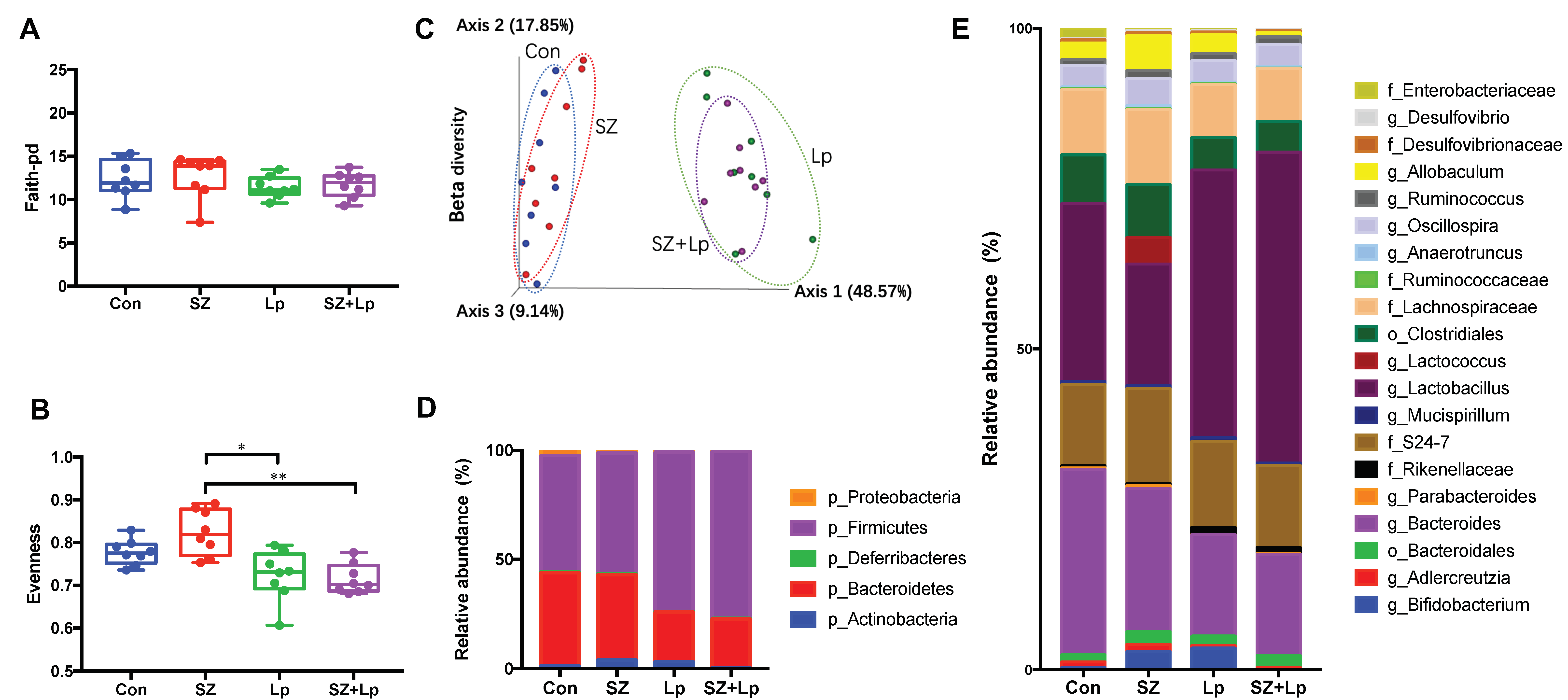
3.4. Changes in the Gut Microbiota after L. plantarum SeZi Administration

Gut microbiota alpha diversities were assessed by richness (Faith-pd) and Pielou’s evenness analyses, which represent the number of species and the degree of species homogeneity, respectively. No significant difference was observed between the groups in the alpha diversity of richness (Figure 3A), while the evenness in the SZ group was significantly larger than Lp and SZ + Lp groups (Figure 3B). The results of beta diversity (Bray-Curtis dissimilarity) indicated that the clustering in microbial communities in the Lp and SZ + Lp group was distinct from that in the control and SZ group (Figure 3C).
The average relative abundances of the final day fecal samples at the phylum level (Figure 3D) and the genus level (Figure 3E) suggested the different microbial compositions amongst the groups after the oral administration of SZ and SZ + Lp. To further evaluate the effects of treatments on microbial compositional changes, the three significantly different genera between the groups were identified and are displayed in Figure 4A–C. The relative abundance of Lactobacillus in the SZ + Lp group was significantly higher than that of the control and SZ groups, and the Lactobacillus level in the Lp group was also significantly higher than the SZ group. Adlercreutzia was significantly abundant in the SZ group compared to the SZ and SZ + Lp groups. Interestingly, Lactococcus was highly enriched only in the SZ group with the relative abundance at 4.15%. In addition, the relative abundance of Allobaculum in the SZ group was much larger than the SZ + Lp group, although the difference was not statistically significant (Figure 4D, p > 0.05).

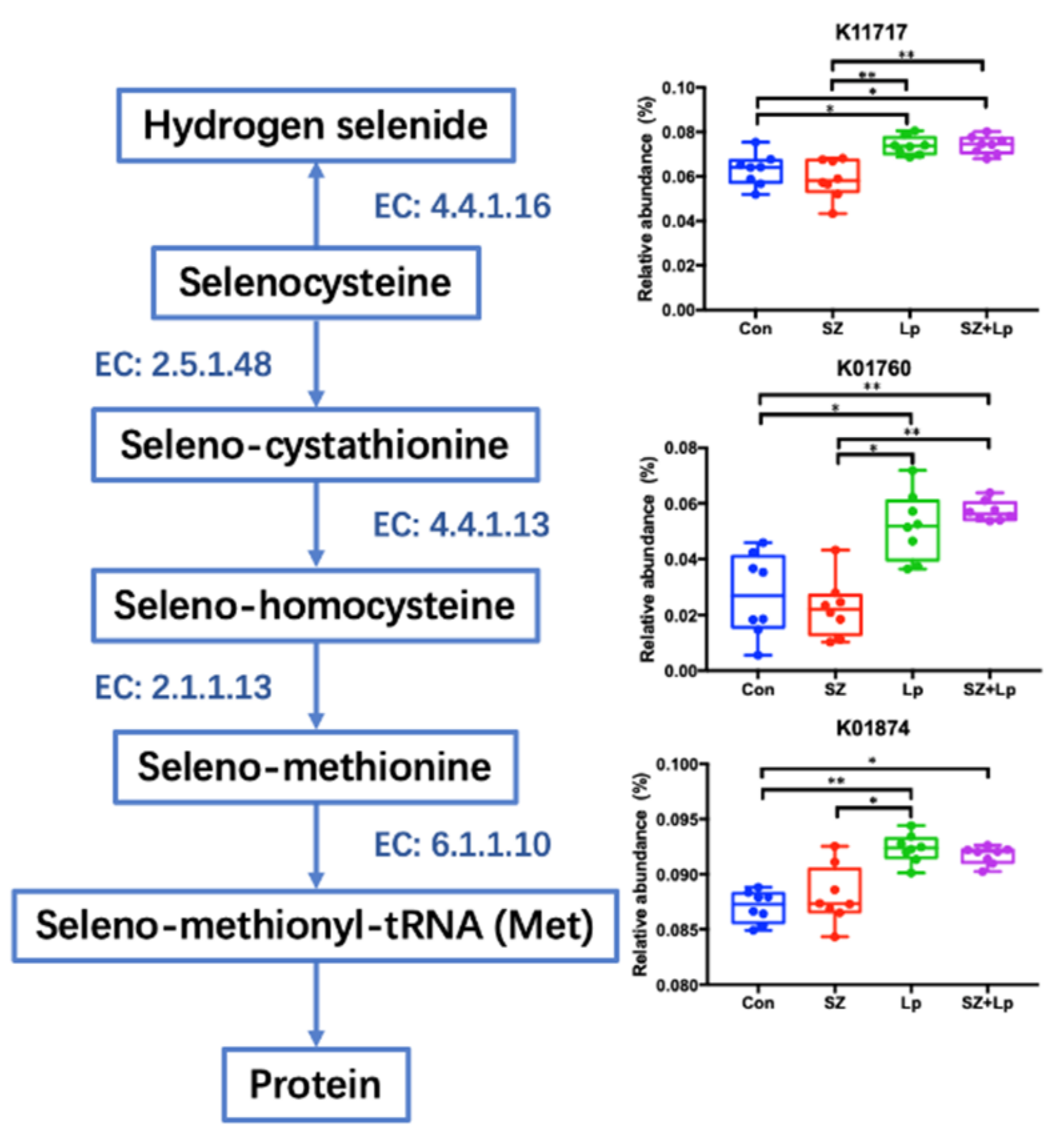
3.5. Microbial Function Analysis Related to Selenocompounds Metabolism

To investigate the functional changes in selenocompounds metabolism of the gut microbiome, KEGG analysis was performed by phylogenetic investigation of the community by reconstruction of unobserved states (PICRUSt).
As presented in Figure 5, L. plantarum SeZi significantly increased the relative abundances of the SCLY gene coding for selenocysteine lyase (EC: 4.4.1.16), CCBL gene coding for cysteine-S-conjugate beta-lyase (EC: 4.4.1.13), and MARS gene coding for methionyl-tRNA synthetase (EC: 6.1.1.10). These selenocompounds metabolism-related genes were responsible for the utilization of selenocysteine, seleno-cystathionine and seleno-methionine, respectively.
To detoxify selenite and selenate in the selenocompounds metabolism pathway, selenite is converted to selenate directly or via an intermediate, and selenate is further metabolized into hydrogen selenide. As shown in Figure 6, the relative abundances of enzymes related to oxidation of selenite to selenate (EC: 2.7.7.4, EC: 1.97.1.9) were significantly reduced in the Lp and SZ + Lp groups.

4. Discussion

In this study, L. plantarum SeZi isolate was screened out from human fecal bacteria by the selenium- and zinc-tolerant abilities. The selenium- and zinc-enriched L. plantarum SeZi strain increased the levels of selenium and zinc and presented antioxidative properties in an ICR mouse model. A thorough search of the literature reporting the bioavailability and functionality of Se- and Zn-enriched microorganisms using a mouse model yielded only one related article [42]. To determine appropriate concentrations of selenium and zinc for mice, we referenced this article and 300 μg of selenium and 1.5 mg of zinc (/kg body weight/day) were used in our study. Yan et al. reported the antioxidant and antitumor activities were significantly increased by supplementation with Se/Zn-enriched mushrooms. However, the in vivo antioxidant activity of Se/Zn-enriched LAB has not been reported yet.
Selenium is an essential element that must be exogenously provided to reach the requirement of human and animal health [43]. The toxicity order of selenium species from high to low is selenate, selenite, nano-selenium, and lactomicro-selenium [44]. The accumulation of selenium in bacteria is processed by extracellular binding via active groups in the cell-membrane conjunction or intracellular binding via ion transportation on the membrane [45]. Many Lactobacillus strains are well-known to accumulate and biotransform toxic selenite into non-toxic seleno-amino acids (i.e., selenocysteine and selenomethionine) and selenoprotein [46,47] The utilization of Se-enriched Lactobacillus possesses unique advantages, including low toxicity, low cost production and additional probiotic effects.
In this study, we focused on the changes in the gut microbial composition as well as functional metabolism associated with Se/Zn uptake and utilization. Interestingly, Se/Zn supplementation greatly induced the enrichment of specific genus, Lactococcus (belonging to LAB), which confirmed the previous reports regarding in vitro tolerance of LAB to selenium [46,47]. However, this indigenously enriched LAB by inorganic Se/Zn supplement showed significantly different patterns in the microbial selenocompounds metabolism compared with Se/Zn-bioaccumulated L. plantarum SeZi strain.
To further investigate strain-specific functions, whole genome sequencing of L. plantarum SeZi strain was conducted and analyzed based on public databases. The genes coding cysA and dedA were observed in the whole genome sequencing of L. plantarum SeZi. Both of the cysA and dedA genes are associated with selenite uptake and detoxification. These genes were abundantly present in L. plantarum SeZi strain, but not observed in Lactococcus or other Lactobacillus spp. According to previous studies, selenite may enter the cells of E. coli through the sulfate permease CysA [48]. DedA can uptake selenite into cells as a direct transporter or a cofactor. Additionally, the dedA gene-contained mutant E. coli displayed selenite resistance [17]. Based on the results of targeted metagenome sequencing, the L. plantarum SeZi strain promoted the utilization of selenocysteine, seleno-cystathionine and seleno-methionine in selenocompounds metabolism pathway of gut microbiome. Besides, the transformation between the toxic inorganic selenium was reduced by L. plantarum SeZi in the detoxification process. These are consistent with the selenium-related function detected in the genome sequencing of L. plantarum SeZi.
Zinc plays an essential role in catalytic, structural, and regulatory functions in enzymes and protein domains [49]. Similar to the other trace elements (i.e., selenium), zinc in the organic forms have more bioavailability its inorganic forms [49]. Pharmacological zinc supplements often have low bioavailability and are easily overdosed [50]. Although internalizing zinc by Lactobacillus has not been studied in depth, the utilization of certain Lactobacillus species can be a promising alternative to deliver zinc in a highly organic form [51].
The selenium and zinc levels in the blood can be affected by dietary supplementation and related metabolism. In this study, the concentrations of selenium and zinc in the Se/Zn-enriched Lactobacillus group were significantly higher than the control or SZ group. This is consistent with a recently published study which indicates that feeding a diet supplemented with Se/Zn-enriched probiotics, 0.3 mg/L selenium, and 100 mg/L zinc significantly enhanced the blood selenium and zinc concentrations in Wistar rats [52]. It is also the only published study referred to Se/Zn-enriched probiotics in a murine model to date. However, the assessment of antioxidant capability and its potential mechanisms was not reported yet.
In this study, the enhancement of antioxidant ability in the SZ + Lp group was probably triggered by the increased selenium level. When selenium is incorporated into the selenoenzymes (i.e., GSH-Px), it enhances the antioxidant activities by suppressing the nuclear factor-kappa B (NF-κB) signal pathway [53]. Most selenoproteins take part in the defense against oxidative stress, protecting tissues and cells from oxidative damages [47]. Although the SZ group was administered with inorganic forms of Se/Zn, the SZ + Lp group was administered with bio-accumulated Se/Zn in bacteria. To understand the mechanisms of bioavailability and functionality related to inorganic/organic forms of trace nutrients, it is necessary to investigate the changes in selenium and zinc metabolites (e.g., selenoproteins, zincproteins, etc.) from blood and fecal samples.
Alteration of microbial communities was evaluated by 16S rRNA community analysis in this study. Up to now, the 16S metagenomic technique is still rarely used in the selenium or zinc-related animal studies. Obviously, the significant surge of Lactobacillus levels in the Lp and SZ + Lp groups was caused by the oral administration of L. plantarum SeZi. The relative abundance of Adlercreutzia was significantly higher in the SZ + Lp group compared to the control and SZ groups. According to the previous studies, a decreased level of Adlercreutzia was observed in multiple sclerosis patients and Alzheimer’s disease patients [54,55]. Amongst Adlercreutzia species, A. equolifaciens is an equol-producing bacteria, promoting intestinal health [56]. Besides, a high relative abundance of Lactococcus (approximately 4.15%) was found in the SZ group, while this bacterium was almost undetectable in the other groups. It might be caused by utilization of selenium and zinc by Lactococcus spp. According to the previous studies, Lactococcus lactis is capable of selenium biotransformation and zinc uptake [57,58]. The relative abundance of Allobaculum spp., which was markedly reduced in the SZ + Lp group compared with other groups, was reported to be adversely associated with mRNA expression levels of tight junction protein genes (zo-1 and occludin) and anti-inflammatory genes (foxp3 and Il-10) in the colon of rats [59]. In some inflammatory bowel disease (IBD) patients, Allobaculum spp. was one of the uniquely observed species [60].
To the best of our knowledge, this study is the first attempt to evaluate the effects of Se/Zn-enriched LAB on in vivo gut microbiome changes. The microbiome analysis suggests a microbial aspect of selenocompound metabolism, however, lacks information on host’s Se/Zn metabolic process. Future studies on evaluating functionality of L. plantarum SeZi in a disease-induced mouse model should include host’s metabolites analysis as well.

5. Conclusions

In conclusion, the selected strain L. plantarum SeZi is able to resist and biotransform inorganic selenium into organic selenium. In the in vivo study, the selenium and zinc-enriched L. plantarum SeZi increased blood selenium level, antioxidant capability and the utilization of seleno-amino acids. Therefore, the L. plantarum SeZi strain is a potential selenium and zinc-enriched probiotic for application as functional food ingredients in the future.

Author Contributions

Conceptualization, S.K., R.L., H.J.Y., and G.E.J.; Investigation, S.K., R.L., and H.J.; Data curation, S.K. and R.L.; Methodology, S.K. and R.L.; Formal analysis, S.K.; Software, S.K.; Visualization, S.K.; writing-original draft preparation, S.K. and R.L.; writing—review and editing, S.K., H.J.Y. and G.E.J.; supervising, H.J.Y. and G.E.J.; funding acquisition, H.J.Y. and G.E.J. All authors have read and agreed to the published version of the manuscript.

Funding

This work was carried out with the support of the Ministry of Small and Medium-sized Enterprises (SMEs) and Startups (MSS), Korea, under the “Regional Specialized Industry Development Program (R&D, Project number S2848321)” supervised by the Korea Institute for Advancement of Technology (KIAT). This work was also supported by the “K-BIO KIURI Center program (Project number 2020M3H1A1073304)”.

Conflicts of Interest

G.E.J. holds BIFIDO Ltd. stocks. Other authors declare no conflict of interest.

References

  1. Mrvčić, J.; Stanzer, D.; Šolić, E.; Stehlik-Tomas, V. Interaction of lactic acid bacteria with metal ions: Opportunities for improving food safety and quality. World J. Microb. Biot. 2012, 28, 2771–2782. [Google Scholar] [CrossRef]
  2. Rayman, M.P. Selenium and adverse health conditions of human pregnancy. In Selenium; Springer: New York, NY, USA, 2011; pp. 531–544. [Google Scholar]
  3. Flores-Mateo, G.; Navas-Acien, A.; Pastor-Barriuso, R.; Guallar, E. Selenium and coronary heart disease: A meta-analysis. Am. J. Clin. Nutr. 2006, 84, 762–773. [Google Scholar] [CrossRef] [PubMed] [Green Version]
  4. Brenneisen, P.; Steinbrenner, H.; Sies, H. Selenium, oxidative stress, and health aspects. Mol. Asp. Med. 2005, 26, 256–267. [Google Scholar] [CrossRef] [PubMed]
  5. Ferguson, L.R.; Karunasinghe, N.; Zhu, S.; Wang, A.H. Selenium and its’ role in the maintenance of genomic stability. Mutat. Res. Fundam. Mol. Mech. Mutagenesis 2012, 733, 100–110. [Google Scholar] [CrossRef]
  6. Rayman, M.P. Selenium and human health. Lancet 2012, 379, 1256–1268. [Google Scholar] [CrossRef]
  7. Arthur, J.R.; Nicol, F.; Beckett, G.J. The role of selenium in thyroid hormone metabolism and effects of selenium deficiency on thyroid hormone and iodine metabolism. Biol. Trace Elem. Res. 1992, 33, 37–42. [Google Scholar] [CrossRef]
  8. Burke, M.P.; Opeskin, K. Fulminant heart failure due to selenium deficiency cardiomyopathy (Keshan disease). Med. Sci. Law 2002, 42, 10–13. [Google Scholar] [CrossRef]
  9. Maksimović, Z.J. Selenium deficiency and Balkan endemic nephropathy. Kidney Int. Suppl. 1991, 34, S12–S14. [Google Scholar] [PubMed]
  10. Gropper, S.S.; Smith, J.L. Advanced Nutrition and Human Metabolism. Wadsworth Cengage Learning: Belmot, CA, USA, 2012. [Google Scholar]
  11. Chimienti, F. Zinc, pancreatic islet cell function and diabetes: New insights into an old story. Nutr. Res. Rev. 2013, 26, 1–11. [Google Scholar] [CrossRef] [PubMed] [Green Version]
  12. Cruz, K.J.C.; de Oliveira, A.R.S.; Morais, J.B.S.; Severo, J.S.; Mendes, P.M.V.; de Sousa Melo, S.R.; de Sousa, G.S.; do Nascimento Marreiro, D. Zinc and insulin resistance: Biochemical and molecular aspects. Biol. Trace Elem. Res. 2018, 186, 407–412. [Google Scholar] [CrossRef] [PubMed]
  13. Islam, M.R.; Attia, J.; Ali, L.; McEvoy, M.; Selim, S.; Sibbritt, D.; Akhter, A.; Akter, S.; Peel, R.; Faruque, O. Zinc supplementation for improving glucose handling in pre-diabetes: A double blind randomized placebo controlled pilot study. Diabetes Res. Clin. Pract. 2016, 115, 39–46. [Google Scholar] [CrossRef] [PubMed]
  14. Afolabi, O.B.; Balogun, B.D.; Oloyede, O.I.; Akinyemi, A.J. Zinc and neurodegenerative disorders. In Handbook of Research on Critical Examinations of Neurodegenerative Disorders; IGI Global: Harrisburg, PA, USA, 2019; pp. 176–193. [Google Scholar]
  15. Gomolak, J.R. Zinc accumulation in the midbrain following methamphetamine exposure as a potential biomarker for neurodegeneration. In Proceedings of the 25th UNG Annual Research Conference, Dahlonega, GA, USA, 11 November 2016. [Google Scholar]
  16. Kieliszek, M.; Błażejak, S. Selenium: Significance, and outlook for supplementation. Nutrition 2013, 29, 713–718. [Google Scholar] [CrossRef] [PubMed]
  17. Ledgham, F.; Quest, B.; Vallaeys, T.; Mergeay, M.; Covès, J. A probable link between the DedA protein and resistance to selenite. Res. Microbiol. 2005, 156, 367–374. [Google Scholar] [CrossRef]
  18. Ayangbenro, A.S.; Babalola, O.O. A new strategy for heavy metal polluted environments: A review of microbial biosorbents. Int. J. Environ. Res. Pub. He. 2017, 14, 94. [Google Scholar] [CrossRef]
  19. Blackwell, K.; Singleton, I.; Tobin, J.M. Metal cation uptake by yeast: A review. Appl. Microbiol. Biot. 1995, 43, 579–584. [Google Scholar] [CrossRef]
  20. Andreoni, V.; Luischi, M.M.; Cavalca, L.; Erba, D.; Ciappellano, S. Selenite tolerance and accumulation in the Lactobacillus species. Ann. Microbiol. 2000, 50, 77–88. [Google Scholar]
  21. Saini, K.; Tomar, S.K. In vitro evaluation of probiotic potential of Lactobacillus cultures of human origin capable of selenium bioaccumulation. LWT 2017, 84, 497–504. [Google Scholar] [CrossRef]
  22. Shu, G.; Mei, S.; Chen, L.; Zhang, B.; Guo, M.; Cui, X.; Chen, H. Screening, identification, and application of selenium--enriched Lactobacillus in goat milk powder and tablet. J. Food Process. Pres. 2020, e14470. [Google Scholar] [CrossRef]
  23. Xia, S.K.; Chen, L.; Liang, J.Q. Enriched selenium and its effects on growth and biochemical composition in Lactobacillus bulgaricus. J. Agric. Food. Chem. 2007, 55, 2413–2417. [Google Scholar] [CrossRef]
  24. Diowksz, A.; Ambroziak, W.; Wlodarczyk, M. Investigation of the ability of selenium accumulation by lactic acid bacteria of Lactobacillus species and yeast Saccharomyces cerevisiae. Pol. J. Food Nutr. Sci. 1999, 1, 17–22. [Google Scholar]
  25. Mudroňová, D.; Gancarčíková, S.; Nemcová, R. Influence of Zinc Sulphate on the Probiotic Properties of Lactobacillus plantarum CCM 7102. Folia Veterinaria 2019, 63, 45–54. [Google Scholar] [CrossRef] [Green Version]
  26. Jin, H.; Jeong, Y.; Yoo, S.H.; Johnston, T.V.; Ku, S.; Ji, G.E. Isolation and characterization of high exopolysaccharide-producing Weissella confusa VP30 from young children’s feces. Microb. Cell Fact. 2019, 18, 110. [Google Scholar] [CrossRef] [Green Version]
  27. Chaney, A.L.; Marbach, E.P. Modified reagents for determination of urea and ammonia. Clin. Chem. 1962, 8, 130–132. [Google Scholar] [CrossRef]
  28. Yoon, S.H.; Ha, S.M.; Kwon, S.; Lim, J.; Kim, Y.; Seo, H.; Chun, J. Introducing EzBioCloud: A taxonomically united database of 16S rRNA gene sequences and whole-genome assemblies. Int. J. Syst. Evol. Microbiol. 2017, 67, 1613. [Google Scholar] [CrossRef]
  29. Schattner, P.; Brooks, A.N.; Lowe, T.M. The tRNAscan-SE, snoscan and snoGPS web servers for the detection of tRNAs and snoRNAs. Nucleic Acids Res. 2005, 33 (Suppl. 2), W686–W689. [Google Scholar] [CrossRef]
  30. Nawrocki, E.P.; Eddy, S.R. Computational identification of functional RNA homologs in metagenomic data. RNA Biol. 2013, 10, 1170–1179. [Google Scholar] [CrossRef]
  31. Hyatt, D.; Chen, G.L.; LoCascio, P.F.; Land, M.L.; Larimer, F.W.; Hauser, L.J. Prodigal: Prokaryotic gene recognition and translation initiation site identification. BMC Bioinform. 2010, 11, 119. [Google Scholar] [CrossRef] [Green Version]
  32. Powell, S.; Forslund, K.; Szklarczyk, D.; Trachana, K.; Roth, A.; Huerta-Cepas, J.; Gabaldon, T.; Rattei, T.; Creevey, C.; Kuhn, M. eggNOG v4. 0: Nested orthology inference across 3686 organisms. Nucleic Acids Res. 2014, 42, D231–D239. [Google Scholar] [CrossRef]
  33. Edgar, R.C. Search and clustering orders of magnitude faster than BLAST. Bioinformatics 2010, 26, 2460–2461. [Google Scholar] [CrossRef] [Green Version]
  34. Consortium, U. UniProt: A hub for protein information. Nucleic Acids Res. 2015, 43, D204–D212. [Google Scholar] [CrossRef]
  35. Kanehisa, M.; Goto, S.; Sato, Y.; Kawashima, M.; Furumichi, M.; Tanabe, M. Data, information, knowledge and principle: Back to metabolism in KEGG. Nucleic Acids Res. 2014, 42, D199–D205. [Google Scholar] [CrossRef] [Green Version]
  36. Overbeek, R.; Begley, T.; Butler, R.M.; Choudhuri, J.V.; Chuang, H.Y.; Cohoon, M.; de Crécy-Lagard, V.; Diaz, N.; Disz, T.; Edwards, R. The subsystems approach to genome annotation and its use in the project to annotate 1000 genomes. Nucleic Acids Res. 2005, 33, 5691–5702. [Google Scholar] [CrossRef] [Green Version]
  37. Kang, S.; You, H.J.; Lee, Y.G.; Jeong, Y.; Johnston, T.V.; Baek, N.I.; Ku, S.; Ji, G.E. Production, structural characterization, and In Vitro assessment of the prebiotic potential of butyl-fructooligosaccharides. Int. J. Mol. Sci. 2020, 21, 445. [Google Scholar] [CrossRef] [Green Version]
  38. Douglas, G.M.; Maffei, V.J.; Zaneveld, J.R.; Yurgel, S.N.; Brown, J.R.; Taylor, C.M.; Huttenhower, C.; Langille, M.G. PICRUSt2 for prediction of metagenome functions. Nat. Biotechnol. 2020, 1–5. [Google Scholar] [CrossRef]
  39. Younus, H. Therapeutic potentials of superoxide dismutase. Int. J. Health Sci. 2018, 12, 88. [Google Scholar]
  40. Blankenberg, S.; Rupprecht, H.J.; Bickel, C.; Torzewski, M.; Hafner, G.; Tiret, L.; Smieja, M.; Cambien, F.; Meyer, J.; Lackner, K.J. Glutathione peroxidase 1 activity and cardiovascular events in patients with coronary artery disease. N. Engl. J. Med. 2003, 349, 1605–1613. [Google Scholar] [CrossRef] [Green Version]
  41. Khoubnasabjafari, M.; Ansarin, K.; Jouyban, A. Reliability of malondialdehyde as a biomarker of oxidative stress in psychological disorders. BioImpacts 2015, 5, 123. [Google Scholar] [PubMed]
  42. Yang, H.; Chang, H. Antioxidant and antitumor activities of selenium and zinc-enriched oyster mushroom in mice. Biol. Trace Elem. Res. 2012, 150, 236–241. [Google Scholar]
  43. Xu, C.; Guo, Y.; Qiao, L.; Ma, L.; Cheng, Y.; Roman, A. Biogenic synthesis of novel functionalized selenium nanoparticles by Lactobacillus casei ATCC 393 and its protective effects on intestinal barrier dysfunction caused by enterotoxigenic Escherichia coli K88. Front. Microbiol. 2018, 9, 1129. [Google Scholar] [CrossRef] [PubMed] [Green Version]
  44. Nagy, G.; Pinczes, G.; Pinter, G.; Pocsi, I.; Prokisch, J.; Banfalvi, G. In situ electron microscopy of lactomicroselenium particles in probiotic bacteria. Int. J. Mol. Sci. 2016, 17, 1047. [Google Scholar] [CrossRef] [Green Version]
  45. Kurek, E.; Ruszczynska, A.; Wojciechowski, M.; Luciuk, A.; Michalska-Kacymirow, M.; Motyl, I.; Bulska, E. Bio-transformation of selenium in Se-enriched bacterial strains of Lactobacillus casei. Roczniki Państwowego Zakładu Higieny 2016, 67, 3. [Google Scholar]
  46. Yazdi, M.H.; Mahdavi, M.; Setayesh, N.; Esfandyar, M.; Shahverdi, A.R. Selenium n6noparticle-enriched Lactobacillus brevis causes more efficient immune responses in vivo and reduces the liver metastasis in metastatic form of mouse breast cancer. DARU 2013, 21, 33. [Google Scholar] [CrossRef] [PubMed] [Green Version]
  47. Qiao, L.; Dou, X.; Yan, S.; Zhang, B.; Xu, C. Biogenic selenium nanoparticles synthesized by Lactobacillus casei ATCC 393 alleviate diquat-induced intestinal barrier dysfunction in C57BL/6 mice through their antioxidant activity. Food Funct. 2020, 11, 3020–3031. [Google Scholar] [CrossRef] [PubMed]
  48. Turner, R.J.; Weiner, J.H.; Taylor, D.E. Selenium metabolism in Escherichia coli. Biometals 1998, 11, 223–227. [Google Scholar] [CrossRef] [PubMed]
  49. Leonardi, A.; Zanoni, S.; de Lucia, M.; Amaretti, A.; Raimondi, S.; Rossi, M. Zinc uptake by lactic acid bacteria. ISRN Biotechnol. 2013, 2013. [Google Scholar] [CrossRef] [Green Version]
  50. Góral, M.; Pankiewicz, U.; Sujka, M.; Kowalski, R. Bioaccumulation of zinc ions in Lactobacillus rhamnosus B 442 cells under treatment of the culture with pulsed electric field. Eur. Food Res. Technol. 2019, 245, 817–824. [Google Scholar] [CrossRef] [Green Version]
  51. Lule, V.K.; Tomar, S.K.; Chawla, P.; Pophaly, S.; Kapila, S.; Arora, S. Bioavailability assessment of zinc enriched lactobacillus biomass in a human colon carcinoma cell line (Caco-2). Food Chem. 2020, 309, 125583. [Google Scholar] [CrossRef]
  52. Malyar, R.M.; Li, H.; Liu, D.; Abdulrahim, Y.; Farid, R.A.; Gan, F.; Ali, W.; Enayatullah, H.; Banuree, S.A.H.; Huang, K. Selenium/Zinc-Enriched probiotics improve serum enzyme activity, antioxidant ability, inflammatory factors and related gene expression of Wistar rats inflated under heat stress. Life Sci. 2020, 248, 117464. [Google Scholar] [CrossRef]
  53. Benstoem, C.; Goetzenich, A.; Kraemer, S.; Borosch, S.; Manzanares, W.; Hardy, G.; Stoppe, C. Selenium and its supplementation in cardiovascular disease—what do we know? Nutrients 2015, 7, 3094–3118. [Google Scholar] [CrossRef] [Green Version]
  54. Fan, Y.; Zhang, J. Dietary modulation of intestinal microbiota: Future opportunities in experimental autoimmune encephalomyelitis and multiple sclerosis. Front. Microbiol. 2019, 10, 740. [Google Scholar] [CrossRef]
  55. Vogt, N.M.; Kerby, R.L.; Dill-McFarland, K.A.; Harding, S.J.; Merluzzi, A.P.; Johnson, S.C.; Carlsson, C.M.; Asthana, S.; Zetterberg, H.; Blennow, K. Gut microbiome alterations in Alzheimer’s disease. Sci. Rep. 2017, 7, 1–11. [Google Scholar] [CrossRef]
  56. Maruo, T.; Sakamoto, M.; Ito, C.; Toda, T.; Benno, Y. Adlercreutzia equolifaciens gen. nov., sp. nov., an equol-producing bacterium isolated from human faeces, and emended description of the genus Eggerthella. Int. J. Syst. Evol. Micr. 2008, 58, 1221–1227. [Google Scholar] [CrossRef]
  57. Martínez, F.G.; Moreno-Martin, G.; Pescuma, M.; Madrid-Albarrán, Y.; Mozzi, F. Biotransformation of selenium by lactic acid bacteria: Formation of seleno-nanoparticles and seleno-amino acids. Front. Bioeng. Biotechnol. 2020, 8, 506. [Google Scholar] [CrossRef]
  58. Llull, D.; Poquet, I. New expression system tightly controlled by zinc availability in Lactococcus lactis. Appl. Environ. Microbiol. 2004, 70, 5398–5406. [Google Scholar] [CrossRef] [Green Version]
  59. Lee, S.M.; Han, H.W.; Yim, S.Y. Beneficial effects of soymilk and fiber on high cholesterol diet-induced alteration of gut microbiota and inflammatory gene expression in rats. Food Funct. 2015, 6, 492–500. [Google Scholar] [CrossRef]
  60. Palm, N.W.; de Zoete, M.R.; Cullen, T.W.; Barry, N.A.; Stefanowski, J.; Hao, L.; Degnan, P.H.; Hu, J.; Peter, I.; Zhang, W. Immunoglobulin A coating identifies colitogenic bacteria in inflammatory bowel disease. Cell 2014, 158, 1000–1010. [Google Scholar] [CrossRef] [Green Version]
Figure 1. The concentrations of blood selenium (A) and zinc (B) in mice after 4 weeks of administration. Data were analyzed by unpaired t-test analysis and expressed as mean ± SD. ** p < 0.01 (n = 8). Con, control; SZ, selenium and zinc supplemented; Lp, Lactobacillus plantarum SeZi; SZ + Lp, selenium- and zinc-enriched L. plantarum SeZi.
Antioxidants 09 01028 g001
Figure 2. Glutathione peroxidase (GSH-Px) activity (A), superoxide dismutase (SOD) activity (B), and lipid oxidation product malondialdehyde (MDA) level (C) in serum of mice at the final day. Treatments with different letters (a, b, c) are significantly different at p < 0.05 (n = 8). Con, control; SZ, selenium and zinc supplemented; Lp, Lactobacillus plantarum SeZi; SZ + Lp, selenium- and zinc-enriched L. plantarum SeZi.
Antioxidants 09 01028 g002
Figure 3. Comparison of diversity indices and microbial compositions amongst groups after 4 weeks of oral administration. Alpha-diversities of microbial communities are shown as (A) richness and (B) evenness. (C) Principal Coordinates Analysis (PCoA) plot represents beta-diversity based on Bray-Curtis dissimilarity. (D) Taxonomic profiles at the phylum level (D) and the genus level (E). Relative Abundance of taxa below 0.1% were excluded prior to analyses. Significance was accepted at * p < 0.05, ** p < 0.01 (n = 8). Con, control; SZ, selenium and zinc supplemented; Lp, Lactobacillus plantarum SeZi; SZ + Lp, selenium- and zinc-enriched L. plantarum SeZi.
Antioxidants 09 01028 g003
Figure 4. Relative abundances of Lactobacillus (A), Adlercreutzia (B), Lactococcus (C), and Allobaculum (D) in fecal samples after 4 weeks of oral administration. Data are expressed as mean ± SD. Significance was accepted at * p < 0.05, ** p < 0.01 (n = 8). Con, control; SZ, selenium and zinc supplemented; Lp, Lactobacillus plantarum SeZi; SZ + Lp, selenium- and zinc-enriched L. plantarum SeZi.
Antioxidants 09 01028 g004
Figure 5. Selenocompounds metabolism pathway. Related enzymes include selenocysteine lyase (EC: 4.4.1.16), cystathionine gamma-synthase (EC: 2.5.1.48), cysteine-S-conjugate beta-lyase (EC: 4.4.1.13), homocysteine methyltransferase (EC: 2.1.1.13), and methionyl-tRNA synthetase (EC: 6.1.1.10). Arrows indicate the related genes that are involved in the corresponding pathway. Error bars represent means ± SD. Significance was accepted at * p < 0.05, ** p < 0.01 (n = 8). Con, control; SZ, selenium and zinc supplemented; Lp, Lactobacillus plantarum SeZi; SZ + Lp, selenium- and zinc-enriched L. plantarum SeZi. K11717, cysteine desulfurase/selenocysteine lyase; K01760, cystathionine beta-lyase; K01874, methionyl-tRNA synthetase.
Antioxidants 09 01028 g005
Figure 6. Detoxification process of inorganic selenium. Related enzymes include 3′-phosphoadenosine 5′-phosphosulfate synthase (EC: 2.7.7.4) and selenate reductase subunit alpha (EC: 1.97.1.9). Arrows indicate the related genes that are involved in the corresponding pathway. Error bars represent means ± SD. Significance was accepted at * p < 0.05, ** p < 0.01 (n = 8). Con, control; SZ, selenium and zinc supplemented; Lp, Lactobacillus plantarum SeZi; SZ + Lp, selenium- and zinc-enriched L. plantarum SeZi. K07310, Tat-targeted selenate reductase subunit YnfF; K07309, Tat-targeted selenate reductase subunit YnfE; K12527, putative selenate reductase; K00956, sulfate adenylyltransferase subunit 1; K00957, sulfate adenylyltransferase subunit 2; K00958, sulfate adenylyltransferase.
Antioxidants 09 01028 g006
Table 1. Generation of biomass and bioconvertion rates of selenium and zinc in the selected SeZn-tolerant probiotic strains in vitro.
StrainsBiomass (g/L)Bioconversion Rate (%)
SeleniumZinc
Lactobacillus plantarum SeZi2.82 ± 0.28 a19.470.35
Lactobacillus pentosus SeZi2.33 ± 0.11 ab8.930.36
Lactobacillus fermentum SeZi1.94 ± 0.34 bc18.040.33
Lactobacillus rhamnosus SeZi1.78 ± 0.34 c6.900.20
Data are expressed as mean ± SD (n = 5). Treatments with different letters are significantly different at p < 0.05 (n = 5).
Table 2. Gene products of selenium resistance gene cluster in Lactobacillus plantarum SeZi.
Coding Region aLength (aa)ProductFunction
264974–265621 (−)648DedA proteinDetoxification and uptake of selenate
193218–193874 (−)657DedA proteinDetoxification and uptake of selenate
19823–20716894Sulfate permease-CysA Detoxification and uptake of selenate
a Genes encoded on the minus strand are indicated with (−).
Table 3. Gene products of zinc resistance gene cluster in Lactobacillus plantarum SeZi.
Coding Region aLength (aa)ProductFunction
237659–238102444ZURZinc uptake regulation
151–966816Multidrug efflux transporter 1 regulatorZinc resistance
31131–31565435Uncharacterized HTH-type transcriptional regulatorZinc resistance
174455–174916462MerR familyZinc resistance
22703–23146444MerR familyZinc resistance
115660–116040381MerR familyZinc resistance
27565–27957 (−)393MerR familyZinc resistance
4022–4399378MerR familyZinc resistance
227–619 (−)393MerR familyZinc resistance
342254–342718465Hypothetical proteinZinc resistance
54502–54756 (−)255Hypothetical proteinZinc resistance
54351–54800450Hypothetical proteinZinc resistance
a Genes encoded on the minus strand are indicated with (−). ZUR: zinc uptake regulation.
Table 4. Other Lactobacillus strains with selenium and/or zinc resistance gene clusters. (From NCBI genome databases).
DedACysAZURMerR Family
L. acidophilus La-14-L.rhamnosus LOCK908L. acidophilus La-14
L. gasseri ATCC 33323 L.rhamnosus LOCK900L. curvatus JCM 1096
L. dekbrueckii subsp. delbrueckii L.rhamnosus LOCK919L. gasseri ATCC 33323
L. salivarius str. Ren L. ruminis ATCC 25644
L. buchneri subsp. silagei CD034 L. buchneri subsp. silagei CD034
L. rhamnosus GG
L. paracasei subsp. paracasei 8700:2
Publisher’s Note: MDPI stays neutral with regard to jurisdictional claims in published maps and institutional affiliations.

