Article

Lacticaseibacillus paracasei JY062 Postbiotic Alleviated 3% DSS-Induced Colitis in Mice via Integrated Antioxidant, Barrier Repair, Immunomodulatory and Microbiota Modulation

1
Key Laboratory of Dairy Science, Ministry of Education, College of Food Science, Northeast Agricultural University, Harbin 150031, China
2
Food Laboratory of Zhongyuan, Luohe 462300, China
*
Authors to whom correspondence should be addressed.
These authors contributed equally to this work and should be considered co-first authors.
Antioxidants 2025, 14(10), 1256; https://doi.org/10.3390/antiox14101256
Submission received: 10 September 2025 / Revised: 1 October 2025 / Accepted: 16 October 2025 / Published: 19 October 2025
(This article belongs to the Topic Functional Foods and Nutraceuticals in Health and Disease)

Abstract

Ulcerative colitis (UC) with multifactorial etiology remains clinically challenging. While current pharmacotherapies alleviate symptoms, their long-term application is constrained by significant side effects. Lacticaseibacillus paracasei JY062 postbiotic (Pa JY062) exerts multi-target therapeutic effects via integrated antioxidant, barrier repair, immunomodulatory and microbiota modulation for UC. Pa JY062 exhibited potent antioxidant capacity and reduced reactive oxygen species (ROS) in Caco-2 cells. In DSS-colitis mice, high-dose Pa JY062 (HP, 800 mg/kg) restored intestinal barrier integrity (Evans blue permeability: 0.0547 vs. 0.107, p < 0.01), enhanced antioxidant defenses (SOD: 9.43 vs. 5.5; GSH-Px: 62.74 vs. 40.94 U/mg protein, p < 0.01), and rebalanced Th1/Th2/Th17/Treg responses. LigiLactobacillus murinus (L. murinus) was enriched, while Bacteroides acidifaciens (B. acidifaciens) was diminished, SCFAs increased (acetate 2772.70 ± 21.08, propionate 669.84 ± 6.79, butyrate 324.14 ± 0.42 μg/g). Spearman correlations linked L. murinus to barrier proteins (ZO-1/Occludin/E-cadherin), contrasting B. acidifaciens–inflammation associations. These findings revealed that Pa JY062 exerted multi-target therapeutic effects on UC, and offered a wider array of options for intestinal health. Pa JY062 represents a promising natural antioxidant-based strategy for UC management.

Graphical Abstract

1. Introduction

UC is a complex form of inflammatory bowel disease (IBD) that emerges from the intricate interaction of genetic susceptibility, environmental factors, disturbances in the intestinal microbiota, and immune system dysfunction [1]. This multifactorial pathogenesis challenges monotherapies targeting isolated pathways. Current standard-of-care treatments, including 5-aminosalicylic acid (5-ASA) for mucosal anti-inflammation, glucocorticoids for acute flare control, and anti-TNFα monoclonal antibodies for systemic immune modulation, effectively suppress acute inflammation but fail to address upstream drivers, leading to high relapse rates and adverse effects (e.g., osteoporosis, opportunistic infections) [2]. Current medications with singular regulatory effects are insufficient to achieve complete resolution of UC, highlighting the need for more comprehensive therapeutic strategies. The restoration of intestinal barrier integrity, increase in intestinal microbial diversity and attenuation of inflammatory responses are fundamental to UC remission. However, targeting the regulation of oxidative stress is also a crucial intervention strategy in the pathogenesis of UC. Research has shown that a nanoparticle targeting the ROS-responsive achieved a synergistic effect in IBD therapy [3].
Consuming an antioxidant-rich diet not only fulfills daily nutritional requirements but also alleviates inflammatory responses. Furthermore, emerging evidence has demonstrated its efficacy in modulating gut homeostasis represents a promising adjunctive strategy, especially for postbiotics [4]. Postbiotic defined as a “preparation of inanimate microorganisms and/or their components that confers a health benefit on the host” [5], postbiotic has gained traction as dietary modulators of gut homeostasis. Postbiotics from Lactobacillus plantarum JM015 [6], extracellular vesicles derived from Bifidobacterium longum NSP001 [7] and montmorillonite composite postbiotics [8] have shown effectiveness in alleviating intestinal inflammation and enhancing gut health. However, while current postbiotic interventions primarily target inflammatory resolution, barrier restoration, and microbiota modulation, their direct impact on oxidative stress remains largely unexplored. This gap in the knowledge highlights the potential of postbiotics with integrated antioxidant properties.
In our previous work, we discovered that Lacticaseibacillus paracasei JY062 can alleviate colitis in mice, and its postbiotic Pa JY062, formulated as a food-grade skimmed-milk-based preparation, possessed potent anti-inflammatory properties and the reestablished the intestinal barrier [9,10]. Therefore, this study aims to comprehensively evaluate the therapeutic potential of Pa JY062 against colitis, with a focus on its antioxidant capacity. We assessed its ability to scavenge ROS in Caco-2 cells in vitro and investigated its effects in a DSS-induced colitis mouse model. The investigation encompassed systemic antioxidant enzymes (SOD, GSH-Px), gut barrier proteins (ZO-1, occludin), immune cytokines (IFN-γ, IL-17A), and microbiota–short-chain fatty acid (SCFA) interactions.

2. Materials and Methods

2.1. Analysis of Antioxidant Capacity

A Lacticaseibacillus rhamnosus GG (LGG) postbiotic was used as a positive control, while Trolox, a water-soluble vitamin E analog, served as the antioxidant standard. The blank was the negative control. The preparation process of postbiotic LGG was the same as that of Postbiotic Pa JY062 [9].
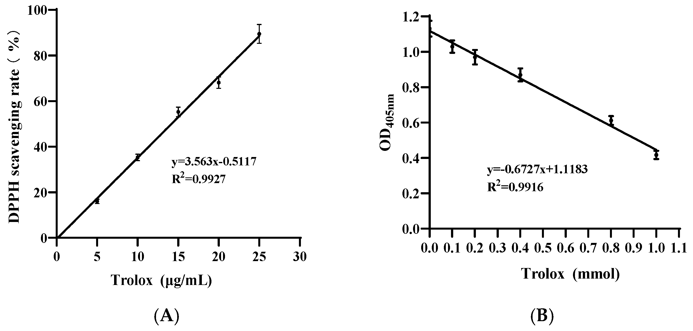
The DPPH radical scavenging activity was assessed utilizing DPPH radical scavenging ability Kit (Nanjing Jiancheng Bioengineering Institute, Nanjing, China). Postbiotics were homogenized in 80% methanol solution at 1:10 (m/v), and centrifuged (12,000 rpm, 10 min, 4 °C) to obtain the supernatant. The remaining procedures, conducted at 4 °C unless otherwise specified, are detailed in Table A1. Briefly, the mixture was incubated at 25 °C for 30 min in the dark and then centrifuged at 4000 rpm for 5 min. OD517nm was measured by a microplate reader (BioTek, WA, USA).
DPPH free radical scavenging rate (%) = (1 – (AdeterminationAcontrol))/(Ablank) × 100
Total antioxidant capacity (T-AOC) was quantified utilizing a T-AOC Assay Kit (Nanjing Jiancheng Bioengineering Institute, Nanjing, China). Postbiotics were homogenized in sterile water at a 1:9 (w/v) ratio and vortex-mixed for complete antioxidant extraction. Following centrifugation (12,000× g, 5 min, 4 °C), supernatants were meticulously collected for subsequent research. The remaining operations were performed according to the Table A2. Reaction mixtures were incubated at 25 °C for 6 min in 96-well plates, after which OD405nm was measured using a microplate reader (BioTek, Shoreline, WA, USA).

2.2. Cell Culture

Caco-2 cells were obtained from the Key Laboratory of Dairy Science at the Ministry of Education, Northeast Agricultural University, Harbin, China. All cells, between passages 5 and 20, were maintained in high-glucose DMEM supplemented with 10% fetal bovine serum and 1% penicillin–streptomycin, then maintained at 37 °C in a 5% CO2-humidified incubator.

2.3. Reactive Oxygen Species Assay

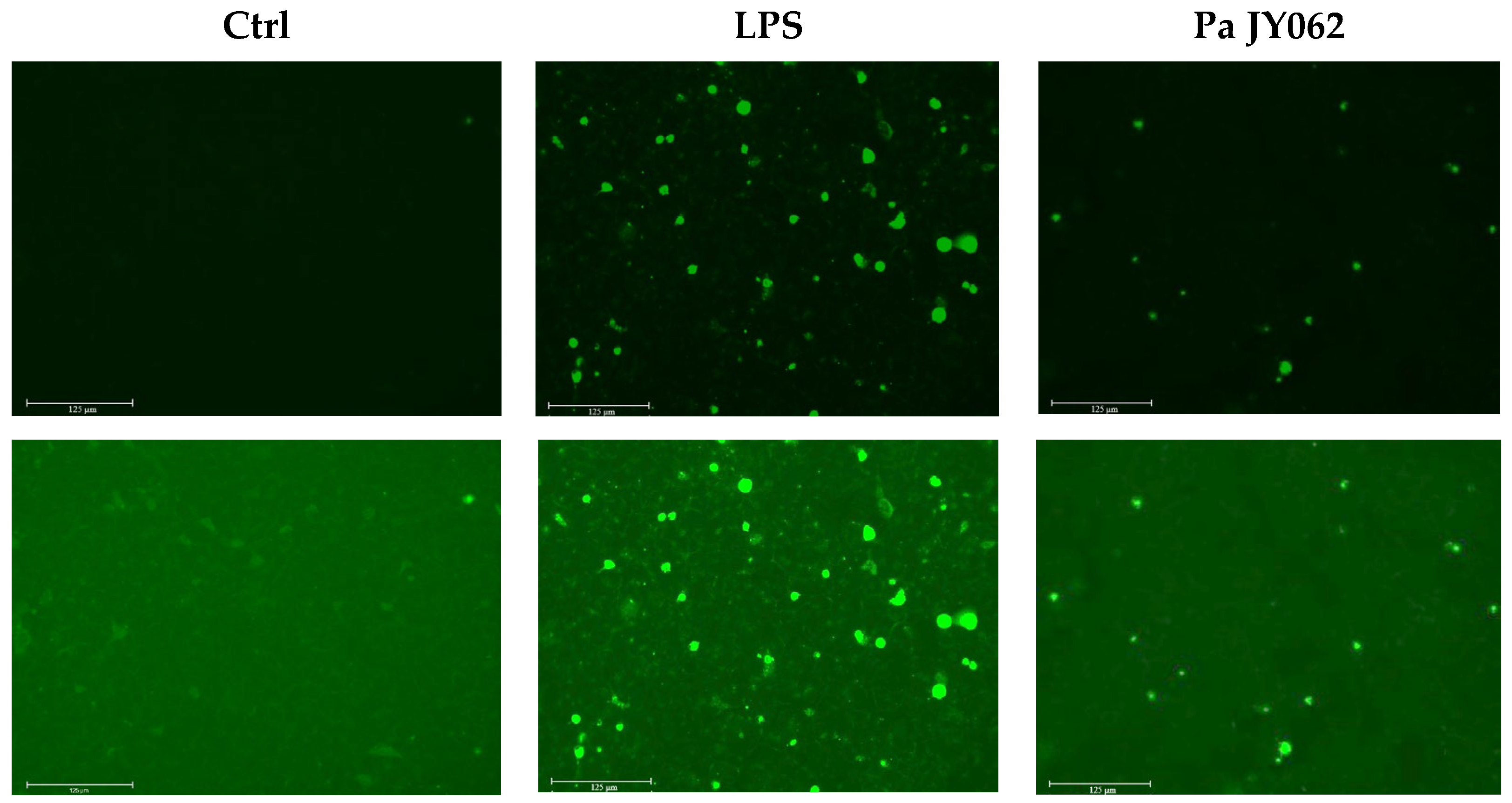
Caco-2 cells were seeded at a density of 1 × 10⁴ cells per well were plated in 96 well plates and treated with LPS (10 μg/mL) for 12 h, followed by Pa JY062 (250 μg/mL) with/without exposure for 24 h. ROS levels were assayed by ROS kit (Beyotime, Shanghai, China). Cells were incubated in DMEM containing DCFH-DA (10 μM) in the dark at 37 °C for 30 min. The ROS images was recorded by the EVOS FL Auto Cell Imaging System (Thermo Fisher Scientific, MA, USA). Unstimulated Caco-2 cells served as negative controls, with three replicates established for each treatment group.

2.4. Analysis of Bioactive Components in Pa JY062

The comprehensive metabolites of postbiotics Pa JY062 was detected using the T500 targeted platform (MetWare, http://www.metware.cn/ (accessed on 15 March 2024)) equipped with AB Sciex QTRAP 6500 LC-MS/MS system, as detailed in our previous publication [9]. This approach identified 198 newly detected components in Pa JY062 postbiotic. A literature search was conducted to identify metabolites with reported bioactivities, including antioxidant, anti-inflammatory, intestinal barrier-repairing, and gut microbiota-modulating properties.

2.5. Animal Experimental Design

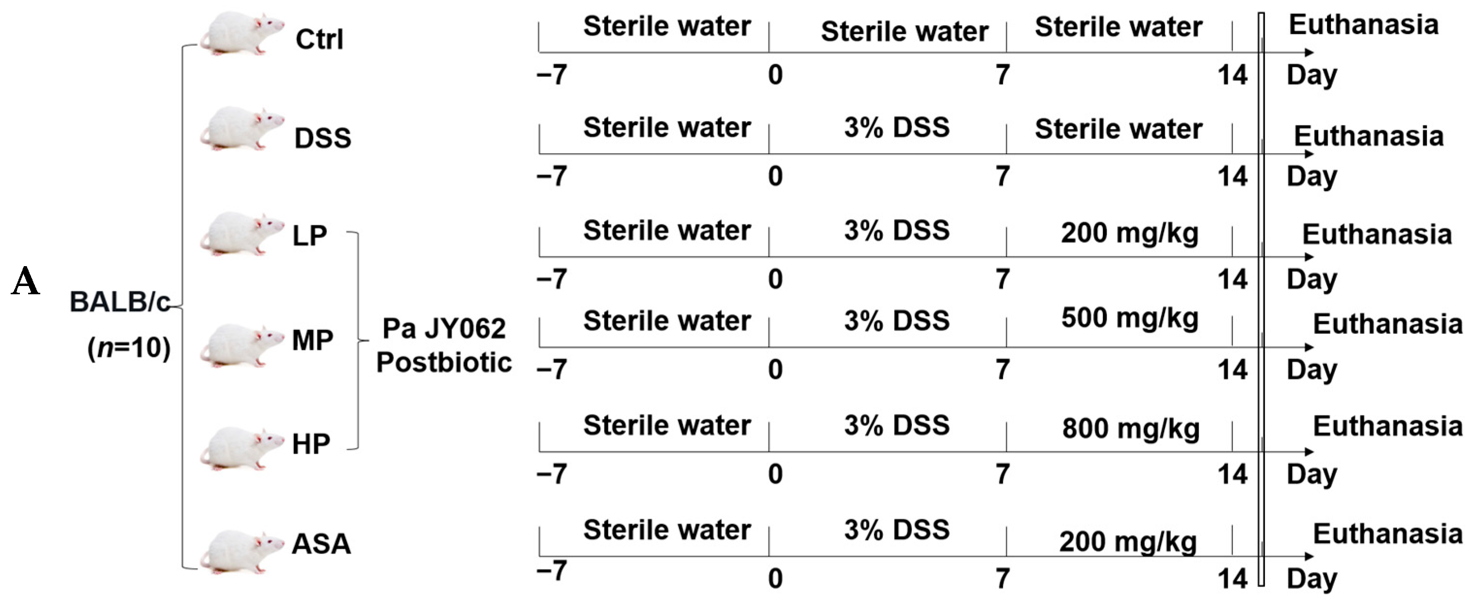
The animal experimentation protocol received approval from the Animal Ethics Committee of Northeast Agricultural University to ensure the utmost adherence to animal welfare, with the experiment designated as NEAUEC20230422, approval on 9 November, 2023. Sixty 8-week-old male BALB/c mice (weight: 20 ± 2 g) were acquired from Chengdu Dossy Experimental Animals Co., Ltd. (Chengdu, China). All mice were randomly divided into 6 groups (n = 10): control group (Ctrl, sterile water), DSS group (DSS, 3% DSS solution), 5-aminosalicylic acid group (ASA, 200 mg/kg), low-dose Pa JY062 (LP, 3.0% DSS + 200 mg/kg Pa JY062), medium-dose Pa JY062 (MP, 3.0% DSS + 500 mg/kg Pa JY062), and high-dose Pa JY062 (HP, 3.0% DSS + 800 mg/kg Pa JY062).
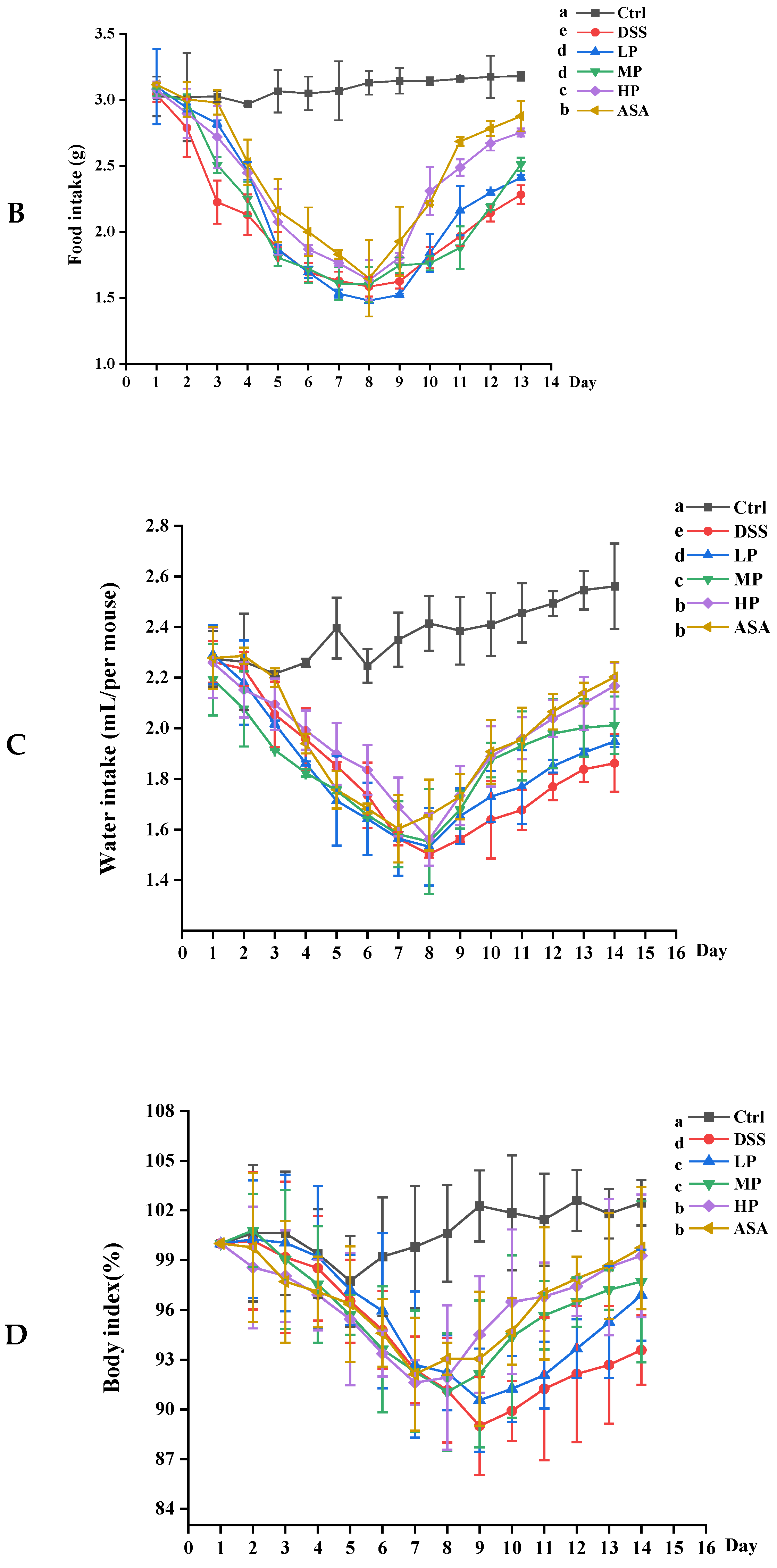
Mice were acclimatized for 7 days under standard conditions: temperature of 23 ± 2 °C, relative humidity of 55 ± 10%, and a 12 h light/dark cycle. Then, all groups, except for the Ctrl group, were granted unrestricted access to 3% DSS to establish a model of colitis (day 0 to day 7). During the remission period (day 8 to day 14), Ctrl and DSS groups received 200 μL of sterile water daily. LP, MP, and HP groups were given daily 200 μL of Pa JY062 solutions. The ASA group was gavaged daily with 200 μL of a 5-ASA solution. The daily food intake, water intake, and body weight changes in all mice were recorded. Fecal samples from each mouse were collected daily into a 1.5 mL sterile centrifuge tube. On day 15, all mice were humanely euthanized under intraperitoneal anesthesia with 10% chloral hydrate. The length of the colon was assessed, and the spleen and thymus were subjected to weighing. Three mice from each group were selected to assess Evans blue colonic permeability. One segment of the fresh colon was frozen at −80 °C, another segment was preserved in 2.5% glutaraldehyde, and the final segment was fixed in 4% paraformaldehyde for subsequent analyses. The spleen, thymus, serum, and cecal contents were harvested and preserved at −80 °C.

2.6. Disease Activity Index

Disease activity index (DAI) was computed according to the methodology outlined by Zhu et al. [11].
DAI = (weight loss score + stool consistency score + bleeding score)/3

2.7. Evans Blue Permeability and Ultrastructural Analysis

The colonic permeability of the mice was evaluated employing the Evans blue method as delineated by Rizvi [12].
For ultrastructural analysis, the distal colon was sectioned into 1 mm2 sections along the intestinal axis and subsequently preserved in precooled 2.5% glutaraldehyde at 4 °C for 12 h. Sample pieces were rinsed 3 times with 0.1 M PBS (15 min/time), stabilized with 1% OsO4 for 30 min, and rinsed again with PBS buffer 3 times. Dehydration was carried using ethanol series. The subsequent procedures followed the procedure detailed by Wang [13].

2.8. Histopathological Staining

A 1 cm2 piece of fresh distal colon was immersed in 4% neutral formaldehyde solution for 24 h. The tissue edges were then neatly trimmed, subjected to gradient dehydration in a dehydrator, and finally immersed in wax for embedding. The wax block was sliced into 4 μm sections, dewaxed in xylene, and rehydrated using a series of ethanol solutions. Colonic tissues were subjected to staining with hematoxylin and eosin (H&E) to evaluate colonic injury [14]. To identify goblet cells, Alcian blue periodic acid–Schiff (AB-PAS) staining was conducted employing the AB-PAS Stain Kit (Solarbio, Beijing, China), following the instructions provided by the manufacturer. The immunohistochemistry (IHC) procedure was carried out following the protocol established in [15].

2.9. Real-Time Quantitative PCR

The colon tissue was cut into 5 mm2 pieces and ground in liquid nitrogen. One part was treated with RNAplus to extract total RNA, another was treated by RIPA lysis buffer (Biosharp, Hefei, China) and preserved at −80 °C for later enzyme-linked immunosorbent assay.
RNA concentration was measured by Nanodrop (Waltham, MA, USA) and recorded in Table A3. The RNA was transcribed into cDNA, and a real-time quantitative polymerase chain reaction (RT-qPCR) was employed to ascertain the transcript levels of colonic genes utilizing the 2−ΔΔCt method. GAPDH served as the housekeeping gene. The primers employed for the RT-qPCR examination are delineated in Table A4.

2.10. Evaluation of Oxidative Stress

The glutathione peroxidase (GSH-PX) test kit (Nanjing Jiancheng Bioengineering Institute, Nanjing, China), and total superoxide dismutase (T-SOD) assay kit (Nanjing Jiancheng Bioengineering Institute, Nanjing, China) were utilized to quantify the levels of GSH-px and T-SOD in the mouse colon. The comprehensive procedural steps are delineated in Appendix A.1.

2.11. Enzyme-Linked Immunosorbent Assay

Colon tissue lysate was centrifuged at 3000× g for 5 min to harvest the supernatant for the assessment of cytokine (IL-2, IL-4, IL-10, IFN-γ) and cell surface adhesion molecules (VCAM-1, ICAM-1) concentrations, in accordance with the manufacturer’s instructions (Xinle, Shanghai, China).

2.12. Gut Microbiota Analysis

Genomic DNA from samples was extracted using the CTAB method. The V3-V4 region of the gut microbiota’s 16S rRNA gene was amplified via PCR. The 1× loading buffer (containing SYBR Green) was combined with the PCR products, which were then subjected to electrophoresis on a 2% agarose gel for visualization. The PCR products were mixed in equimolar ratios. This mixture was purified using the Qiagen Gel Extraction Kit (Qiagen, Germany). Sequencing libraries were prepared in accordance with the manufacturer’s guidelines with the TruSeq® DNA PCR-Free Sample Preparation Kit (Illumina, CA, USA), and index codes were incorporated. The quality of the libraries was evaluated using the Qubit@ 2.0 Fluorometer (Thermo Scientific, MA, USA) and the Agilent Bioanalyzer 2100 system. Sequencing was conducted using the Illumina NovaSeq system, generating 250 bp paired-end reads. For details on 16S rRNA analysis, see Appendix A.2.

2.13. Detection of Short-Chain Fatty Acids

SCFAs levels were analyzed by MetWare (http://www.metware.cn/ (accessed on 15 March 2024)) utilizing the Agilent 8890-7000D GC-MS/MS system. For detailed operation steps, see Appendix A.3.

2.14. Statistical Analysis

Statistical analyses were carried out with IBM SPSS Statistics 25.0. For data exhibiting a normal distribution (mean ± SEM), comparisons were made via one-way ANOVA, followed by Duncan’s post hoc test. For microbial abundance (phylum/genus/species), nonparametric Kruskal–Wallis tests were applied. Spearman’s correlation assessed associations.

3. Results

3.1. Antioxidant Capacity of Pa JY062 Postbiotics

The antioxidant capacity of Pa JY062 was systematically evaluated through DPPH radical scavenging and T-AOC assays. As shown in Figure 1A, the DPPH inhibition rate of Pa JY062 (36.54 ± 2.9%) was 2.95-fold higher than that of the blank control (Ctrl, 12.36 ± 0.83%; p < 0.01) and statistically indistinguishable from the LGG postbiotic positive control (35.46 ± 2.4%). The potent antioxidant activity of Pa JY062 was further confirmed by the T-AOC assay (Figure 1B). The T-AOC value of Pa JY062 (0.82 ± 0.01 mmol Trolox equiv/g) was significantly higher (p < 0.05) than both the LGG postbiotic (0.65 ± 0.01 mmol/g) and the blank control (0.55 ± 0.02 mmol/g).

3.2. The Influence of Pa JY062 on Reactive Oxygen Species

To investigate the protective effect of Pa JY062 against oxidative stress, intracellular ROS levels in Caco-2 cells were assessed by fluorescence imaging. The LPS resulted in a significant increase in green fluorescence in Caco-2 cells compared to the Ctrl. Pa JY062 markedly reduced the green fluorescence relative to LPS (Figure 2, p < 0.001).

3.3. Bioactive Ingredients of Pa JY062

T500-targeted metabolomic profiling of Pa JY062 identified 198 metabolites (Table 1). Compositional analysis revealed that the profile was quantitatively dominated by five major five classes. Nucleotides and their metabolites (18.69%), amino acids (12.12%), organic acids and derivatives (11.11%), amino acid derivatives (9.09%), and small peptides (6.57%) (Figure 3). Based on the established literature, 31 of these metabolites were annotated with putative functions falling into four primary categories: antioxidant activity, anti-inflammatory effects, intestinal barrier repair, and gut microbiota modulation. Notable functional metabolites included amino acids (e.g., histidine) and organic acid derivatives (e.g., allantoin, citric acid, 3-phenyllactic acid). Together, these two classes accounted for 42% of all annotated functional metabolites (Table A5).

3.4. Pa JY062 Mitigated DSS-Induced Colitis Progression

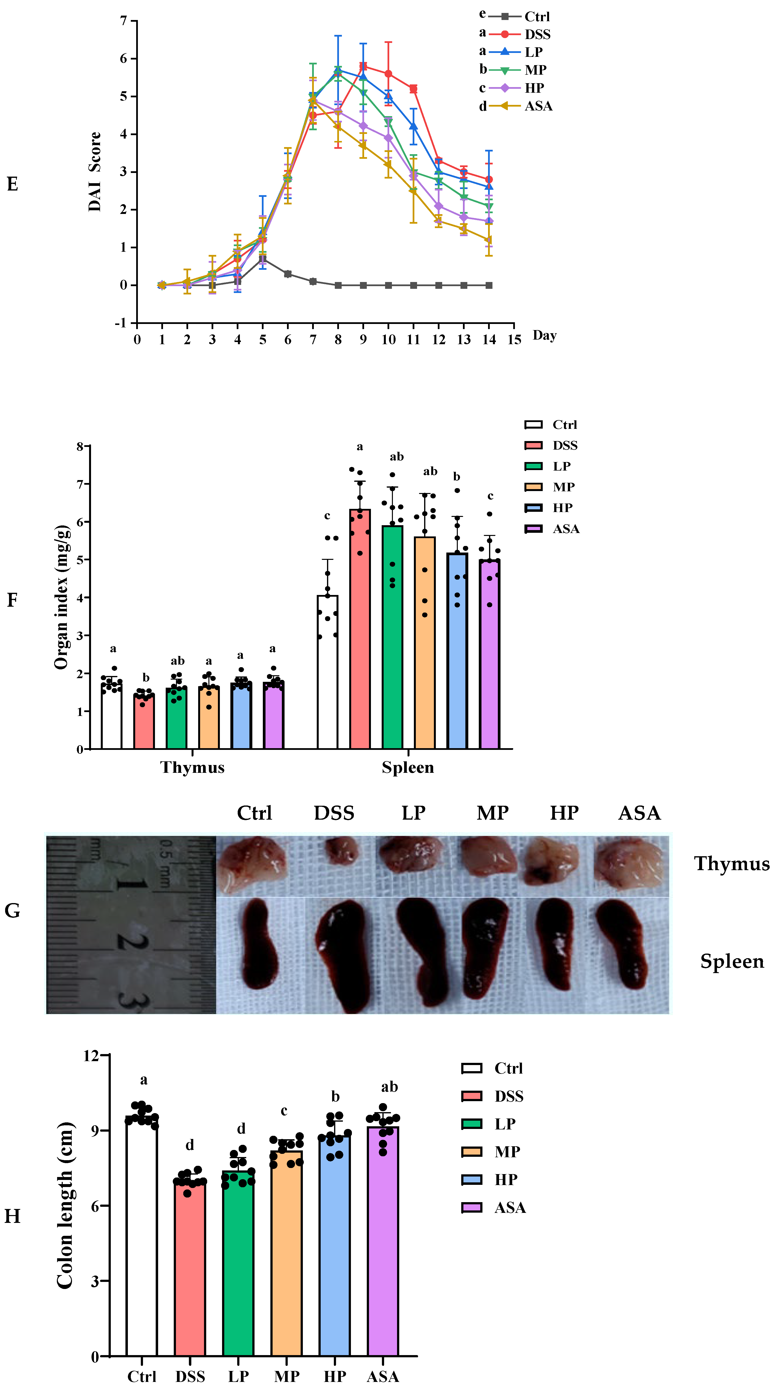
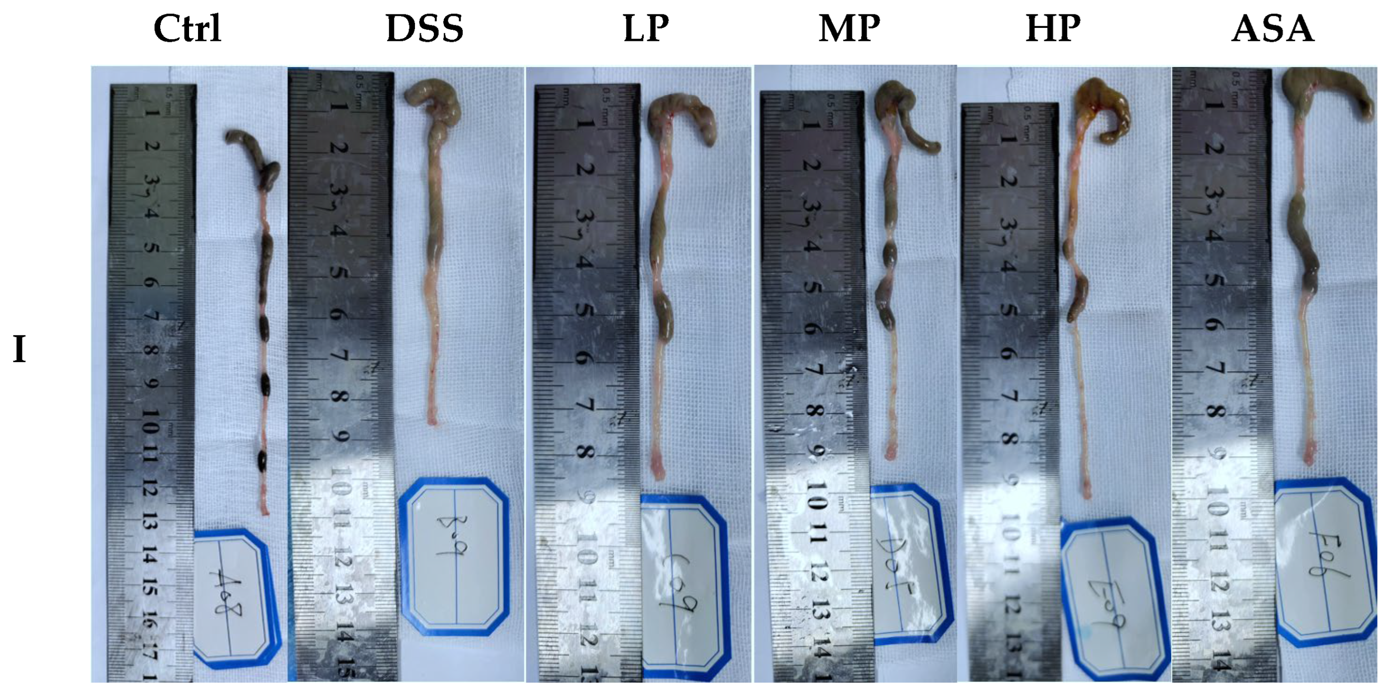
Pa JY062 significantly mitigated the progression of DSS-induced colitis, as schematically outlined in the experimental timeline (Figure 4A). The administration of DSS successfully induced colitis, as evidenced by progressive clinical manifestations such as significant body weight loss, elevated disease activity index (DAI), and reductions in food and water intake (Figure 4B–E). Following DSS withdrawal (Day 8), mice treated with Pa JY062, particularly at a high dose (HP), exhibited a significantly accelerated recovery in these clinical parameters compared to the DSS group. The HP group demonstrated comparable therapeutic efficacy to the positive control ASA (Figure 4B–E, Day 13, p < 0.001). Furthermore, Pa JY062 attenuated DSS-induced immune organ pathology, ameliorating splenomegaly (spleen index: HP 5.18 ± 95 mg/g vs. DSS 6.34 ± 0.73 mg/g, p < 0.01) and thymic atrophy (thymus index: HP 1.75 ± 0.15 mg/g vs. DSS 1.41 ± 0.11 mg/g, p < 0.01; Figure 4F,G). The HP group also exhibited significant prevention of DSS-induced colon shortening (colon length: HP 8.80 ± 0.57 cm vs. DSS 7.01 ± 0.26 cm, p < 0.01; Figure 4H,I). Hence, Pa JY062 mitigated 3% DSS-induced colitis progression.

3.5. Pa JY062 Mitigated Damage to the Colonic Barrier in DSS Mice

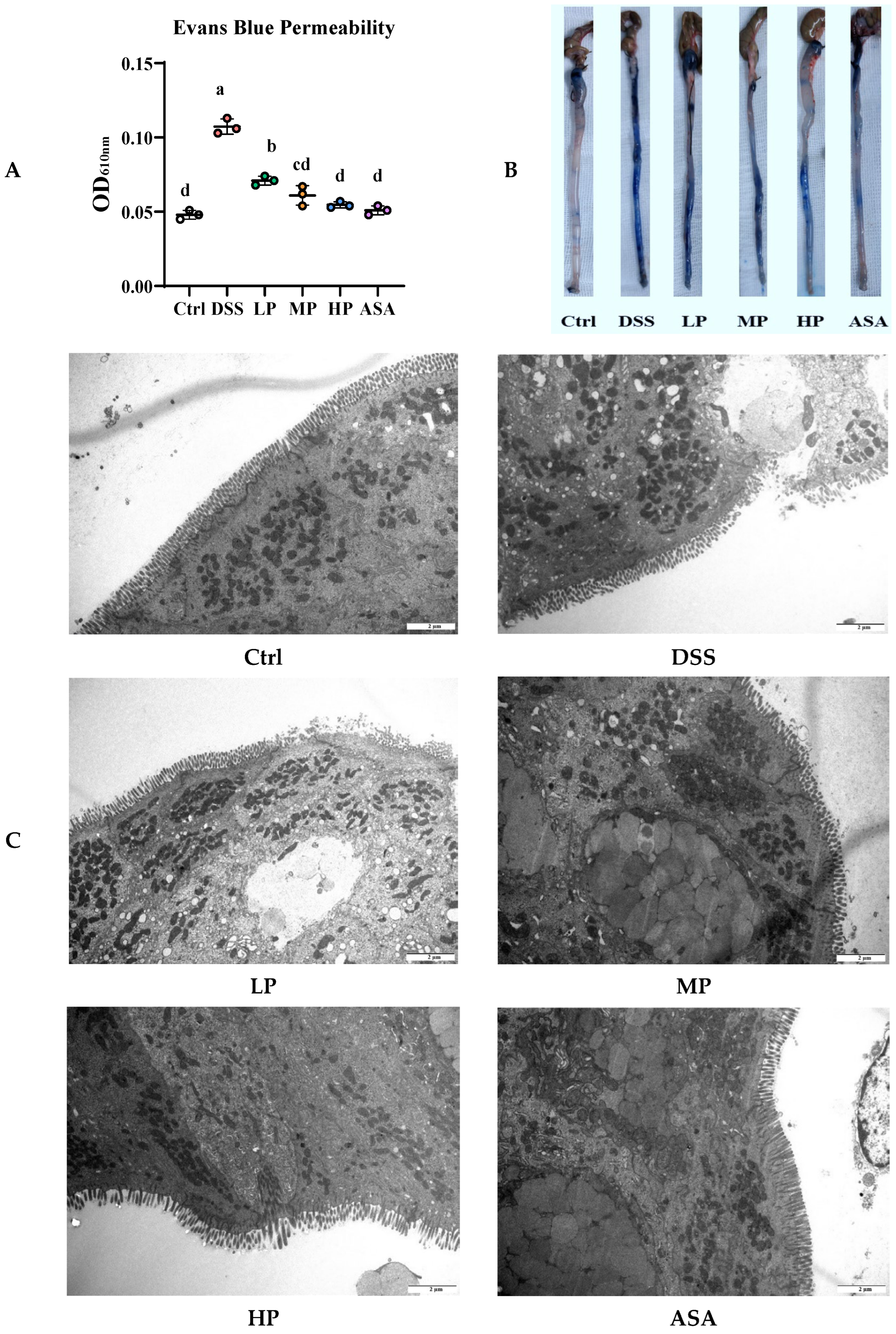
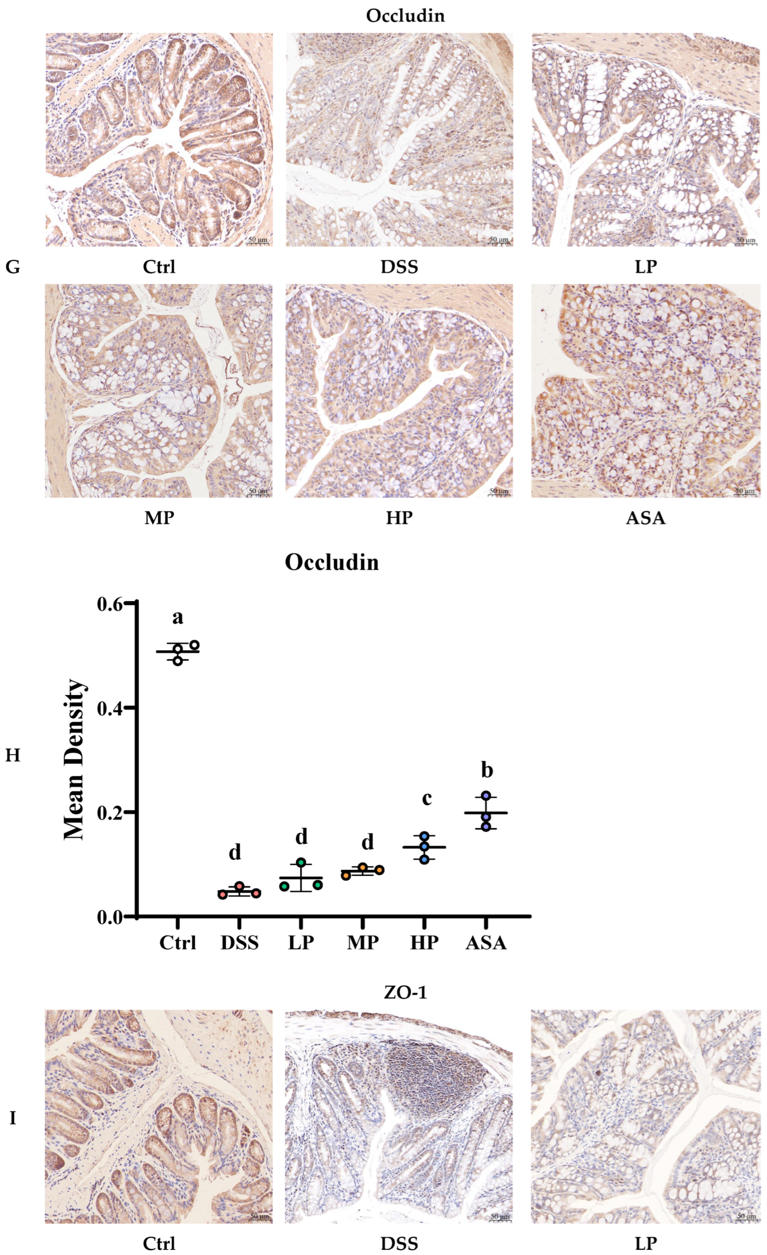
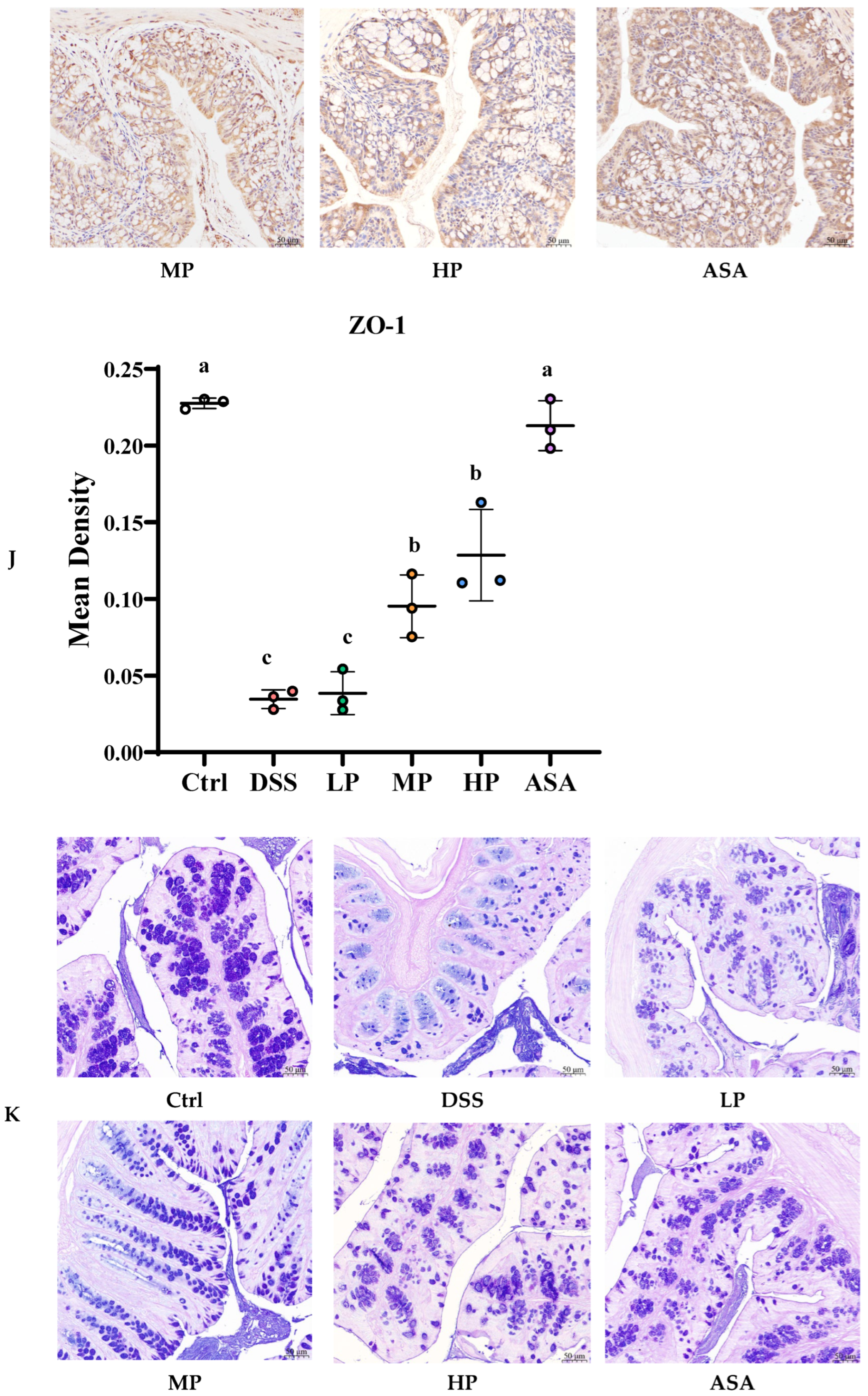
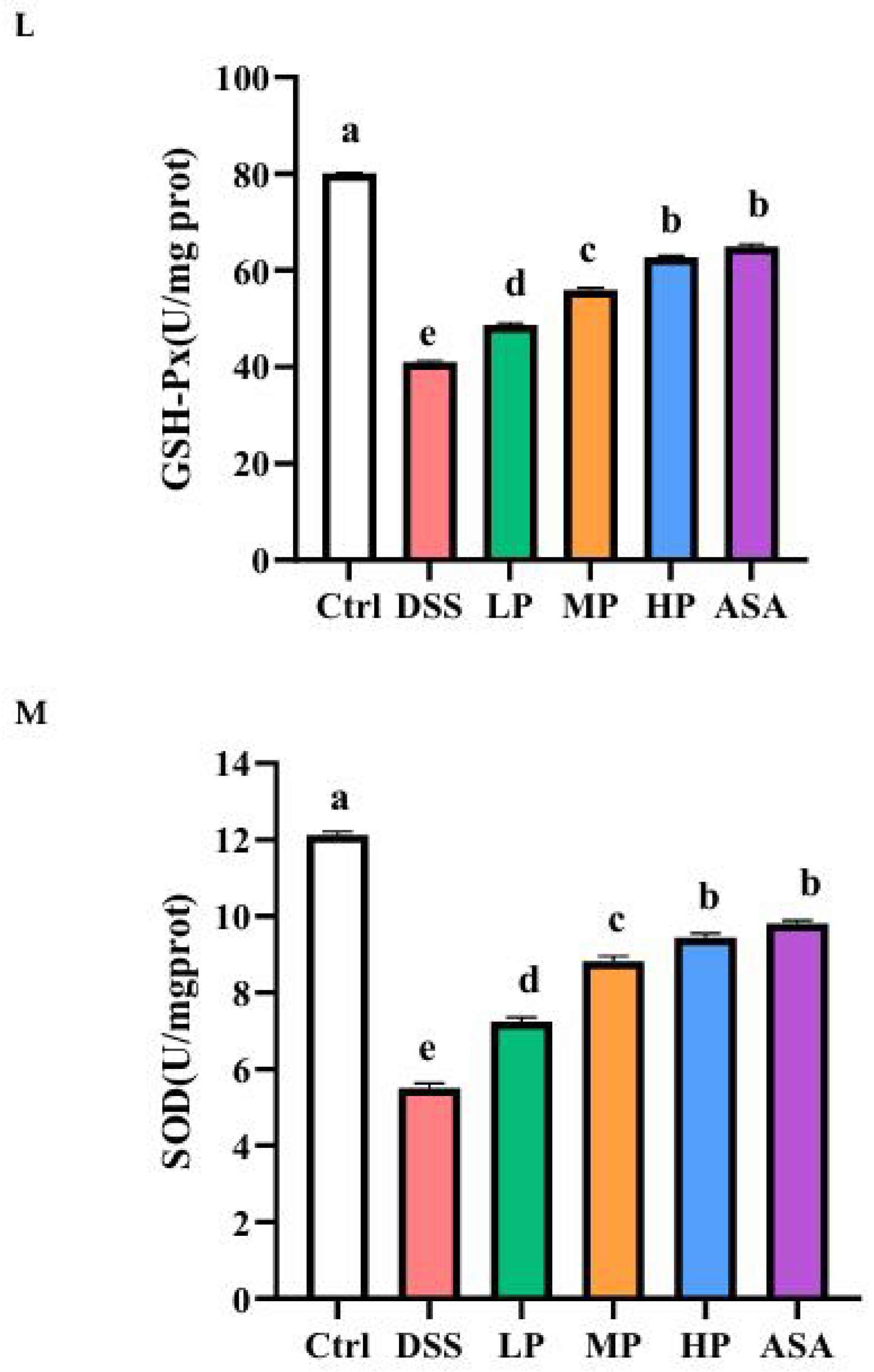
The integrity of the intestinal barrier is regulated by multiple factors. Therefore, we analyzed the protective effect of Pa JY062 on 3% DSS-induced colonic barrier damage from multiple perspectives, including Evans blue permeability, microstructure, expression of barrier regulatory proteins, and oxidative stress. Compared with the Ctrl group, the Evans blue permeability of the DSS group increased significantly (DSS: 0.1073 ± 0.005 vs. Ctrl: 0.048 ± 0.003, p < 0.01, Figure 5A,B). TEM revealed that the structural integrity of the intestinal barrier junctions was compromised, accompanied by a loss of the brush border (Figure 5C). RT-qPCR exhibited the expression of barrier junction proteins (ZO-1, Occludin, Claudin-1 and E-cadherin) decreased (Figure 5D). Immunohistochemistry (IHC) analyses further demonstrated that the Ctrl group exhibited the strongest positive staining (brown-yellow) for ZO-1, Occludin, and Claudin-1, whereas the DSS group showed a substantial reduction in the expression of these proteins (Figure 5E–J). Meanwhile, the number of goblet cells decreased (Figure 5K), and the enzyme activities of GSH-Px (DSS: 40.94 ± 0.34 U/mg prot vs. Ctrl: 80.12 ± 0.17 U/mg prot, p < 0.01, Figure 5L) and SOD (DSS: 5.5 ± 0.13 U/mg prot vs. Ctrl: 12.11 ± 0.10 U/mg prot, p < 0.01, Figure 5M) decreased significantly. The protective effect of Pa JY062 against DSS-induced intestinal barrier damage was dose-dependent, with the most pronounced amelioration observed in the high-dose (HP) group. The Evans blue permeability in the HP group was markedly reduced compared to the DSS group (HP: 0.0547± 0.0021 vs. DSS: 0.1073 ± 0.005, p < 0.01, Figure 5A,B), the colonic microvilli showed neat brush-like edges, which was similar to the ASA and Ctrl groups (Figure 5C), the expression of barrier junction proteins increased (Figure 5D, p < 0.01), the number of goblet cells increased (Figure 5K), and the enzyme activities of GSH-Px (HP: 62.74 ± 0.32 U/mg prot vs. DSS: 40.94 ± 0.34 U/mg prot, p < 0.01, Figure 5L) and SOD (HP: 9.43 ± 0.11 U/mg prot vs. DSS: 5.5 ± 0.13 U/mg prot, p < 0.01, Figure 5M) increased significantly. In conclusion, Pa JY062 alleviated DSS-induced intestinal damage by restoring microvilli architecture, enhancing the expression of junctional proteins, and reducing permeability, suggesting its therapeutic potential for maintaining intestinal homeostasis.

3.6. Pa JY062 Alleviated the Inflammatory Response Induced by DSS in Mice

UC involves a self-amplifying inflammatory cascade characterized by inflammatory infiltration, release of pro-inflammatory factors, and dysregulation of T-helper (Th) cell responses. Here, we investigated the multi-faceted mechanisms by which Pa JY062 interrupts this vicious cycle. H&E staining revealed severe mucosal damage in DSS-treated mice, characterized by crypt loss, glandular architecture disruption, and inflammatory infiltration into the basal layer (Figure 6A). Pa JY062 dose-dependently restored crypt integrity, increased goblet cell density, and attenuated leukocyte infiltration, with the HP group showing maximal efficacy. Consistent with these observations, the histopathological score of colon tissue was significantly lower in the HP group compared to the DSS group (Figure 6B, p < 0.01).
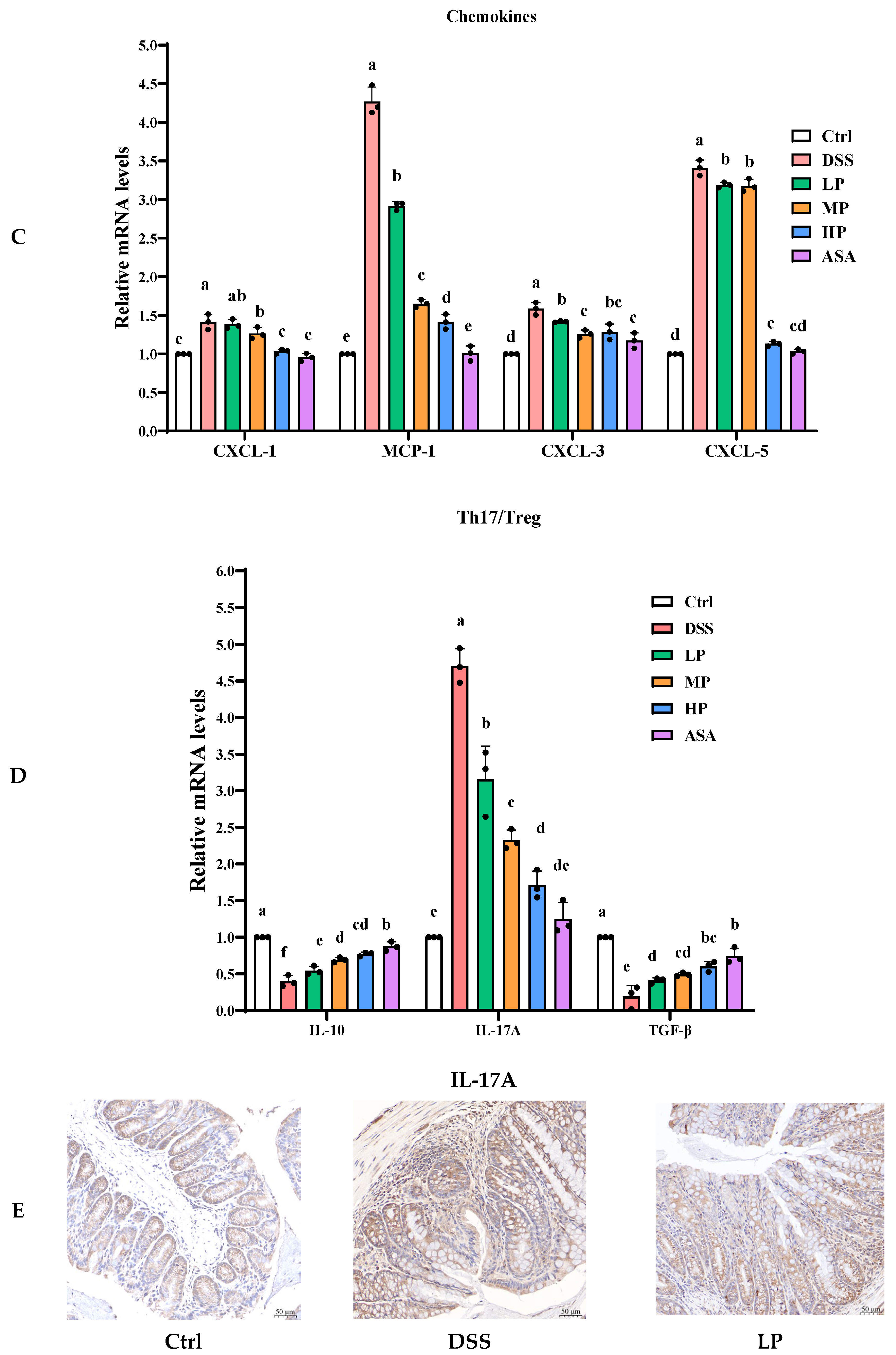
The transcript levels of chemokines demonstrated DSS-induced overexpression of CXCL-1 (1.42 ± 0.1 fold), MCP-1 (4.27 ± 0.19 fold), CXCL-3 (1.59 ± 0.08 fold), and CXCL-5 (3.41 ± 0.1 fold) versus Ctrl. Both ASA and Pa JY062 decreased chemokine hyperactivation (HP group reductions: CXCL-1 27%, MCP-1 67%, CXCL-3 19%, CXCL-5 67%; p < 0.01; Figure 6C).
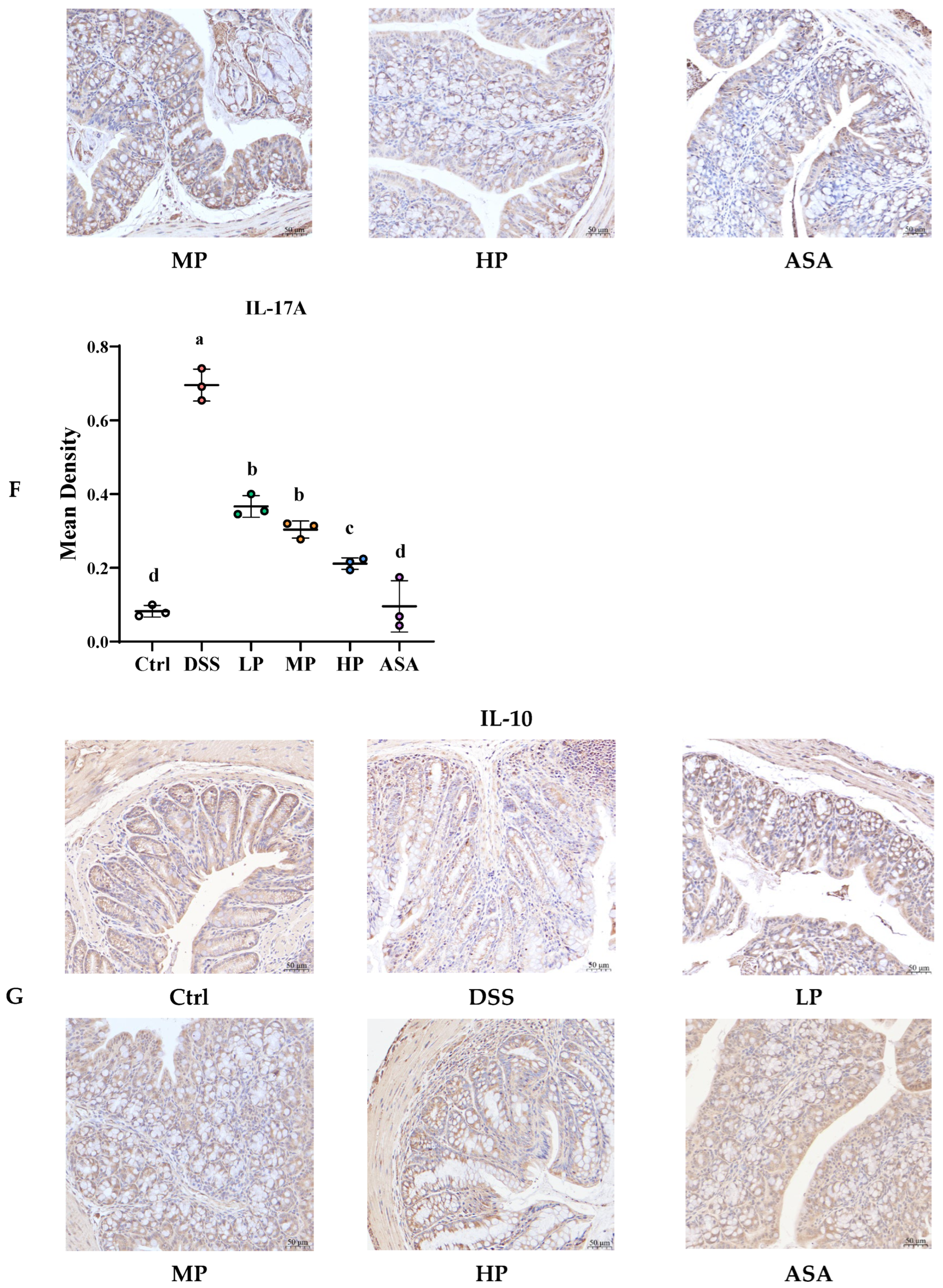
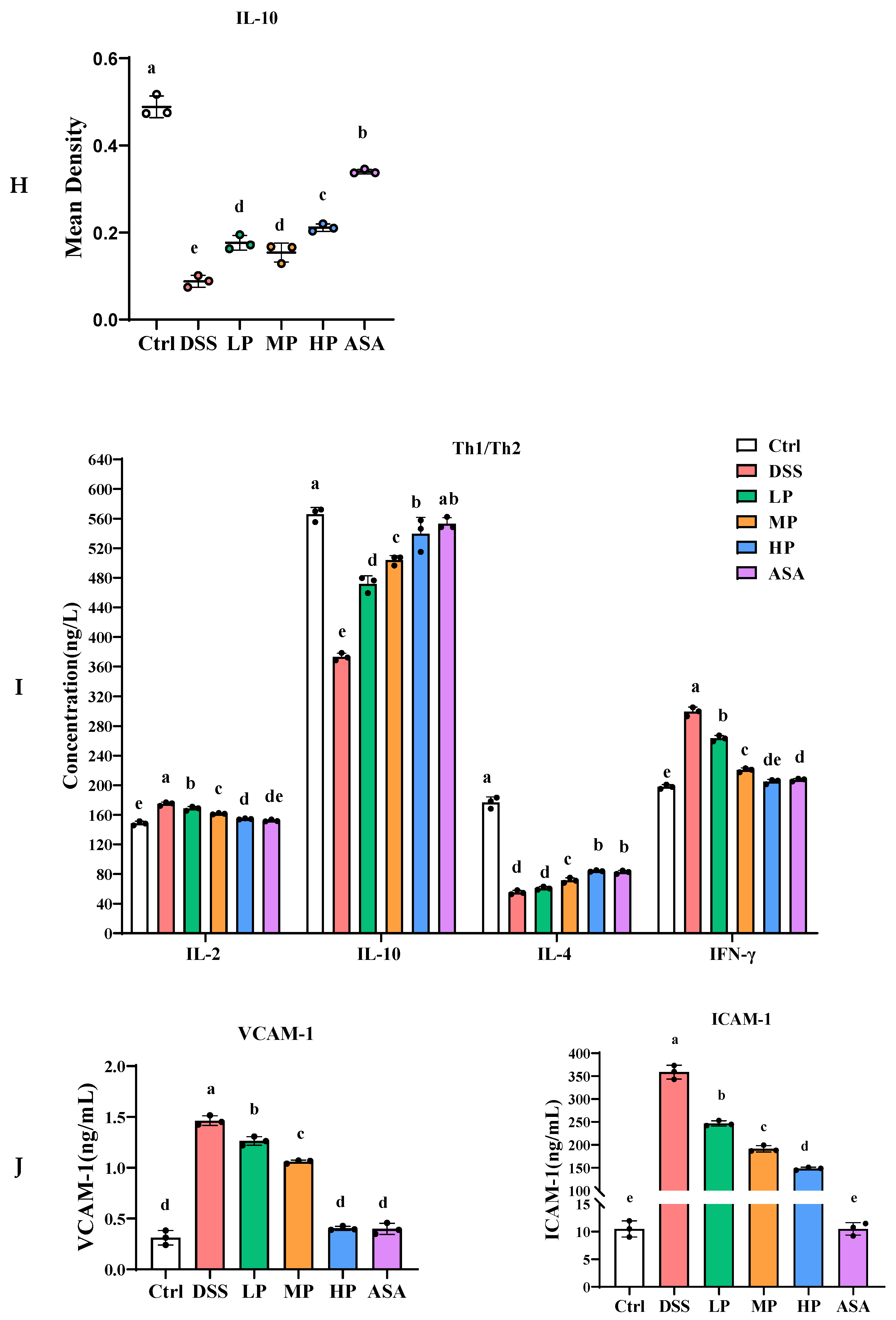
The DSS group exhibited Th17/Treg imbalance, marked by elevated IL-17A (4.7-fold vs. Ctrl) and suppressed IL-10 (0.4-fold) and TGF-β (0.2-fold). Pa JY062 restored cytokine equilibrium (HP group: IL-17A 0.4-fold, IL-10 1.9-fold, TGF-β 3.2-fold vs. DSS; p < 0.001; Figure 6D). IHC confirmed these findings, and the DSS group showed intense IL-17A immunostaining, whereas HP treatment reduced IL-17A positivity and enhanced IL-10 expression (Figure 6E–H).
ELISA further revealed a Th1/Th2 imbalance in DSS mice, indicated by significantly elevated serum levels of the Th1 cytokines IL-2 (174.89 ± 2.43 ng/L) and IFN-γ (299.44 ± 6.10 ng/L) compared to the Ctrl group (148.50 ± 2.82 and 197.89 ± 2.92 ng/L, respectively; p < 0.01). Pa JY062 normalized Th1 cytokines (HP group: IL-2 58 ± 6 ng/L, IFN-γ 35 ± 4 ng/L; p < 0.05) and elevated Th2 mediators (IL-4: 84.09 ± 1.01 ng/L; IL-10: 539.63 ± 22.03 ng/L vs. DSS 55.20 ± 2.72 and 373.08 ± 5.02 ng/L; p < 0.001; Figure 6I).
Adhesion molecule analysis showed DSS-induced upregulation of ICAM-1 (34.17-fold) and VCAM-1 (4.68-fold) compared with Ctrl, which Pa JY062 dose-dependently suppressed (HP group: ICAM-1 0.41-fold, VCAM-1 0.28-fold vs. DSS; p < 0.001; Figure 6J).
Collectively, these results demonstrate that Pa JY062 alleviates DSS-induced colitis by suppressing leukocyte infiltration, rebalancing cytokine homeostasis, and rectifying T-helper cell dysregulation.

3.7. Pa JY062 Alleviated DSS-Induced Dysbiosis of the Intestinal Microbiota and Depletion of SCFAs in Mice

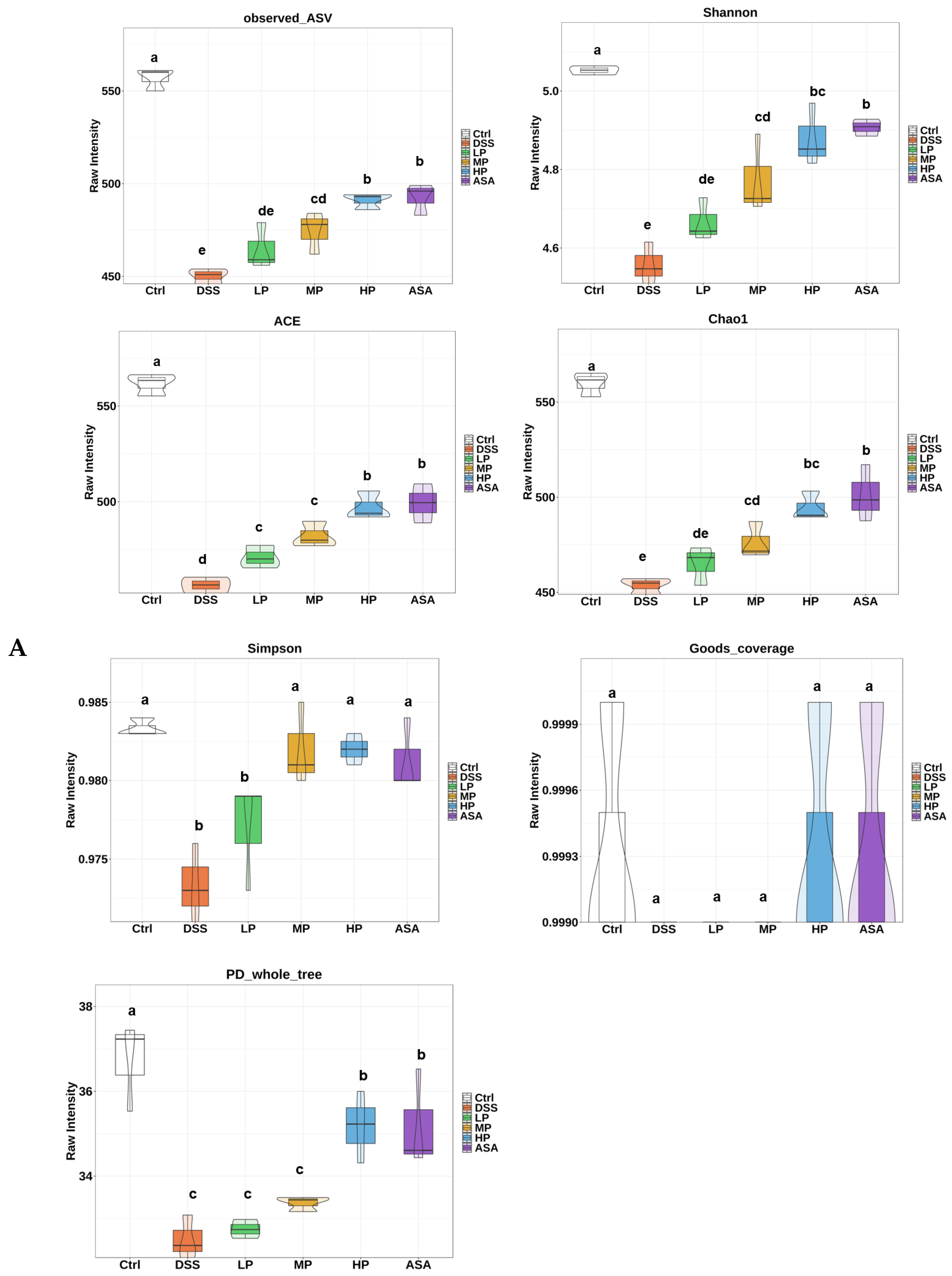
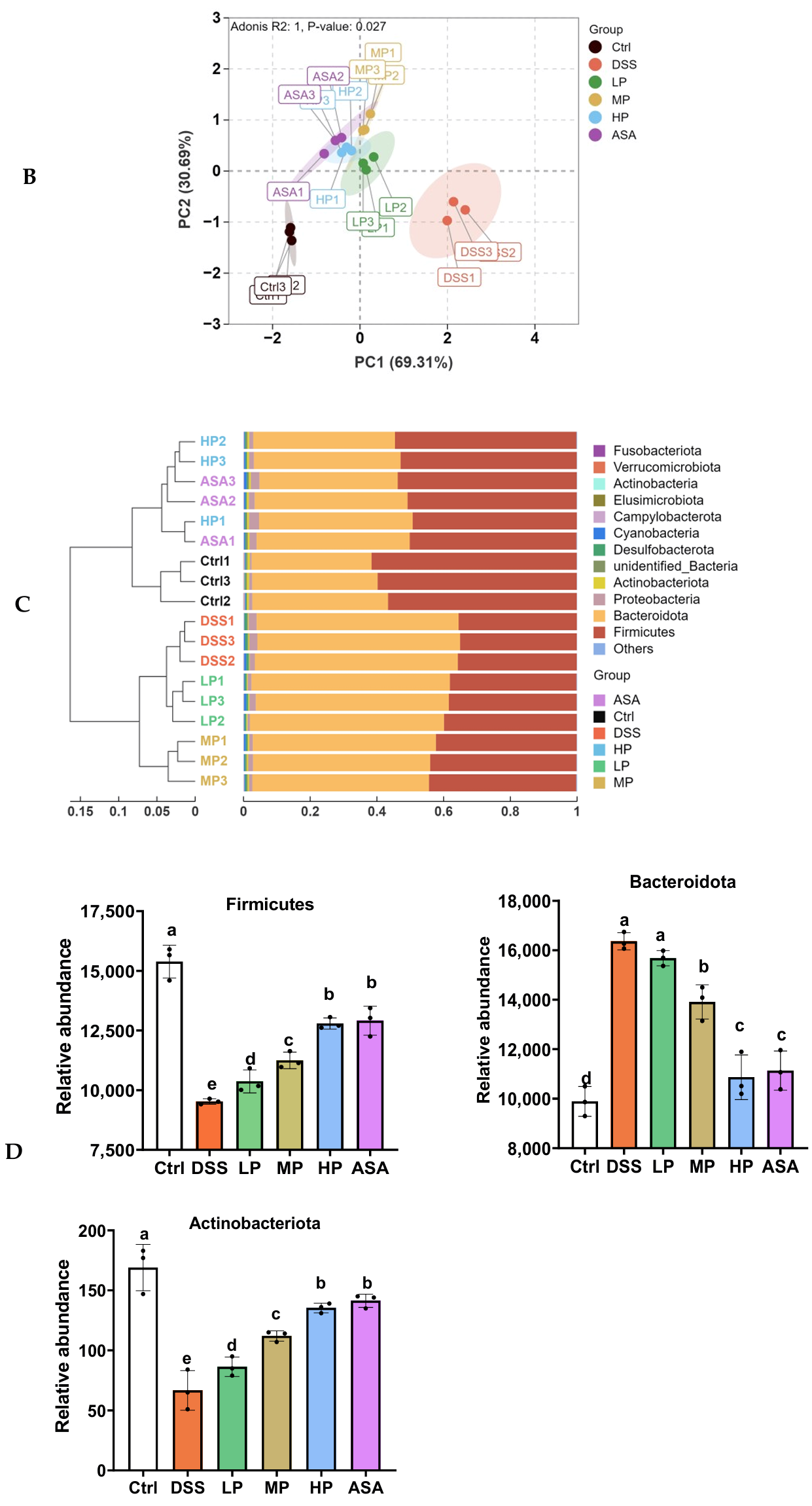
To comprehensively assess the modulatory influence of Pa JY062 on UC in mice, we investigated the changes in gut microbiota and SCFAs. The alpha diversity analysis (Figure 7A) indicated that the DSS group displayed the lowest indices of species richness (Observed ASV, Shannon, Simpson, Chao1, ACE) compared to the Ctrl group. Pa JY062 treatment dose-dependently restored these indices, with the HP group showing the most pronounced restoration (p < 0.05). The phylogenetic diversity index (PD whole tree) followed a similar trend, while goods coverage remained consistent across groups, confirming adequate sequencing depth. Beta diversity analysis via principal component analysis (PCA) demonstrated distinct clustering between the DSS and Ctrl groups. In contrast, the LP, MP, HP, and ASA groups exhibited closer proximity to the Ctrl group, with intra-group homogeneity, indicating Pa JY062 effectively mitigated DSS-induced beta diversity disruption (Figure 7B).
At the phylum level, DSS treatment significantly altered the microbial composition, notably increasing the relative abundance of Bacteroidota and decreasing that of Firmicutes and Actinobacteriota compared to the Ctrl group (p < 0.01; Figure 7C,D). These alterations were reversed in the HP group, achieving a profile comparable to the ASA group (Figure 7C,D).
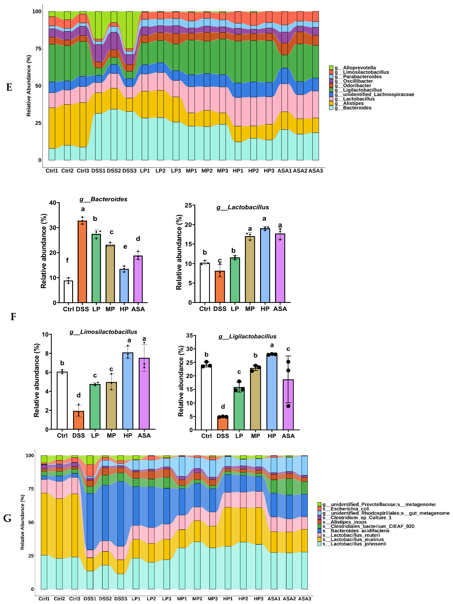
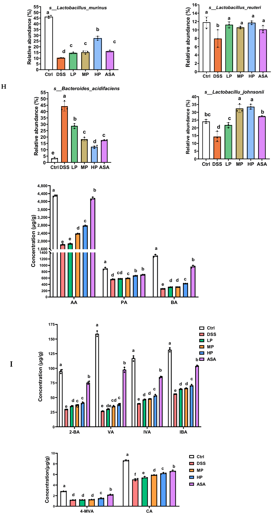
At the genus level, Pa JY062 (HP) significantly ameliorated the DSS-induced depletion of beneficial genera including Limosilactobacillus (4.18-fold), Lactobacillus (2.34-fold), and Ligilactobacillus (5.74-fold) (p < 0.05). Concurrently, it reduced the overgrowth of Bacteroides (DSS:32.78% vs. HP: 13.49%, p < 0.01) (Figure 7E,F). Species-level analysis further confirmed that HP treatment enhanced the abundance of Lactobacillus murinus (2.68-fold), Lactobacillus johnsonii (2.34-fold), and Limosilactobacillus reuteri (1.47-fold), while suppressing Bacteroides acidifaciens (27% reduction vs. DSS, p < 0.01; Figure 7G,H).
SCFAs quantification revealed that DSS treatment drastically decreased fecal acetate (AA: 1819.99 ± 22.83 vs. Ctrl 4292.28 ± 41.49 μg/g), propionate (PA: 556.65 ± 10.68 vs. 893.81 ± 34.60 μg/g), and butyrate (BA: 260.11 ± 2.42 vs. 1299.15 ± 38.11 μg/g) (p < 0.001). HP treatment restored these levels (AA: 2772.70 ± 21.08, PA: 669.84 ± 6.79, BA: 324.14 ± 0.42 μg/g) and significantly increased 2-methylbutyrate (2-BA), valerate (VA), and caproate (CA) (p < 0.01 vs. DSS) (Figure 7I).

3.8. Correlation Analysis Between Gut Microbiota and Intestinal Homeostasis

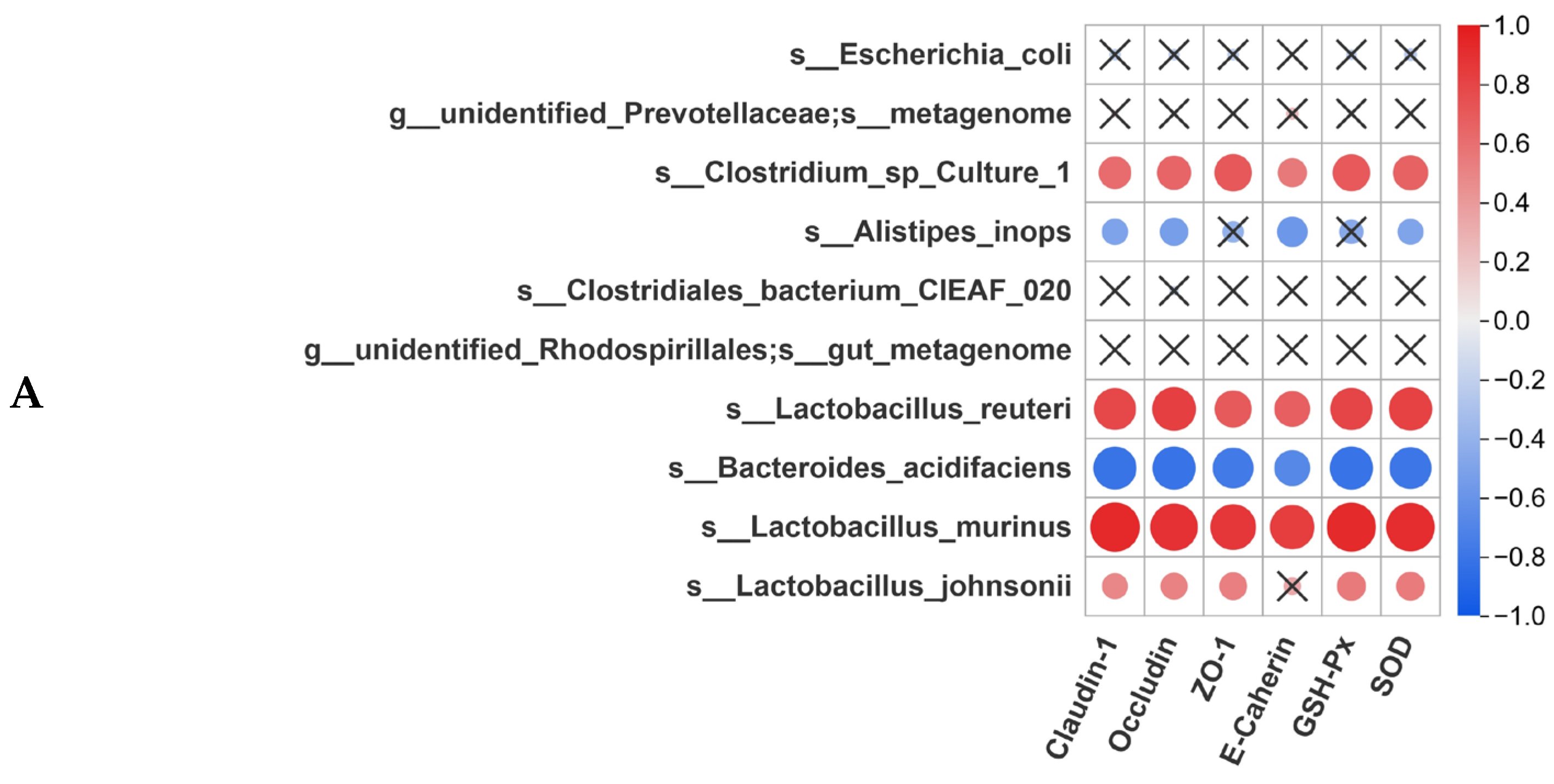
To elucidate the interplay among gut microbiota, intestinal barrier function, inflammatory factors, and SCFAs, Spearman correlation analysis was performed on 10 key microbial species, intestinal barrier proteins (Claudin-1, Occludin, ZO-1, E-Cadherin), antioxidant enzymes (GSH-Px, SOD), inflammatory factors, and SCFA levels. Claudin-1, Occludin, ZO-1, E-Cadherin, GSH-Px, and SOD exhibited strong positive correlations with L. murinus (r = 0.93–0.97, p < 0.01) and L. reuteri (r = 0.73–0.88, p < 0.01). These markers showed significant negative correlations with B. acidifaciens (r = −0.87 to −0.69, p < 0.01) (Figure 8A).
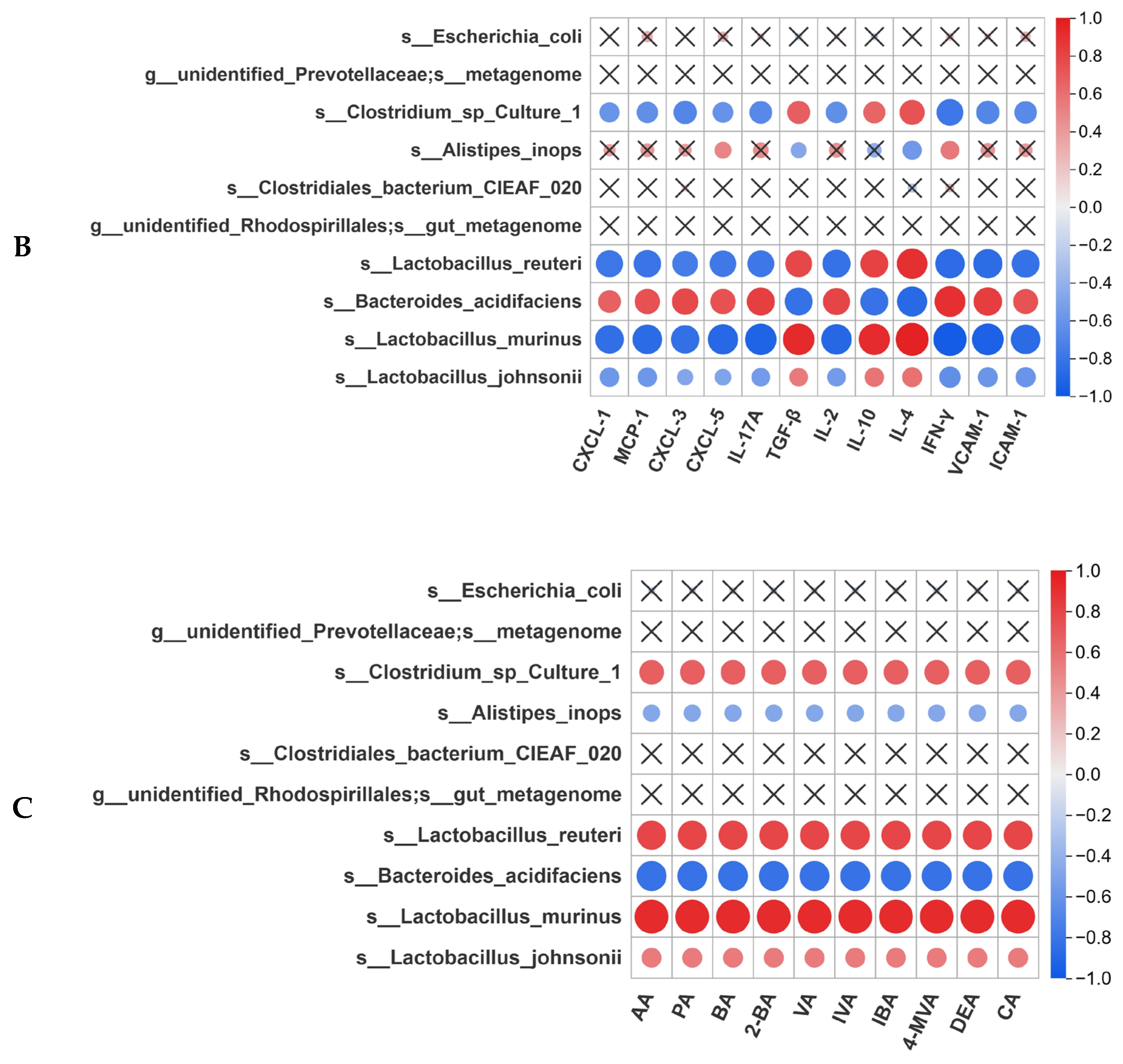
Pro-inflammatory factors (CXCL-1, MCP-1, CXCL-3, CXCL-5, IL-2, IL-17A, IFN-γ, VCAM-1 and ICAM-1) were negatively associated with L. murinus (r = −0.93 to −0.81) and L. reuteri (r = −0.77 to −0.69), while positively linked to B. acidifaciens (r = 0.63–0.88) (p < 0.001). Anti-inflammatory cytokines (IL-4, IL-10, TGF-β) were positively correlated with L. murinus (r = 0.92–0.90) and L. reuteri (r = 0.63–0.88), while negatively correlated with B. acidifaciens (r = −0.89 to −0.81) (p < 0.01) (Figure 8B).
SCFAs concentrations positively correlated with L. murinus (r = 0.91–0.95) and L. reuteri (r = 0.81–0.85), but negatively with B. acidifaciens (r = −0.94 to −0. 89) (p < 0.01 for all) (Figure 8C). These results indicate that DSS-induced colitis disrupts the proliferation of beneficial lactobacilli (L. murinus, L. reuteri) and promotes the expansion of B. acidifaciens. These collective changes are associated with impaired intestinal barrier integrity and antioxidant capacity. High-dose Pa JY062 likely alleviates colitis by restoring probiotic (L. murinus, L. reuteri), enhancing SCFAs production, and rebalancing pro-/anti-inflammatory cytokine networks (Th1/Th2 and Th17/Treg balance).

4. Discussion

UC is a multifactorial intestinal disorder mechanistically linked to excessive oxidative stress [16], compromised epithelial barrier integrity, dysregulated inflammatory cascades, and gut microbiota dysbiosis [17]. Our findings demonstrated postbiotic Pa JY062 alleviated colitis through multidimensional restoration. Pa JY062 exhibits potent antioxidant efficacy across in vitro and in vivo systems. It displayed exceptional radical scavenging capacity in vitro, as evidenced by its DPPH radical scavenging activity and total antioxidant capacity (T-AOC) (Figure 1A,B), while also maintaining low intracellular ROS levels in Caco-2 cells (Figure 2A,B). In vivo, Pa JY062 administration significantly enhanced colonic antioxidant defenses in DSS-treated mice, elevating the activities of SOD and GSH-Px (Figure 5L,M).
The DPPH radical scavenging rate of the Pa JY062 postbiotic (36.54 ± 2.9%) was significantly higher than that of other reported postbiotics, including a lysed cell preparation of Levilactobacillus brevis BK3 (30.97 ± 5.56%) [18] and a cell-free fermented liquid of Weissella cibaria ETE (35.65 ± 2.08%) [19]. This enhanced antioxidant activity can be attributed to the unique composite nature of Pa JY062. Unlike the singular compositions of the comparator postbiotics, Pa JY062 is a powdered product comprising the entire fermented material containing both inactivated bacterial cells and metabolites from the skim milk medium. We propose that this integrative composition results in a synergistic interplay among its dominant components (Table A5, Figure 3), leading to a more potent and multifaceted antioxidant capacity. Emerging evidence indicates that various organic acid metabolites, including allantoin [20], citric acid [21], 3-phenyllactic acid [22], salicylic acid [23], quinic acid [24], and stachydrine hydrochloride [25], have antioxidant properties. Furthermore, as vital constituents of Pa JY062, histidine [26], L-norvaline [27], phenylalanine [28], glycine [29], tryptophan [30], threonine [31], and serine [32] have been observed in prior studies to possess significant antioxidant activity. Furthermore, the efficacy of Pa JY062 may be augmented by its intrinsic betaine content. Betaine is a well-established osmoprotectant that safeguards intestinal epithelial cells by maintaining ion homeostasis [33]. Importantly, it also exhibits anti-inflammatory properties by inhibiting the NF-κB pathway, subsequently suppressing the expression of pro-inflammatory mediators including IL-1β, COX-2, and iNOS [34].
Since oxidative stress can damage the intestinal mucosa, we further investigated the protective effects of Pa JY062. Our findings demonstrate that 800 mg/kg Pa JY062 integrates epithelial restitution, ultrastructural repair, and junctional reinforcement into a coordinated action (Figure 5), which collectively underlies its therapeutic effect against UC. For a mouse dose of 800 mg/kg, the HED is approximately 65 mg/kg, equating to a total daily dose of 3.9 g for a 60 kg adult. A previous animal study utilizing postbiotics from Lactobacillus helveticus KLDS 1.8701 reported an effective dose of 1200 mg/kg/d in mice, which translates to a human-relevant dose of approximately 6 g/day [35]. A randomized, double-blind, crossover clinical trial demonstrated the safety and efficacy of a postbiotic preparation (Probio-Eco®) administered at a dose of 7.2 g/day for 21 days [36]. This clinical dose is significantly higher than our calculated HED of ~4 g/day, providing strong evidence for the practical acceptability and safety of our proposed dosage range in humans.
The preservation of intestinal barrier integrity is intricately associated with the accurate migration of epithelial cells [37]. Our results demonstrate that Pa JY062 not only ameliorated the DSS-induced damage and loss of microvilli (Figure 5C), but also enhanced intestinal barrier integrity by upregulating the expression of tight junction proteins (ZO-1, Occludin, Claudin-1) and the adherens junction protein E-cadherin (Figure 5D–J), while reducing Evans blue permeability (Figure 5A,B). Microvilli restoration is not merely structural, but functionally pivotal. Increasing the density and length of microvilli expands the surface area of the apical membrane, enhancing the nutrient absorption of mature intestinal epithelial cells [38]. The physical barrier, constituting the architectural core of intestinal barrier integrity, comprises polarized enterocytes interconnected via specialized junctional complexes—including TJs, AJs, and desmosomes [39]. While intestinal epithelial cells govern transcellular permeability through selective solute/water transport, TJs localized at the apical intercellular membrane domains serve as gatekeepers of paracellular permeability by forming size- and charge-selective ion channels [40]. AJs, primarily mediated by E-cadherin, orchestrate intestinal epithelial cell adhesion dynamics, epithelial restitution through coordinated proliferation/migration and barrier maintenance [41]. Notably, Lactococcus lactis HF08-derived postbiotic (P-HF08) [42], Lacticaseibacillus paracasei SNB-derived postbiotic [43], and L. reuteri DS0384 [44] also have the effects of promoting intestinal epithelial cell proliferation and repairing intestinal barrier damage. These parallels suggest a conserved barrier-repair capacity among phylogenetically distinct strains.
The compromised intestinal barrier in UC initiates a self-reinforcing inflammatory loop. Luminal pathogen translocation activates lamina propria macrophages, which secrete CXCL-1 and CXCL-5 (Figure 6C) to recruit neutrophils via ICAM-1/VCAM-1-mediated adhesion (Figure 6J). This cascade is further amplified by DSS-induced epithelial damage (Figure 6A,B). While neutrophils rapidly clear pathogens, their prolonged activation exacerbates mucosal injury through ROS overproduction [45], creating a niche for monocyte-derived macrophages to perpetuate inflammation via IL-1β. Notably, DSS disrupts the physiological inflammation–resolution axis, skewing immunity toward Th17-dominant acute inflammation (IL-17A up 4.7-fold, Figure 6D) that transitions to Th1-driven chronicity (IFN-γ increased, Figure 6I). IFN-γ binding to interferon-γ receptor (IFNGR) triggers a Janus kinase (JAK)-signal transducer and activator of transcription 1 (STAT1) signaling cascade, where JAK phosphorylates STAT1, thereby guiding macrophage polarization toward the pro-inflammatory M1 phenotype [46]. These alterations were clinically manifested as colonic shortening (Figure 4H,I), splenomegaly, and thymic involution (Figure 4F,G), reflecting systemic immune exhaustion and immunosuppression [47]. Our study elucidates that the 67% reduction in MCP-1 and CXCL-5 (Figure 6C) aligns with Pa JY062’s high salicylic acid, which inhibits NF-κB translocation by blocking IκBα phosphorylation [48]. 5-ASA is a salicylic acid derivative with anti-inflammatory properties [49]. Notably, Pa JY062 had similar effects to 5-ASA in suppressing CXCL-1/CXCL-3 (Figure 6C). Pa JY062 selectively suppressed Th17 responses (decreased IL-17A) while promoting Treg maintenance, as evidenced by elevated TGF-β levels (Figure 6D). It also fostered the differentiation of monocytes into regulatory macrophages (increased IL-10), thereby rectifying the Th17/Treg imbalance and resolving acute inflammation (Figure 6E). These findings are consistent with our previous report [10]. The clinical potential of Pa JY062 is supported by favorable comparisons with natural products that have established efficacy in UC management. For instance, in our model, Pa JY062 induced IL-10 levels (~540 ng/L) substantially exceeding those reported for curcumin (~120 ng/L) [50], which has demonstrated clinical benefits in mild-to-moderate UC [51]. Furthermore, the efficacy of natural indigo (IN) in clinical trials [52], and its mechanistic role in suppressing macrophage inflammation and MCP-1 production in mice [53], align with our finding that Pa JY062 significantly reduces colonic MCP-1 (Figure 6C). Given that Pa JY062 outperforms a clinically relevant benchmark (curcumin) in a key immunoregulatory aspect and shares a mechanistic pathway with another effective agent (IN), we posit that Pa JY062 holds significant promise for clinical translation in UC therapy.
The Pa JY062 group exhibited concurrent reduction in Th1-associated cytokines (IFN-γ and IL-2; Figure 6I) alongside an increase in Th2 markers (IL-4 and IL-10), indicating a potential shift from cell-mediated toward humoral immunity during intestinal inflammation. This rebalancing of the Th1/Th2 axis likely represents a compensatory anti-inflammatory response to mucosal damage, wherein the well-established immunosuppressive cytokine IL-10 functions to limit excessive inflammation [54]. The parallel suppression of ICAM-1/VCAM-1 (Figure 6J) further highlights its endothelial-protective role. The suppression of ICAM-1 and VCAM-1 by Pa JY062 (Figure 6J) likely confers similar therapeutic benefits: reduced VCAM-1 attenuates α4 integrin-mediated immune cell recruitment [55], while ICAM-1 inhibition suppresses neutrophil infiltration and promotes anti-inflammatory macrophage polarization [56], collectively mitigating chronic inflammation.
Meanwhile, our study provides evidence that Pa JY062 dose-dependently restores DSS-induced gut microbiota dysbiosis. The high-dose intervention not only normalized alpha and beta diversity indices (Figure 7A,B), but also reversed specific pathogenic shifts at the phylum level, namely the increased Bacteroidota/Firmicutes ratio (Figure 7C,D), a signature commonly observed in clinical UC patients [57,58]. A particularly noteworthy finding was the 5.74-fold increase in Ligilactobacillus (Figure 7E,F), a genus with well-documented anti-colitic properties. Notably, Ligilactobacillus salivarius CCFM 1266 [59], Ligilactobacillus salivarius Li01 [60], and Ligilactobacillus acidipiscis YJ5 [61] demonstrated significant therapeutic efficacy in alleviating colitis. Concurrently, Limosilactobacillus and Lactobacillus exhibited significant increases. Species-level analysis further identified L. murinus (2.68-fold), L. johnsonii (2.34-fold), and L. reuteri (1.47-fold) as the predominant species enhanced by Pa JY062 treatment (Figure 7G, H). The dominance of Lactobacillus johnsonii in HP group correlates with its unique capacity to polarize resident macrophages toward an immunoregulatory CD206+ phenotype and mediates IL-10 secretion via the TLR1/2-STAT3 signaling axis to ameliorate experimental colitis [62]. Crucially, thermal inactivation of L. johnsonii abolished its therapeutic efficacy in ameliorating murine colitis [63]. L. reuteri potentiated PD-1+ T follicular helper (Tfh) cell-dependent IgA responses, thereby restructuring the gut microbiota (e.g., Akkermansia muciniphila, AKK) and ameliorating DSS- triggered colitis and microbial imbalance [64,65]. Consistent with our findings, prior studies have demonstrated that combinatorial administration of LGG with an anti-PD-1 antibody synergistically enhanced the abundance of L. murinus. Notably, L. murinus was mechanistically linked to the activation of dendritic cells (DCs) in the murine intestinal microenvironment [66], indicating that L. murinus is important for the restoration of intestinal immunity in colitis. Hence, L. murinus, L. johnsonii, and L. reuteri play critical roles in Pa JY062-regulated colitis. SCFAs, which are metabolites produced by the gut microbiota, vary in concentration in response to alterations in microbial composition. We observed a recovery of SCFAs, particularly butyrate (from 260.11 to 324.14 μg/g; Figure 7I). The restoration of butyrate, which synergistically contributes to intestinal homeostasis [67], likely links the Pa JY062-induced microbial shifts to the observed barrier repair.
Our systematic correlation analysis establishes that Pa JY062 achieves multimodal colitis amelioration through precision remodeling of the “gut microbiota–metabolite–oxidative stress-barrier–immunity network”, distinguishing it from single-target anti-inflammatory or antibacterial strategies. L. murinus serves as a pivotal bacterium for mucosal restoration. Its abundance exhibits a robust positive correlation with barrier proteins (Claudin-1/ZO-1, r = 0.93–0.97) and antioxidant markers (Figure 8A), implying that this strain may mitigate mucosal injury by reinforcing mucus layer integrity and scavenging intestinal free radicals. This aligns with documented Lactobacillus mechanisms, including antioxidants and the modulation of the Nrf2 antioxidant pathway [68,69]. The strong correlation of both L. reuteri and L. murinus with the upregulation of tight junction proteins (ZO-1, Occludin) and E-cadherin (Figure 8A) suggests they may act in concert to reinforce the epithelial barrier.
The strong negative correlation between L. murinus abundance and pro-inflammatory factors (e.g., IL-17A and IFN-γ; r = −0.93 to −0.81, Figure 8B) suggests its potential as a quantifiable biomarker for monitoring inflammatory activity. This finding provides a new perspective for clinical prediction of inflammatory status by detecting the abundance of commensal bacteria, which is could offer a more gut-specific assessment than measuring systemic inflammatory markers in serum. The strong positive correlation between SCFAs and L. murinus/L. reuteri, coupled with the negative correlation with B. acidifaciens (Figure 8C), suggests a positive ecological feedback loop: Pa JY062 promotes beneficial lactobacilli, which produce SCFAs that in turn inhibit pathogens and further foster a healthy microbial environment. This self-reinforcing cycle may represent an advantage over some traditional probiotics, potentially moving beyond a simple “occupancy effect” by actively reshaping the microbial niche through metabolic output. Pa JY062’s therapeutic efficacy arises not from isolated mechanisms but through a trans-hierarchical cascade: Lactobacillus enrichment, oxidative stress mitigation, barrier restoration, SCFA elevation, anti-inflammatory resolution. This systems-level mode of action establishes a novel paradigm for IBD management. Critically, such multi-dimensional postbiotic targeting may mitigate therapeutic escape and systemic toxicity risks inherent to single-pathway pharmacotherapies.
While our findings provide strong preclinical evidence for the efficacy of postbiotic Pa JY062, several limitations inherent to our experimental models must be acknowledged in terms of clinical translation. First, the DSS-induced colitis model, while excellent for studying acute epithelial injury and inflammation, does not fully recapitulate the chronic, relapsing–remitting nature and complex immune dysregulation of human UC. Second, the Caco-2 monoculture system, although valuable for initial barrier assessment, lacks the immune compartment and microbiome cues of the human gut, limiting the full validation of the immunomodulatory mechanisms proposed. These limitations are compounded by the inherent complexity of the postbiotic mixture itself, which precludes attributing effects to a single constituent—a common challenge in postbiotic research.
To bridge this translational gap, we propose a clear pathway for future research, progressing from human-relevant systems like patient-derived organoids to well-controlled clinical trials in human subjects to assess safety, dosage, and therapeutic potential. Notwithstanding these limitations, our work establishes a robust foundational framework that justifies and guides these essential next steps toward clinical application.

5. Conclusions

This research illustrated that postbiotic Pa JY062 conferred multidimensional repair against DSS-induced colitis by regulating the balance of intestinal microecology. Specifically, the mRNA expression of intestinal barrier proteins (ZO-1, Claudin-1, Occludin and E-cadherin) were elevated, and the activities of antioxidant enzymes (GSH-Px, SOD) in the colon were increased, it exhibited a mitigating influence on the symptoms of colitis in mice. Meanwhile, it also decreases the infiltration of inflammatory, restoring cytokine homeostasis, and correcting Th cell dysregulation (Th1/Th2 and Treg/Th17). Further analyses indicated that Pa JY062 selectively promotes the proliferation of beneficial bacteria (L. murinus and L. reuteri) while suppressing B. acidifaciens growth, concomitant with increased SCFAs production. These findings provide compelling evidence for the probiotic potential of postbiotics in gut health and pave the way for broader therapeutic applications. However, current research remains largely confined to laboratory-scale investigations, with limited studies on large-scale industrial production optimization and cost-effectiveness. Addressing these gaps is crucial to facilitate the commercialization and widespread adoption of postbiotic-based interventions.

Author Contributions

Conceptualization, Y.Z. (Yu Zhang) and Y.J.; methodology, H.Z.; software, Y.S. (Yilin Sun) and Y.Z. (Ying Zhao); validation, K.G., H.Z. and Y.S. (Yilin Sun); formal analysis, K.G. and H.Z.; investigation, J.G.; resources, Y.S. (Yilin Sun) and (Ying Zhao); data curation, K.G.; writing—original draft preparation, J.G.; writing—review and editing, Y.S. (Yue Su); visualization, J.G.; supervision, Y.Z. (Yu Zhang) and Y.J.; project administration, Y.Z. (Yu Zhang); funding acquisition, (Yu Zhang). All authors have read and agreed to the published version of the manuscript.

Funding

This research received financial backing from the Heilongjiang Provincial Natural Science Foundation Project (LH2023C033).

Institutional Review Board Statement

The animal study protocol was approved by the Animal Ethics Committee of Northeast Agricultural University (approval number: NEAUEC20230422, approval date: 2023-11-09), ensuring strict observance of animal welfare and ethical standards.

Data Availability Statement

Data collection for this project is ongoing; however, all datasets utilized in this research can be obtained by reaching out to the authors.

Acknowledgments

The authors gratefully acknowledge Jie Cui from the College of Food Science and Light Industry, Nanjing Tech University for her careful review and constructive comments on this manuscript.

Conflicts of Interest

The authors declare no conflicts of interest.

Abbreviations

The following abbreviations are used in this manuscript:
UCUlcerative colitis
IBDInflammatory bowel disease
Pa JY062Lacticaseibacillus paracasei JY062 postbiotic
ROSReactive oxygen species
CtrlControl
DSSDextran sulfate sodium
LPLow-dose Pa JY062
MPMedium-dose Pa JY062
HPHigh-dose Pa JY062
T-SODTotal superoxide dismutase
GSH-PxGlutathione peroxidase
Th1/Th2Helper T 1 cell/Helper T 2 cell
Th17/TregHelper T 17 cell/Regulatory T cells
ZO-1Zona occludens 1
DAIDisease activity index
SCFAsShort-chain fatty acids
5-ASA5-aminosalicylic acid
TNF-αTumor necrosis factor-α
IFN-γInterferon-γ
IL-2Interleukin 2
IL-4Interleukin 4
IL-10Interleukin 10
IL-17AInterleukin 17A
VCAM-1Vascular cell adhesion molecule 1
ICAM-1Intercellular cell adhesion molecule-1
LGGLacticaseibacillus rhamnosus GG
DPPH1,1-diphenyl-2-picryl-hydrazyl radical
ODOptical density
T-AOCTotal Antioxidant Capacity
DMEMDulbecco’s modification of Eagle’s medium
DCFH-DA2′,7′-Dichlorodihydrofluorescein diacetate
PBSPhosphate-buffer saline
H&EHematoxylin and eosin
AB-PASAlcian blue periodic acid–Schiff
IHCImmunohistochemistry
RT-qPCRReal-time quantitative polymerase chain reaction
GAPDHGlyceraldehyde-3-phosphate dehydrogenase
HRPHorseradish peroxidase
CXCL-1/3/5C-X-C Motif Chemokine Ligand 1/3/5
MCP-1Monocyte chemoattractant protein-1
TGF-βTransforming growth factor-β
ELISAEnzyme-linked immunosorbent assay
PCAPrincipal component analysis
IECsIntestinal epithelial cells

Appendix A

Appendix A.1. Evaluation of Oxidative Stress

Appendix A.1.1. Assay of Glutathione Peroxidase (GSH-PX)

Colon tissues were dissected into 5 mm2 fragments, flash-frozen in liquid nitrogen, and homogenized in ice-cold physiological saline (1:9 w/v ratio). After low-temperature centrifugation (3500 rpm, 10 min), the supernatant was collected. Protein concentrations were quantified via a bicinchoninic acid (BCA) assay kit.
For enzymatic assays, 0.2 mL of 1 mmol/L GSH and 0.2 mL homogenate were added to the enzyme group, while the non-enzyme control received only 0.2 mL GSH. Both tubes were preheated (37 °C, 5 min), followed by sequential addition of 0.1 mL Reagent 1 (37 °C incubation, 5 min) and 2 mL Reagent 2. Post-centrifugation (4000 rpm, 10 min), 1 mL of supernatant was transferred for chromogenic reaction. Blank (1.0 mL standard solvent), standard (1.0 mL 20 μmol/L GSH), and sample tubes (1.0 mL supernatant) were treated with Reagents 3 (1.0 mL), 4 (0.25 mL), and 5 (0.05 mL), incubated at 25 °C for 15 min, and quantified via OD412nm measurements.
GSH-Px activity was calculated as
Activity (U/mg protein) = (Anon-enzymeAenzyme/(AstandardAblank) × (Cstandard × N)/(T × (Vsample × Cpr))
Parameters:
Cstandard: GSH standard solution concentration (20 μmol/L) in chromogenic reaction
N: dilution factor (fixed at 5);
T: enzymatic reaction duration (5 min);
Vsample: homogenate volume in reaction (0.2 mL);
Cpr: homogenate protein concentration (mg protein/mL).

Appendix A.1.2. Assay of Superoxide Dismutase (SOD)

Colon tissues were processed identically to the GSH-Px assay. Assay tubes received sequential additions of 1.0 mL reagent 1, 0.05 mL sample, and 0.1 mL each of reagents 2–4, whereas control tubes substituted the sample with 0.05 mL distilled water. Samples were vortex-mixed thoroughly and incubated at 37 °C for 40 min. Post-incubation, 2 mL chromogenic developer was added to each tube with immediate mixing, followed by 10 min stabilization at 20–25 °C. Absorbance at 550 nm was measured against a distilled water blank.
Total SOD activity (U/mgprot) = (ODcontrolODdetermination)/(ODcontrol) × (Vtotal)/(50% × Vsample × Cpr)
Parameters:
Vtotal: total reaction volume (mL)
Vsample: sample volume in reaction (mL)
Cpr: homogenate protein concentration (mgpro/mL)

Appendix A.2. 16S rRNA Analysis

Appendix A.2.1. Alpha Diversity

Alpha diversity is applied in analyzing complexity of species diversity for a sample through 6 indices, including observed-species, Chao1, Shannon, Simpson, ACE, good-coverage. All these indices in our samples were calculated with QIIME 2 (version 2024.5) and displayed with R software (Version 4.1.2).
Two indices were selected to identify community richness:
Chao—the Chao1 estimator (http://www.mothur.org/wiki/Chao (accessed on 25 July 2024));
ACE—the ACE estimator (http://www.mothur.org/wiki/Ace (accessed on 25 July 2024));
Two indices were used to identify community diversity:
Shannon—the Shannon index (http://www.mothur.org/wiki/Shannon (accessed on 25 July 2024));
Simpson—the Simpson index (http://www.mothur.org/wiki/Simpson (accessed on 25 July 2024));
One indice was used to characterized Sequencing depth:
Coverage—the Good’s coverage (http://www.mothur.org/wiki/Coverage (accessed on 25 July 2024)).

Appendix A.2.2. Beta Diversity

Beta diversity analysis was used to evaluate differences in samples in species complexity, Beta diversity on both weighted and unweighted unifrac were calculated by QIIME software. Cluster analysis was preceded by principal component analysis (PCA), which was applied to reduce the dimension of the original variables using the stats package and ggplot2 package in R software. Unweighted pair-group method with arithmetic means (UPGMA) clustering was performed as a type of hierarchical clustering method to interpret the distance matrix using average linkage and was conducted by QIIME software.

Appendix A.2.3. Analysis of the Relative Abundance

The relative abundance of gut microbes across experimental groups at the phylum, genus, and species levels were conducted on the Metware Cloud platform (https://cloud.metware.cn (accessed on 15 March 2024)) using R v4.1.2 and ggplot2 v3.3.5.

Appendix A.3. Detection of Short-Chain Fatty Acids

Appendix A.3.1. Chemicals and Reagents

Methyl tert-butyl ether (MTBE) were purchased from CNW (CNW Technologies, Düsseldorf, Germany). MilliQ water (Millipore, PA, USA) was used in all experiments. All of the standards were purchased from CNW (CNW, Beijing, China) or aladdin (Shanghai, China). The stock solutions of standards were prepared at the concentration of 1 mg/mL in MTBE. All stock solutions were stored at −20 °C. The stock solutions were diluted with MTBE to working solutions before analysis.

Appendix A.3.2. Sample Preparation and Extraction

A total of 20 mg of fecal sample were accurately weighed and placed in a 2 mL EP tube. 1 mL of phosphoric acid (0.5% v/v) solution and a small steel ball were added to the EP tube. The samples were ground uniformly, then vortexed for 10 min and ultrasonicated for 5 min. 100 μL of supernatant was moved into 1.5 mL centrifugal tube after the mixture was centrifuged with a speed of 12,000 r/min for 10 min at 4 °C. 500 μL of MTBE (containing internal standard) solution was added to the centrifugal tube and the mixture was vortexed for 3 min followed by ultrasonicating for 5 min. After that, the mixture was centrifuged with a speed of 12,000 r/min for 10 min at 4 °C. The supernatant was collected and used for GC-MS/MS analysis.

Appendix A.3.3. GC–MS Analysis

An Agilent 7890B gas chromatograph coupled to a 7000D mass spectrometer with a DB-FFAP column (30 m length × 0.25 mm i.d. × 0.25 μm film thickness, J&W Scientific, CA, USA) was employed for GC-MS/MS analysis of SCFAs. Helium was used as carrier gas, at a flow rate of 1.2 mL/min. Injection was made in the split mode with a split ratio 5:1 and the injection volume was 1 μL. The oven temperature was held at 50 °C for 1 min, raised to 220 °C at a rate of 18 °C/min and held for 5 min. All samples were analyzed in multiple reaction monitoring mode. The injector inlet and transfer line temperature were 250 °C and 230 °C, respectively.
Table A1. Subsequent operations of total antioxidant capacity (T-AOC).
Title 1Blank WellStandard WellTest Well
Sterile water (μL)10  
0.1, 0.2, 0.4, 0.8, and 1.0 mM Trolox (μL) 10 
Samples (μL)  10
Reagent IV application solution (μL)202020
ABTS working solution (μL)170170170
Table A2. Operation steps of DPPH radical scavenging activity in EP tube.
Title 1Control TubeSample TubeBlank Tube
Sample (μL)400400 
80% methanol (μL)600 400
Working solution (μL) 600600
Table A3. RNA concentration.
SamplesRNA Concentration (ng/μL)
Ctrl 1875
Ctrl 2937
Ctrl 3911
LPS 11062
LPS 21369
LPS 31136
LP 1854
LP 2761
LP31329
MP 11291
MP 21365
MP 31555
HP 1955
HP 21029
HP 31422
ASA-1852
ASA-21036
ASA-31112
Table A4. RT-qPCR primers.
Primer NameForward Primer (5′-3′)Reverse Primer (5′-3′)
Zo-1TGAGGCAGCTCACATAATGCGGTCTCTGCTGGCTTGTTTC
OccludinAAAGGGCATTGCTCATCCTGAACAATGGCAATGGCAATTCATC
Claudin-1CCAGTCAATGCCAGGTACGAATGGCCTTGGTGTTGGGTAAGA
E-cadherinCCCAAACGTAACGAGGGTATCGGCAGCTTGAAGTGGTAGAAGT
CXCL-1TGCACCCAAACCGAAGTCATACTTGGGGACACCTTTTAGCAT
MCP-1CAGGTCCCTGTCATGCTTCTCCCATTCCTTCTTGGGGTCA
CXCL-3TGAGGCAGTATTCCTTGGCTGACCGGCATGACCTTGTTTGT
CXCL-5TCCTCAGTCATAGCCGCAACTAGCTTTCTTTTTGTCACTGCCC
IL-10GGGTTGCCAAGCCTTGTCTGAGCCTTGATGTCTGGGTCTTGGTTCTC
IL-17AGTTAGGGTGCTTTAGGTCCTAACAATGAGTTTCTGTACG
TGF-βTACAGCAACAATTCCTGGCGATACCCTCAACCACTGCCGCACAACTC
GAPDHGAGAAGGCTGGGGCTCATTTTAAGCAGTTGGTGGTGCAGG
Table A5. Postbiotic components of PaJY062 with active function.
CompoundsFunctionReferences
1AllantoinAntioxidant; anti-inflammatory[70]
2TrehaloseAntioxidant; anti-inflammatory; repair the intestinal barrier[71]
3HistidineAntioxidant; anti-inflammatory; repair the intestinal barrier[26,72]
4L-NorvalineAnti-inflammatory[73]
5PhenylalanineAntioxidant; anti-inflammatory; repair the intestinal barrier[74]
6GlycineAntioxidant; anti-inflammatory[75,76]
7TryptophanAnti-inflammatory[77]
8BetaineAntioxidant; anti-inflammatory; repair the intestinal barrier[78,79]
9Citric acidAntioxidant[80]
103-phenyllactic acidAntioxidant[81]
11ThreonineAntioxidant; anti-inflammatory; repair the intestinal barrier[82]
12Benzoic acidRepair the intestinal barrier[83]
13BiotinAnti-inflammatory; repair the intestinal barrier[84]
14SerineAnti-inflammatory; repair the intestinal barrier[85]
15MannoseAntioxidant; anti-inflammatory; repair the intestinal barrier; modulate the gut microbiota[86]
16OleamideAnti-inflammatory[87]
17GlutamineAnti-inflammatory; repair the intestinal barrier; modulate the gut microbiota[88]
18TaurineAntioxidant; anti-inflammatory; repair the intestinal barrier[89]
19Salicylic acidAnti-inflammatory; repair the intestinal barrier[90]
20HexadecanamideAnti-inflammatory[91]
21AdenosineAntioxidant; anti-inflammatory; repair the intestinal barrier[92]
22Quinic acidAntioxidant; anti-inflammatory; repair the intestinal barrier[93]
23HypotaurineAntioxidant[94]
24Stachydrine hydrochlorideAnti-inflammatory[95]
25DimethylglycineAnti-inflammatory; repair the intestinal barrier; modulate the gut microbiota[96]
26TrigonellineAnti-inflammatory[97]
27AnserineAntioxidant[98]
28NiacinamideAntioxidant; anti-inflammatory[99]
294-hydroxybenzaldehydeAntioxidant[100]
3017α-estradiolAnti-inflammatory[101]
31Palmitoyl ethanolamideAnti-inflammatory[102]

Appendix B

Figure A1. The standard curve. (A) DPPH scavenging rate. (B) total antioxidant capacity.
Antioxidants 14 01256 g0a1

References

  1. Arnauts, K.; Sudhakar, P.; Verstockt, S.; Lapierre, C.; Potche, S.; Caenepeel, C.; Verstockt, B.; Raes, J.; Vermeire, S.; Sabino, J.; et al. Microbiota, not host origin drives ex vivo intestinal epithelial responses. Gut Microbes 2022, 14, 2089003. [Google Scholar] [CrossRef] [PubMed]
  2. Zhou, X.; Yu, M.; Ma, L.; Fu, J.; Guo, J.; Lei, J.; Fu, Z.; Fu, Y.; Zhang, Q.; Zhang, C.Y.; et al. In vivo self-assembled siRNA as a modality for combination therapy of ulcerative colitis. Nat. Commun. 2022, 13, 5700. [Google Scholar] [CrossRef]
  3. Wang, D.; Jiang, Q.; Shen, R.; Peng, L.; Zhou, W.; Meng, T.; Hu, F.; Wang, J.; Yuan, H. ROS-responsive nanoparticles targeting inflamed colon for synergistic therapy of inflammatory bowel disease via barrier repair and anti-inflammation. Nano Res. 2024, 17, 5409–5423. [Google Scholar] [CrossRef]
  4. Peluzio, M.d.C.G.; Martinez, J.A.; Milagro, F.I. Postbiotics: Metabolites and mechanisms involved in microbiota-host interactions. Trends Food Sci. Technol. 2021, 108, 11–26. [Google Scholar] [CrossRef]
  5. Salminen, S.; Collado, M.C.; Endo, A.; Hill, C.; Lebeer, S.; Quigley, E.M.; Sanders, M.E.; Shamir, R.; Swann, J.R.; Szajewska, H. The International Scientific Association of Probiotics and Prebiotics (ISAPP) consensus statement on the definition and scope of postbiotics. Nat. Rev. Gastroenterol. Hepatol. 2021, 18, 649–667. [Google Scholar] [CrossRef]
  6. Pang, L.; Huang, Y.; Li, R.; Guo, L.; Man, C.; Yang, X.; Jiang, Y. Effects of postbiotics produced by Lactobacillus plantarum JM015 isolated from traditional fermented dairy products on Salmonella-induced intestinal inflammation: A preventive strategy. Food Chem. 2025, 469, 142549. [Google Scholar] [CrossRef]
  7. Nie, X.; Li, Q.; Ji, H.; Zhang, S.; Wang, Y.; Xie, J.; Nie, S. Bifidobacterium longum NSP001-derived extracellular vesicles ameliorate ulcerative colitis by modulating T cell responses in gut microbiota-(in)dependent manners. npj Biofilms Microbiomes 2025, 11, 27. [Google Scholar] [CrossRef] [PubMed]
  8. Fang, Z.; Yang, X.; Shang, L. Microfluidic-derived montmorillonite composite microparticles for oral codelivery of probiotic biofilm and postbiotics. Sci. Adv. 2025, 11, eadt2131. [Google Scholar] [CrossRef]
  9. Guo, J.; Zhao, Y.; Guo, W.; Sun, Y.; Zhang, W.; Zhao, Q.; Zhang, Y.; Jiang, Y. Effects of Lactobacillus paracei JY062 Postbiotic on Intestinal Barrier, Immunity, and Gut Microbiota. Nutrients 2025, 17, 1272. [Google Scholar] [CrossRef]
  10. Su, Y.; Cui, Z.; Yang, X.; Jiang, Y.; Zhang, W.; Zhang, Y.; Man, C. Lactobacillus paracasei JY062 and its exopolysaccharide enhance the intestinal barrier through macrophage polarization and Th17/Treg cell balance. Food Res. Int. 2024, 197, 115235. [Google Scholar] [CrossRef] [PubMed]
  11. Zhu, S.; Zhang, J.; Jiang, X.; Wang, W.; Chen, Y.Q. Free fatty acid receptor 4 deletion attenuates colitis by modulating Treg Cells via ZBED6-IL33 pathway. EBioMedicine 2022, 80, 104060. [Google Scholar] [CrossRef]
  12. Rizvi, Z.A.; Dalal, R.; Sadhu, S.; Kumar, Y.; Kumar, S.; Gupta, S.K.; Tripathy, M.R.; Rathore, D.K.; Awasthi, A. High-salt diet mediates interplay between NK cells and gut microbiota to induce potent tumor immunity. Sci. Adv. 2021, 7, eabg5016. [Google Scholar] [CrossRef]
  13. Wang, Y.; Lai, W.; Zheng, X.; Li, K.; Zhang, Y.; Pang, X.; Gao, J.; Lou, Z. Linderae Radix extract attenuates ulcerative colitis by inhibiting the JAK/STAT signaling pathway. Phytomedicine 2024, 132, 155868. [Google Scholar] [CrossRef]
  14. Shi, C.; Dawulieti, J.; Shi, F.; Yang, C.; Qin, Q.; Shi, T.; Wang, L.; Hu, H.; Sun, M.; Ren, L.; et al. A nanoparticulate dual scavenger for targeted therapy of inflammatory bowel disease. Sci. Adv. 2022, 8, eabj2372. [Google Scholar] [CrossRef] [PubMed]
  15. de Jesus, L.C.L.; Freitas, A.d.S.; Dutra, J.d.C.F.; Campos, G.M.; Américo, M.F.; Laguna, J.G.; Dornelas, E.G.; Carvalho, R.D.d.O.; Vital, K.D.; Fernandes, S.O.A.; et al. Lactobacillus delbrueckii CIDCA 133 fermented milk modulates inflammation and gut microbiota to alleviate acute colitis. Food Res. Int. 2024, 186, 114322. [Google Scholar] [CrossRef]
  16. Zhang, L.; Gui, S.; Xu, Y.; Zeng, J.; Wang, J.; Chen, Q.; Su, L.; Wang, Z.; Deng, R.; Chu, F.; et al. Colon tissue-accumulating mesoporous carbon nanoparticles loaded with Musca domestica cecropin for ulcerative colitis therapy. Theranostics 2021, 11, 3417–3438. [Google Scholar] [CrossRef]
  17. Li, H.; Zhang, Y.; Liu, M.; Fan, C.; Feng, C.; Lu, Q.; Xiang, C.; Lu, H.; Yang, X.; Wu, B.; et al. Targeting PDE4 as a promising therapeutic strategy in chronic ulcerative colitis through modulating mucosal homeostasis. Acta Pharm. Sinica. B 2022, 12, 228–245. [Google Scholar] [CrossRef] [PubMed]
  18. Lee, Y.S.; Lee, S.J.; Jang, W.J.; Lee, E.W. Protective Effects of the Postbiotic Levilactobacillus brevis BK3 against H(2)O(2)-Induced Oxidative Damage in Skin Cells. J. Microbiol. Biotechnol. 2024, 34, 1401–1409. [Google Scholar] [CrossRef] [PubMed]
  19. Aydın, B.; Çiydem, T.; Kaya, E.; Açık, L. Evaluation of the antioxidant effects of postbiotics and paraprobiotics in lactic acid bacteria isolated from traditional fermented sausages. Avrupa Bilim Ve Teknol. Derg. 2021, 849–852. [Google Scholar] [CrossRef]
  20. Wang, J.; Li, Y.; Wang, Y.; Du, F.; Zhang, Y.; Yin, M.; Zhao, X.; Xu, J.; Yang, Y.; Wang, W.; et al. Transcriptome and Metabolome Analyses Reveal Complex Molecular Mechanisms Involved in the Salt Tolerance of Rice Induced by Exogenous Allantoin. Antioxidants 2022, 11, 2045. [Google Scholar] [CrossRef]
  21. Ryabova, A.; Cornette, R.; Cherkasov, A.; Watanabe, M.; Okuda, T.; Shagimardanova, E.; Kikawada, T.; Gusev, O. Combined metabolome and transcriptome analysis reveals key components of complete desiccation tolerance in an anhydrobiotic insect. Proc. Natl. Acad. Sci. USA 2020, 117, 19209–19220. [Google Scholar] [CrossRef]
  22. Martí-Quijal, F.J.; Tornos, A.; Príncep, A.; Luz, C.; Meca, G.; Tedeschi, P.; Ruiz, M.J.; Barba, F.J. Impact of Fermentation on the Recovery of Antioxidant Bioactive Compounds from Sea Bass Byproducts. Antioxidants 2020, 9, 239. [Google Scholar] [CrossRef]
  23. Mohd, S.; Qazi, F.; Christian Danve, M.C. Salicylic acid: A key regulator of redox signalling and plant immunity. Plant Physiol. Biochem. 2021, 168, 381–397. [Google Scholar] [CrossRef]
  24. El Din, R.H.; Thabit, S. Quinic acid protects against the development of Huntington’s disease in Caenorhabditis elegans model. BMC Complement. Med. Ther. 2024, 24, 377. [Google Scholar] [CrossRef]
  25. Fu, M.; Guo, S.; Yang, S.; Yang, K.; Li, R.; Shan, X.; Zhao, P.; Zhang, C.; Guo, W.; Xu, M.; et al. Stachydrine hydrochloride reduces NOX2 activity to suppress oxidative stress levels to improve cardiac insufficiency. Phytomedicine 2025, 140, 156621. [Google Scholar] [CrossRef]
  26. Zeng, W.; Wu, J.; Xie, H.; Xu, H.; Liang, D.; He, Q.; Yang, X.; Liu, C.; Gong, J.; Zhang, Q.; et al. Enteral nutrition promotes the remission of colitis by gut bacteria-mediated histidine biosynthesis. EBioMedicine 2024, 100, 104959. [Google Scholar] [CrossRef]
  27. Javrushyan, H.; Nadiryan, E.; Grigoryan, A.; Avtandilyan, N.; Maloyan, A. Antihyperglycemic activity of L-norvaline and L-arginine in high-fat diet and streptozotocin-treated male rats. Exp. Mol. Pathol. 2022, 126, 104763. [Google Scholar] [CrossRef]
  28. Kumar Patel, M.; Fanyuk, M.; Feyngenberg, O.; Maurer, D.; Sela, N.; Ovadia, R.; Oren-Shamir, M.; Alkan, N. Phenylalanine induces mango fruit resistance against chilling injuries during storage at suboptimal temperature. Food Chem. 2023, 405, 134909. [Google Scholar] [CrossRef]
  29. Rom, O.; Liu, Y.; Finney, A.C.; Ghrayeb, A.; Zhao, Y.; Shukha, Y.; Wang, L.; Rajanayake, K.K.; Das, S.; Rashdan, N.A.; et al. Induction of glutathione biosynthesis by glycine-based treatment mitigates atherosclerosis. Redox Biol. 2022, 52, 102313. [Google Scholar] [CrossRef]
  30. Ahmed, I.; Ahmad, I.; Malla, B.A. Effects of dietary tryptophan levels on growth performance, plasma profile, intestinal antioxidant capacity and growth related genes in rainbow trout (Oncorhynchus mykiss) fingerlings. Aquaculture 2024, 585, 740710. [Google Scholar] [CrossRef]
  31. Worlanyo, H.G.; Jiang, S.; Yu, Y.; Liu, B.; Zhou, Q.; Sun, C.; Miao, L.; Lin, Y.; Zheng, X.; Saidyleigh, M.; et al. Effects of dietary threonine on growth and immune response of oriental river prawn (Macrobrachium nipponense). Fish Shellfish Immunol. 2022, 128, 288–299. [Google Scholar] [CrossRef]
  32. Li, K.; Wu, J.L.; Qin, B.; Fan, Z.; Tang, Q.; Lu, W.; Zhang, H.; Xing, F.; Meng, M.; Zou, S.; et al. ILF3 is a substrate of SPOP for regulating serine biosynthesis in colorectal cancer. Cell Res. 2020, 30, 163–178. [Google Scholar] [CrossRef]
  33. Kempson, S.A.; Vovor-Dassu, K.; Day, C. Betaine Transport in Kidney and Liver: Use of Betaine in Liver Injury. Cell. Physiol. Biochem. 2013, 32, 32–40. [Google Scholar] [CrossRef]
  34. Go, E.K.; Jung, K.J.; Kim, J.Y.; Yu, B.P.; Chung, H.Y. Betaine suppresses proinflammatory signaling during aging: The involvement of nuclear factor-kappaB via nuclear factor-inducing kinase/IkappaB kinase and mitogen-activated protein kinases. J. gerontology. Ser. A Biol. Sci. Med. Sci. 2005, 60, 1252–1264. [Google Scholar] [CrossRef]
  35. Xin, Y.; Hu, C.; Li, Y.; Yang, Z.; Zhang, L.; Li, A.; Li, C.; Liu, L.; Du, P. Immunomodulatory potential of Lactobacillus helveticus KLDS 1.8701 postbiotics: By regulating the Th17/Treg balance. Food Biosci. 2024, 61, 104842. [Google Scholar] [CrossRef]
  36. Guo, S.; Ma, T.; Kwok, L.Y.; Quan, K.; Li, B.; Wang, H.; Zhang, H.; Menghe, B.; Chen, Y. Effects of postbiotics on chronic diarrhea in young adults: A randomized, double-blind, placebo-controlled crossover trial assessing clinical symptoms, gut microbiota, and metabolite profiles. Gut Microbes 2024, 16, 2395092. [Google Scholar] [CrossRef]
  37. Patankar, J.V.; Becker, C. Cell death in the gut epithelium and implications for chronic inflammation. Nat. Rev. Gastroenterol. Hepatol. 2020, 17, 543–556. [Google Scholar] [CrossRef]
  38. Postema, M.M.; Grega-Larson, N.E.; Neininger, A.C.; Tyska, M.J. IRTKS (BAIAP2L1) Elongates Epithelial Microvilli Using EPS8-Dependent and Independent Mechanisms. Curr. Biol. CB 2018, 28, 2876–2888.e2874. [Google Scholar] [CrossRef]
  39. Kwak, S.Y.; Jang, W.I.; Park, S.; Cho, S.S.; Lee, S.B.; Kim, M.J.; Park, S.; Shim, S.; Jang, H. Metallothionein 2 activation by pravastatin reinforces epithelial integrity and ameliorates radiation-induced enteropathy. EBioMedicine 2021, 73, 103641. [Google Scholar] [CrossRef]
  40. Zhou, Y.; Ji, H.; Xu, Q.; Zhang, X.; Cao, X.; Chen, Y.; Shao, M.; Wu, Z.; Zhang, J.; Lu, C.; et al. Congenital biliary atresia is correlated with disrupted cell junctions and polarity caused by Cdc42 insufficiency in the liver. Theranostics 2021, 11, 7262–7275. [Google Scholar] [CrossRef]
  41. Neurath, M.F.; Artis, D.; Becker, C. The intestinal barrier: A pivotal role in health, inflammation, and cancer. Lancet Gastroenterol. Hepatol. 2025, 10, 573–592. [Google Scholar] [CrossRef]
  42. Liu, X.; Ma, Y.; Guan, K.; Liu, R.; Mao, K.; Xu, X.; Li, Q.; Wang, R. Intestinal barrier, immunity and gut microbiota-based protective effects of Lactococcus lactis HF08 and its postbiotic derivative on aging and aging colitis mice. Food Res. Int. 2024, 197, 115164. [Google Scholar] [CrossRef]
  43. Xiao, L.; Zhang, C.; Zhang, X.; Zhao, X.; Chaeipeima Mahsa, G.; Ma, K.; Ji, F.; Azarpazhooh, E.; Ajami, M.; Rui, X.; et al. Effects of Lacticaseibacillus paracasei SNB-derived postbiotic components on intestinal barrier dysfunction and composition of gut microbiota. Food Res. Int. 2023, 175, 113773. [Google Scholar] [CrossRef]
  44. Lee, H.; Jung, K.B.; Kwon, O.; Son, Y.S.; Choi, E.; Yu, W.D.; Son, N.; Jeon, J.H.; Jo, H.; Yang, H.; et al. Limosilactobacillus reuteri DS0384 promotes intestinal epithelial maturation via the postbiotic effect in human intestinal organoids and infant mice. Gut Microbes 2022, 14, 2121580. [Google Scholar] [CrossRef]
  45. Herro, R.; Grimes, H.L. The diverse roles of neutrophils from protection to pathogenesis. Nat. Immunol. 2024, 25, 2209–2219. [Google Scholar] [CrossRef]
  46. Chen, S.; Saeed, A.F.U.H.; Liu, Q.; Jiang, Q.; Xu, H.; Xiao, G.G.; Rao, L.; Duo, Y. Macrophages in immunoregulation and therapeutics. Signal Transduct. Target. Ther. 2023, 8, 207. [Google Scholar] [CrossRef]
  47. Kimura, S.; Nakamura, Y.; Kobayashi, N.; Shiroguchi, K.; Kawakami, E.; Mutoh, M.; Takahashi-Iwanaga, H.; Yamada, T.; Hisamoto, M.; Nakamura, M.; et al. Osteoprotegerin-dependent M cell self-regulation balances gut infection and immunity. Nat. Commun. 2020, 11, 234. [Google Scholar] [CrossRef]
  48. Ciceroni, C.; Bonelli, M.; Mastrantoni, E.; Niccolini, C.; Laurenza, M.; Larocca, L.M.; Pallini, R.; Traficante, A.; Spinsanti, P.; Ricci-Vitiani, L.; et al. Type-3 metabotropic glutamate receptors regulate chemoresistance in glioma stem cells, and their levels are inversely related to survival in patients with malignant gliomas. Cell Death Differ. 2013, 20, 396–407. [Google Scholar] [CrossRef]
  49. Chu, S.; Sun, R.; Gu, X.; Chen, L.; Liu, M.; Guo, H.; Ju, S.; Vatsalya, V.; Feng, W.; McClain, C.J.; et al. Inhibition of Sphingosine-1-Phosphate-Induced Th17 Cells Ameliorates Alcohol-Associated Steatohepatitis in Mice. Hepatology 2021, 73, 952–967. [Google Scholar] [CrossRef]
  50. Wei, C.; Wang, J.Y.; Xiong, F.; Wu, B.H.; Luo, M.H.; Yu, Z.C.; Liu, T.T.; Li, D.F.; Tang, Q.; Li, Y.X.; et al. Curcumin ameliorates DSS-induced colitis in mice by regulating the Treg/Th17 signaling pathway. Mol. Med. Rep. 2021, 23, 34. [Google Scholar] [CrossRef]
  51. Sadeghi, N.; Mansoori, A.; Shayesteh, A.; Hashemi, S.J. The effect of curcumin supplementation on clinical outcomes and inflammatory markers in patients with ulcerative colitis. Phytother. Res. PTR 2020, 34, 1123–1133. [Google Scholar] [CrossRef]
  52. Uchiyama, K.; Takami, S.; Suzuki, H.; Umeki, K.; Mochizuki, S.; Kakinoki, N.; Iwamoto, J.; Hoshino, Y.; Omori, J.; Fujimori, S.; et al. Efficacy and safety of short-term therapy with indigo naturalis for ulcerative colitis: An investigator-initiated multicenter double-blind clinical trial. PLoS ONE 2020, 15, e0241337. [Google Scholar] [CrossRef]
  53. Xiao, H.-T.; Peng, J.; Hu, D.-D.; Lin, C.-Y.; Du, B.; Tsang, S.-W.; Lin, Z.-S.; Zhang, X.-J.; Lueng, F.-P.; Han, Q.-B.; et al. Qing-dai powder promotes recovery of colitis by inhibiting inflammatory responses of colonic macrophages in dextran sulfate sodium-treated mice. Chin. Med. 2015, 10, 29. [Google Scholar] [CrossRef]
  54. Chen, S.J.; Chen, L.H.; Yeh, Y.M.; Lin, C.K.; Lin, P.C.; Huang, H.W.; Shen, M.R.; Lin, B.W.; Lee, J.C.; Lee, C.C.; et al. Targeting lysosomal cysteine protease cathepsin S reveals immunomodulatory therapeutic strategy for oxaliplatin-induced peripheral neuropathy. Theranostics 2021, 11, 4672–4687. [Google Scholar] [CrossRef]
  55. Pickett, J.R.; Wu, Y.; Ta, H.T. VCAM-1 as a common biomarker in inflammatory bowel disease and colorectal cancer: Unveiling the dual anti-inflammatory and anti-cancer capacities of anti-VCAM-1 therapies. Cancer Metastasis Rev. 2025, 44, 40. [Google Scholar] [CrossRef]
  56. Xiao, Y.; Peng, X.; Peng, Y.; Zhang, C.; Liu, W.; Yang, W.; Dou, X.; Jiang, Y.; Wang, Y.; Yang, S.; et al. Macrophage-derived extracellular vesicles regulate follicular activation and improve ovarian function in old mice by modulating local environment. Clin. Transl. Med. 2022, 12, e1071. [Google Scholar] [CrossRef] [PubMed]
  57. Henn, M.R.; O’Brien, E.J.; Diao, L.; Feagan, B.G.; Sandborn, W.J.; Huttenhower, C.; Wortman, J.R.; McGovern, B.H.; Wang-Weigand, S.; Lichter, D.I.; et al. A Phase 1b Safety Study of SER-287, a Spore-Based Microbiome Therapeutic, for Active Mild to Moderate Ulcerative Colitis. Gastroenterology 2021, 160, 115–127.e130. [Google Scholar] [CrossRef]
  58. Schirmer, M.; Garner, A.; Vlamakis, H.; Xavier, R.J. Microbial genes and pathways in inflammatory bowel disease. Nat. Rev. Microbiol. 2019, 17, 497–511. [Google Scholar] [CrossRef]
  59. Yan, S.; Yu, L.; Tian, F.; Zhao, J.; Chen, W.; Chen, H.; Zhai, Q. Ligilactobacillus salivarius CCFM 1266 modulates gut microbiota and GPR109a-mediated immune suppression to attenuate immune checkpoint blockade-induced colitis. Food Funct. 2023, 14, 10549–10563. [Google Scholar] [CrossRef]
  60. Huang, Y.; Yang, C.; Fu, B.; Guo, H.; Chen, Y.; Xu, D. Impact of Ligilactobacillus salivarius Li01 on benzo[a]pyrene-induced colitis, based on host-microbiome interactions in Mongolian gerbils. Front. Nutr. 2025, 12, 1494525. [Google Scholar] [CrossRef]
  61. Shen, F.; Wang, Q.; Ullah, S.; Pan, Y.; Zhao, M.; Wang, J.; Chen, M.; Feng, F.; Zhong, H. Ligilactobacillus acidipiscis YJ5 modulates the gut microbiota and produces beneficial metabolites to relieve constipation by enhancing the mucosal barrier. Food Funct. 2024, 15, 310–325. [Google Scholar] [CrossRef]
  62. Jia, D.-J.-C.; Qi-Wen, W.; Ying-Ying, H.; Jia-Min, H.; Qi-Wei, G.; Ya-Dong, Q.; Lu-Yi, C.; Ying, Z.; Li-Na, F.; Yi-Feng, L.; et al. Lactobacillus johnsonii alleviates colitis by TLR1/2-STAT3 mediated CD206+ macrophagesIL-10 activation. Gut Microbes 2022, 14, 2145843. [Google Scholar] [CrossRef] [PubMed]
  63. Zhang, S.; Nie, Q.; Sun, Y.; Zuo, S.; Chen, C.; Li, S.; Yang, J.; Hu, J.; Zhou, X.; Yu, Y.; et al. Bacteroides uniformis degrades β-glucan to promote Lactobacillus johnsonii improving indole-3-lactic acid levels in alleviating colitis. Microbiome 2024, 12, 177. [Google Scholar] [CrossRef] [PubMed]
  64. Liu, H.Y.; Giraud, A.; Seignez, C.; Ahl, D.; Guo, F.; Sedin, J.; Walden, T.; Oh, J.H.; van Pijkeren, J.P.; Holm, L.; et al. Distinct B cell subsets in Peyer’s patches convey probiotic effects by Limosilactobacillus reuteri. Microbiome 2021, 9, 198. [Google Scholar] [CrossRef]
  65. Yue, N.; Zhao, H.; Hu, P.; Zhang, Y.; Tian, C.; Kong, C.; Mai, Z.; Huang, L.; Luo, Q.; Wei, D.; et al. Real-world of Limosilactobacillus reuteri in mitigation of acute experimental colitis. J. Nanobiotechnol. 2025, 23, 65. [Google Scholar] [CrossRef]
  66. Si, W.; Liang, H.; Bugno, J.; Xu, Q.; Ding, X.; Yang, K.; Fu, Y.; Weichselbaum, R.R.; Zhao, X.; Wang, L. Lactobacillus rhamnosus GG induces cGAS/STING- dependent type I interferon and improves response to immune checkpoint blockade. Gut 2022, 71, 521–533. [Google Scholar] [CrossRef]
  67. Wang, R.X.; Lee, J.S.; Campbell, E.L.; Colgan, S.P. Microbiota-derived butyrate dynamically regulates intestinal homeostasis through regulation of actin-associated protein synaptopodin. Proc. Natl. Acad. Sci. USA 2020, 117, 11648–11657. [Google Scholar] [CrossRef] [PubMed]
  68. Jones, R.M.; Desai, C.; Darby, T.M.; Luo, L.; Wolfarth, A.A.; Scharer, C.D.; Ardita, C.S.; Reedy, A.R.; Keebaugh, E.S.; Neish, A.S. Lactobacilli Modulate Epithelial Cytoprotection through the Nrf2 Pathway. Cell Rep. 2015, 12, 1217–1225. [Google Scholar] [CrossRef]
  69. Wu, L.; Xie, X.; Li, Y.; Liang, T.; Zhong, H.; Yang, L.; Xi, Y.; Zhang, J.; Ding, Y.; Wu, Q. Gut microbiota as an antioxidant system in centenarians associated with high antioxidant activities of gut-resident Lactobacillus. npj Biofilms Microbiomes 2022, 8, 102. [Google Scholar] [CrossRef] [PubMed]
  70. Dinica, R.M.; Sandu, C.; Dediu Botezatu, A.V.; Cazanevscaia Busuioc, A.; Balanescu, F.; Ionica Mihaila, M.D.; Dumitru, C.N.; Furdui, B.; Iancu, A.V. Allantoin from Valuable Romanian Animal and Plant Sources with Promising Anti-Inflammatory Activity as a Nutricosmetic Ingredient. Sustainability 2021, 13, 10170. [Google Scholar] [CrossRef]
  71. Mizunoe, Y.; Kobayashi, M.; Sudo, Y.; Watanabe, S.; Yasukawa, H.; Natori, D.; Hoshino, A.; Negishi, A.; Okita, N.; Komatsu, M.; et al. Trehalose protects against oxidative stress by regulating the Keap1-Nrf2 and autophagy pathways. Redox Biol. 2018, 15, 115–124. [Google Scholar] [CrossRef]
  72. Liang, H.; Xu, P.; Xu, G.; Zhang, L.; Huang, D.; Ren, M.; Zhang, L. Histidine Deficiency Inhibits Intestinal Antioxidant Capacity and Induces Intestinal Endoplasmic-Reticulum Stress, Inflammatory Response, Apoptosis, and Necroptosis in Largemouth Bass (Micropterus salmoides). Antioxidants 2022, 11, 2399. [Google Scholar] [CrossRef]
  73. Polis, B.; Srikanth, K.D.; Elliott, E.; Gil-Henn, H.; Samson, A.O. L-Norvaline Reverses Cognitive Decline and Synaptic Loss in a Murine Model of Alzheimer’s Disease. Neurotherapeutics 2018, 15, 1036–1054. [Google Scholar] [CrossRef]
  74. Feng, L.; Li, W.; Liu, Y.; Jiang, W.D.; Kuang, S.Y.; Jiang, J.; Tang, L.; Wu, P.; Tang, W.N.; Zhang, Y.A.; et al. Dietary phenylalanine-improved intestinal barrier health in young grass carp (Ctenopharyngodon idella) is associated with increased immune status and regulated gene expression of cytokines, tight junction proteins, antioxidant enzymes and related signalling molecules. Fish Shellfish Immunol. 2015, 45, 495–509. [Google Scholar] [CrossRef]
  75. Meyer, K.F.; Martins, J.L.; de Freitas Filho, L.G.; Oliva, M.L.; Patrício, F.R.; Macedo, M.; Wang, L. Glycine reduces tissue lipid peroxidation in hypoxia-reoxygenation-induced necrotizing enterocolitis in rats. Acta Cir. Bras. 2006, 21, 161–167. [Google Scholar] [CrossRef]
  76. Stoffels, B.; Türler, A.; Schmidt, J.; Nazir, A.; Tsukamoto, T.; Moore, B.A.; Schnurr, C.; Kalff, J.C.; Bauer, A.J. Anti-inflammatory role of glycine in reducing rodent postoperative inflammatory ileus. Neurogastroenterol. Motil. 2011, 23, 76–87. [Google Scholar] [CrossRef]
  77. Seo, S.K.; Kwon, B. Immune regulation through tryptophan metabolism. Exp. Mol. Med. 2023, 55, 1371–1379. [Google Scholar] [CrossRef]
  78. Mohamed, G.A.E.; Ali, N.I.; Torra, D.; Amen, O.A. The effect of betaine on broilers infected experimentally with Clostridium perfringens. SVU-Int. J. Vet. Sci. 2022, 5, 174–192. [Google Scholar] [CrossRef]
  79. Zhao, N.; Yang, Y.; Chen, C.; Jing, T.; Hu, Y.; Xu, H.; Wang, S.; He, Y.; Liu, E.; Cui, J. Betaine supplementation alleviates dextran sulfate sodium-induced colitis via regulating the inflammatory response, enhancing the intestinal barrier, and altering gut microbiota. Food Funct. 2022, 13, 12814–12826. [Google Scholar] [CrossRef]
  80. Singh, S.K.; Kaldate, R.; Bisht, A. Chapter4.5–Citric acid, antioxidant effects in health. In Antioxidants Effects in Health; Nabavi, S.M., Silva, A.S., Eds.; Elsevier: Amsterdam, The Netherlands, 2022; pp. 309–322. [Google Scholar] [CrossRef]
  81. Kim, J.; Jo, Y.; Lim, G.; Ji, Y.; Roh, J.H.; Kim, W.G.; Yi, H.S.; Choi, D.W.; Cho, D.; Ryu, D. A microbiota-derived metabolite, 3-phenyllactic acid, prolongs healthspan by enhancing mitochondrial function and stress resilience via SKN-1/ATFS-1 in C. elegans. Nat. Commun. 2024, 15, 10773. [Google Scholar] [CrossRef]
  82. Wang, C.; Yang, Y.; Gao, N.; Lan, J.; Dou, X.; Li, J.; Shan, A. L-Threonine upregulates the expression of β-defensins by activating the NF-κB signaling pathway and suppressing SIRT1 expression in porcine intestinal epithelial cells. Food Funct. 2021, 12, 5821–5836. [Google Scholar] [CrossRef]
  83. Liu, R.; Tang, R.; Li, Y.; Zhong, Q.; Cao, Y.; Yang, Q. A novel function of benzoic acid to enhance intestinal barrier defense against PEDV infection in Piglets. Vet. Microbiol. 2024, 295, 110152. [Google Scholar] [CrossRef] [PubMed]
  84. Skupsky, J.; Sabui, S.; Hwang, M.; Nakasaki, M.; Cahalan, M.D.; Said, H.M. Biotin Supplementation Ameliorates Murine Colitis by Preventing NF-κB Activation. Cell. Mol. Gastroenterol. Hepatol. 2020, 9, 557–567. [Google Scholar] [CrossRef] [PubMed]
  85. Zhou, X.; Zhang, Y.; He, L.; Wan, D.; Liu, G.; Wu, X.; Yin, Y. Serine prevents LPS-induced intestinal inflammation and barrier damage via p53-dependent glutathione synthesis and AMPK activation. J. Funct. Foods 2017, 39, 225–232. [Google Scholar] [CrossRef]
  86. Dong, L.; Xie, J.; Wang, Y.; Jiang, H.; Chen, K.; Li, D.; Wang, J.; Liu, Y.; He, J.; Zhou, J.; et al. Mannose ameliorates experimental colitis by protecting intestinal barrier integrity. Nat. Commun. 2022, 13, 4804. [Google Scholar] [CrossRef]
  87. Moon, S.-M.; Lee, S.A.; Hong, J.H.; Kim, J.-S.; Kim, D.K.; Kim, C.S. Oleamide suppresses inflammatory responses in LPS-induced RAW264.7 murine macrophages and alleviates paw edema in a carrageenan-induced inflammatory rat model. Int. Immunopharmacol. 2018, 56, 179–185. [Google Scholar] [CrossRef] [PubMed]
  88. Deters, B.J.; Saleem, M. The role of glutamine in supporting gut health and neuropsychiatric factors. Food Sci. Hum. Wellness 2021, 10, 149–154. [Google Scholar] [CrossRef]
  89. Zheng, J.; Zhang, J.; Zhou, Y.; Zhang, D.; Guo, H.; Li, B.; Cui, S. Taurine Alleviates Experimental Colitis by Enhancing Intestinal Barrier Function and Inhibiting Inflammatory Response through TLR4/NF-κB Signaling. J. Agric. Food Chem. 2024, 72, 12119–12129. [Google Scholar] [CrossRef]
  90. Xiang, S.; Xiao, J. Protective effects of syringic acid on inflammation, apoptosis and intestinal barrier function in Caco-2 cells following oxygen-glucose deprivation/reoxygenation-induced injury. Exp. Ther. Med. 2022, 23, 66. [Google Scholar] [CrossRef]
  91. Bao, L.; Sun, H.; Zhao, Y.; Feng, L.; Wu, K.; Shang, S.; Xu, J.; Shan, R.; Duan, S.; Qiu, M.; et al. Hexadecanamide alleviates Staphylococcus aureus-induced mastitis in mice by inhibiting inflammatory responses and restoring blood-milk barrier integrity. PLoS Pathog. 2023, 19, e1011764. [Google Scholar] [CrossRef]
  92. Ercan, G.; Yigitturk, G.; Erbas, O. Therapeutic effect of adenosine on experimentally induced acute ulcerative colitis model in rats. Acta Cir. Bras. 2020, 34, e201901204. [Google Scholar] [CrossRef]
  93. Ghasemi-Dehnoo, M.; Lorigooini, Z.; Amini-Khoei, H.; Sabzevary-Ghahfarokhi, M.; Rafieian-Kopaei, M. Quinic acid ameliorates ulcerative colitis in rats, through the inhibition of two TLR4-NF-κB and NF-κB-INOS-NO signaling pathways. Immun. Inflamm. Dis. 2023, 11, e926. [Google Scholar] [CrossRef]
  94. Wan, Q.L.; Fu, X.; Meng, X.; Luo, Z.; Dai, W.; Yang, J.; Wang, C.; Wang, H.; Zhou, Q. Hypotaurine promotes longevity and stress tolerance via the stress response factors DAF-16/FOXO and SKN-1/NRF2 in Caenorhabditis elegans. Food Funct. 2020, 11, 347–357. [Google Scholar] [CrossRef]
  95. Shang, Y.; Zhang, Z.; Tian, J.; Li, X. Anti-Inflammatory Effects of Natural Products on Cerebral Ischemia. Front. Pharmacol. 2022, 13, 914630. [Google Scholar] [CrossRef] [PubMed]
  96. Wang, Z.; Shao, D.; Wu, S.; Song, Z.; Shi, S. Heat stress-induced intestinal barrier damage and dimethylglycine alleviates via improving the metabolism function of microbiota gut brain axis. Ecotoxicol. Environ. Saf. 2022, 244, 114053. [Google Scholar] [CrossRef]
  97. Chowdhury, A.A.; Gawali, N.B.; Munshi, R.; Juvekar, A.R. Trigonelline insulates against oxidative stress, proinflammatory cytokines and restores BDNF levels in lipopolysaccharide induced cognitive impairment in adult mice. Metab. Brain Dis. 2018, 33, 681–691. [Google Scholar] [CrossRef] [PubMed]
  98. Kohen, R.; Yamamoto, Y.; Cundy, K.C.; Ames, B.N. Antioxidant activity of carnosine, homocarnosine, and anserine present in muscle and brain. Proc. Natl. Acad. Sci. USA 1988, 85, 3175–3179. [Google Scholar] [CrossRef] [PubMed]
  99. Madaan, P.; Sikka, P.; Malik, D.S. Cosmeceutical Aptitudes of Niacinamide: A Review. Recent Adv. Anti-Infect. Drug Discov. 2021, 16, 196–208. [Google Scholar] [CrossRef]
  100. Jadhav, R.V.; Redasani, V.; Kalbhare, S.B.; Yadav, K.; Langeh, A.; Tidke, A. Evaluation of anti-ulcer activity of 4-hydrooxy benzalydehide against NSAIDs induced ulcers in rats. Int. J. Res. Med. Sci. 2021, 9, 1412. [Google Scholar] [CrossRef]
  101. Bubak, M.P.; Mann, S.N.; Borowik, A.K.; Pranay, A.; Batushansky, A.; Vieira de Sousa Neto, I.; Mondal, S.A.; Doidge, S.M.; Davidyan, A.; Szczygiel, M.M.; et al. 17α-Estradiol alleviates high-fat diet-induced inflammatory and metabolic dysfunction in skeletal muscle of male and female mice. Am. J. physiology. Endocrinol. Metab. 2024, 326, E226–E244. [Google Scholar] [CrossRef]
  102. Gabrielsson, L.; Gouveia-Figueira, S.; Häggström, J.; Alhouayek, M.; Fowler, C.J. The anti-inflammatory compound palmitoylethanolamide inhibits prostaglandin and hydroxyeicosatetraenoic acid production by a macrophage cell line. Pharmacol. Res. Perspect. 2017, 5, e00300. [Google Scholar] [CrossRef] [PubMed]
Figure 1. The antioxidant capacity of postbiotic Pa JY062 (n = 3 biological replicates). (A) The DPPH scavenging rate (%) of postbiotic Pa JY062. (B) The total antioxidant capacity of postbiotic Pa JY062. The different letters indicate statistically significant differences (p < 0.05).
Antioxidants 14 01256 g001
Figure 2. The effect of PaJY062 on ROS production in Caco-2 cell (n = 3 biological replicates). DCFH-labeled Caco-2 cell fluorescence microscopy treated for 24 h by Pa JY062 (250 μg/mL).
Antioxidants 14 01256 g002
Figure 3. The pie chart of Pa JY062 postbiotic components classification and the percentage of each substance.
Antioxidants 14 01256 g003
Figure 4. Efficacy of postbiotic Pa JY062 on 3% dextran sodium sulfate (DSS)-induced acute colitis (n = 10). (A) Experimental protocol design. (B) Food intake. (C) Water intake. (D) Body index. (E) Disease activity index (DAI). (F) Organ index. (G) Physical picture in the thymus and spleen. (H) Colon length. (I) Physical picture of colon length. The different letters indicate statistically significant differences (p < 0.05).
Antioxidants 14 01256 g004aAntioxidants 14 01256 g004bAntioxidants 14 01256 g004cAntioxidants 14 01256 g004d
Figure 5. Relief effect of postbiotic Pa JY062 on 3% dextran sodium sulfate (DSS)-induced colonic barrier injury (n = 3). (A) Evans blue permeability. (B) Physical picture of Evans blue permeability. (C) Colon microstructure. (D) The mRNA expression of intestinal barrier proteins (Claudin-1, Occludin, ZO-1 and E-Cadherin). (E) Immunohistochemical images of Claudin-1. (F) Image J was used to quantify the immunohistochemical images of Claudin-1. (G) Immunohistochemical images of Occludin. (H) Image J was used to quantify the immunohistochemical images of Occludin. (I) Immunohistochemical images of ZO-1. (J) Image J was used to quantify the immunohistochemical images of ZO-1. (K) Representative AB-PAS pictures. (L) Glutathione peroxidase (GSH-Px). (M) Superoxide dismutase (SOD). The different letters indicate statistically significant differences (p < 0.05).
Antioxidants 14 01256 g005aAntioxidants 14 01256 g005bAntioxidants 14 01256 g005cAntioxidants 14 01256 g005dAntioxidants 14 01256 g005e
Figure 6. Pa JY062 alleviated the gut microbiota induced by 3% DSS in mice (n = 3). (A) H&E staining. (B) Histologic scores. (C) Relative mRNA levels of chemokines. (D) The changes in Th17/Treg balance. (E) Immunohistochemical images of IL-17A. (F) Image J was used to quantify the immunohistochemical images of IL-17A. (G) Immunohistochemical images of IL-10. (H) Image J quantification of protein expression of IL-10. (I) The changes in Th1/Th2 balance. (J) Adhesion molecule analysis. The different letters indicate statistically significant differences (p < 0.05).
Antioxidants 14 01256 g006aAntioxidants 14 01256 g006bAntioxidants 14 01256 g006cAntioxidants 14 01256 g006d
Figure 7. Pa JY062 alleviated the imbalance of intestinal microbiota homeostasis induced by 3% DSS in mice (n = 3). (A) Alpha diversity analysis indices (Observed_ASV, Shannon, Simpson, Chao1, ACE, Goods_coverage, PD_whole_tree). (B) Principal component analysis (PCA) of beta diversity. (C) Unweighted pair-group method with arithmetic mean (UPGMA) clustering tree. (D) Relative abundance of Firmicutes, Bacteroidetes, and Actinobacteriota based on the UPGMA clustering tree. (E) Relative abundance at genus level. (F) Relative abundance of significantly different genera. (G) Relative abundance at species level. (H) Relative abundance of significantly different species. (I) Short-chain fatty acid analysis.The different letters indicate statistically significant differences (p < 0.05).
Antioxidants 14 01256 g007aAntioxidants 14 01256 g007bAntioxidants 14 01256 g007cAntioxidants 14 01256 g007d
Figure 8. Spearman correlation analysis between species-level abundance and short-chain fatty acids, intestinal barrier proteins, and inflammatory factors.(A) Correlation between key gut barrier markers and gut microbiota. (B) Correlation between intestinal inflammation markers and gut microbiota. (C) Correlation between short-chain fatty acids (SCFA) and gut microbiota. In the panels, the ‘X’ symbol indicates no significant association; red and blue circles denote positive and negative correlations, respectively; the size of the circle represents the strength of the correlation.
Antioxidants 14 01256 g008aAntioxidants 14 01256 g008b
Table 1. T500-targeted metabolomics identified 198 previously unreported components in PaJY062.
CompoundsClassChemical
Formula
Content (ng/mL)
12-thiocytidineNucleotide and its metabolomicsC9H13N3O4S39,470.82
2UridineNucleotide and its metabolomicsC9H12N2O611,549.76
3UMPNucleotide and its metabolomicsC9H13N2O9P4668.834
4CytidineNucleotide and its metabolomicsC9H13N3O53045.108
5UracilNucleotide and its metabolomicsC4H4N2O22854.398
6GuanosineNucleotide and its metabolomicsC10H13N5O52787.54
7XanthosineNucleotide and its metabolomicsC10H12N4O62771.946
8InosineNucleotide and its metabolomicsC10H12N4O52457.132
95-carboxycytidineNucleotide and its metabolomicsC10H13N3O72209.53
105′-deoxy-5′-methylthioadensineNucleotide and its metabolomicsC11H15N5O3S2207.274
11N4-acetyl-2′-O-methylcytidineNucleotide and its metabolomicsC12H17N3O61948.782
12N6-threonylcarbamoyladenosineNucleotide and its metabolomicsC15H20N6O81913.694
135′-cytidylic acidNucleotide and its metabolomicsC9H14N3O8P1047.882
14IMPNucleotide and its metabolomicsC10H13N4O8P1018.416
15AdenosineNucleotide and its metabolomicsC10H13N5O4805.098
16AMPNucleotide and its metabolomicsC10H14N5O7P663.306
17PseudouridineNucleotide and its metabolomicsC9H12N2O6598.8162
182′-O-methylguanosineNucleotide and its metabolomicsC11H15N5O5561.0024
192′-deoxycytidineNucleotide and its metabolomicsC9H13N3O4318.0354
20dCMPNucleotide and its metabolomicsC9H14N3O7P301.2276
21GuanineNucleotide and its metabolomicsC5H5N5O299.766
222′-O-methyluridineNucleotide and its metabolomicsC10H14N2O6254.6922
23N,N-dimethylguanosineNucleotide and its metabolomicsC12H17N5O5150.2892
245-hydroxymethylcytidineNucleotide and its metabolomicsC10H15N3O6131.3736
25CytosineNucleotide and its metabolomicsC4H5N3O101.3124
261-methylhistamineNucleotide and its metabolomicsC6H13Cl2N379.2288
27N2,N2,7-trimethylguanosineNucleotide and its metabolomicsC13H19N5O575.4542
285-hydroxymethyluracilNucleotide and its metabolomicsC5H6N2O359.51946
29S-adenosyl-L-methioninateNucleotide and its metabolomicsC15H22N6O5S58.54818
30dTMPNucleotide and its metabolomicsC10H15N2O8P54.00894
31N6-methyladenosineNucleotide and its metabolomicsC11H15N5O449.44744
321-methyladenosineNucleotide and its metabolomicsC11H15N5O447.67612
337-methylguanosineNucleotide and its metabolomicsC11H15N5O530.29154
341-methylguanosineNucleotide and its metabolomicsC11H15N5O518.14586
35N6, N6-dimethyladenosineNucleotide and its metabolomicsC12H17N5O417.21268
361-methylinosineNucleotide and its metabolomicsC11H14N4O52.482842
37AdenineNucleotide and its metabolomicsC5H5N51.93587
38HistidineAmino acidsC6H9N3O285,330.8
39L-norvalineAmino acidsC5H11NO285,231.8
40PhenylalanineAmino acidsC9H11NO281,640.2
41SarcosineAmino acidsC3H7NO277,844
42TyrosineAmino acidsC9H11NO377,773.2
43L-prolineAmino acidsC5H9NO273,215
44L-methionineAmino acidsC5H11NO2S73,150.8
45GlycineAmino acidsC2H5NO271,337.6
46TryptophanAmino acidsC11H12N2O261,059
47L-leucineAmino acidsC6H13NO259,032.8
48L-isoleucineAmino acidsC6H13NO249,271.58
49ArginineAmino acidsC6H14N4O219,382.1
50LysineAmino acidsC6H14N2O213,724.82
51ThreonineAmino acidsC4H9NO311,059.86
52L-aspartateAmino acidsC4H7NO410,520.7
53SerineAmino acidsC3H7NO37868.04
54L-homocystineAmino acidsC8H16N2O4S22382.066
55L-homocitrullineAmino acidsC7H15N3O31522.74
56HomoserineAmino acidsC4H9NO31503.816
57OrnithineAmino acidsC5H12N2O21352.352
58L-asparagineAmino acidsC4H8N2O31192.662
59L-citrullineAmino acidsC6H13N3O31018.074
60H-Lys(Me)-OH chloride hydrochloride saltAmino acidsC9H21ClN2O2761.61
61Homo-L-arginineAmino acidsC7H16N4O2331.8738
62AllantoinOrganic acid and its derivativesC4H6N4O399,550.2
63CreatinineOrganic acid and its derivativesC4H7N3O61,650.6
644-aminobutyric acidOrganic acid and its derivativesC4H9NO259,846.52
65Hydroxyphenyllactic acidOrganic acid and its derivativesC9H10O433,299.94
66CreatineOrganic acid and its derivativesC4H9N3O224,808.56
673-hydroxyisovaleric acidOrganic acid and its derivativesC5H10O316,237.26
68N-formylkynurenineOrganic acid and its derivativesC11H12N2O413,108.98
69Citric acidOrganic acid and its derivativesC6H8O712,422.58
703-phenyllactic acidOrganic acid and its derivativesC9H10O311,733.18
713-hydroxybutyric acidOrganic acid and its derivativesC4H8O36365.04
722-hydroxyisovaleric acidOrganic acid and its derivativesC5H10O36087.54
73Potassium phenyl sulfateOrganic acid and its derivativesC6H5KO4S3206.172
74Sebacic acidOrganic acid and its derivativesC10H18O42042.904
75Salicylic acidOrganic acid and its derivativesC7H6O31362.336
76Urocanic acidOrganic acid and its derivativesC6H6N2O21306.572
77Alpha-aminoadipic acidOrganic acid and its derivativesC6H11NO4848.568
78Quinic acidOrganic acid and its derivativesC7H12O6782.604
79Guanidinoacetic acidOrganic acid and its derivativesC3H7N3O2487.7532
80Stachydrine hydrochlorideOrganic acid and its derivativesC7H14NO2287.0406
812,4-Hexadienoic acidOrganic acid and its derivativesC6H8O2157.9614
82Sumiki’s acidOrganic acid and its derivativesC6H6O458.19304
83Acetoacetic acid sodium saltOrganic acid and its derivativesC4H5NaO356.4012
84N-acetylputrescineOrganic acid and its derivativesC6H14N2O7.49262
85Cyclamic acidOrganic acid and its derivativesC6H13NO3S0.3217764
86Trans-4-hydroxy-L-prolineAmino acid derivativesC5H9NO315,543.54
87GlutamineAmino acid derivativesC5H10N2O33402.09
88AceglutamideAmino acid derivativesC7H12N2O42753.85
893-hydroxymethylglutaric acidAmino acid derivativesC6H10O51395.51
90Methionine sulfoxideAmino acid derivativesC5H11NO3S936.9
912-aminoisobutyric acidAmino acid derivativesC4H9NO2569.7132
92Kynurenic acidAmino acid derivativesC10H7NO3538.8558
93N-A-acetyl-L-arginineAmino acid derivativesC8H16N4O3253.0788
94DimethylglycineAmino acid derivativesC4H9NO2196.1832
95N6-acetyl-L-lysineAmino acid derivativesC8H16N2O3176.6814
96L-cystathionineAmino acid derivativesC7H14N2O4S137.0454
97N-acetyl-L-tyrosineAmino acid derivativesC11H13NO4130.8588
983-NmMethyl-L-histidineAmino acid derivativesC7H11N3O260.3822
99S-(5-adenosy)-L-homocysteineAmino acid derivativesC14H20N6O5S43.86558
1003-hydroxyhippuric acidAmino acid derivativesC9H9NO438.94438
1015-hydroxylysineAmino acid derivativesC6H14N2O337.97568
102L-tyrosine methyl esterAmino acid derivativesC10H13NO39.99594
103Xanthurenic acidAmino acid derivativesC10H7NO40.859944
104Phe MetSmall peptideC14H20N2O3S31,754.34
105Gamma-Glu-MetSmall peptideC10H18N2O5S8757.42
106H-Glu(Leu-OH)-OHSmall peptideC11H20N2O52768.154
107L-alpha-aspartyl-L-phenylalanineSmall peptideC13H16N2O51252.206
108Gamma-glutamylalanineSmall peptideC8H14N2O51129.818
109ProlylhydroxyprolineSmall peptideC10H16N2O4704.256
110Glycyl-L-ProlineSmall peptideC7H12N2O3647.298
111Gamma-Glu-PheSmall peptideC14H18N2O5179.1786
112L-2-phenylglycineSmall peptideC8H9NO2120.2412
113D-alanyl-D-alanineSmall peptideC6H12N2O366.7956
114AnserineSmall peptideC10H16N4O359.87802
115Leu-AlaSmall peptideC9H18N2O357.22938
116Phe ValSmall peptideC14H20N2O31.918536
117BiotinCoenzymes and vitaminsC10H16N2O3S10,261.5
118NadideCoenzymes and vitaminsC21H27N7O14P29476.7
119Pyridoxal hydrochlorideCoenzymes and vitaminsC8H10ClNO32699.274
120Riboflavin sodium phosphateCoenzymes and vitaminsC17H20N4NaO9P389.1276
121vitamin B1Coenzymes and vitaminsC12H17ClN4OS362.4606
122Flavin mononucleotideCoenzymes and vitaminsC17H21N4O9P230.5356
123TrigonellineCoenzymes and vitaminsC7H7NO2150.8532
124DethiobiotinCoenzymes and vitaminsC10H18N2O3142.0878
125Pyridoxamine dichlorohydrateCoenzymes and vitaminsC8H14Cl2N2O254.82812
126PantothenolCoenzymes and vitaminsC9H19NO449.37106
127NiacinamideCoenzymes and vitaminsC6H6N2O28.4085
128Acetyl-carnitineCARC9H17NO47657.56
1292-methylbutyroylcarnitineCARC12H23NO47291.62
130DL-carnitineCARC7H15NO36279.66
131Isovaleryl-carnitineCARC12H23NO45110.812
132Isobutyryl-L-carnitineCARC11H21NO41932.33
133L-palmitoylcarnitineCARC23H46NO48.33856
134Octanoyl-carnitineCARC15H29NO44.018992
135Myristoyl-L-carnitineCARC21H42ClNO41.626294
136DodecanoylcarnitineCARC19H38NO40.2799738
137Decanoyl-carnitineCARC17H33NO40.135042
138UreaAminesCH4N2O44,732.22
139Trimethylamine N-oxideAminesC3H9NO6001.5
140OleamideAminesC18H35NO4558.662
141HexadecanamideAminesC16H33NO946.986
1425-methoxytryptamineAminesC11H14N2O479.9064
1433-methyl-1-butylamineAminesC5H13N51.13974
144BenzamideAminesC7H7NO7.99776
145N-cyclohexylformamideAminesC7H13NO2.7777
1461-monopalmitoylglycerolFFAC19H38O451,437.4
147Apigenic acidFFAC18H34O28156.88
148Transvaccenic acid (C18:1T)FFAC18H34O23299.142
149Octadecanedioate (C18)FFAC18H34O4338.8098
1502-hydroxyhexadecanoic acidFFAC16H32O322.30626
151Benzoic acidBenzene and substituted derivativesC7H6O210,863.42
152TyramineBenzene and substituted derivativesC8H11NO625.092
1532-phenylacetamideBenzene and substituted derivativesC8H9NO25.30776
154PhenylethylamineBenzene and substituted derivativesC8H11N25.27692
1554-hydroxybenzaldehydeBenzene and substituted derivativesC7H6O224.20568
156Cholic acidBile acidsC24H40O5256.8258
157Taurocholic acidBile acidsC26H45NO7S1.507548
158Glycocholic acidBile acidsC26H43NO61.366428
159LumichromeHeterocyclic compoundsC12H10N4O2293.6082
1602-aminobenzoic acidPhenolic acidsC7H7NO2267.9906
1622-piperidoneHeterocyclic compoundsC5H9NO247.3776
163BiopterinPteridines and derivativesC9H11N5O359.94504
1644-hydroxybenzoic acidPhenolic acidsC7H6O358.10418
165Betaine aldehyde chlorideOthersC5H12ClNO45.2907
166Pyrrole-2-carboxylic acidHeterocyclic compoundsC5H5NO220.29596
167DihydrotestosteroneHormones and hormone-related compoundsC19H30O219.09512
168Indole-3-carboxaldehydeIndole and its derivativesC9H7NO18.5718
16917a-EstradiolHormones and hormone-related compoundsC18H24O27.79946
170Palmitoyl ethanolamideLPEC18H37NO26.8454
1711-octadecylglycero-3-phosphocholineOthersC26H56NO6P5.111292
1721-palmitoyl-sn-glycero-3-phosphocholineLPCC24H50NO7P4.477656
173Quinoline-2-carboxylic acidPteridines and derivativesC10H7NO21.756626
174Taurocholic acidBile acidsC26H45NO7S1.507548
175Glycocholic acidBile acidsC26H43NO61.366428
1761-hexadecanoyl-sn-glycero-3-phosphoethanolamineLPEC21H44NO7P0.737412
177L-pipecolic AcidHeterocyclic compoundsC6H11NO21895.538
178AcetamideCarboximidic acidsCH3CONH241,799.18
179NADHDinucleotidesC21H29N7O14P222,004.58
180OleoylethanolamideFatty acylC20H39NO20.2143722
181DL-mevalonic acid lactoneEstersC6H10O31863.324
182Indolelactic acidIndole and its derivativesC11H11NO33846.162
183D-glucose 6-phosphatePhosphate sugarsC6H13O9P3526.254
184TaurineSulfonic acidsC2H7NO3S2789.094
1851,5-anhydro-D-glucitolSugar alcoholsC6H12O51732.818
186TryptopholIndole and its derivativesC10H11NO438.7266
1872-picolinic acidPteridines and derivativesC6H5NO2805.35
188HypotaurineSulfonic acidsC2H7NO2S652.476
189PhosphorylethanolamineLPEC2H8NO4P543.3204
190dUMPNucleotide metabolomicsC9H13N2O8P807.042
191Glycerol 3-phosphatePhosphoric acidsC3H9O6P87,501.6
192D-erythrose 4-phosphatePhosphate sugarsC4H9O7P75,813
193Xyluose-5-phosphatePhosphate sugarsC5H11O8P14,043.66
194EthanolaminePolyaminesC2H7NO11,848.26
1952-methylbutanoic acidFatty acidsC5H10O211,774.04
1962-(formylamino)benzoic acidPhenolic acidsC8H7NO311,033.76
197MannoseSugarsC6H12O66619.68
1982-amino-2-deoxymannoseSugarsC6H13NO54629.444
Disclaimer/Publisher’s Note: The statements, opinions and data contained in all publications are solely those of the individual author(s) and contributor(s) and not of MDPI and/or the editor(s). MDPI and/or the editor(s) disclaim responsibility for any injury to people or property resulting from any ideas, methods, instructions or products referred to in the content.