Article

Impacts of Fortifying Nile Tilapia (Oreochromis niloticus) Diet with Different Strains of Microalgae on Its Performance, Fillet Quality and Disease Resistance to Aeromonas hydrophila Considering the Interplay between Antioxidant and Inflammatory Response

by
Doaa Ibrahim
1,*,
Marwa I. Abd El-Hamid
2,
Mayasar I. Al-Zaban
3,
Mohamed ElHady
4,
Mona M. El-Azzouny
5,
Tamer Mohamed ElFeky
6,
Gehan M. Al Sadik
5,
Omima M. Samy
7,
Thoria A. Hamed
8,
Fauzeya Mateq Albalwe
9,
Muneefah Abdullah Alenezi
9 and
Anaam E. Omar
1
1
Department of Nutrition and Clinical Nutrition, Faculty of Veterinary Medicine, Zagazig University, Zagazig 44511, Egypt
2
Department of Microbiology, Faculty of Veterinary Medicine, Zagazig University, Zagazig 44511, Egypt
3
Department of Biology, College of Science, Princess Nourah Bint Abdulrahman University, P.O. Box 84428, Riyadh 11671, Saudi Arabia
4
Department of Fish Diseases and Management, Faculty of Veterinary Medicine, Zagazig University, Zagazig 44511, Egypt
5
Department of Bacteriology, Animal Health Research Institute (AHRI), Zagazig Branch, Agriculture Research Center (ARC), Zagazig 44511, Egypt
6
Department of Bacteriology, Animal Health Research Institute (AHRI), Mansura Lab, Agriculture Research Center (ARC), Mansura 35516, Egypt
7
Department of Pathology and Clinical Pathology, Animal Health Research Institute (AHRI), Zagazig Branch, Agriculture Research Center (ARC), Zagazig 44511, Egypt
8
Department of Biochemistry, Animal Health Research Institute (AHRI), Zagazig Branch, Agriculture Research Center (ARC), Zagazig 44511, Egypt
9
Department of Biology, Faculty of Science, Tabuk University, Tabuk 71491, Saudi Arabia
*
Author to whom correspondence should be addressed.
Antioxidants 2022, 11(11), 2181; https://doi.org/10.3390/antiox11112181
Submission received: 25 September 2022 / Revised: 22 October 2022 / Accepted: 28 October 2022 / Published: 3 November 2022
(This article belongs to the Special Issue Antioxidants Benefits in Aquaculture)

Abstract

:
The oxidative stress facing fish during intensive production brings about diseases and mortalities that negatively influence their performance. Along with that, the increased awareness of omega-3 polyunsaturated fatty acids (omega-3-PUFAs) health benefits has been triggered the introduction of alternative additives in aqua feed that cause not only modulation in fish immune response but also fortification of their fillet. In this context, the role of microalgae mix (NSS) containing Nannochloropsis oculate and Schizochytrium and Spirulina species, which were enriched with bioactive molecules, especially EPA and DHA, was assessed on Nile tilapia’s performance, fillet antioxidant stability, immune response, and disease resistance. Varying levels of NSS (0.75%, 1.5%, and 3%) were added to Nile tilapia’s diet for 12 weeks and then a challenge of fish with virulent Aeromonas hydrophila (A. hydrophila) was carried out. Results showed that groups fed NSS, especially at higher levels, showed an improved WG and FCR, which corresponded with enhanced digestive enzymes’ activities. Higher T-AOC was detected in muscle tissues of NSS3.0% fed fish with remarkable reduction in ROS, H2O2, and MDA contents, which came in parallel with upregulation of GSH-Px, CAT, and SOD genes. Notably, the contents of EPA and DHA in fillet were significantly increased with increasing the NSS levels. The mean log10 counts of pathogenic Vibrio and Staphylococcus species were reduced, and conversely, the populations of beneficial Lactobacillus and Bacillus species were increased more eminent after supplementation of NSS3.0% and NSS1.5%. Moreover, regulation of the immune response (lysozyme, IgM, ACH50, NO, and MPO), upregulation of IL-10, TGF-β, and IgM, and downregulation of IL-, TNF-α, HSP70, and COX-2 were observed following dietary higher NSS levels. After challenge, reduction in A. hydrophila counts was more prominent, especially in NSS3.0% supplemented group. Taken together, the current study encourages the incorporation of such microalgae mix in Nile tilapia’s diet for targeting maximum performance, superior fillet quality, and protection against A. hydrophila.

1. Introduction

Due to the intensification of production, Nile tilapia (Oreochromis niloticus) is usually exposed to multiple biological, physical, environmental, and chemical stressors that can impair their health, reduce their overall performance, and increase their susceptibility to diseases. Moreover, exposure to extrinsic stressors such as changes in water temperature, salinity, pH values, and dissolved oxygen level or chemical toxicants (e.g., heavy metals, fungicides, herbicides, insecticide, etc.) can promote extreme production of reactive oxygen species (ROS), which induces oxidative stress [1,2]. At a high concentration of ROS together with their great reactivity, they can attack the cellular components leading to lipid peroxidation, DNA damage, impaired cell function, and ultimately necrosis or apoptosis [3]. Oxidative stress, identified by cell/tissue injury and attendant oxidative macromolecule damage, results from an imbalance between ROS production and their elimination by protective antioxidant defense mechanisms [4,5]. The cellular antioxidant defense mechanism comprises antioxidant enzymes; superoxide dismutase (SOD), catalase (CAT), glutathione peroxidase (GSH-Px), and other nonenzymatic molecules that can neutralize ROS effects and stabilize cellular functions [1]. Thus, a balance between ROS and cellular antioxidant systems is essential for cell function, regulation, and adaptation to diverse conditions (Nordberg and Arnér, 2001). On the other hand, excessive ROS production can play a vital role in the progression of many inflammatory disorders and regulate various types of transcription factors related to the activation of pro-inflammatory genes [6,7]. To alleviate the impact of stressors and/or to keep the balance between the released ROS/free radicals and cellular antioxidant defense, a great research interest has been focused to find new, safe, and inexpensive dietary supplements with potent antioxidant characteristics [8,9].
Microalgae could be promising feed additives for aquaculture because of their bioactive phytochemicals that exhibit strong antioxidant, anti-inflammatory, and immunomodulatory properties [10]. Recently, increasing attention has focused on microalgae for aquafeeds because of their nutritional quality, especially their elevated fatty acids concentration [11]. Microalgae are relatively high in essential long chain omega-3 polyunsaturated fatty acids (PUFAs) such as docosahexaenoic acid (DHA) and eicosapentaenoic acid (EPA), which are important for maintaining fish health and imparting excellent health benefits to human consumers [11]. Furthermore, utilization of microalgae as hopeful alternatives in combination with diet can aid to improve the immune response and physiological status of larval, juvenile, and adult fish and crustacean species [12]. Additionally, it was proved that microalgae had antimicrobial features against the bacterial fish pathogens, especially Aeromonas hydrophila (A. hydrophila) [13] owing to the antibacterial components produced by microalgae cells [14]. Virulent A. hydrophila is responsible for hemorrhagic septicemia and causes high levels of mortality and significant economic losses in freshwater fish crustaceans and occasionally marine fish [15,16]. This crisis has grown and become more difficult owing to the emergence of multidrug-resistance phenomenon leading to failure in management approaches [17,18,19].
The Schizochytrium species, a marine microalga, is recognized as an important, sustainable, and alternative source of oils rich in long-chain omega-3 PUFAs [20]. Schizochytrium species contain 18–22% DHA of their dry matter [21] and its supplementation in aquafeed can improve total long-chain omega-3 PUFAs including DHA in the fish fillet [22]. Moreover, Schizochytrium species is a prospective source of natural antioxidants as carotenoid and astaxanthin pigments, which could be readily accumulated in fish tissues and strengthen their oxidative stability [11]. Dietary Schizochytrium species could maintain/improve the fish lipid metabolism, and their antioxidant, immune, and anti-inflammatory capacities [20].
Spirulina is among the widely distributed cultured filamentous microalgae at the commercial scale [23]. Furthermore, feeding on spirulina-enriched diets revealed positive effects on growth performance, carcass composition, immune status, and disease resistance of various fish species [24] owing to its high content of several bioactive molecules with antioxidant and anti-inflammatory activities [24,25].
Nannochloropsis oculata (N. oculata), an eukaryotic unicellular microalga, is broadly used in aquaculture industry with an important nutritional value due to its elevated contents of proteins and PUFAs, particularly EPA [26]. The feeding of fish on N. oculata supplemented diets has possibly been considered to improve the growth performance, feed utilization, immune response, anti-inflammatory activity, antioxidant capacity, and resistance against pathogenic bacterial species [27,28,29] and mitigate the oxidative stress [30].
Although many previous studies have investigated the impacts of dietary application of the three-abovementioned microalgae (Schizochytrium and Spirulina species and N. oculata) separately on fish performance, immune response, oxidative stress, flesh quality, and disease resistance, the current work is, the first, conducted to evaluate the effectiveness of a combination of these microalgae as a dietary supplement for fish. Therefore, we assessed the positive roles of microalgae mix (NSS) on the growth performance and fillet fatty acid profile considering the crosstalk between the oxidative and inflammatory status of Nile tilapia. Moreover, their effects on the population of some beneficial and harmful bacteria in addition to their protective roles against A. hydrophila challenge in Nile tilapia were explored.

2. Materials and Methods

2.1. Ethical Approval

All experimental techniques were accompanied in agreement with the rules and recommendations permitted by the Institutional Animal Care and Use Committee (IACUC), Faculty of Veterinary Medicine, Zagazig University, Egypt, under the reference number of ZUIACUC/2F/197/2022.

2.2. Fish Maintenance

Four hundred uniformly sized Nile tilapia; Oreochromis niloticus (O. niloticus) weighing 23.87 ± 0.5 g were procured from El-Abassa Fish Hatchery, Sharkia, Egypt. They were then transported to the Fish Research Unit at Faculty of Veterinary Medicine, Zagazig University, Egypt. Prior to the beginning of the experiment, fish were acclimated to the laboratory rearing conditions for two weeks and received the control diet twice daily (Table 1). After that, the experimental fish were allocated in 20 glass aquaria; 20 fish per aquarium and each aquarium was supplemented with dechlorinated tap water. Along the acclimation and experiment periods, all aquaria were kept in constant rearing conditions involving dissolved oxygen (6.7 ± 0.5 mg/L), which was adjusted via an oxygen meter (YSI Company model 56, Yellow Springs, OH, USA), pH (7.2 ± 0.1), which was measured by pH meter (Orion, Thermo Fisher, San Francisco, CA, USA), temperature (24 ± 2 °C), nitrate (5.4 mg/L), nitrite (0.034 mg/L), ammonium (0.23 mg/L), and photoperiod (12 h light: 12 h darkness). Moreover, the water quality parameters recommended by the American Public Health Association were considered.

2.3. Microalgae and Diets Formulation

Nannochloropsis oculate and Schizochytrium, and Spirulina species dried powders were provided by National Research Centre, Cairo, Egypt. Four experimental diets were prepared at the Fish Research Unit, Faculty of Veterinary Medicine, Zagazig University, Egypt. The three microalgae (NSS) were added together as a microalgae mixture containing equal proportions (1:1:1) of three different levels (0.25%, 0.5%, and 1%) to make a final concentration of 0.75%, 1.5%, and 3% (w/w) in the experimental diets. A microalgae-free diet was prepared and used as a control. The formulation and chemical composition of the diets are shown in Table 1. Feed ingredients were ground with thorough mixing and then water was added to make homogeneous dough. The diets were pelleted (2 mm diameter) using an electric meat mincer, air-dried at room temperature, and then kept in sealed dry plastic bags at 4 °C until use.

2.4. Experimental Design

After the two-week acclimation period, fish were randomly distributed in four groups with five replicates (100 fish per group, 20 fish per replicate). Group 1, namely NSS-0 was fed the microalgae-free diet and kept as a control, whereas groups 2–4, namely NSS-0.75, NSS-1.5, and NSS-3 were fed the diets supplemented with the microalgae mixture at 0.75%, 1.5%, and 3%, respectively. Fish were fed until apparent satiation twice daily (9:00 AM and 3:00 PM) for a period of 12 weeks. Fish were weighed at the beginning of the experiment, and then every two weeks until the end of the experiment (12 weeks) to calculate their mean body weight and the biomass present in each aquarium. Fish excreta were carefully siphoned out daily and nearly 75% water was exchanged every day throughout the experiment period.

2.5. Growth Performance

Growth and feed performance parameters were assessed basing on initial weight (Wi), final weight (Wf), weight gain (WG), specific growth rate (SGR), and feed conversion ratio (FCR), as described formerly [8,31,32]. The following formulas were used:
WG (g) = Wf, g − Wi, g
SGR (%/day) = [(Ln Wf − Ln Wi)/t] × 100,
where (Ln Wf) and (Ln Wi) are the natural logarithm of final and initial weights (g), respectively, and (t) is the experiment period (days)
FCR = Feed intake, g/WG, g
Protein efficiency ratio (PER) = WG, g/protein intake, g
Survival rate, % = (fish number at the end of experiment /initial fish number) × 100

2.6. Blood and Tissue Sampling

At the end of the experiment, five fish from each replicate of the different experimental groups (25 fish per group) were randomly selected for sampling. Blood samples were collected in two different tubes. The first one contained an anticoagulant and the blood in this tube was used to determine the white blood cells (WBCs), red blood cells (RBCs), hematocrit (Ht), and hemoglobin (Hb) concentrations according to Blaxhall and Daisley [33]. The second tube, which did not contain any anticoagulant, was left at room temperature for 2 h allowing the blood to clot, and then it was centrifuged at 1400× g for 10 min to obtain serum, which was kept at −20 °C until use for subsequent biochemical and immunological analysis. Furthermore, fresh samples of fish spleen, and musculature were immediately taken from euthanized fish following the guidelines for the Use of Fishes in Research [34] and then stored at −80 °C until use.

2.7. Digestive and Liver Enzymes’ Activities

Using commercially available kits acquired from Sigma-Aldrich (Sigma-Aldrich, St. Louis, MO, USA), amylase, chymotrypsin, protease, and lipase were analyzed following the manufacturers’ instructions. Activities of aspartate aminotransferase (AST) and alanine aminotransferase (ALT) were determined as affirmed by the protocols of Reitman and Frankel [35].

2.8. Fatty Acid Profile and Oxidative/Antioxidant Status in Serum and Muscle Tissues

Extraction of lipid from fish musculature samples was done following an earlier method detailed previously [36]. Briefly, 0.5 g of muscle samples were added to 2.5 mL of chloroform, 0.4 mL of water, and 5 mL of methanol, and then the mixture was subjected to mechanical shaking for 1 h. Subsequently, Na2SO4 solution (1.5%) and chloroform (2.5 mL each) were added and then the prepared mixture was centrifuged for 3 min at 2000× g. To prepare fatty acid methyl esters, hexane and methanolic solution were added, and finally, fatty acid analysis was conducted via gas chromatography (Varian, Palo Alto, CA, USA).
Antioxidant enzymes involving superoxide dismutase (SOD), catalase (CAT), and glutathione peroxidase (GSH-Px) were assayed in fish serum following the methods described previously [36]. Serum levels of malondialdehyde (MDA) were estimated using commercial kits (Nanjing Bioengineering Institute, Nanjing, China). Total antioxidant capacity (T-AOC) was determined in fish muscle tissues via equivalent diagnostic kits (Nanjing Jiancheng Bioengineering Institute, China) following the company’s guidelines. To estimate meat ROS contents, an oxidation technique was utilized [37]. Muscle hydrogen peroxide (H2O2) amounts were calculated adopting the methods described elsewhere [38] and their values were estimated as μmoL/g of tissue. Moreover, malondialdehyde (MDA) values were evaluated in fish muscle through the thiobarbituric acid reaction according to Livingstone et al. [39].

2.9. Assessment of Serum Immune-Mediated Biomarkers

The serum lysozyme activity was measured by a turbidimetric assay depending on the lysis of Gram-positive bacterium Micrococcus lysodeikticus [40]. Nitric oxide (NO) level was assayed using the colorimetric method described elsewhere [41,42]. The total myeloperoxidase (MPO) content was estimated adopting the protocol described by Suzuki et al. [43]. Alternative complement pathway activity (ACH50) was determined using rabbit red blood cells as target cells for hemolysis following the method defined by Sunyer and Tort [44]. Immunoglobulin M (IgM) was evaluated via an enzyme-linked immunosorbent assay kit (Sigma Aldrich, MO, USA). The serum cortisol amount was determined following the method described previously [45]. C-reactive protein (CRP) was evaluated by latex advanced nephelometry based on phosphocholine interaction [46].

2.10. Gene Expression Analysis

The mRNA levels of SOD, CAT, GSH-Px, heat shock protein 70, HSP70 and cyclooxygenase-2, COX-2 genes were assessed in the fish muscle and those of interleukin; IL-, IL-10, tumor necrosis factor alpha, TNF-α, IgM, and transforming growth factor beta, TGF-β genes were evaluated in the fish spleen. Total RNA was extracted from the tissue samples using the QIAamp RNeasy Mini Kit (Qiagen, Hilden, Germany) according to the manufacturer’s protocol. RNA concentration was measured using a nanodrop spectrophotometer and gel electrophoresis was used to assess the RNA integrity. Subsequently, RNA was reverse-transcribed into cDNA using QuantiTect Reverse Transcription Kits (Qiagen, Hilden, Germany) following the manufacturer’s instructions. The quantitative reverse-transcription PCR (qRT-PCR) of a housekeeping gene and the target genes was carried out on a Rotor-Gene Q cycler (Qiagen, Hilden, Germany) using specific primers (Table 2). QuantiTect SYBR Green PCR Kits (Qiagen, Hilden, Germany) were used in all reactions. To calculate and analyze the relative gene expression according to the 2−ΔΔCT method [47], the cycle threshold (Ct) values were detected and β-actin was used as the housekeeping gene.

2.11. Real-Time PCR for Quantitative Detection of Fish Bacterial Species

Quantification of some beneficial and pathogenic bacterial species including Lactobacillus, Bacillus, Vibrio, and Staphylococcus was carried out by quantitative real-time PCR (RT-PCR) technique at 4, 8, and 12 weeks of age. DNA was extracted from intestinal samples of fish (5 per group) using the commercial Qiagen QIAamp DNA kit (Qiagen, Germany) according to the manufacturer’s directions. The concentration and quality of extracted DNA were determined using the Nano Drop TM 2000 spectrophotometer (Thermo Fisher Scientific Inc., Waltham, MA, USA) and purified DNA was stored at −80 °C until further analysis. The populations of the investigated bacterial species were calculated, in triplicate, via RT-PCR assay carried out on Stratagene MX3005P machine using SYBR® Premix Ex Taq™ kit (TaKaRa, Kyoto, Japan) and previously designed Lactobacillus, Vibrio and Staphylococcus species 16S rRNA and Bacillus species 16S-23S rRNA specific primers (Table 2) adopting the manufacturer’s recommendations. To construct standard curves, DNA samples extracted from pure bacterial cultures were ten-fold serially diluted and quantified in real-time PCR runs to detect their related Ct values. The concentrations of target bacterial species in the examined samples were calculated in respect of log10 CFU per gram of fish intestine.

2.12. Challenge Test

A well-characterized virulent and multidrug-resistant A. hydrophila strain isolated from diseased fish was used for the challenge model to evaluate the effectiveness of the microalgae blend. Prior to the challenge, PCR was utilized to verify the identification of A. hydrophila strain using one set of primers targeted gyrB gene as previously described [53]. The virulence of the challenging strain was confirmed via PCR amplification of aerolysin (aer) and haemolysin (hyl) virulence genes [54]. Before a challenge test, fish were examined to be free from A. hydrophila infection.
To reveal the in vivo effect of the microalgae blend supplementation on A. hydrophila infection, 15 fish/replicate were injected with A. hydrophila culture at the median lethal dose, via intraperitoneal injection (0.2 mL/fish) after the end of feeding trial (12 weeks) as previously stated [5,55]. Injected fish were kept under observation for two weeks from the day of A. hydrophila injection and immediate clinical signs, post-mortem changes, and mortality were recorded. Liver, kidney, gut, spleen, and skin tissue samples of dead fish were subjected to re-isolation and identification of A. hydrophila challenging strain to confirm the presence of A. hydrophila. Moreover, quantification of A. hydrophila DNA copies in splenic tissue samples was conducted adopting the previously reported protocol at 5, 10, and 15 days post-experimental infection [52].

2.13. Statistical Analysis

Statistical analysis was carried out with PASW Statistics 18 (SPSS, Inc., Chicago, IL, USA). The data analysis was conducted using general linear model procedure after testing the homogeneity of variance of the achieved results among experimental fish groups via Levene’s test and normality via Shapiro–Wilk’s test. The Tukey’s test was utilized to detect the significance (p < 0.05) among the supplemented groups. The yielded graphs were prepared using GraphPad Prism software (San Diego, CA, USA).

3. Results

3.1. Effect of NSS on Fish Growth Performance

The dietary addition of a microalgae mixture (NSS) containing equal proportions (1:1:1) of N. oculate and Schizochytrium and Spirulina species for Nile tilapia at 0.75, 1.5, and 3% improved their growth performance parameters in a dose-dependent manner (Table 3). Notably, NSS fed groups, especially NSS3.0% and NSS1.5% showed significant (p < 0.05) improvements in final body weight (FBW), WG, SGR, and FCR when compared to NSS0.0% group, which was fed the microalgae-free diet. Moreover, NSS3.0% group recorded the most significant (p < 0.05) improvements in FBW, WG, FCR, and PER (Table 3).

3.2. Effect of NSS on Digestive and Liver Enzymes

As presented in Table 4, significant (p < 0.05) elevations in the levels of chymotrypsin, amylase, and protease digestive enzymes were noted in NSS-received fish compared to non-received control fish (NSS0.0%). No significant (p > 0.05) differences were detected in the levels of these enzymes among NSS1.5% and NSS3.0% groups, except for protease, which was significantly higher in the NSS3.0% group. No significant (p > 0.05) changes were noticed in serum ALT and AST levels among the NSS fed groups and the control one (NSS0.0%) (Table 4).

3.3. Effect of NSS on Hematological, Immunological, and Antioxidant Status of Fish

As shown in Table 5, the highest RBCs’ counts were recorded in NSS1.5% and NSS3.0% fed groups. In contrast, the results of other hematological parameters revealed no remarkable variations among NSS fed groups and the control one (NSS0.0%). The dietary supplementation of NSS at 0.75%, 1.5%, and 3% significantly (p < 0.05) enhanced the serum lysozyme activity in a dose-dependent way when compared to NSS0.0% group, which fed the microalgae-free diet (Table 5). Moreover, NO and ACH50 levels were increased with increasing the concentration of NSS mixture. However, only NSS3.0% group exhibited the highest significant (p < 0.05) elevation in MPO activity and IgM level and, conversely, the lowest significant (p < 0.05) CRP level. Feeding on NSS supplemented diets enhanced the antioxidant defense system of fish (Table 5). Activities of CAT, SOD, and GSH-Px enzymes were prominently (p < 0.05) boosted with the increase of dietary NSS content, and the highest antioxidant enzymes’ activities were reported in NSS3.0% group. Correspondingly, the serum level of the lipid peroxidation marker (MDA) was dramatically (p < 0.05) reduced in NSS-received groups with the rise of NSS inclusion level. No significant (p > 0.05) differences were noticed in serum cortisol levels among the NSS fed groups and the control one (Table 5).

3.4. Effect of NSS on Oxidative/Antioxidant Status and Fatty Acid Profile in Muscle Tissues

A noticeable reduction in ROS, H2O2, and the oxidative stress marker (MDA) levels was observed in the muscle tissues of NSS fed fish, especially with higher levels (Table 6). Furthermore, a significantly (p < 0.05) higher T-AOC level was reported in NSS3.0% fed group, followed by NSS1.5% and NSS0.75% groups when compared to the control one (NSS0.0%). Influence of NSS supplementation on muscle fatty acid profile (Table 6) revealed that the lowest significant (p < 0.05) total saturated fatty acids level (Σ SFAs) was detected in NSS3.0% supplemented group. Moreover, the concentration of total monounsaturated fatty acids (Σ MUSFAs) was remarkably (p < 0.05) decreased, and the content of total polyunsaturated fatty acids (Σ PUFAs) was significantly (p < 0.05) increased post-supplementation with increasing NSS levels. Another remarkable observation that emerged from data analysis was the significant (p < 0.05) dose-dependent elevation in the DHA and EPA contents in NSS fed groups. Correspondingly, the concentration of Σn−3 fatty acid was significantly (p < 0.05) increased with increasing the NSS supplementation levels. Inversely, the content of Σn−6 fatty acid and the Σn−6/Σn−3 ratio were remarkably (p < 0.05) reduced in all experimental fish groups in a dose-dependent way when compared with the control one (Table 6).

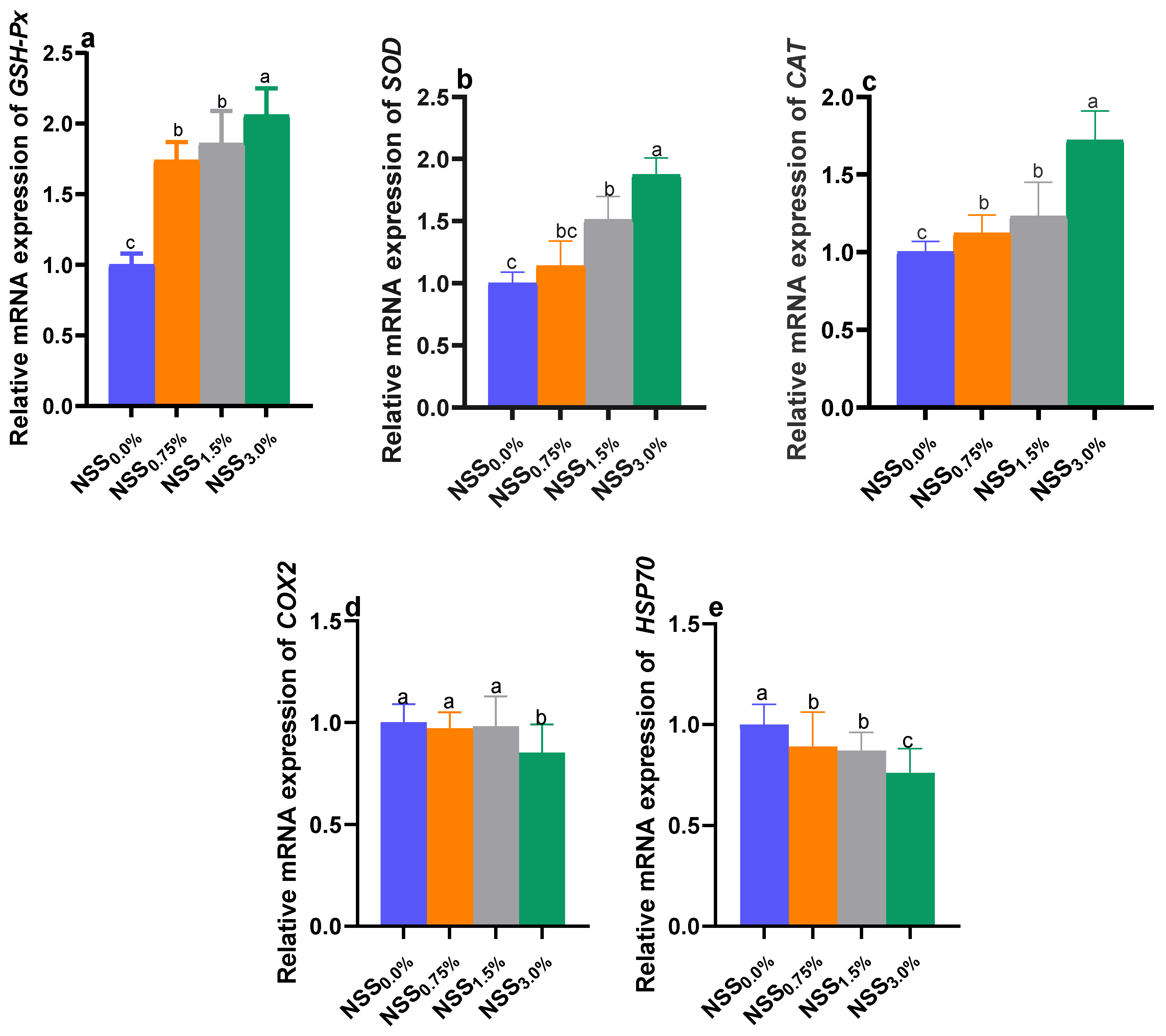
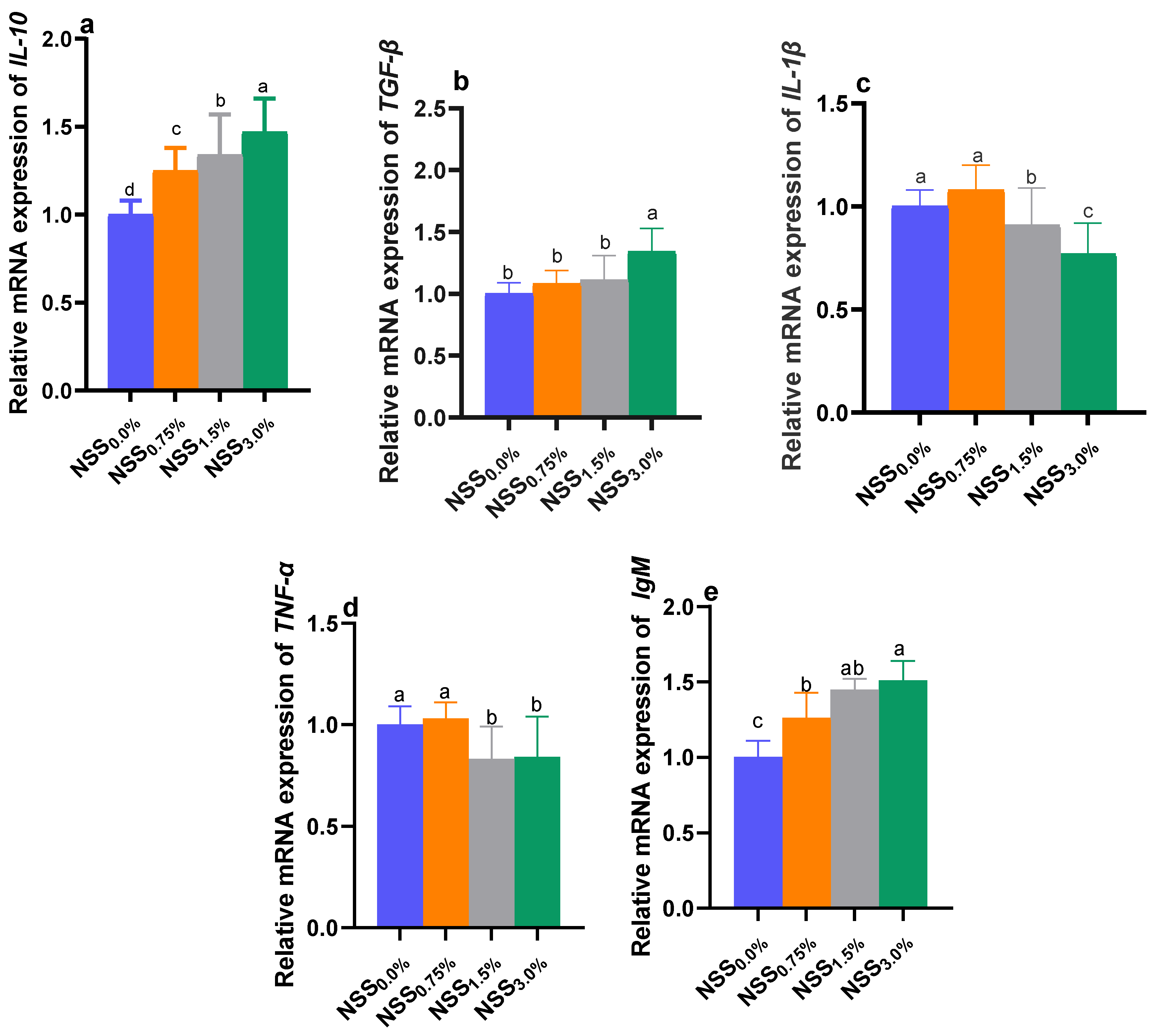
3.5. Modulation of Genes Expression by NSS

Dietary NSS administration influenced the relative expression of selected antioxidant, immune-linked, and stress-related genes of O. niloticus (Figure 1 and Figure 2). The expression analysis of genes encoding antioxidant enzymes; CAT, SOD, and GSH-Px (Figure 1), immunoglobulin M; IgM and the anti-inflammatory cytokine; IL-10 (Figure 2) reveled the highest significant (p < 0.05) upregulation in NSS3.0% fed fish. Moreover, TGF-β gene was upregulated significantly (p < 0.05) only in the NSS3.0% group unlike the control one. The genes of proinflammatory cytokines; IL- and TNF-α were significantly (p < 0.05) downregulated in NSS1.5% and NSS3.0% fish groups compared to the NSS0.0% one. Meanwhile, the group fed NSS0.75% showed no significant changes in the expression of IL- and TNF-α genes when compared to the control group (NSS0.0%). Notably, the expression of the inflammatory mediator; COX-2 gene, was slightly downregulated in NSS fed groups with no significant variations (p > 0.05) compared to the control one (NSS0.0%), except for NSS3.0% fish group. The stress-related gene; HSP70 was significantly (p < 0.05) downregulated in fish received NSS supplemented diets when compared to those received the microalgae-free diet. Group NSS3.0% showed a significantly (p < 0.05) lower HSP70 expression rate than other NSS fed groups (NSS1.5% and NSS0.75%), which displayed non-significant (p > 0.05) variations between each other (Figure 1).

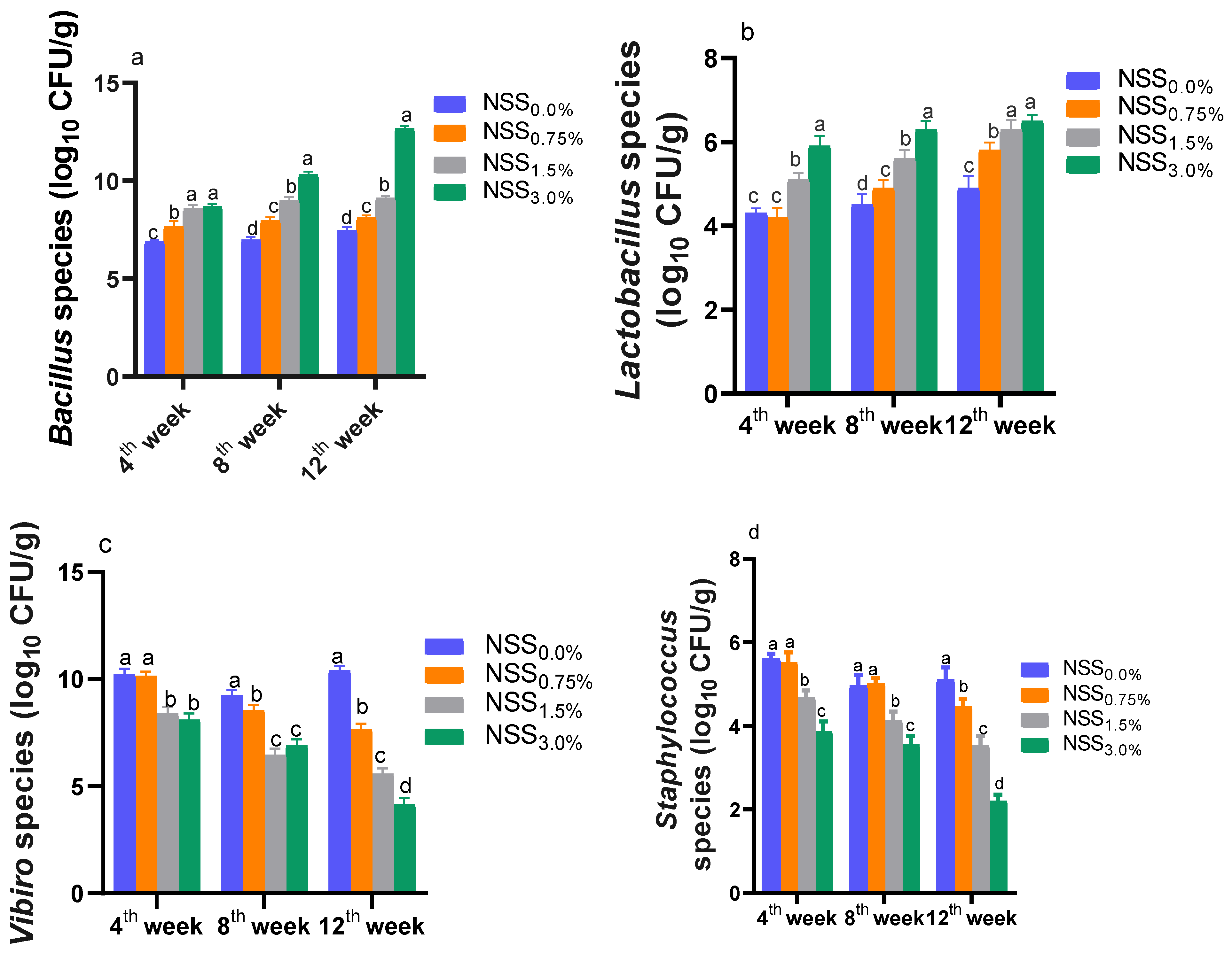
3.6. Effect of NNS on Some Intestinal Microbiota

As illustrated in Figure 3, the inclusion of NSS in fish diet for 12 weeks reduced Vibrio and Staphylococcus populations and increased Lactobacillus and Bacillus copies with respect to the control group (NSS0.0%). At 4 weeks of age, fish fed NSS1.5% and NSS3.0% had considerable (p < 0.05) lower Vibrio and Staphylococcus counts and higher Lactobacillus and Bacillus number of copies when compared to the control group with a trend towards significant differences between both levels considering Lactobacillus and Staphylococcus populations. At 8 weeks of age, dietary supplementation of NSS at different levels increased Lactobacillus and Bacillus counts in a dose-dependent manner compared to the control group. Meanwhile, statistically significant (p < 0.05) decreases in Vibrio and Staphylococcus numbers in relation to the NSS0.0% group were recorded for NSS1.5% and NSS3.0% and NSS3.0% groups, respectively. At 12 weeks of age, there were dose-dependent rises in Bacillus populations and reductions in Staphylococcus and Vibrio counts post-NSS supplementation in fish diet. Moreover, Lactobacillus counts were markedly (p < 0.05) increased in NSS1.5% and NSS3.0% groups unlike the control group.

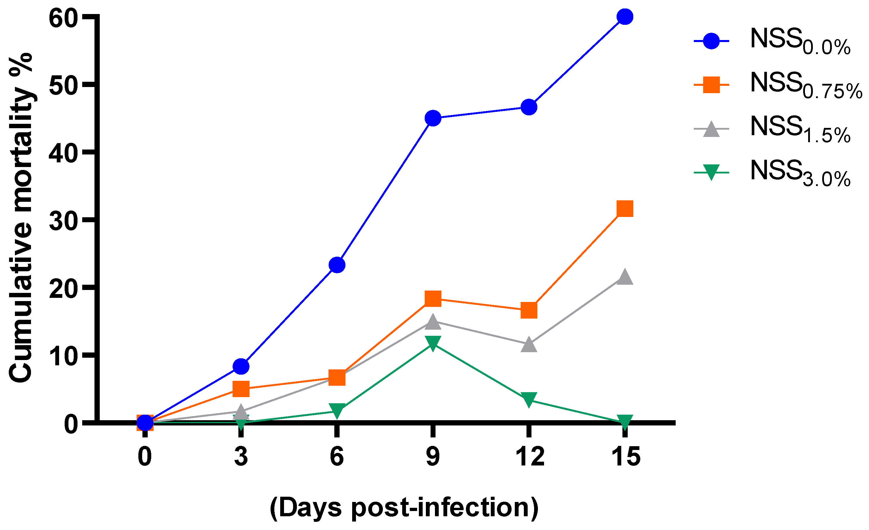
3.7. Effect of NNS on Aeromonas Hydrophila Population and Cumulative Mortality Rates

As shown in Figure 4 and Figure 5, supplementing fish diet with varying levels of NSS led to a considerable (p < 0.05) reduction in A. hydrophila counts and cumulative mortality percentages at various time intervals post-infection unlike the free microalgae-challenged group. Of note, fish fed NSS3.0% had the most remarkable (p < 0.05) lower A. hydrophila counts and cumulative mortality percentages at 15 days post-infection.

4. Discussion

Ongoing rapid development of aquaculture has been accompanied by more stressful conditions, which have impaired immune response and augmented disease outbreaks among farmed fish [56]. Concerning these issues that threaten aquaculture industry, the application of natural functional nutrients enriched with omega-3 (n-3) fatty acids not only has significant regulatory effects on inflammatory response in fish [57], but also offers healthy food choice for consumers with numerous health outcomes [58]. It has been shown that many microalgae species have a number of health-promoting impacts, particularly in the prevention and treatment of several diseases owing to their composition from natural antioxidant compounds or production of long-chain PUFAs [59,60,61]. Among these important microalgae are Nannochloropsis, Spirulina, and Schizochytrium species, which are fairly high in n-3 PUFAs such as EPA (C20:5n3) and DHA (C22:6n3), and are important for sustaining fish health and imparting neurological, cardiovascular, and anticancer benefits to humans [11,62,63]. However, the purpose from continuous feeding of such combination of microalgae and investigating the molecular basis clearing their roles on fish health and quality was not properly investigated until now. Therefore, our study is focused on the prospective application of microalgae mix in fish diet to promote its growth and flesh quality and to act as immunostimulant and antioxidant agents with a promising role in protecting against A. hydrophila infection. In the current study, after a 12-week feeding period on N. oculate and Schizochytrium and Spirulina species combination enriched with omega-3, we achieved our targeted goal concerning maximum fish production and an enhanced fillet quality with omega-3 (n-3) fatty acids. In accordance, dietary feeding on Schizochytrium species for Nile tilapia [11] and Nannochloropsis species for European sea bass [64] improved their growth rates to certain limits, while feeding of Nile tilapia in our study on a combination of selected microalgae enhanced the growth performance parameters of Nile tilapia more prominently, especially with increasing their inclusion levels (up to 3%). Besides, more efficient digestion due to higher digestive enzymes’ activities was detected in groups exhibiting improved growth-related parameters and those fed higher levels of microalgae mixtures, which came in the same line with the findings stated previously [65,66,67,68,69]. These positive outcomes in allover growth performance parameters could be attributed to the high contents of microalgae DHA and EPA fatty acids those are linked with the improved health condition of fish, especially those reared under intensive farming conditions [70]. Additionally, DHA and EPA fatty acids are engaged in important roles such as activation of insulin-like growth factor-1 and Akt-mTOR-p70S6K pathway [71] that positively impacted the growth and metabolic regulation of fish. Furthermore, it was proved that improved fish growth due to omega-3 supplementation may be associated with the prompted health condition [70].
Shifting from Nile tilapia traditional feeding to those enriched with functional nutrients comes with the same consumers’ needs targeting good quality fillet [72]. Moreover, due to the limited ability of the human body to change alpha-linolenic acid into longer chain omega-3 fatty acids; DHA, EPA, and DPA (less than 10%), it is critical to supply significant amounts of long chain omega-3 fatty acids in the food [73,74]. In the current study, fortification of Nile tilapia fillet by n3-PUFAs was prominent in groups fed higher levels of microalgae mixtures enriched with these healthy fatty acids. Our findings of beneficial impacts of including NSS microalgae mixture in Nile tilapia diets on deposition of DHA and EPA (two important omega-3 PUFAs) are also consistent with a previous observation [75], where higher PUFAs were observed with increasing the supplementation level of Schizochytrium species enriched with DHA fatty acid. Moreover, incorporation of microalgae enriched with DHA and EPA increased their contents in copepods [76]. Additionally, feeding of sea bream on diets enriched with microalgae blends including Nannochloropsis, oculata, and Schizocthytrium species displayed an increased long-chain n-3-PUFAs, DHA, and EPA levels [77]. Obviously, these potential benefits are attributed to the higher contents of n-3 PUFA, which can inhibit LDL-C and VLDL uptake and degradation [78,79]. Similarly, dietary supplementation of omega-3 reduced cholesterol, triglyceride, and VLDL levels [80].
Fish health and immunity are greatly connected to the antioxidant defense system. Exposing fish to stressful conditions those are associated with oxidative stress under intensive farming can trigger the higher ROS production resulting in extensive cell damage. The antioxidant defense system supports fish to retain endogenous ROS at quite minimal levels and to mitigate the oxidative damage provoked by ROS high reactivity [81]. Under normal physiological circumstances, the concentration of free radicals in fish is kept under a dynamic equilibrium due to their constant generation and clearness by its antioxidant system [82]. Conversely, increased ROS production can stimulate cell membranes’ lipids peroxidation and negatively impact fish performance and health [79]. Antioxidant enzymes such as GSH-Px, CAT, and SOD are considered main defense lines against the generation of toxic ROS leading to direct detoxification [83,84]. Fish antioxidant system can be coordinated by dietary enriched antioxidants that can scavenge free radicals. In this context, microalgae are enriched with natural antioxidants; however, searching on the mechanisms by which their impacts on the fish antioxidant system and whether their combination will add an additional benefit for strengthening this function is still scarce and needs more investigation. Herein, activation of antioxidant enzymatic mechanisms in groups fed higher levels of NSS microalgae blend was prominent, as detected by higher serum levels of CAT, SOD, and GSH-Px, and upregulation of their expression in fish muscle. The T-AOC is considered an index to mirror the antioxidant status of the body [85]. Notably, higher T-AOC and reduced fish fillet ROS and H2O2 levels following supplementation of NSS microalgae mixture implies decreased free radical contents and lipid damage. Similarly, the activities of GSH-Px and SOD antioxidant enzymes’ in the plasma and liver of turbot were enhanced after dietary Nannochloropsis species supplementation [86,87]. The higher antioxidant capacity of NSS microalgal mixture in the present study may be attributed to their higher contents of DHA and EPA those possess excellent antioxidant properties [88]. Additionally, increasing dietary levels of omega-3 can reduce ROS production [80] via strengthening cellular ability against oxidative stress. Moreover, S. platensis is declared to have pigments those possess antioxidative properties and are capable of scavenging peroxide radicals [89]. On the other hand, higher free radicals result in MDA overproduction, which is one of the end products of lipid peroxidation inside the cells; therefore, the MDA level is generally identified as a marker of oxidative stress [90]. Herein, the contents of MDA in fish fillet were greatly reduced after inclusion of higher levels of NSS microalgae mixture. In accordance, dietary inclusion of algal Schizochytrium species augmented the antioxidant status of Micropterus salmoides and reduced MDA tissues levels [20]. Taken together, a great deal of researches has claimed the antioxidant functions of several microalgae owing to tocopherols, phenolic compounds, and carotenoids those account for free radical scavenging pursuits supplying a considerable amelioration to oxidative stress responses in different fish species [91,92,93].
An alteration of the redox status and the dysregulation of the immune system during exposure to infectious agents result in an elevation of inflammatory systemic response [94]. Considering that, the inflammatory status prompted by the infectious stimulation is characterized by the reciprocal control of major mediators (COX-2, NO, ROS, and the antioxidant glutathione). COX-2 is an enzyme, which mediates the bioconversion of arachidonic acid to inflammatory prostaglandins with a consequent release of cytokines [40] [95]. After dietary feeding of higher levels of NSS microalgae mixture in our study, an inverse trend was found between the relative expressions of TGF-β and IL-10 and TNF-α and IL-B genes. As evidenced in our study, the regulation of the expression of these inflammatory markers could be mediated by depressing production of ROS and downregulation of COX-2 gene, which are the main messengers those modulate the expression of various genes involved in inflammation [96]. Moreover, lysozymes are ubiquitous defense anti-microbial proteins of the immune system those are associated with the first barrier of innate immunity in fish and have lysis activities against pathogenic bacteria [97]. Additionally, immunoglobulins have very important roles in the defense mechanism via killing microbes and pathogens and restricting the spread of infectious agents [18]. Herein, our consequences cleared that using various levels of NSS microalga mixture enhanced the Nile tilapia immune system (IgM, lysozymes, and MPO) prior to the challenge as previously declared elsewhere [30,98]. Similarly, dietary supplementation with microalgae blends comprising Schizochytrium species, Spirulina platensis, Chloroella sorokiniana, and Chromochloris zofingiensis significantly decreased the genes expression of pro-inflammatory cytokines; IL-8, IL-6, and IL-1β and increased the lysozyme activity in zebra fish [99]. Moreover, dietary supplementation of 5 or 10% of S. platensis significantly boosted lysozyme, serum IgM and total protein levels, thereby enhancing the sturgeon ability to resist various pathogens [100]. Notably, it has been shown that dietary fatty acid composition prompted the non-specific immunity (e.g., serum lysozyme, phagocytosis, and respiratory burst), specific immunity (e.g., antibody production and resistance to pathogens), eicosanoid production, and immune-related genes expression in fish [101,102,103]. Moreover, dietary feeding on omega-3 fatty acids improved immunity of the fish as detected by increasing MPO and total immunoglobulin levels [80], and these positive effects may be related to the reduction in the synthesis of omega-6-derived metabolites, which promote the inflammation [104]. Additionally, omega-3 fatty acids have the ability to reduce inflammation via reducing the production and secretion of cytokines and chemokines by macrophages [105]. Recently, the beneficial roles of dietary omega-3 enriched oils in modulating the expression of cytokines-related genes against mixovirus in marine fish were proved [106]. The feeding of sea bream (Sparidentex hasta) on supplemental DHA could enhance the serum immunological parameters like lysozyme and phagocytic activity and modulated the expression of IL-1B, IL-6, and IL-10 genes [107]. Furthermore, Nile tilapia fed 10% Nannochloropsis oculata exhibited significant upregulation of TGF-β and IL-10 and marked downregulation of IL- and TNF-α genes [30]. In this regard, the anti-inflammatory properties of Nannochloropsis oculata could be attributed to its role as a good potential source of EPA and its high contents of pigments such as zeaxanthin, chlorophyll, astaxanthin, and canthaxanthin [108]. In the same context, Schizochytrium limacinum is rich in DHA, which enhances the immune function of white shrimp and golden pompano [109,110]. These positive findings after dietary intake of microalga mixture are resulted from higher contents of n-3-PUFAs, particularly EPA and DHA those could boost an anti-inflammatory environment within the fish body and in that way, they could strengthen its combat against infectious diseases aiming for maximum production [111].
Heat shock proteins (HSPs) are stress-associated keys those play a vital role in adaptive and innate immune responses in fish, and they are strong candidates for the progression of new approaches for preventing the fish diseases [112]. Commonly, HSP70 is expressed in low levels, but its expression rises in reaction to ecological and biological stress conditions [32,113]. Over-expression of HSP70 gene was observed in sea bream liver tissue post-infection with Vibrio alginolyticus [114]. This study denoted that Nile tilapia groups fed NSS microalgae blends supplemented diet showed HSP70 low expression levels. In accordance, the expression levels of HSP70 were downregulated with increasing levels of Nannochloropsis oculate in Nile tilapia subjected to air stress [30].
It has been established that gut microbiota heavily affects the health status of aquaculture species regarding digestion, nutrient absorption, immunity, metabolism, and biological antagonism [115]. Microalgae could control the homeostasis of probiotic and harmful bacteria implying a positive impact on the fish health. Regarding beneficial bacteria (Bacillus and Lactobacillus species), supplementing fish with NSS microalgae blends showed an improvement in their counts with a direct relationship between their populations and high NSS doses, which were illustrated in the form of significant increases as compared to the control group. Numerous studies explored the effects of microalgae such as Chlorella, Tetraselmis, Schizochytrium, and Nannochloropsis species on farmed fish microbial ecology [113,116,117]. Kulshreshtha et al. [118] concluded that Spirulina species is advantageous for the beneficial intestinal microflora. Lactobacillus and Bacillus species can be used as growth promoters and immunostimulants as they improved the Nile tilapia immune response and disease resistance [119]. Concomitantly, beneficial Lactobacillus genus was enriched in zebrafish fed a Schizochytrium species supplemented diet [116]. Moreover, other investigators in previous teams [120,121] established positive effects of dietary Spirulina platensis to birds on boosting Lactobacillus counts in the intestine. On the other hand, dietary supplementation with NSS microalgae blends significantly decreased Vibrio counts. Moreover, supplementing diets with the NSS microalgae mixture revealed valuable inhibition against Staphylococcus species. The same achievements were reported for Chlorella salina and Tetraselmis chuii studied previously [13], where they had the most positive records against the fish indicator pathogens such as Vibrio species. Concurrently, the results of KoKou et al. [122] demonstrated also that the microalgae Nannochloropsis species, Tetraselmis chuii, Isochrysis species, Arthrospira platensis, and Cocculinella minutissima cultures inhibited the growth of Vibrio species comparing with the control treatments. There were previously similar findings with certain microalgae against Staphylococcus species [123].
Infections caused by pathogenic bacteria such as A. hydrophila could induce changes in the components of gut microbiota and trigger the malfunction of the physiochemical activities leading to diseases [124]. Moreover, A. hydrophila is responsible for hemorrhagic septicemia and causes high levels of mortality and significant economic loss in fish [15,16]. Our results proved the good antibacterial activity of the used NSS microalgae mixture against the challenging A. hydrophila strain. Interestingly, Nile tilapia supplemented with higher levels of NSS microalgae blend showed lower cumulative mortality rates that came in accordance with the remarkable reduction in A. hydrophila counts. These enhanced survival rates could be ascribed to the beneficial effects of NSS microalgae blend on both immune and antioxidant functions of Nile tilapia. These finding are in harmony with that of Neveen and Ibraheem [125] suggesting that feeding of microalgae enhances the fish immune response. Thus, the positive effects of adding microalgae in the diet proved to be a practical and simple approach to decrease the pathogenic microbial loads in fish. Antimicrobial features of microalgae cultures have been demonstrated in earlier studies [13,14,122]. These higher antimicrobial activities against the strongest fish pathogens could be attributed to the competition of the bacterial populations associated with microalgae cultures [126] or the production of antibacterial components by microalgal cells. These compounds belonged to various chemical classes such as terpenes, phenols, volatile halogenated hydrocarbons, indoles, fatty acids, and acetogenins [127]. Additionally, the microalgae antimicrobial activity may be related to the antimicrobial proteins, oxygen free radicals and associated microflora produced by microalgae cells [122]. Earlier findings illustrated that DHA and EPA contents and carotenoids could control the immune system of fish in response to invasion of harmful microorganisms [106,128,129]. Our outcomes showed, for the first time, that DHA and EPA components of the used NSS microalgae blend have antibacterial properties against A. hydrophila. Likewise, dietary feeding on omega-3 fatty acids decreased the infection against A. hydrophila in catfish [80]. Moreover, higher levels of EPA and DHA could inhibit bacterial growth and boost secretion of anti-inflammatory cytokines, thereby protecting zebrafish from Vibrio vulnificus infection [130]. It is likely that the high contents of unsaturated fatty acid affect the intestinal membrane structure and function that may influence the attachment sites of the gut mucosa [123].

5. Conclusions

Considered together, our results suggested that dietary inclusion of microalgae mix containing Nannochloropsis oculate and Schizochytrium and Spirulina species could display beneficial properties that modulate the composition of the intestinal microbiota and contribute to unique immunomodulation and disease tolerance with consequences for superior fish growth and quality. Therefore, our findings have located the selected microalgae in a unique situation in the aquaculture industry. Despite the outstanding achievement of microalgae mix in protecting the health of fish, there are important challenges to be evaluated considering the proposed mechanisms beyond their beneficial effects. Moreover, it would be very valuable to generalize the positive findings beyond the study’s parameters using microalgae mix. Therefore, conducting more in vivo studies those are required to assess the protective effects of microalgae mix against other pathogenic bacterial species threatening fish farming is an interesting idea for further researches.

Author Contributions

Conceptualization, D.I. and M.I.A.E.-H.; methodology, D.I., M.I.A.E.-H., M.E., M.M.E.-A., T.M.E., G.M.A.S., O.M.S., T.A.H. and A.E.O.; software D.I., M.I.A.E.-H., M.I.A.-Z., M.E., M.M.E.-A., T.M.E., G.M.A.S., O.M.S., T.A.H., F.M.A., M.A.A. and A.E.O., validation, D.I., M.I.A.E.-H., M.E., M.M.E.-A., T.M.E., G.M.A.S., O.M.S., T.A.H., A.E.O. and M.A.A.; formal analysis, D.I., M.I.A.E.-H., M.I.A.-Z., M.E., M.M.E.-A., T.M.E., G.M.A.S., O.M.S., T.A.H., F.M.A., M.A.A. and A.E.O.; resources, D.I., M.I.A.E.-H., M.E., M.M.E.-A., T.M.E., G.M.A.S., O.M.S., T.A.H., A.E.O., F.M.A. and M.A.A.; data curation, D.I. and M.I.A.E.-H.; writing—original draft preparation, D.I., M.I.A.E.-H., M.I.A.-Z., M.E., M.M.E.-A., T.M.E., G.M.A.S., O.M.S., T.A.H., A.E.O., F.M.A. and M.A.A.; writing—review and editing, D.I. and M.I.A.E.-H.; visualization, D.I., M.I.A.E.-H., M.I.A.-Z., M.E., M.M.E.-A., T.M.E., G.M.A.S., O.M.S., T.A.H., F.M.A., M.A.A. and A.E.O.; supervision, D.I., M.I.A.E.-H., M.I.A.-Z., M.E., M.M.E.-A., T.M.E., G.M.A.S., O.M.S., T.A.H., F.M.A., M.A.A. and A.E.O.; project administration, D.I., M.I.A.E.-H., M.I.A.-Z., M.E., M.M.E.-A., T.M.E., G.M.A.S., O.M.S., T.A.H., F.M.A., M.A.A. and A.E.O.; funding acquisition, D.I., M.I.A.E.-H., M.I.A.-Z., M.E., M.M.E.-A., T.M.E., G.M.A.S., O.M.S., T.A.H., F.M.A., M.A.A. and A.E.O. All authors have read and agreed to the published version of the manuscript.

Funding

This research was funded by Princess Nourah Bint Abdulrahman University Researchers Supporting Project number (PNURSP2022R84), Princess Nourah Bint Abdulrahman University, Riyadh, Saudi Arabia.

Institutional Review Board Statement

Animal care and management, and experimental measures were in conformity with the guidelines and ethics of the Institutional Animal Care and Use Committee of the Faculty of Veterinary Medicine at Zagazig University.

Informed Consent Statement

Not applicable.

Data Availability Statement

The data presented in this study are available upon request from the corresponding author.

Acknowledgments

We want to thank Zagazig University. And we also extend our thanks to the Princess Nourah bint Abdulrahman University Researchers Supporting Project number (PNURSP2022R84), Princess Nourah bint Abdulrahman University, Riyadh, Saudi Arabia.

Conflicts of Interest

The authors declare no conflict of interest.

References

  1. Lushchak, V.I. Environmentally induced oxidative stress in aquatic animals. Aquat. Toxicol. 2011, 101, 13–30. [Google Scholar] [CrossRef] [PubMed]
  2. Birnie-Gauvin, K.; Costantini, D.; Cooke, S.J.; Willmore, W.G. A comparative and evolutionary approach to oxidative stress in fish: A review. Fish Fish. 2017, 18, 928–942. [Google Scholar] [CrossRef]
  3. Nordberg, J.; Arnér, E.S. Reactive oxygen species, antioxidants, and the mammalian thioredoxin system. Free Radic. Biol. Med. 2001, 31, 1287–1312. [Google Scholar] [CrossRef]
  4. Hussain, T.; Tan, B.; Yin, Y.; Blachier, F.; Tossou, M.C.; Rahu, N. Oxidative stress and inflammation: What polyphenols can do for us? Oxid. Med. Cell. Longev. 2016, 2016. [Google Scholar] [CrossRef] [PubMed] [Green Version]
  5. Ibrahim, D.; Kishawy, A.T.; Khater, S.I.; Khalifa, E.; Ismail, T.A.; Mohammed, H.A.; Elnahriry, S.S.; Tolba, H.A.; Sherief, W.R.; Farag, M.F. Interactive effects of dietary quercetin nanoparticles on growth, flesh antioxidant capacity and transcription of cytokines and Aeromonas hydrophila quorum sensing orchestrating genes in Nile tilapia (Oreochromis niloticus). Fish Shellfish Immunol. 2021. [Google Scholar] [CrossRef]
  6. Mittal, M.; Siddiqui, M.R.; Tran, K.; Reddy, S.P.; Malik, A.B. Reactive oxygen species in inflammation and tissue injury. Antioxid. Redox Signal. 2014, 20, 1126–1167. [Google Scholar] [CrossRef] [Green Version]
  7. Ranneh, Y.; Ali, F.; Akim, A.M.; Hamid, H.A.; Khazaai, H.; Fadel, A. Crosstalk between reactive oxygen species and pro-inflammatory markers in developing various chronic diseases: A review. Appl. Biol. Chem. 2017, 60, 327–338. [Google Scholar] [CrossRef]
  8. Abd El-Hamid, M.I.; Ibrahim, S.M.; Eldemery, F.; El-Mandrawy, S.A.; Metwally, A.S.; Khalifa, E.; Elnahriry, S.S.; Ibrahim, D. Dietary cinnamaldehyde nanoemulsion boosts growth and transcriptomes of antioxidant and immune related genes to fight Streptococcus agalactiae infection in Nile tilapia (Oreochromis niloticus). Fish Shellfish Immunol. 2021, 113, 96–105. [Google Scholar] [CrossRef]
  9. Ibrahim, D.; Arisha, A.H.; Khater, S.I.; Gad, W.M.; Hassan, Z.; Abou-Khadra, S.H.; Mohamed, D.I.; Ahmed Ismail, T.; Gad, S.A.; Eid, S.A. Impact of Omega-3 Fatty Acids Nano-Formulation on Growth, Antioxidant Potential, Fillet Quality, Immunity, Autophagy-Related Genes and Aeromonas hydrophila Resistance in Nile Tilapia (Oreochromis niloticus). Antioxidants 2022, 11, 1523. [Google Scholar] [CrossRef]
  10. Abdel-Latif, H.M.; El-Ashram, S.; Yilmaz, S.; Naiel, M.A.; Kari, Z.A.; Hamid, N.K.A.; Dawood, M.A.; Nowosad, J.; Kucharczyk, D. The effectiveness of Arthrospira platensis and microalgae in relieving stressful conditions affecting finfish and shellfish species: An overview. Aquac. Rep. 2022, 24, 101135. [Google Scholar] [CrossRef]
  11. Sarker, P.K.; Kapuscinski, A.R.; Lanois, A.J.; Livesey, E.D.; Bernhard, K.P.; Coley, M.L. Towards sustainable aquafeeds: Complete substitution of fish oil with marine microalga Schizochytrium sp. improves growth and fatty acid deposition in juvenile Nile tilapia (Oreochromis niloticus). PLoS One 2016, 11, e0156684. [Google Scholar] [CrossRef] [PubMed] [Green Version]
  12. Shah, M.R.; Lutzu, G.A.; Alam, A.; Sarker, P.; Chowdhury, K.; Parsaeimehr, A.; Liang, Y.; Daroch, M. Microalgae in aquafeeds for a sustainable aquaculture industry. J. Appl. Phycol. 2018, 30, 197–213. [Google Scholar] [CrossRef]
  13. El-Sayed, H.S.; Ibrahim, H.A.; Beltagy, E.A.; Khairy, H.M. Effects of short term feeding of some marine microalgae on the microbial profile associated with Dicentrarchus labrax post larvae. Egypt. J. Aquat. Res. 2014, 40, 251–260. [Google Scholar] [CrossRef] [Green Version]
  14. Austin, B.; Baudet, E.; Stobie, M. Inhibition of bacterial fish pathogens by Tetraselmis suecica. J. Fish Dis. 1992, 15, 55–61. [Google Scholar] [CrossRef]
  15. Vivas, J.; Carracedo, B.; Riano, J.; Razquin, B.E.; López-Fierro, P.; Acosta, F.; Naharro, G.; Villena, A.J. Behavior of an Aeromonas hydrophila aroA live vaccine in water microcosms. Appl. Environ. Microbiol. 2004, 70, 2702–2708. [Google Scholar] [CrossRef] [Green Version]
  16. Ardó, L.; Jeney, Z.; Adams, A.; Jeney, G. Immune responses of resistant and sensitive common carp families following experimental challenge with Aeromonas hydrophila. Fish Shellfish Immunol. 2010, 29, 111–116. [Google Scholar] [CrossRef]
  17. Ammar, A.M.; El-Naenaeey, E.-S.Y.; Abd El-Hamid, M.I.; El-Gedawy, A.A.; Elmalt, R.M. Campylobacter as a Major Foodborne Pathogen: A Review of Its Characteristics, Pathogenesis, Antimicrobial Resistance and Control. J. Microbiol. Biotechnol. Food Sci. 2021, 10, 609–619. [Google Scholar] [CrossRef]
  18. Ammar, A.M.; El-Naenaeey, E.-S.Y.; El-Malt, R.; El-Gedawy, A.A.; Khalifa, E.; Elnahriry, S.S.; El-Hamid, A.; Marwa, I. Prevalence, antimicrobial susceptibility, virulence and genotyping of Campylobacter jejuni with a special reference to the anti-virulence potential of Eugenol and beta-resorcylic acid on some multi-drug resistant isolates in Egypt. Animals 2021, 11, 3. [Google Scholar] [CrossRef]
  19. Ammar, A.; El-Hamid, M.; Eid, S.E.; El Oksh, A.S. Insight into antimicrobial resistance and virulence genes of emergent multidrug resistant avian pathogenic Escherichia coli in Egypt: How closely related are they. Rev. Med. Vet. 2015, 166, 304–314. [Google Scholar]
  20. Habte-Tsion, H.-M.; Kolimadu, G.D.; Rossi, W.; Filer, K.; Kumar, V. Effects of Schizochytrium and micro-minerals on immune, antioxidant, inflammatory and lipid-metabolism status of Micropterus salmoides fed high-and low-fishmeal diets. Sci. Rep. 2020, 10, 1–13. [Google Scholar] [CrossRef]
  21. Bélanger, A.; Sarker, P.K.; Bureau, D.P.; Chouinard, Y.; Vandenberg, G.W. Apparent digestibility of macronutrients and fatty acids from microalgae (Schizochytrium sp.) fed to rainbow trout (Oncorhynchus mykiss): A potential candidate for fish oil substitution. Animals 2021, 11, 456. [Google Scholar] [CrossRef] [PubMed]
  22. Cardona, E.; Segret, E.; Cachelou, Y.; Vanderesse, T.; Larroquet, L.; Hermann, A.; Surget, A.; Corraze, G.; Cachelou, F.; Bobe, J. Effect of micro-algae Schizochytrium sp. supplementation in plant diet on reproduction of female rainbow trout (Oncorhynchus mykiss): Maternal programming impact of progeny. J. Anim. Sci. Biotechnol. 2022, 13, 1–18. [Google Scholar] [CrossRef] [PubMed]
  23. Priyadarshani, I.; Rath, B. Commercial and industrial applications of micro algae—A review. J. Algal Biomass Util. 2012, 3, 89–100. [Google Scholar]
  24. Zhang, F.; Man, Y.B.; Mo, W.Y.; Wong, M.H. Application of Spirulina in aquaculture: A review on wastewater treatment and fish growth. Rev. Aquac. 2020, 12, 582–599. [Google Scholar] [CrossRef]
  25. Wu, Q.; Liu, L.; Miron, A.; Klímová, B.; Wan, D.; Kuča, K. The antioxidant, immunomodulatory, and anti-inflammatory activities of Spirulina: An overview. Arch. Toxicol. 2016, 90, 1817–1840. [Google Scholar] [CrossRef]
  26. Sharifah, E.N.; Eguchi, M. The phytoplankton Nannochloropsis oculata enhances the ability of Roseobacter clade bacteria to inhibit the growth of fish pathogen Vibrio anguillarum. PLoS ONE 2011, 6, e26756. [Google Scholar] [CrossRef] [Green Version]
  27. Mounes, H.A.M.; Mansour, E.G.; Ahmed, K.M. Effect of Azolla pinnata and Nannochloropsis oculata on growth performance and immunoresponse of Nile tilapia (Oreochromis niloticus) and its resistance to bacterial infection. Egypt. J. Aquac. 2020, 10, 43–62. [Google Scholar] [CrossRef]
  28. Md, A.; Jin, F.; Jeong, U.-C.; Choi, J.-K.; Lee, D.-I.; Yu, H.S.; Kang, S.-J. Effects of Nannochloropsis concentration in diet on growth, survival and anti-inflammatory cytokine (Interleukin-10) production of the sea cucumber Apostichopus japonicus. Turkish J. Fish. Aquat. Sci. 2018, 18, 567–575. [Google Scholar]
  29. Abdelghany, M.F.; El-Sawy, H.B.; Abd El-Hameed, S.A.; Khames, M.K.; Abdel-Latif, H.M.; Naiel, M.A. Effects of dietary Nannochloropsis oculata on growth performance, serum biochemical parameters, immune responses, and resistance against Aeromonas veronii challenge in Nile tilapia (Oreochromis niloticus). Fish Shellfish Immunol. 2020, 107, 277–288. [Google Scholar] [CrossRef]
  30. Zahran, E.; Elbahnaswy, S.; Ibrahim, I.; Khaled, A.A. Nannochloropsis oculata enhances immune response, transcription of stress, and cytokine genes in Nile tilapia subjected to air exposure stress. Aquac. Rep. 2021, 21, 100911. [Google Scholar] [CrossRef]
  31. Ibrahim, D.; Nem, A.N.A.; Ibrahim, S.M.; Eissa, H.M.; Fawzey, M.; Mostafa, D.I.; Abd El-Kader, S.A.; Khater, S.; Khater, S.I. Dual effect of Selenium loaded Chitosan Nanoparticles on growth, antioxidant, immune related genes expression, transcriptomics modulation of caspase 1, cytochrome P450 and heat shock protein and Aeromonas hydrophila resistance of Nile Tilapia (Oreochromis niloticus). Fish Shellfish Immunol. 2021. [Google Scholar]
  32. Kishawy, A.T.; Mohammed, H.A.; Zaglool, A.W.; Attia, M.S.; Hassan, F.A.; Roushdy, E.M.; Ismail, T.A.; Ibrahim, D. Partial defatted black solider larvae meal as a promising strategy to replace fish meal protein in diet for Nile tilapia (Oreochromis niloticus): Performance, expression of protein and fat transporters, and cytokines related genes and economic efficiency. Aquaculture 2022, 555, 738195. [Google Scholar] [CrossRef]
  33. Blaxhall, P.; Daisley, K. Routine haematological methods for use with fish blood. J. Fish Biol. 1973, 5, 771–781. [Google Scholar] [CrossRef]
  34. Jenkins, J.A.; Bart Jr, H.; Bowker, J.D.; Bowser, P.; MacMillan, J.; Nickum, J.; Rose, J.; Sorensen, P.; Whitledge, G.; Rachlin, J.W. Guidelines for the Use of Fishes in Research. American Fisheries Society: Bethesda, MD, USA, 2014. [Google Scholar]
  35. Reitman, S.; Frankel, S. A colorimetric method for the determination of serum glutamic oxalacetic and glutamic pyruvic transaminases. Am. J. Clin. Pathol. 1957, 28, 56–63. [Google Scholar] [CrossRef]
  36. Aebi, H. Catalase in vitro. In Methods Enzymol; Elsevier: Amsterdam, The Netherlands, 1984; Volume 105, pp. 121–126. [Google Scholar]
  37. LeBel, C.P.; Ischiropoulos, H.; Bondy, S.C. Evaluation of the probe 2’,7’-dichlorofluorescin as an indicator of reactive oxygen species formation and oxidative stress. Chem. Res. Toxicol. 1992, 5, 227–231. [Google Scholar] [CrossRef] [Green Version]
  38. Loreto, F.; Velikova, V. Isoprene produced by leaves protects the photosynthetic apparatus against ozone damage, quenches ozone products, and reduces lipid peroxidation of cellular membranes. Plant Physiol. 2001, 127, 1781–1787. [Google Scholar] [CrossRef]
  39. Livingstone, D.; Martinez, P.G.; Michel, X.; Narbonne, J.; O’hara, S.; Ribera, D.; Winston, G. Oxyradical production as a pollution-mediated mechanism of toxicity in the common mussel, Mytilus edulis L., and other molluscs. Funct. Ecol. 1990, 415–424. [Google Scholar] [CrossRef]
  40. Shah, S.; Pal, A.; Kaushik, V.; Devi, S. Preparation and characterization of venlafaxine hydrochloride-loaded chitosan nanoparticles and in vitro release of drug. J. Appl. Polym. Sci. 2009, 112, 2876–2887. [Google Scholar] [CrossRef]
  41. Bryan, N.S.; Grisham, M.B. Methods to detect nitric oxide and its metabolites in biological samples. Free Radic. Biol. Med. 2007, 43, 645–657. [Google Scholar] [CrossRef] [Green Version]
  42. Grisham, M.B.; Johnson, G.G.; Lancaster, J.R., Jr. Quantitation of nitrate and nitrite in extracellular fluids. In Methods Enzymol; Elsevier: Amsterdam, The Netherlands, 1996; Volume 268, pp. 237–246. [Google Scholar]
  43. Suzuki, K.; Ota, H.; Sasagawa, S.; Sakatani, T.; Fujikura, T. Assay method for myeloperoxidase in human polymorphonuclear leukocytes. Anal. Biochem. 1983, 132, 345–352. [Google Scholar] [CrossRef]
  44. Sunyer, J.O.; Tort, L. Natural hemolytic and bactericidal activities of sea bream Chlorella vulgaris serum are effected by the alternative complement pathway. Vet. Immunol. Immunopathol. 1995, 45, 333–345. [Google Scholar] [CrossRef]
  45. Chen, X.-M.; Guo, G.-L.; Sun, L.; Yang, Q.-S.; Wang, G.-Q.; Qin, G.-X.; Zhang, D.-M. Effects of Ala-Gln feeding strategies on growth, metabolism, and crowding stress resistance of juvenile Cyprinus carpio var. Jian. Fish Shellfish Immunol. 2016, 51, 365–372. [Google Scholar] [CrossRef]
  46. Drieghe, S.A.; Alsaadi, H.; Tugirimana, P.L.; Delanghe, J.R. A new high-sensitive nephelometric method for assaying serum C-reactive protein based on phosphocholine interaction. Clin. Chem. Lab. Med. (CCLM) 2014, 52, 861–867. [Google Scholar] [CrossRef] [PubMed] [Green Version]
  47. Livak, K.J.; Schmittgen, T.D. Analysis of relative gene expression data using real-time quantitative PCR and the 2−ΔΔCT method. Methods 2001, 25, 402–408. [Google Scholar] [CrossRef] [PubMed]
  48. McOrist, A.L.; Jackson, M.; Bird, A.R. A comparison of five methods for extraction of bacterial DNA from human faecal samples. J. Microbiol. Methods 2002, 50, 131–139. [Google Scholar] [CrossRef]
  49. Solichová, K.; Němečková, I.; Šviráková, E.; Horáčková, Š. Novel identification methods including a species-specific PCR for hazardous Bacillus species. Acta Aliment. 2019, 48, 415–422. [Google Scholar] [CrossRef] [Green Version]
  50. Zhang, K.; Sparling, J.; Chow, B.L.; Elsayed, S.; Hussain, Z.; Church, D.L.; Gregson, D.B.; Louie, T.; Conly, J.M. New quadriplex PCR assay for detection of methicillin and mupirocin resistance and simultaneous discrimination of Staphylococcus aureus from coagulase-negative staphylococci. J. Clin. Microbiol. 2004, 42, 4947–4955. [Google Scholar] [CrossRef] [Green Version]
  51. Thompson, J.R.; Randa, M.A.; Marcelino, L.A.; Tomita-Mitchell, A.; Lim, E.; Polz, M.F. Diversity and dynamics of a North Atlantic coastal Vibrio community. Appl. Environ. Microbiol. 2004, 70, 4103–4110. [Google Scholar] [CrossRef] [Green Version]
  52. Sebastião, F.; Lemos, E.M.; Pilarski, F. Development of an absolute quantitative real-time PCR (qPCR) for the diagnosis of Aeromonas hydrophila infections in fish. Acta Sci. Microbiol. 2018, 1, 23–29. [Google Scholar] [CrossRef]
  53. Algammal, A.M.; Mohamed, M.F.; Tawfiek, B.A.; Hozzein, W.N.; El Kazzaz, W.M.; Mabrok, M. Molecular typing, antibiogram and PCR-RFLP based detection of Aeromonas hydrophila complex isolated from Oreochromis niloticus. Pathogens 2020, 9, 238. [Google Scholar] [CrossRef] [Green Version]
  54. El-Gohary, F.A.; Zahran, E.; Abd El-Gawad, E.A.; El-Gohary, A.H.M.; Abdelhamid, F.; El-Mleeh, A.; Elmahallawy, E.K.; Elsayed, M.M. Investigation of the prevalence, virulence genes, and antibiogram of motile Aeromonads isolated from Nile tilapia fish farms in Egypt and assessment of their water quality. Animals 2020, 10, 1432. [Google Scholar] [CrossRef] [PubMed]
  55. Phumkhachorn, P.; Rattanachaikunsopon, P. Use of Bacteriophage to Control Experimental Aeromonas hydrophila Infection in Tilapia (Oreochromis niloticus). Pak. J. Biol. Sci. PJBS 2020, 23, 1659–1665. [Google Scholar] [CrossRef] [PubMed]
  56. Abd El-Hamid, M.; Abd El-Aziz, N.; Ali, H. Protective potency of clove oil and its transcriptional down-regulation of Aeromonas sobria virulence genes in African catfish (Clarias gariepinus L.). Cell. Mol. Biol. 2016, 62, 49–54. [Google Scholar] [PubMed]
  57. Nakharuthai, C.; Rodrigues, P.M.; Schrama, D.; Kumkhong, S.; Boonanuntanasarn, S. Effects of different dietary vegetable lipid sources on health status in Nile tilapia (Oreochromis niloticus): Haematological indices, immune response parameters and plasma proteome. Animals 2020, 10, 1377. [Google Scholar] [CrossRef] [PubMed]
  58. Kris-Etherton, P.M.; Harris, W.S.; Appel, L.J. Fish consumption, fish oil, omega-3 fatty acids, and cardiovascular disease. Circulation 2002, 106, 2747–2757. [Google Scholar] [CrossRef] [PubMed]
  59. Pulz, O.; Gross, W. Valuable products from biotechnology of microalgae. Appl. Microbiol. Biotechnol. 2004, 65, 635–648. [Google Scholar] [CrossRef]
  60. Wells, M.L.; Potin, P.; Craigie, J.S.; Raven, J.A.; Merchant, S.S.; Helliwell, K.E.; Smith, A.G.; Camire, M.E.; Brawley, S.H. Algae as nutritional and functional food sources: Revisiting our understanding. J. Appl. Phycol. 2017, 29, 949–982. [Google Scholar] [CrossRef]
  61. Olofsson, M.; Lamela, T.; Nilsson, E.; Bergé, J.-P.; Del Pino, V.; Uronen, P.; Legrand, C. Combined effects of nitrogen concentration and seasonal changes on the production of lipids in Nannochloropsis oculata. Mar. Drugs 2014, 12, 1891–1910. [Google Scholar] [CrossRef] [Green Version]
  62. Peet, M.; Stokes, C. Omega-3 fatty acids in the treatment of psychiatric disorders. Drugs 2005, 65, 1051–1059. [Google Scholar] [CrossRef]
  63. Brasky, T.M.; Till, C.; White, E.; Neuhouser, M.L.; Song, X.; Goodman, P.; Thompson, I.M.; King, I.B.; Albanes, D.; Kristal, A.R. Serum phospholipid fatty acids and prostate cancer risk: Results from the prostate cancer prevention trial. Am. J. Epidemiol. 2011, 173, 1429–1439. [Google Scholar] [CrossRef] [Green Version]
  64. Pascon, G.; Messina, M.; Petit, L.; Valente, L.M.P.; Oliveira, B.; Przybyla, C.; Dutto, G.; Tulli, F. Potential application and beneficial effects of a marine microalgal biomass produced in a high-rate algal pond (HRAP) in diets of European sea bass, Dicentrarchus labrax. Environ. Sci. Pollut. Res. 2021, 28, 62185–62199. [Google Scholar] [CrossRef] [PubMed]
  65. Sarker, P.K.; Kapuscinski, A.R.; McKuin, B.; Fitzgerald, D.S.; Nash, H.M.; Greenwood, C. Microalgae-blend tilapia feed eliminates fishmeal and fish oil, improves growth, and is cost viable. Sci. Rep. 2020, 10, 1–14. [Google Scholar]
  66. Elmowalid, G.A.E.; Ahmad, A.A.M.; El-Hamid, M.I.A.; Ibrahim, D.; Wahdan, A.; El Oksh, A.S.; Yonis, A.E.; Elkady, M.A.; Ismail, T.A.; Alkhedaide, A.Q. Nigella sativa Extract Potentially Inhibited Methicillin Resistant Staphylococcus aureus Induced Infection in Rabbits: Potential Immunomodulatory and Growth Promoting Properties. Animals 2022, 12, 2635. [Google Scholar] [CrossRef]
  67. Hashem, Y.M.; Abd El-Hamid, M.I.; Awad, N.F.; Ibrahim, D.; Elshater, N.S.; El-Malt, R.M.; Hassan, W.H.; Abo-Shama, U.H.; Nassan, M.A.; El-Bahy, S.M. Insights into growth-promoting, anti-inflammatory, immunostimulant, and antibacterial activities of Toldin CRD as a novel phytobiotic in broiler chickens experimentally infected with Mycoplasma gallisepticum. Poult. Sci. 2022, 101, 102154. [Google Scholar] [CrossRef]
  68. Ibrahim, D.; Eldemery, F.; Metwally, A.S.; Abd-Allah, E.M.; Mohamed, D.T.; Ismail, T.A.; Hamed, T.A.; Al Sadik, G.M.; Neamat-Allah, A.N.; Abd El-Hamid, M.I. Dietary eugenol nanoemulsion potentiated performance of broiler chickens: Orchestration of digestive enzymes, intestinal barrier functions and cytokines related gene expression with a consequence of attenuating the severity of E. coli O78 infection. Front. Vet. Sci. 2022, 9. [Google Scholar] [CrossRef] [PubMed]
  69. Ibrahim, D.; Ismail, T.A.; Khalifa, E.; El-Kader, A.; Shaimaa, A.; Mohamed, D.I.; Mohamed, D.T.; Shahin, S.E.; El-Hamid, A.; Marwa, I. Supplementing Garlic Nanohydrogel Optimized Growth, Gastrointestinal Integrity and Economics and Ameliorated Necrotic Enteritis in Broiler Chickens Using a Clostridium perfringens Challenge Model. Animals 2021, 11, 2027. [Google Scholar] [CrossRef]
  70. Wei, H.-K.; Deng, Z.; Jiang, S.-Z.; Song, T.-X.; Zhou, Y.-F.; Peng, J.; Tao, Y.-X. Eicosapentaenoic acid abolishes inhibition of insulin-induced mTOR phosphorylation by LPS via PTP1B downregulation in skeletal muscle. Mol. Cell. Endocrinol. 2017, 439, 116–125. [Google Scholar] [CrossRef]
  71. Gingras, A.A.; White, P.J.; Chouinard, P.Y.; Julien, P.; Davis, T.A.; Dombrowski, L.; Couture, Y.; Dubreuil, P.; Myre, A.; Bergeron, K. Long-chain omega-3 fatty acids regulate bovine whole-body protein metabolism by promoting muscle insulin signalling to the Akt–mTOR–S6K1 pathway and insulin sensitivity. J. Physiol. 2007, 579, 269–284. [Google Scholar] [CrossRef]
  72. Stoneham, T.R.; Kuhn, D.D.; Taylor, D.P.; Neilson, A.P.; Smith, S.A.; Gatlin, D.M.; Chu, H.S.S.; O’Keefe, S.F. Production of omega-3 enriched tilapia through the dietary use of algae meal or fish oil: Improved nutrient value of fillet and offal. PLoS ONE 2018, 13, e0194241. [Google Scholar]
  73. Burdge, G. Metabolism of α-linolenic acid in humans. Prostaglandins Leukot. Essent. Fat. Acids 2006, 75, 161–168. [Google Scholar] [CrossRef]
  74. Sinclair, A.J.; Attar-Bashi, N.M.; Li, D. What is the role of α-linolenic acid for mammals? Lipids 2002, 37, 1113–1123. [Google Scholar] [CrossRef] [PubMed]
  75. dos Santos, S.K.A.; Schorer, M.; Moura, G.d.S.; Lanna, E.A.T.; Pedreira, M.M. Evaluation of growth and fatty acid profile of Nile tilapia (Oreochromis niloticus) fed with Schizochytrium sp. Aquac. Res. 2019, 50, 1068–1074. [Google Scholar] [CrossRef]
  76. Dayras, P.; Bialais, C.; Sadovskaya, I.; Lee, M.-C.; Lee, J.-S.; Souissi, S. Microalgal Diet Influences the Nutritive Quality and Reproductive Investment of the Cyclopoid Copepod Paracyclopina nana. Front. Mar. Sci. 2021, 1147. [Google Scholar] [CrossRef]
  77. Eryalçın, K.M.; Yıldız, M. Effects of long-term feeding with dried microalgae added microdiets on growth and fatty acid composition of gilthead sea bream (Chlorella vulgaris L., 1758). Turkish J. Fish. Aquat. Sci. 2015, 15, 905–915. [Google Scholar]
  78. Mozanzadeh, M.T.; Marammazi, J.G.; Yavari, V.; Agh, N.; Mohammadian, T.; Gisbert, E. Dietary n− 3 LC-PUFA requirements in silvery-black porgy juveniles (Sparidentex hasta). Aquaculture 2015, 448, 151–161. [Google Scholar] [CrossRef]
  79. Peng, M.; Xu, W.; Tan, P.; Du, J.; Mai, K.; Zhou, H.; Zhang, Y.; Nian, R.; Macq, B.; Ai, Q. Effect of dietary fatty acid composition on growth, fatty acids composition and hepatic lipid metabolism in juvenile turbot (Scophthalmus maximus L.) fed diets with required n3 LC-PUFAs. Aquaculture 2017, 479, 591–600. [Google Scholar] [CrossRef]
  80. Kumar, N.; Chandan, N.K.; Gupta, S.K.; Bhushan, S.; Patole, P.B. Omega-3 fatty acids effectively modulate growth performance, immune response, and disease resistance in fish against multiple stresses. Aquaculture 2022, 547, 737506. [Google Scholar] [CrossRef]
  81. Wilhelm Filho, D.; Tribess, T.; Gaspari, C.; Claudio, F.; Torres, M.; Magalhaes, A. Seasonal changes in antioxidant defenses of the digestive gland of the brown mussel (Perna perna). Aquaculture 2001, 203, 149–158. [Google Scholar] [CrossRef]
  82. Winston, G.W.; Di Giulio, R.T. Prooxidant and antioxidant mechanisms in aquatic organisms. Aquat. Toxicol. 1991, 19, 137–161. [Google Scholar] [CrossRef]
  83. Vickers, N.J. Animal communication: When i’m calling you, will you answer too? Curr. Biol. 2017, 27, R713–R715. [Google Scholar] [CrossRef]
  84. Martínez-Álvarez, R.M.; Morales, A.E.; Sanz, A. Antioxidant defenses in fish: Biotic and abiotic factors. Rev. Fish Biol. Fish. 2005, 15, 75–88. [Google Scholar] [CrossRef]
  85. Mahfouz, R.; Sharma, R.; Sharma, D.; Sabanegh, E.; Agarwal, A. Diagnostic value of the total antioxidant capacity (TAC) in human seminal plasma. Fertil. Steril. 2009, 91, 805–811. [Google Scholar] [CrossRef] [PubMed]
  86. Qiao, H.; Hu, D.; Ma, J.; Wang, X.; Wu, H.; Wang, J. Feeding effects of the microalga Nannochloropsis sp. on juvenile turbot (Scophthalmus maximus L.). Algal Res. 2019, 41, 101540. [Google Scholar] [CrossRef]
  87. Castro, C.; Coutinho, F.; Iglesias, P.; Oliva-Teles, A.; Couto, A. Chlorella sp. and Nannochloropsis sp. inclusion in plant-based diets modulate the intestine and liver antioxidant mechanisms of European sea bass juveniles. Front. Vet. Sci. 2020, 7, 607575. [Google Scholar] [CrossRef]
  88. Wu, K.; Cleveland, B.M.; Portman, M.; Sealey, W.M.; Lei, X.G. Supplemental microalgal DHA and astaxanthin affect astaxanthin metabolism and redox status of Juvenile Rainbow trout. Antioxidants 2020, 10, 16. [Google Scholar] [CrossRef]
  89. Palmegiano, G.B.; Gai, F.; Daprà, F.; Gasco, L.; Pazzaglia, M.; Peiretti, P.G. Effects of Spirulina and plant oil on the growth and lipid traits of white sturgeon (Acipenser transmontanus) fingerlings. Aquac. Res. 2008, 39, 587–595. [Google Scholar] [CrossRef]
  90. Gaweł, S.; Wardas, M.; Niedworok, E.; Wardas, P. Malondialdehyde (MDA) as a lipid peroxidation marker. Wiad. Lek. Wars. Pol. 2004, 57, 453–455. [Google Scholar]
  91. Sharma, S.; Shah, E.; Davla, D.; Dixit, G.; Patel, A.; Kumar, A.K. Effect of microalga-based diet on oxidative stress enzymes of African catfish, Clarias gariepinus. Int. Aquat. Res. 2019, 11, 377–387. [Google Scholar] [CrossRef]
  92. Ahmad, M.T.; Shariff, M.; Md. Yusoff, F.; Goh, Y.M.; Banerjee, S. Applications of microalga Chlorella vulgaris in aquaculture. Rev. Aquac. 2020, 12, 328–346. [Google Scholar] [CrossRef]
  93. Safafar, H.; Van Wagenen, J.; Møller, P.; Jacobsen, C. Carotenoids, phenolic compounds and tocopherols contribute to the antioxidative properties of some microalgae species grown on industrial wastewater. Mar. Drugs 2015, 13, 7339–7356. [Google Scholar] [CrossRef] [Green Version]
  94. Tan, B.L.; Norhaizan, M.E.; Liew, W.-P.-P.; Sulaiman Rahman, H. Antioxidant and oxidative stress: A mutual interplay in age-related diseases. Front. Pharmacol. 2018, 9, 1162. [Google Scholar] [CrossRef] [PubMed] [Green Version]
  95. Yu, H.Y.; Kim, K.-S.; Lee, Y.-C.; Moon, H.-I.; Lee, J.-H. Oleifolioside A, a new active compound, attenuates LPS-stimulated iNOS and COX-2 expression through the downregulation of NF-κB and MAPK activities in RAW 264.7 macrophages. Evid. Based Complement. Alternat. Med. 2012, 2012. [Google Scholar] [CrossRef] [PubMed] [Green Version]
  96. Finkel, T.; Holbrook, N.J. Oxidants, oxidative stress and the biology of ageing. Nature 2000, 408, 239–247. [Google Scholar] [CrossRef] [PubMed]
  97. Uribe, C.; Folch, H.; Enríquez, R.; Moran, G. Innate and adaptive immunity in teleost fish: A review. Vet. Med. 2011, 56, 486. [Google Scholar] [CrossRef] [Green Version]
  98. Cerezuela, R.; Guardiola, F.A.; Meseguer, J.; Esteban, M. Enrichment of gilthead seabream (Chlorella vulgaris L.) diet with microalgae: Effects on the immune system. Fish Physiol. Biochem. 2012, 38, 1729–1739. [Google Scholar] [CrossRef]
  99. Ma, K.; Chen, S.; Wu, Y.; Ma, Y.; Qiao, H.; Fan, J.; Wu, H. Dietary supplementation with microalgae enhances the zebrafish growth performance by modulating immune status and gut microbiota. Appl. Microbiol. Biotechnol. 2022, 1–16. [Google Scholar] [CrossRef] [PubMed]
  100. Chen, Y.-Y.; Chen, J.-C.; Tayag, C.M.; Li, H.-F.; Putra, D.F.; Kuo, Y.-H.; Bai, J.-C.; Chang, Y.-H. Spirulina elicits the activation of innate immunity and increases resistance against Vibrio alginolyticus in shrimp. Fish Shellfish Immunol. 2016, 55, 690–698. [Google Scholar] [CrossRef]
  101. Zuo, R.; Ai, Q.; Mai, K.; Xu, W.; Wang, J.; Xu, H.; Liufu, Z.; Zhang, Y. Effects of dietary n-3 highly unsaturated fatty acids on growth, nonspecific immunity, expression of some immune related genes and disease resistance of large yellow croaker (Larmichthys crocea) following natural infestation of parasites (Cryptocaryon irritans). Fish Shellfish Immunol. 2012, 32, 249–258. [Google Scholar]
  102. Benítez-Dorta, V.; Caballero, M.J.; Izquierdo, M.; Manchado, M.; Infante, C.; Zamorano, M.J.; Montero, D. Total substitution of fish oil by vegetable oils in Senegalese sole (Solea senegalensis) diets: Effects on fish performance, biochemical composition, and expression of some glucocorticoid receptor-related genes. Fish Physiol. Biochem. 2013, 39, 335–349. [Google Scholar] [CrossRef]
  103. Kiron, V.; Thawonsuwan, J.; Panigrahi, A.; Scharsack, J.; Satoh, S. Antioxidant and immune defences of rainbow trout (Oncorhynchus mykiss) offered plant oils differing in fatty acid profiles from early stages. Aquacult. Nutr. 2011, 17, 130–140. [Google Scholar] [CrossRef]
  104. Gutiérrez, S.; Svahn, S.L.; Johansson, M.E. Effects of omega-3 fatty acids on immune cells. Int. J. Mol. Sci. 2019, 20, 5028. [Google Scholar] [CrossRef] [PubMed] [Green Version]
  105. Talukdar, S.; Bae, E.J.; Imamura, T.; Morinaga, H.; Fan, W.; Li, P.; Lu, W.J.; Watkins, S.M.; Olefsky, J.M. GPR120 is an omega-3 fatty acid receptor mediating potent anti-inflammatory and insulin-sensitizing effects. Cell 2010, 142, 687–698. [Google Scholar]
  106. Montero, D.; Mathlouthi, F.; Tort, L.; Afonso, J.; Torrecillas, S.; Fernández-Vaquero, A.; Negrin, D.; Izquierdo, M. Replacement of dietary fish oil by vegetable oils affects humoral immunity and expression of pro-inflammatory cytokines genes in gilthead sea bream Chlorella vulgaris. Fish Shellfish Immunol. 2010, 29, 1073–1081. [Google Scholar] [CrossRef] [PubMed]
  107. Hossain, M.; Al-Adul-Elah, K.; Azad, I.; Alzalzalah, A.; Alnuiami, S. High DHA Algae Meal as Cost-effective Alternative to High DHA Fish Oil in Finisher Feed for Sobaity Sea Bream (Sparidentex hasta). Anim. Feed Sci. Technol. 2022, 115209. [Google Scholar] [CrossRef]
  108. Scarpa, R.; Hutchinson, W.G.; Chilton, S.M.; Buongiorno, J. Importance of forest attributes in the willingness to pay for recreation: A contingent valuation study of Irish forests. For. Policy Econ. 2000, 1, 315–329. [Google Scholar] [CrossRef]
  109. Xie, J.; Fang, H.; Liao, S.; Guo, T.; Yin, P.; Liu, Y.; Tian, L.; Niu, J. Study on Schizochytrium sp. improving the growth performance and non-specific immunity of golden pompano (Trachinotus ovatus) while not affecting the antioxidant capacity. Fish Shellfish Immunol. 2019, 95, 617–623. [Google Scholar] [CrossRef]
  110. Wang, Y.; Li, M.; Filer, K.; Xue, Y.; Ai, Q.; Mai, K. Evaluation of Schizochytrium meal in microdiets of Pacific white shrimp (Litopenaeus vannamei) larvae. Aquac. Res. 2017, 48, 2328–2336. [Google Scholar] [CrossRef]
  111. Lin, F.; Xu, J.; Shi, J.; Li, H.; Li, B. Molecular cloning and characterization of a novel glyoxalase I gene TaGly I in wheat (Triticum aestivum L.). Mol. Biol. Rep. 2010, 37, 729–735. [Google Scholar] [CrossRef]
  112. Baharloei, M.; Heidari, B.; Zamani, H.; Ghafouri, H.; Hadavi, M. Effects of heat shock protein inducer on Hsp70 gene expression and immune parameters during Streptococcus iniae infection in a Persian sturgeon fry. Vet. Res. Forum 2021, 12, 473–479. [Google Scholar]
  113. Cerezuela, R.; Fumanal, M.; Tapia-Paniagua, S.T.; Meseguer, J.; Moriñigo, M.Á.; Esteban, M. Histological alterations and microbial ecology of the intestine in gilthead seabream (Chlorella vulgaris L.) fed dietary probiotics and microalgae. Cell Tissue Res. 2012, 350, 477–489. [Google Scholar] [CrossRef]
  114. Deane, E.E.; Li, J.; Woo, N.Y. Modulated heat shock protein expression during pathogenic Vibrio alginolyticus stress of sea bream. Dis. Aquat. Organ. 2004, 62, 205–215. [Google Scholar] [CrossRef] [PubMed]
  115. Li, E.; Xu, C.; Wang, X.; Wang, S.; Zhao, Q.; Zhang, M.; Qin, J.G.; Chen, L. Gut microbiota and its modulation for healthy farming of Pacific white shrimp Litopenaeus vannamei. Rev. Fish. Sci. Aquac. 2018, 26, 381–399. [Google Scholar] [CrossRef]
  116. Shi, Y.; Cao, X.; Ye, Z.; Xu, Y.; Wang, Y.; Li, Z.; Hang, W.; He, N. Role of dietary Schizochytrium sp. in improving disease resistance of zebrafish through metabolic and microbial analysis. Aquaculture 2021, 539, 736631. [Google Scholar] [CrossRef]
  117. Bravo-Tello, K.; Ehrenfeld, N.; Solís, C.J.; Ulloa, P.E.; Hedrera, M.; Pizarro-Guajardo, M.; Paredes-Sabja, D.; Feijóo, C.G. Effect of microalgae on intestinal inflammation triggered by soybean meal and bacterial infection in zebrafish. PLoS ONE 2017, 12, e0187696. [Google Scholar] [CrossRef] [PubMed] [Green Version]
  118. Kulshreshtha, A.; Jarouliya, U.; Bhadauriya, P.; Prasad, G.; Bisen, P. Spirulina in health care management. Curr. Pharm. Biotechnol. 2008, 9, 400–405. [Google Scholar] [CrossRef] [PubMed] [Green Version]
  119. Rahman, A.; Shefat, S.H.T.; Chowdhury, M.A. Effects of Probiotic Bacillus on Growth Performance, Immune Response and Disease Resistance in Aquaculture. Preprints 2021, 2021030075, 1–25. [Google Scholar]
  120. Bhowmik, D.; Dubey, J.; Mehra, S. Probiotic efficiency of Spirulina platensis-stimulating growth of lactic acid bacteria. World J. Dairy Food Sci. 2009, 4, 160–163. [Google Scholar]
  121. Mariey, Y.; Samak, H.; Ibrahem, M. Effect of using Spirulina platensis algae as afeed additive for poultry diets: 1-Productive and reproductive performances of local laying hens. Egypt. Poult. Sci. J. 2012, 32, 201–215. [Google Scholar]
  122. Kokou, F.; Makridis, P.; Kentouri, M.; Divanach, P. Antibacterial activity in microalgae cultures. Aquac. Res. 2012, 43, 1520–1527. [Google Scholar] [CrossRef]
  123. Kankaanpää, P.E.; Salminen, S.J.; Isolauri, E.; Lee, Y.K. The influence of polyunsaturated fatty acids on probiotic growth and adhesion. FEMS Microbiol. Lett. 2001, 194, 149–153. [Google Scholar] [CrossRef]
  124. Ringø, E.; Zhou, Z.; Vecino, J.G.; Wadsworth, S.; Romero, J.; Krogdahl, Å.; Olsen, R.E.; Dimitroglou, A.; Foey, A.; Davies, S. Effect of dietary components on the gut microbiota of aquatic animals. A never-ending story? Aquacult. Nutr. 2016, 22, 219–282. [Google Scholar] [CrossRef] [Green Version]
  125. Abdel-Raouf, N.; Ibraheem, I.B. Antibiotic activity of two Anabaena species against four fish pathogenic Aeromonas species. Afr. J. Biotechnol. 2008, 7, 2644–2648. [Google Scholar]
  126. Dopazo, C.; Lemos, M.; Lodeiros, C.; Bolinches, J.; Barja, J.; Toranzo, A.E. Inhibitory activity of antibiotic-producing marine bacteria against fish pathogens. J. Appl. Bacteriol. 1988, 65, 97–101. [Google Scholar] [CrossRef] [PubMed]
  127. Cardozo, K.H.; Guaratini, T.; Barros, M.P.; Falcão, V.R.; Tonon, A.P.; Lopes, N.P.; Campos, S.; Torres, M.A.; Souza, A.O.; Colepicolo, P. Metabolites from algae with economical impact. Comp. Biochem. Physiol. Part C Toxicol. Pharmacol. 2007, 146, 60–78. [Google Scholar] [CrossRef] [PubMed]
  128. Yu, H.; Gao, Q.; Dong, S.; Zhou, J.; Ye, Z.; Lan, Y. Effects of dietary n-3 highly unsaturated fatty acids (HUFAs) on growth, fatty acid profiles, antioxidant capacity and immunity of sea cucumber Apostichopus japonicus (Selenka). Fish Shellfish Immunol. 2016, 54, 211–219. [Google Scholar] [CrossRef] [PubMed]
  129. Wall, R.; Ross, R.P.; Fitzgerald, G.F.; Stanton, C. Fatty acids from fish: The anti-inflammatory potential of long-chain omega-3 fatty acids. Nutr. Rev. 2010, 68, 280–289. [Google Scholar] [CrossRef]
  130. Cheng, C.-L.; Huang, S.-J.; Wu, C.-L.; Gong, H.-Y.; Ken, C.-F.; Hu, S.-Y.; Wu, J.-L. Transgenic expression of omega-3 PUFA synthesis genes improves zebrafish survival during Vibrio vulnificus infection. J. Biomed. Sci. 2015, 22, 1–13. [Google Scholar] [CrossRef]
Figure 1. Effect of supplementing diets with varying levels of a microalgae mix (NSS) containing Nannochloropsis oculate and Schizochytrium and Spirulina species at equal proportions (1:1:1) for 12 weeks on relative expression of antioxidant-related genes; GSH-Px: glutathione peroxidase (a), SOD: superoxide dismutase (b) and CAT: catalase (c) and stress-related genes; COX-2: cyclooxygenase-2 (d) and HSP70: heat shock protein 70 (e) in Nile tilapia fillet. Data are expressed as means ± SE. Bars with different letters denote significant differences (p < 0.05). NSS: microalgae mix containing Nannochloropsis oculate and Schizochytrium and Spirulina species at equal proportions (1:1:1), NSS0.0%: control group fed basal diet free from NSS, NSS0.75%: basal diet supplemented with 0.75% of NSS, NSS1.5%: basal diet supplemented with 1.5% of NSS, NSS3.0%: basal diet supplemented with 3% of NSS.
Antioxidants 11 02181 g001
Figure 2. Effect of supplementing diets with varying levels of a microalgae mix (NSS) containing Nannochloropsis oculate and Schizochytrium and Spirulina species at equal proportions (1:1:1) for 12 weeks on relative expression of interleukin (IL)-10 (a), transforming growth factor beta; TGF-β (b), IL- (c), tumor necrosis factor alpha; TNF-α (d) and Immunoglobulin M; IgM (e) genes in Nile tilapia spleen. Data are expressed as means ± SE. Bars with different letters denote significant differences (p < 0.05). NSS: microalgae mix containing Nannochloropsis oculate and Schizochytrium and Spirulina species at equal proportions (1:1:1), NSS0.0%: control group fed basal diet free from NSS, NSS0.75%: basal diet supplemented with 0.75% of NSS, NSS1.5%: basal diet supplemented with 1.5% of NSS, NSS3.0%: basal diet supplemented with 3% of NSS.
Antioxidants 11 02181 g002
Figure 3. Effect of supplementing diets with varying levels of a microalgae mix (NSS) containing Nannochloropsis oculate and Schizochytrium and Spirulina species at equal proportions (1:1:1) for 12 weeks on the population of some beneficial (Bacillus, (a) and Lactobacillus, (b)) and pathogenic (Vibrio, (c) and Staphylococcus, (d)) species in Nile tilapia intestinal samples at 4, 8, and 12 weeks of age. Data are expressed as means ± SE. Bars with different letters denote significant differences (p < 0.05). NSS: microalgae mix containing Nannochloropsis oculate and Schizochytrium and Spirulina species at equal proportions (1:1:1), NSS0.0%: control group fed basal diet free from NSS, NSS0.75%: basal diet supplemented with 0.75% of NSS, NSS1.5%: basal diet supplemented with 1.5% of NSS, NSS3.0%: basal diet supplemented with 3% of NSS.
Antioxidants 11 02181 g003
Figure 4. Effect of supplementing diets with varying levels of a microalgae mix (NSS) containing Nannochloropsis oculate and Schizochytrium and Spirulina species at equal proportions (1:1:1) for 12 weeks on the population of Aeromonas hydrophila at 5, 10, and 15 days post-infection (dpi). Data are expressed as means ± SE. Bars with different letters denote significant differences (p < 0.05). NSS: microalgae mix containing Nannochloropsis oculate and Schizochytrium and Spirulina species at equal proportions (1:1:1), NSS0.0%: control group fed basal diet free from NSS, NSS0.75%: basal diet supplemented with 0.75% of NSS, NSS1.5%: basal diet supplemented with 1.5% of NSS, NSS3.0%: basal diet supplemented with 3% of NSS.
Antioxidants 11 02181 g004
Figure 5. Effect of supplementing diets with varying levels of a microalgae mix (NSS) containing Nannochloropsis oculate and Schizochytrium and Spirulina species at equal proportions (1:1:1) for 12 weeks on the cumulative mortality percentages of Nile tilapia after challenge with Aeromonas hydrophila. Data are expressed as means ± SE. Bars with different letters denote significant differences (p < 0.05). NSS: microalgae mix containing Nannochloropsis oculate and Schizochytrium and Spirulina species at equal proportions (1:1:1), NSS0.0%: control group fed basal diet free from NSS, NSS0.75%: basal diet supplemented with 0.75% of NSS, NSS1.5%: basal diet supplemented with 1.5% of NSS, NSS3.0%: basal diet supplemented with 3% of NSS.
Antioxidants 11 02181 g005
Table 1. Ingredients and chemical composition of the basal diet.
Item
Ingredient%
Fish meal21.5
Soybean meal24.00
Yellow corn33.30
Corn gluten5.50
Rice bran10.00
Fish oil2.80
Lysine 0.10
DL-methionine (98%)0.20
Threonine0.10
Di-calcium phosphate1.20
* Vitamins and minerals premix1.20
Chemical analysis
Digestible energy (kcal/kg)2904
Crude protein, %32.00
Ether extract, %7.91
Nitrogen free extract, %45.81
Calcium, %0.90
Available phosphorus, %0.45
Lysine, %2.00
Methionine, %0.88
* Vitamins and minerals/kg of product: 125 mg biotin, 200 mg folic acid, 28 mg cobalt, 5000 mg pantothenic acid, 2500 mg copper, 0.50 g antioxidant, 75 mg selenium, 17,500 mg zinc, 820 mg iron, 100 mg iodine, 3750 mg manganese, 5000 mg niacin, 1,000,000 IU vitamin A, 1250 mg vitamin B1, 2500 mg vitamin B2, 2485 mg vitamin B6, 3750 mg vitamin B12, 28,000 mg vitamin C, 500,000 IU vitamin D3, 20,000 IU vitamin E, and 500 mg vitamin K.
Table 2. Primer sequences utilized for PCR analysis.
Target GenePrimer Sequence (5′–3′)Accession Number/Reference
SODF-GACGTGACAACACAGGTTGC
R-TACAGCCACCGTAACAGCAG
XM_003449940.5
CATF-TCAGCACAGAAGACACAGACA
R-GACCATTCCTCCACTCCAGAT
XM_031754288.1
GSH-PxF-F-CCAAGAGAACTGCAAGAACGA
R-CAGGACACGTCATTCCTACAC
NM_001279711.1
TGF-βF-GTTTGAACTTCGGCGGTACTG
R-TCCTGCTCATAGTCCCAGAGA
XM_003459454.2
IL-10F-CTGCTAGATCAGTCCGTCGAA
R-GCAGAACCGTGTCCAGGTAA
XM_013269189.3
IgMF: AGGAGACAGGACTGGAATGCACAA
R: GGAGGCAGTATAGGTATCATCCTC
XM_025906584.1
IL-F-TGCTGAGCACAGAATTCCAG
R-GCTGTGGAGAAGAACCAAGC
XM_019365841.2
TNF-αF-GAGGTCGGCGTGCCAAGA
R-TGGTTTCCGTCCACAGCGT
NM_001279533.1
HSP70F-TGGAGTCCTACGCCTTCAACA
R-CAGGTAGCACCAGTGGGCAT
XM_003442456.5
COX-2F-GGCCGGGTGTAGTCACAAAT
R-CGACCACTACCTACACGCTC
XM_003445052
β-actinF-CAGCAAGCAGGAGTACGATG
R-TGTGTGGTGTGTGGTTGTTTTG
XM_031749543.1
16s rRNA/genus LactobacillusF-TGGAAACAGGTGCTAATACCG
R-CCATTGTGGAAGATTCCC
[48]
16S-23S rRNA/Bacillus speciesF-GCTGGTTAGAGCGCACGCCTGATA
R-CATCCACCGTGCGCCCTTTCTAAC
[49]
16S rRNA/genus StaphylococcusF-AACTCTGTTATTAGGGAAGAACA
R-CCACCTTCCTCCGGTTTGTCACC
[50]
16S rRNA/genus
Vibrio
F-GGCGTAAAGCGCATGCAGGT
R-GAAATTCTACCCCCCTCTACAG
[51]
ahaI/Aeromonas hydrophilaF-GAGAAGGTGACCACCAAGAACA
R-GAGATGTCAGCCTTGTAGAGCT
[52]
SOD: superoxide dismutase, CAT: catalase, GSH-Px: glutathione peroxidase, TGF-β: transforming growth factor beta, IL: interleukin, IgM: Immunoglobulin M: TNF-α: tumor necrosis factor alpha, HSP70: heat shock protein 70, COX-2: cyclooxygenase-2.
Table 3. Growth performance parameters of Nile tilapia (O. niloticus) fed diets enriched with different levels of a microalgae mix (NSS) containing Nannochloropsis oculate and Schizochytrium and Spirulina species at equal proportions (1:1:1) for 12 weeks.
ParameterExperimental Groupp ValueSEM
NSS0.0%NSS0.75%NSS1.5%NSS3.0%
IBW (g/fish23.9023.8623.6824.050.870.07
FBW (g/fish)75.60 c76.93 c87.07 b96.03 a<0.043.02
WG (g/fish)51.70 c53.07 c63.38 b71.98 a<0.0017.50
WG (%)216.36 b222.35 b267.60 a299.21 a0.00116.43
Feed intake (g/fish)84.96 a84.57 a85.13 a75.70 b0.034.01
FCR1.65 a1.60 a1.34 b1.05 c<0.0060.01
SGR (%)1.37 b1.39 b1.55 a1.65 a<0.0010.00
PER1.90 b1.96 b2.32 b2.98 a<0.0010.02
IBW: initial body weight, FBW: final body weight, WG: weight gain, FCR: feed conversion ratio, SGR: specific growth rate, PER: protein efficiency ratio, SEM: standard error of the mean. Mean values with different letters in the same row differ significantly at p < 0.05. NSS: microalgae mix containing Nannochloropsis oculate and Schizochytrium and Spirulina species at equal proportions (1:1:1), NSS0.0%: control group fed basal diet free from NSS, NSS0.75%: basal diet supplemented with 0.75% of NSS, NSS1.5%: basal diet supplemented with 1.5% of NSS, NSS3.0%: basal diet supplemented with 3% of NSS.
Table 4. Digestive and liver enzymes of Nile tilapia (O. niloticus) fed diets enriched with different levels of a microalgae mix (NSS) containing Nannochloropsis oculate and Schizochytrium and Spirulina species at equal proportions (1:1:1) for 12 weeks.
ParameterExperimental Groupp ValueSEM
NSS0.0%NSS0.75%NSS1.5%NSS3.0%
Chymotrypsin (U/L)24.60 c26.30 b27.57 a27.83 a0.0080.03
Amylase (U/L)28.23 c30.73 b32.33 a32.30 a0.0090.26
Lipase (U/L)25.67 b26.40 ab27.83 a28.43 a<0.010.08
Protease (U/L)28.47 d29.27 c30.53 b33.20 a0.020.17
ALT (U/L)60.4060.0360.0359.930.680.54
AST(U/L)17.2717.0717.4017.070.090.16
ALT: alanine transaminase, AST: aspartate transaminase, SEM: standard error of the mean. Mean values with different letters in the same row differ significantly at p < 0.05. NSS: microalgae mix containing Nannochloropsis oculate and Schizochytrium and Spirulina species at equal proportions (1:1:1), NSS0.0%: control group fed a basal diet free from NSS, NSS0.75%: basal diet supplemented with 0.75% of NSS, NSS1.5%: basal diet supplemented with 1.5% of NSS, NSS3.0%: basal diet supplemented with 3% of NSS.
Table 5. Hematological, immunological, and antioxidant markers of Nile tilapia (O. niloticus) fed diets enriched with different levels of a microalgae mix (NSS) containing Nannochloropsis oculate and Schizochytrium and Spirulina species at equal proportions (1:1:1) for 12 weeks.
ParameterExperimental Groupp ValueSEM
NSS0.0%NSS0.75%NSS1.5%NSS3.0%
RBCs (×106/μL)2.36 b2.38 b2.41 ab2.55 a0.020.07
Ht (%)32.4333.0032.5532.530.090.57
Hb (g/dL)7.217.377.357.420.110.09
WBCs (×103/μL)6.936.816.486.880.080.16
Lysozyme (μg/mL)0.89 d1.16 c1.42 b1.55 a<0.0010.13
NO (μmol/L)0.40 c0.68 b0.74 b0.88 a<0.0010.06
ACH50 (u/mL)258.00 c328.67 b341.33 b382.00 a<0.0017.16
MPO (μmoL/L)0.67 b0.64 b0.73 ab0.82 a<0.0010.25
IgM (μg/mL)28.50 b28.27 b28.90 b29.78 a<0.0011.38
MDA (nmoL/mL)9.50 a8.07 b6.23 c4.07 d<0.0010.06
CAT (U/L)78.93 c90.63 b93.33 ab96.53 a0.020.96
SOD (μ/mL)11.23 c14.80 b16.67 a17.70 a0.030.14
GSH-Px (μmoL/mg)4.37 c4.43 c5.80 b7.90 a<0.0010.04
CRP (ng/mL)8.97 a7.1333 b7.0333 b5.367 c<0.0010.09
Cortisol (nmol/L)5.886.055.916.000.060.25
RBCs: red blood cells, Ht: hematocrit, Hb: hemoglobin, WBCs: white blood cells, NO: nitric oxide, ACH50: alternative complement pathway activity, MPO: myeloperoxidase, IgM: immunoglobulin M, MDA: malondialdehyde, CAT: catalase, SOD: superoxide dismutase, GSH-Px: glutathione peroxidase, CRP: C-reactive protein, SEM: standard error of the mean. Mean values with different letters in the same row differ significantly at p < 0.05. NSS: microalgae mix containing Nannochloropsis oculate and Schizochytrium and Spirulina species at equal proportions (1:1:1), NSS0.0%: control group fed basal diet free from NSS, NSS0.75%: basal diet supplemented with 0.75% of NSS, NSS1.5%: basal diet supplemented with 1.5% of NSS, NSS3.0%: basal diet supplemented with 3% of NSS.
Table 6. Oxidative/antioxidant status and fatty acid profile in muscle tissues of Nile tilapia (O. niloticus) fed diets supplemented with varying levels of a microalgae mix (NSS) containing Nannochloropsis oculate and Schizochytrium and Spirulina species at equal proportions (1:1:1) for 12 weeks.
ParameterExperimental Groupsp ValueSEM
NSS0.0%NSS0.75%NSS1.5%NSS3.0%
MDA (nmol/g tissue)23.63a22.83 ab21.80 b20.73 b<0.010.09
ROS 90.00 a88.83 a81.63 b76.87 c<0.010.90
H2O2 (μmoL/g tissue)2.99 a2.86 b2.46 c2.22 d0.031.30
T-AOC (U/mg prot)1.54 c1.80 b1.86 b2.38 a0.040.17
Σ SFAs37.60 a35.11 b34.12 b31.92 c<0.0010.39
Σ MUSFAs44.13 a38.59 b31.96 c29.10 d0.030.29
Σ PUFAs48.69 c52.05 b55.62 a58.90 a<0.010.16
EPA0.76 d0.91 c1.86 b2.01 a0.041.34
DHA1.23 d1.45 c3.27 b3.86 a<0.010.96
Σn−34.10 d5.30 c6.30 b8.10 a0.030.22
Σn−642.69 a40.25 b38.69 c35.4 d0.010.35
Σn−6/Σn−37.62 a5.51 b4.35 c2.85 d0.020.17
MDA: malondialdehyde, ROS: reactive oxygen species, H2O2: hydrogen peroxide, T-AOC: total antioxidant capacity, Σ SFAs: total saturated fatty acids, Σ MUFAs: total monounsaturated fatty acids, Σ PUFAs: total polyunsaturated fatty acids, EPA: eicosapentaenoic acid, DHA: docosahexaenoic acid, SEM: standard error of the mean. Mean values with different letters in the same row differ significantly at p < 0.05. NSS: microalgae mix containing Nannochloropsis oculate and Schizochytrium and Spirulina species at equal proportions (1:1:1), NSS0.0%: control group fed basal diet free from NSS, NSS0.75%: basal diet supplemented with 0.75% of NSS, NSS1.5%: basal diet supplemented with 1.5% of NSS, NSS3.0%: basal diet supplemented with 3% of NSS.
Publisher’s Note: MDPI stays neutral with regard to jurisdictional claims in published maps and institutional affiliations.

