Article

Pressurized Hot Liquid Extraction with 15% v/v Glycerol-Water as An Effective Environment-Friendly Process to Obtain Durvillaea incurvata and Lessonia spicata Phlorotannin Extracts with Antioxidant and Antihyperglycemic Potential

by
Fernanda Erpel
1,
María Salomé Mariotti-Celis
2,
Javier Parada
3,
Franco Pedreschi
1 and
José Ricardo Pérez-Correa
1,*
1
Chemical and Bioprocess Engineering Department, School of Engineering, Pontificia Universidad Católica de Chile, Santiago 7820436, Chile
2
Escuela de Nutrición y Dietética, Universidad Finis Terrae, Santiago 7501015, Chile
3
Institute of Food Science and Technology, Faculty of Agricultural and Food Sciences, Universidad Austral de Chile, Valdivia 5110566, Chile
*
Author to whom correspondence should be addressed.
Antioxidants 2021, 10(7), 1105; https://doi.org/10.3390/antiox10071105
Submission received: 17 June 2021 / Revised: 6 July 2021 / Accepted: 7 July 2021 / Published: 10 July 2021
(This article belongs to the Section Extraction and Industrial Applications of Antioxidants)

Abstract

:
Brown seaweed phlorotannins have shown the potential to promote several health benefits. Durvillaea incurvata and Lessonia spicata—species that are widely distributed in central and southern Chile—were investigated to obtain phlorotannin extracts with antioxidant and antihyperglycemic potential. The use of an environmentally friendly and food-grade glycerol-based pressurized hot liquid extraction (PHLE) process (15% v/v glycerol water) was assessed for the first time to obtain phlorotannins. Multiple effects were analyzed, including the effect of the species, harvesting area (Las Cruces and Niebla), and anatomical part (holdfast, stipe, and frond) on the extracts’ polyphenol content (TPC), antioxidant capacity (AC), and carbohydrate-hydrolyzing enzyme—α-glucosidase and α-amylase—inhibitory activity. Contaminants, such as mannitol, heavy metals (As, Cd, Pb, Hg, and Sn), and 5-hydroxymethylfurfural (HMF), were also determined. The anatomical part used demonstrated a significant impact on the extracts’ TPC and AC, with holdfasts showing the highest values (TPC: 95 ± 24 mg phloroglucinol equivalents/g dry extract; DPPH: 400 ± 140 μmol Trolox equivalents/g dry extract; ORAC: 560 ± 130 μmol TE/g dry extract). Accordingly, holdfast extracts presented the most potent α-glucosidase inhibition, with D. incurvata from Niebla showing an activity equivalent to fifteen times that of acarbose. Only one frond and stipe extract showed significant α-glucosidase inhibitory capacity. No α-amylase inhibition was found in any extract. Although no HMF was detected, potentially hazardous cadmium levels (over the French limit) and substantial mannitol concentrations—reaching up to 50% of the extract dry weight—were found in most seaweed samples and extracts. Therefore, further purification steps are suggested if food or pharmaceutical applications are intended for the seaweed PHLE extracts obtained in this study.

Graphical Abstract

1. Introduction

Seaweeds, or marine macroalgae, are a valuable source of nutrients, such as amino acids, dietary fiber (e.g., fucoidan and carrageenan), vitamins (A, B, C, D, and E), minerals (e.g., Ca, P, and I), and essential fatty acids (ω-3 and ω-6) [1]. Southern Chilean coastal populations (Monte Verde, 14,600 years b.p.) included them as part of their diet and medicines [2], similarly to eastern Asian cultures, which have been consuming them for hundreds of years [3]. In the Occident, seaweeds are mainly used to produce gelling or thickening agents for the food and pharmaceutical industries (e.g., carrageenan and alginates); however, their use as “functional foods” is growing, given the accumulating evidence in the literature for their potential health-promoting effects, such as antidiabetic [4], anticancer [5], and antibacterial [6] effects. Nevertheless, since some species accumulate heavy metals and possess high iodine concentrations, there is increasing concern about algae consumption’s potential health risks [7].
Seaweeds are classified into three types: Chlorophyta (green algae), Phaeophyta (brown algae), and Rhodophyta (red algae). Brown algae have attracted greater interest due to their high content of bioactive compounds [8]. Phlorotannins, a group of polyphenols only found in Phaeophyta, stand out due to their superior antioxidant capacity and valuable biological activities [9,10]. Phlorotannins are phloroglucinol (1,3,5-trihydroxy benzene) polymers with chain or net-like structures [11]. They are located in cell vesicles called physodes, in a soluble form and are strongly associated with the cell wall’s proteins and alginates [12]. Phlorotannins’ primary function is to protect seaweeds from stress; hence, their concentrations vary according to abiotic and biotic factors (e.g., UV radiation, temperature, and herbivores) [13]. Antidiabetic capacity is one of the most frequently explored bioactivities of phlorotannins. Phlorotannin-rich extracts and isolated phlorotannins (e.g., dieckol) from algae of the genera Ecklonia and Ishige have shown antihyperglycemic potential similar to that of acarbose, a commercial antidiabetic drug [14,15]. This response is mainly attributed to their capacity to inhibit the carbohydrate-hydrolyzing enzymes of the gut—α-glucosidase and α-amylase—and to modulate crucial enzymes in glucose metabolism in the liver and muscles [16,17].
Different extraction processes have been tested to obtain phlorotannin-rich extracts with potential food and pharmaceutical applications. As phlorotannins are moderately polar compounds, high efficiencies have been achieved using maceration with acetone, ethanol, or solvent-water mixtures. However, maceration involves high solvent volumes and long extraction times [10,18]. Centrifugal partition extraction with 50:50 ethyl acetate-water has also shown high efficiency, with increased productivity [19]. Phlorotannin-rich extracts have also been prepared through environmentally friendly extraction techniques, such as microwave-assisted extraction, ultrasound-assisted extraction, and pressurized hot liquid extraction (PHLE) [19,20].
PHLE with green solvents has been postulated as the most favorable environmentally friendly method for extracting various compounds from plants and algae [21]. PHLE involves applying solvents at temperatures higher than their boiling point under high pressure (1500 psi) to keep them in a liquid state. These conditions enhance the analyte’s solubility and mass transfer rate, thus reducing extraction times and solvent consumption. Additionally, as PHLE reduces the dielectric constant of water, extractions with water or hydroalcoholic mixtures (e.g., water-glycerol) effectively extract polar and moderately polar compounds. PHLE with hydroalcoholic mixtures has been successfully applied to obtain food-grade polyphenols from plant and seaweed matrices [21,22]. However, the harsh operating conditions of PHLE might cause the co-extraction of undesirable compounds, such as heavy metals and reducing sugars, and the neoformation of potential human carcinogens, such as 5-hydroxymethylfurfural (HMF) [23,24]. Therefore, PHLE extracts could not only present technological problems but also pose a risk for consumers’ health.
This study aimed to obtain phlorotannin-rich extracts with antioxidant and antihyperglycemic potential from seaweeds that are broadly distributed in central and southern Chile—Durvillaea incurvata and Lessonia spicata—using PHLE with 15% v/v glycerol-water as a food-grade and environment-friendly extraction approach [25]. The effects of sepcies, harvesting area, and the anatomical part used (i.e., holdfast, stipe, or frond) on the extracts’ polyphenol content, antioxidant capacity, and carbohydrate-hydrolyzing enzyme inhibitory activity were assessed. In our previous PHLE study, 15% v/v glycerol-water was shown to be the best solvent, compared with 15% v/v ethanol-water and water, to maximize the polyphenol recovery from grape pomace [26]. PHLE with ethanol-water mixtures has been extensively employed to obtain polyphenols from seaweed [19,20,27]; to the best of our knowledge, this study is the first to report on the performance of a glycerol-based PHLE process for this purpose. The current study also raises awareness about the potentially toxic heavy metal content of seaweed extracts through evidencing cadmium levels higher than those permitted by French legislation in some phlorotannin extracts. Thus, the characterization presented here provides further information for the effective design of purification processes intended to remove such contaminants.

2. Materials and Methods

2.1. Chemicals and Reagents

Most chemicals were acquired from Merck, Germany, including (a) analytical grade solvents: glycerol, acetone, and methanol; (b) HPLC-grade acetonitrile; and (c) analytical grade reagents: Folin-Ciocalteu phenol reagent, 85% ortho-phosphoric acid, glacial acetic acid, NaOH, soluble starch, and the salts KH2PO4, K2HPO4, K4[Fe(CN)6]⋅3H2O, ZnSO4⋅7H2O, KNaC4H4O6⋅4H2O, and NaCl. Other materials were purchased from Sigma-Aldrich, USA, including (a) 2,2-diphenyl-1-picrylhydrazyl (DPPH), fluorescein, 2,2’-Azobis(2-amidinopropane) dihydrochloride (AAPH), 3,5-dinitrosalicylic acid, and p-Nitrophenyl-α-D-glucopyranoside (PNPG); (b) the enzymes α-glucosidase from S. cerevisiae (type I, G5003-100UN) and α-amylase from porcine pancreas (type VI-B, A3176-5MU); and (c) the standards phloroglucinol, Trolox, mannitol, and HMF.
Dinitrosalicylic acid color reagent was prepared by dissolving 5 g of 3,5-dinitrosalicylic acid in 400 mL of deionized water. Then, 150 g of potassium sodium tartrate tetrahydrate (KNaC4H4O6⋅4H2O) and 8 g of NaOH were slowly added under agitation at 70 °C. The solution was made up to 500 mL with deionized water.

2.2. Seaweed Samples

Five replicates of D. incurvata (Suhr) Macaya [28]—ex D. antarctica—and five replicates of L. spicata (Suhr) Santelices [29]—ex L. nigrescens—were collected in spring (November 2018) from Las Cruces (33°30′09″ S 71°37′59″ W), Valparaíso, and Niebla (39°52′12″ S 73°23′55″ O), Valdivia, Chile. They were morphologically identified by considering taxonomical characteristics, such as thallus shape, the number of pyrenoids, the presence/absence of marginal teeth, and the cross-section thickness.
The samples were rinsed with tap water and deionized water to remove epiphytes, sand, and salt. They were dried with absorbent paper and split into the three thallus parts: holdfast, stipe, and frond (blades). Each seaweed part was cut into small pieces and frozen at −80 °C until freeze-drying (FDT-8620, Operon, Gimpo, Korea). Finally, the dried samples were milled and stored at −20 °C before the extractions. The twelve studied groups (five replicates each) are shown in Table 1.

2.3. Liquid Extraction Methods: PHLE and Maceration with 60% Acetone

All samples were extracted using PHLE. Additionally, one randomly selected sample per species/thallus part combination was extracted by maceration with 60% acetone used as a reference [10]. The PHLE process effectivity was determined through a comparison against the acetone control, considering the polyphenol content and the extracts’ antioxidant capacity. The mean solid:liquid ratio of both extraction procedures was 1:12 w/v. Some extractions were carried out in triplicate in both methods to confirm low variability (CV < 10%).
For PHLE, 10 g of ground sample was mixed with (i) 60 g of quartz sand or (ii) 20 g of diatomaceous earth (stipe and frond of D. incurvata) and poured into a 100-mL stainless steel extraction cell previously filled with 45 g of quartz sand. The extraction was performed in an accelerated solvent extractor (Dionex ASE 150, Thermo Fisher Scientific, Waltham, MA, USA) at 150 °C and 1500 psi, using a 15% v/v glycerol-water solution as a solvent; these operational conditions maximized the recovery of grape pomace polyphenols in a previous optimization study [26]. Two extraction cycles of 5 min were sequentially applied, followed by rinsing with 100 mL of solvent and purging with pressurized nitrogen (total extraction time: 20 min). For maceration, 1 g of sample was extracted with 12 mL of 60% v/v acetone-water solution at 30 °C for 1 h in an orbital shaker incubator (ZWYR-240, LABWIT, Melbourne, Australia). The extract was filtered; deionized water was added to reach 50 mL of solution. All extracts were stored at −20 °C until analysis.

2.4. Total Solids Content of the PHLE Extracts

A gravimetric method was applied to determine the dry matter content of the PHLE extracts. About 1 g (0.9 mL) of the extract was evenly distributed in the tared dish of an infrared moisture analyzer (MS-70, A&D Weighing, San Jose, CA, USA) and allowed to dry at 150 °C until reaching a constant weight (15–20 min). The final weight corresponded to the dry matter content in 0.9 mL of extract. Measurements were performed in triplicate. Total solids were used to calculate the extraction yields on a dry extract basis (% w/w, g of extracted solids per g dry seaweed).

2.5. Total Polyphenol Content

Phlorotannin content was estimated using the Folin–Ciocalteu method [30]. In brief, 0.5 mL of extract, standard, or extraction solvent (blank), along with 0.25 mL of Folin–Ciocalteu reagent (diluted 1:2 with deionized water), was sequentially transferred to 3.75 mL of deionized water. The solution was homogenized, and 0.5 mL of 10% w/v Na2CO3 was added. The solution was mixed and left to stand in the dark for 1 h at room temperature. The absorbances were measured at 750 nm (Genesys 150 UV-Vis, Thermo Fisher Scientific, Waltham, MA, USA) and compared to a phloroglucinol calibration curve (15–150 mg/L). All analyses were carried out in duplicate. The results were expressed as mg of phloroglucinol equivalents (mg PE) per g of dry seaweed and g of dry extract.

2.6. Antioxidant Capacity

2.6.1. DPPH Radical Scavenging Activity (DPPH RSA)

The DPPH method was performed as described by other authors [31,32], with modifications. First, a 0.238 mg/mL DPPH stock was prepared in methanol and maintained at 4 °C. Before each batch, a working solution (0.048 mg/mL) was prepared by diluting the DPPH stock (1 in 5). For the analysis, 0.5 mL of the DPPH working solution was added to 0.5 mL of methanol diluted extract or standard. The solution was homogenized and left in the dark for 30 min at room temperature. Sample blanks were prepared by mixing 0.5 mL of the extract with 0.5 mL of methanol to eliminate the extract color interference. The absorbances were read at 520 nm (Genesys 150 UV-Vis, Thermo Fisher Scientific, Waltham, MA, USA) and compared with a Trolox curve (2.5–8 mg/L). All analyses were performed in duplicate. Results were expressed as μmol of Trolox equivalents (μmol TE) per g of dry seaweed and g of dry extract.

2.6.2. Oxygen Radical Absorbance Capacity (ORAC)

ORAC analysis was carried out as reported previously [33], with some modifications. In a 96-well microplate, 250 μL of 55 nM fluorescein prepared in 75 mM phosphate buffer pH 7.4 was added to 25 μL of extract or standard (diluted in the buffer). The microplate was incubated for 30 min at 37 °C, and the reaction was initiated by adding 25 μL of freshly prepared 153 mM AAPH solution (in the phosphate buffer). Fluorescence (excitation: 485 nm; emission: 528 nm) was recorded every 1 min for 1 h at 37 °C in a Synergy HTX multi-mode microplate reader (Biotek Instruments, Winooski, VT, USA). All analyses were performed in triplicate. Gen5 Data Analysis Software (Biotek Instruments, Winooski, VT, USA) was used to determine the area under the curve (AUC) of the samples, standards, and phosphate buffer blanks (AUC blank). The net AUC values of samples or standards were calculated as follows:
  Net   AUC = AUC sample   or   Std . AUC blank
ORAC values of the samples were interpolated from the curve Net AUC Std. vs. Trolox Concentration (2–10 mg/L) and were expressed as μmol of Trolox equivalents (μmol TE) per g of dry seaweed and g of dry extract.

2.7. Inhibition of Carbohydrate-Hydrolyzing Enzymes

2.7.1. Sample Preparation

Equivolumetric mixtures of the five replicates per sample group were prepared and freeze-dried (FreeZone 4.5 L -50 Freeze Dryer, Labconco, Kansas City, MO, USA). Stock assay solutions (10 mg/mL) were prepared by re-suspending the dried samples in each enzyme buffer. Both inhibition analyses were performed in duplicate, as previously reported [34].

2.7.2. Inhibition of α-Glucosidase Activity

First, sample dilutions (10–4500 μg/mL), PNPG (5 mM), and α-glucosidase from S. cerevisiae (0.1 U/mL) were prepared in 100 mM phosphate buffer pH 6.9. In a 96-well microplate, equal volumes (50 μL) of each sample dilution and 5mM PNPG were mixed and incubated at 37 °C for 5 min. The reaction was initiated by adding 100 μL of 0.1 U/mL α-glucosidase. The absorbances (405 nm) were recorded every 3 min for 9 min at 37 °C (Gemini XPS, Molecular Devices, San Jose, CA, USA). The commercial antidiabetic drug acarbose was also assayed as a positive control. Enzyme activities (%) were determined by subtracting sample blank readings (no enzyme, Abs blank) from reaction absorbances (Abs sample), and results were compared to the control (no sample, Abs control) as follows:
  % Activity = Abs sample   Abs blank   Abs control × 100
IC50 values (μg/mL) were also determined as the extract concentration that produced 50% inhibition of the enzyme.

2.7.3. Inhibition of α-Amylase Activity

Prior to the analysis, sample dilutions (10–4500 μg/mL), starch solution (0.5% w/v), and porcine pancreatic α-amylase (0.5 mg/mL) were prepared in 20 mM phosphate buffer pH 6.9 (with 6 mM sodium chloride). Then, equal volumes (100 μL) of each sample dilution and 0.5% starch were mixed and incubated at 25 °C for 10 min in a water bath. An aliquot of 100 μL of 0.5 mg/mL α-amylase was added to the mixtures and incubated again at 25 °C for 10 min. The reactions were stopped by adding 200 μL of dinitrosalicylic acid color reagent and incubating at 100 °C for 5 min. Once room temperature was reached, 50 μL of the samples were transferred to a 96-well microplate and diluted with 200 μL of deionized water. The absorbances were read at 540 nm (Gemini XPS, Molecular Devices, San Jose, CA, USA). The commercial antidiabetic drug acarbose was also assayed as the positive control. Enzyme activities (%) were calculated using equation 2.

2.8. Contaminant Content

2.8.1. Mannitol

Mannitol was quantified using high-performance liquid chromatography coupled with refractive index detection (HPLC-RI). In brief, 850 μL of deionized water was added to 650 μL of extract and homogenized with a vortex. Then, 3.5 mL of acetonitrile was added to the diluted extract and vortexed again. The mixture was centrifuged for 15 min at 6000 rpm and 20 °C (MIKRO 220R, Hettich, Kirchlengern, Germany) and passed through a 0.22-μm Nylon syringe filter. Then, 20 μL of the sample was injected into an HPLC-IR system (Dionex Ultimate 3000, Thermo Fisher Scientific, Waltham, MA, USA), equipped with a standard phase amino column (LiChroCART® 250-4,6 Purospher® STAR NH₂ (5 µm), Merck, Darmstadt, Germany). Chromatographic separation was carried out at 1 mL/min and 50 °C for 18 min, using 80:20 v/v acetonitrile/250 mM H3PO4 in water as the mobile phase. Under these operating conditions, mannitol eluted at 7.4 min. Analyses were performed in duplicate, and results were expressed as mg of mannitol per g of dry seaweed and g of dry extract.
Before the analyses, the method was validated for linearity, specificity, accuracy, and precision [35]. Linearity was assessed in the range of 100–1000 mg/L and expressed by the coefficient of determination (Table 2). The limit of detection (LOD) and limit of quantification (LOQ) were determined using the calibration curve, according to the following equations:
                                                                    LOD = ( 3.3 × RSD ) /   b
  LOQ = ( 10 × RSD ) /   b
where RSD is the residual standard deviation of the linear regression and b is the slope of the regression line.

2.8.2. Hydroxymethylfurfural

HMF was determined through HPLC coupled with diode array detection (HPLC-DAD), based on a previously described method [36], with modifications. In summary, 1 mL of 1:99 v/v acetic acid in deionized water was added to 1 mL of extract and homogenized. Then, 300 μL of carrez I (15% w/v K4[Fe(CN)6]⋅3H2O in water) and 300 μL of carrez II (30% w/v ZnSO4⋅7H2O in water) were poured into the solution. The mixture was vortexed for 30 s and centrifugated for 15 min at 6000 rpm and 20 °C (MIKRO 220R, Hettich, Kirchlengern, Germany). The supernatant was transferred to a 5-mL volumetric flask, and the pellet was re-suspended with 2 mL of deionized water and centrifugated. Both supernatants were combined, and water was added to reach the desired volumes. The solution was passed through a 0.22-μm nylon syringe filter, and 20 μL of the sample was injected into an HPLC-DAD system (Dionex Ultimate 3000, Thermo Fisher Scientific, Waltham, MA, USA), equipped with a reverse-phase C18 column (PerfectSil® ODS-3, 125 × 4.6 mm (4 μm), MZ Analysentechnik, Mainz, Germany). The mobile phases consisted of (A) 1:99 v/v acetic acid in water and (B) acetonitrile, eluted at 1 mL/min and 30 °C according to the following gradient: 0 min, 95% A; 8 min, 95% A; 20 min, 70% A; 25 min, 95 % A; 38 min, 95% A. The retention time of HMF was 5.5 min. The standard curve was prepared in 0.2 % v/v acetic acid in water. All analyses were performed in duplicate.
Before the analyses, the method was validated for linearity, specificity, accuracy, and precision [35]. Linearity was assessed in the range of 25–250 μg/L and expressed using the coefficient of determination (Table 2). LOD and LOQ were determined using the calibration curve, according to Equations (3) and (4).

2.8.3. Heavy Metals

Total arsenic (As), cadmium (Cd), lead (Pb), mercury (Hg), and tin (Sn) were quantified in the dry seaweeds and in the extracts’ equivolumetric mixtures (see Section 2.7.1). Total As and Hg were analyzed using atomic absorption spectroscopy and Cd, Sn, and Pb using inductively coupled plasma atomic emission spectroscopy (ICP-AES). Dry seaweeds were processed using the TMECC 04.14 and 04.12-B methods [37] (LOQ (mg/kg): As, 1.50; Cd, 2.00; Pb, 1.00; Hg, 0.20; Sn, 2.00) and results were expressed as mg of metal per kg of dry weight. Extracts were analyzed according to the methods ME-12 [38], SM 3030 C and E, and 3120 B [39] (LOQ (mg/L): As, 0.006; Cd, 0.0015; Pb, 0.003; Hg, 0.001; Sn, 0.01), and results were expressed as mg of metal per kg of dry weight. Analyses were performed in duplicate.

2.9. Statistical Analysis

Total polyphenol content, antioxidant capacity, and mannitol analysis results are presented as means ±SD of five replicates (n = 5) per species, area, and thallus part combination. Carbohydrate-hydrolyzing enzyme activity and heavy metal assay results came from equivolumetric mixtures of the five replicates per sample group. STATGRAPHICS Centurion XVI software (Statgraphics Technologies, The Plains, VA, USA) was used to compare means through the analysis of variance (ANOVA) and Tukey post hoc tests. Correlations between variables were assessed using the Pearson correlation coefficient and the Steiger z-test. A 95% confidence level (α = 0.05) was used to determine the statistical significance in all analyses.

3. Results and Discussion

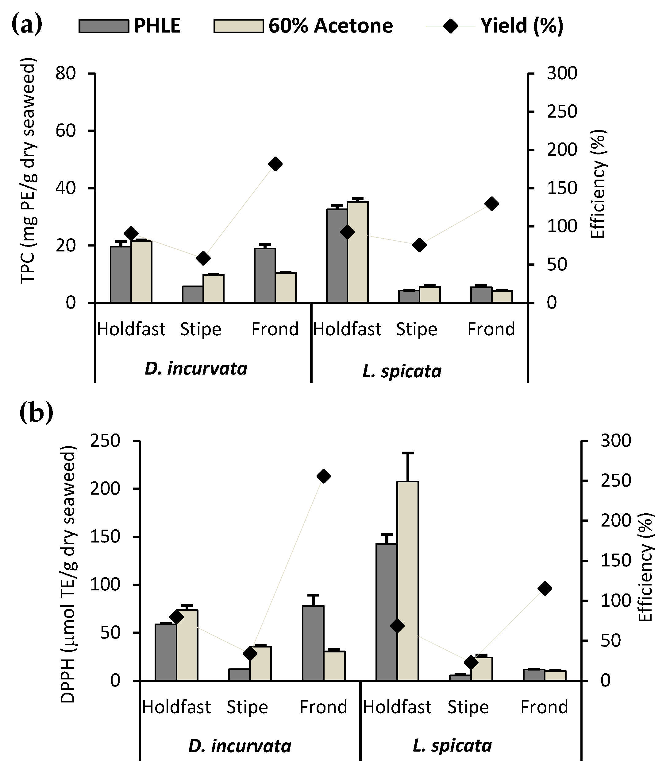
3.1. Effectivity of PHLE with 15% v/v Glycerol-Water against Maceration with 60% Acetone

Glycerol is a low-cost, food-grade, and environmentally friendly (bioderived, recyclable, and degradable) solvent [25]. Glycerol-water mixtures have been proven quite effective in extracting polyphenols from different plant matrices [22,25,26]. Therefore, we used a previously optimized glycerol-water PHLE process (15% v/v, 150 °C, 1500 psi) [26] to extract phlorotannins from D. incurvata and L. spicata. Maceration with 60% acetone (30 °C, 1 h) was used as the reference method because of its capacity to achieve high polyphenol yields from seaweeds [10,18].
In terms of TPC, the PHLE efficiencies (TPC PHLE extracts/TPC acetone extracts) varied according to the seaweed part used, showing a similar trend in both species: frond > holdfast > stipe (Figure 1a). The frond of D. incurvata presented a PHLE-TPC efficiency close to 180%, whereas that of L. spicata fronds was close to 130%. Both holdfasts showed a PHLE-TPC efficiency close to 100%, whereas that of the stipes were about 60%. A similar tendency was shown in the PHLE-AC efficiencies, measured as DPPH RSA (Figure 1b). The discrepancies among anatomical parts could be attributed to the solvents’ capacity to differentially extract distinct phlorotannin types according to their polarities and the unequal distribution of each phlorotannin type in the seaweed thallus. It has been proven that acetone increases the extraction efficiency of tannins associated with proteins because it can inhibit the phlorotannin–protein interaction [40].
The 15% v/v glycerol-water PHLE process can be considered an effective method to obtain phlorotannin extracts from the different thallus parts of D. incurvata and L. spicata, as it showed extraction efficiencies ranging from moderate to far above those of the comparison method, accomplished in a third of the time.

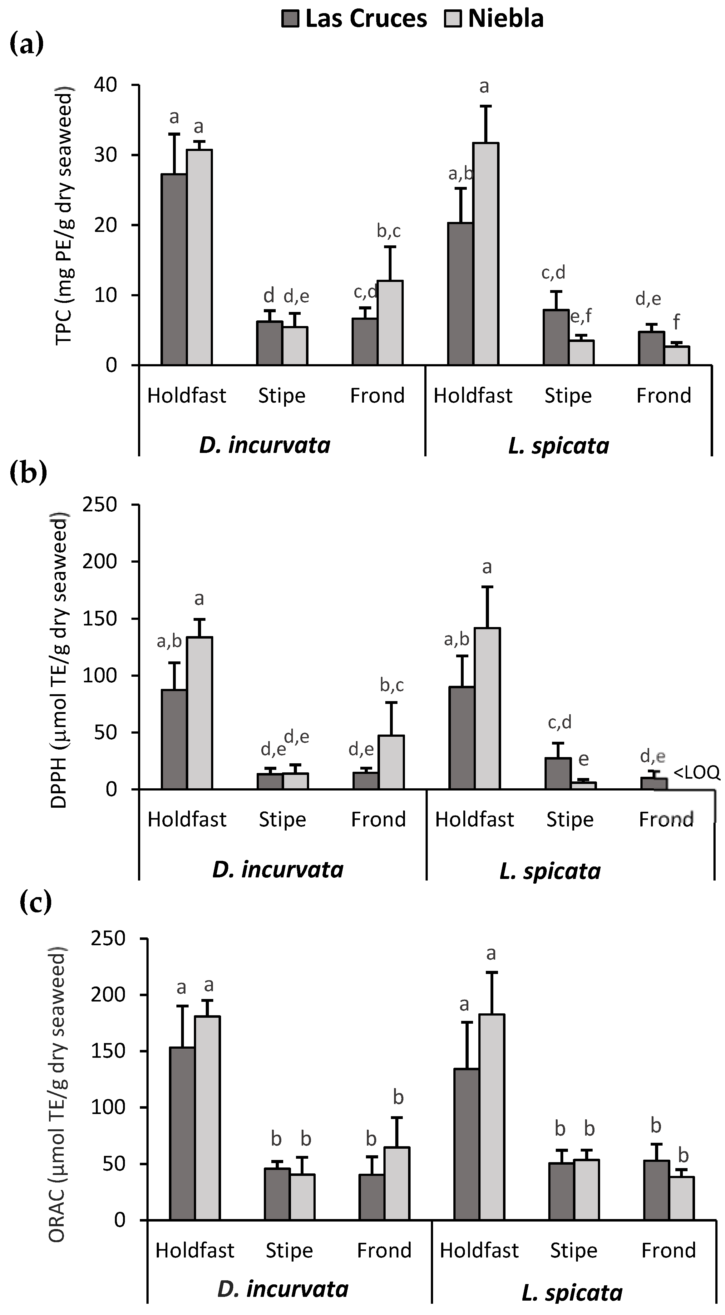
3.2. Effect of the Species, Harvesting Area, and Thallus Part Used on the TPC and AC of D. incurvata and L. spicata

This study’s goal was to investigate the conditions for the potential production of food-grade and high-phlorotannin-content extracts from D. incurvata and L. spicata using a previously optimized 15% v/v glycerol-water PHLE procedure. The influence of the species, harvesting area, and thallus part used on the TPC and AC of the extracts were explored.
As noted in Figure 2a, the factor that exerted the most significant influence on the TPC was the anatomical part used, with holdfasts showing the highest values (27.5 ± 6.3 mg PE/g dry seaweed). As expected, the same occurred for both AC measurements, DPPH RSA and ORAC (Figure 2b,c), with holdfasts showing mean values of 113 ± 25 and 163 ± 38 μmol TE/g dry seaweed, respectively. As verified by three-way ANOVA, the species also significantly affected TPC and DPPH RSA values, with D. incurvata showing the highest values. No statistically significant effect of harvesting area on TPC or AC was found; however, significant second-order interactions were detected (Tables S1–S3).
DPPH RSA and ORAC presented the same trends. The correlation coefficients of both variables with TPC were high and not statistically different (0.96 vs. 0.97). DPPH RSA quantifies the antioxidants’ capacity to scavenge the artificial radical DPPH in organic media (e.g., methanol), whereas ORAC measures their ability to inhibit peroxyl radicals generated in aqueous media under physiological conditions. Thus, as peroxyl radicals are the predominant free radicals in biological systems, ORAC analysis is thought to be more suitable than DPPH RSA for determining the AC of compounds for pharmaceutical or food applications [41]. Another disadvantage of the DPPH RSA assay is related to the structure of DPPH; since the DPPH molecule is sterically hindered, high-molecular-weight compounds, such as phlorotannins, do not have full access to the radical site. Moreover, other compounds can impede the antioxidant from reaching the DPPH molecule [42].
In summary, regardless of the species (D. incurvata and L. spicata) and area (Las Cruces and Niebla), the holdfast showed the highest TPC and AC values. Hence, according to bio-ecological recommendations for a sustainable seaweed fishery, the entire alga’s utilization, including the holdfast, is suggested in order to recover the most phlorotannins from these species [43].

3.3. Extraction Yield, TPC, and AC of the PHLE Extract

TPC and AC were also expressed on a dry extract basis to determine the D. incurvata and L. spicata PHLE extracts’ technological value (Table 3). The extracts’ total solids were quantified, and results are presented as extraction yield values (% w/w, g extracted solids/100 g dry seaweed). Extraction yields were similar among the twelve groups, with only DLH and LNF being significantly different, with 36.8% ± 6.6% and 22.9% ± 3.7%, respectively. Yields in a similar range (20.4%–36.9%) were obtained with an optimized ultrasound-assisted extraction (UAE) procedure (50% v/v ethanol-water, 35 kHz, 30 min, 1:10 w/v) applied in ten Phaeophyta species. When the same samples were extracted with 50% ethanol-water using a conventional method (200 rpm, 20 °C, 4 h, 2 cycles, 1:15 w/v), yields were 50% smaller [44]. Using a UAE treatment similar to that of Ummat et al. (2020) [44], Agregán et al. (2018) [45] reported high yields in Ascophyllum nodosum and Bifurcaria bifurcata extracts (25.9% and 35.9%, respectively). Lower yields (15.5%–31.6%) were obtained using PHLE (95% v/v ethanol-water, 1500 psi, 160 °C, 20 min) to extract phlorotannins from S. muticum collected on the North Atlantic coasts [27]. Applying conventional methods (200 rpm, 24 h, 1:25 w/v), extraction yields from F. vesiculosus of 12.0% with 80% ethyl acetate at room temperature and 37.8% with water at 70 °C were obtained [10].
The values of TPC and AC expressed in terms of the extracts’ dry weight presented a variability similar to TPC and AC expressed as dry seaweed. Holdfast extracts showed the highest TPCs (mean ± SD: 95 ± 24 mg PE/g dry extract), with values ranging from 75 ± 14 to 129 ± 30 mg PE/g dry extract. Fronds and stipes ranged from 11.7 ± 1.8 to 41 ± 21 mg PE/g dry extract. Holdfast extracts’ TPCs were similar to those previously quantified in 95% ethanol-water PHLE extracts of S. muticum (75.43–148.97 mg PE/g dry extract or 1.02–4.70 g PE/100 g dry seaweed) [27]. Nevertheless, they were much lower (2–5-fold) than those systematically reported for other brown seaweed extracts, such as F. vesiculosus and A. nodosum acetone- or ethanol-water extracts prepared using maceration [10,46] or UAE [44,45]. D. antarctica and L. spicata extracts analyzed in previous studies presented TPC values similar to or lower than those obtained in this study. Conventional 50% ethanol-water extracts from D. antarctica and L. spicata collected in summer showed TPCs of 0.5 and 0.1 g PE/100 g dry seaweed [47], whereas 70% acetone-water extracts from fronds and stipes of L. spicata harvested in spring showed values of 28.02 and 40.20 mg PE/g dry extract [48].
Holdfast extracts showed the highest values in both AC assays; DPPH RSA values ranged from 238 ± 50 to 580 ± 180 μmol TE/g dry extract (mean ± SD: 400 ± 140 μmol TE/g dry extract) and ORAC values from 416 ± 73 to 740 ± 190 μmol TE/g dry extract (mean ± SD: 560 ± 130 μmol TE/g dry extract). Frond and stipe extract AC values were much lower; DPPH RSA results ranged from <LOQ to 160 ± 120 μmol TE/g dry extract and ORAC values from 126 ± 36 to 220 ± 110 μmol TE/g dry extract. The DPPH activities of the holdfast extracts described here were comparable to those of 50% ethanol-water UAE extracts from high-polyphenol-content Phaeophytas species, such as F. vesiculosus, A. nodosum, and B. bifurcata, which showed values in the range of 100–400 μmol TE/g dry extract [44,45]. However, their ORAC values were lower than those described for UAE extracts (50% ethanol-water), ranging from 1100 to 1600 μmol TE/g dry extract [45], and those reported for conventional methanolic extracts, which showed values between 1200 and 4200 μmol TE/g dry extract [49]. Our results were comparable to conventional 50% ethanol-water extracts obtained from brown seaweeds from the Northwest Mexican Pacific coast (190–820 μmol TE/g dry extract) [50]. All PHLE extracts characterized here presented higher ORAC values than those previously reported for D. antarctica and L. spicata 50% ethanol-water extracts [47].
The D. incurvata and L. spicata PHLE extracts presented similar yields and DPPH RSA values but lower TPCs than food-grade extracts of some well-studied Fucales species. Moreover, they showed comparable or higher TPC and AC values than those previously reported for D. antarctica and L. spicata conventional extracts. It is worth pointing out that direct comparisons among species or studies are not entirely accurate. Differences in the extraction conditions, the harvesting area and season, and other environmental factors to which the seaweeds were exposed could affect the polyphenol content and the antioxidant performance of the extracts.

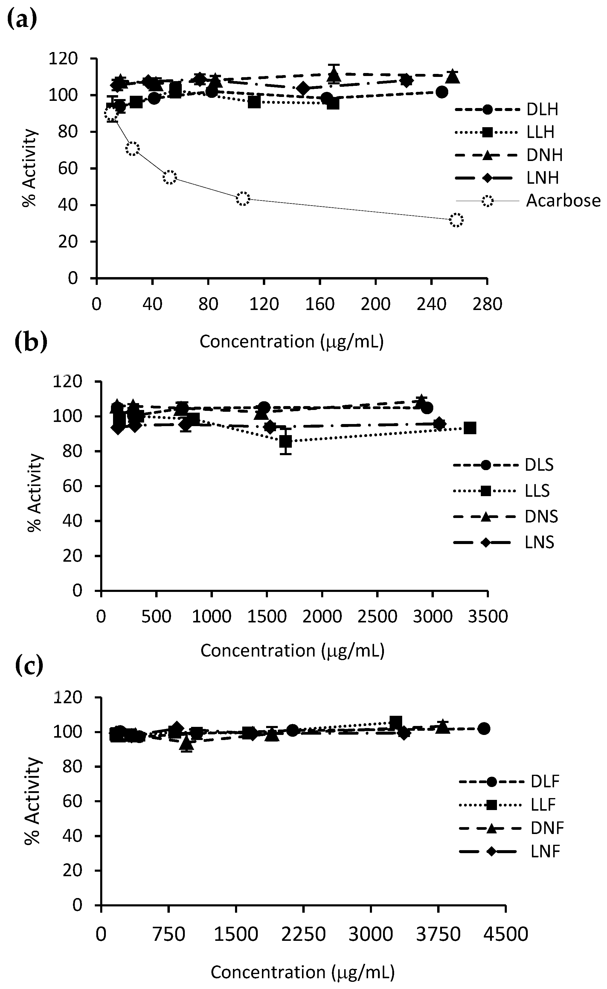
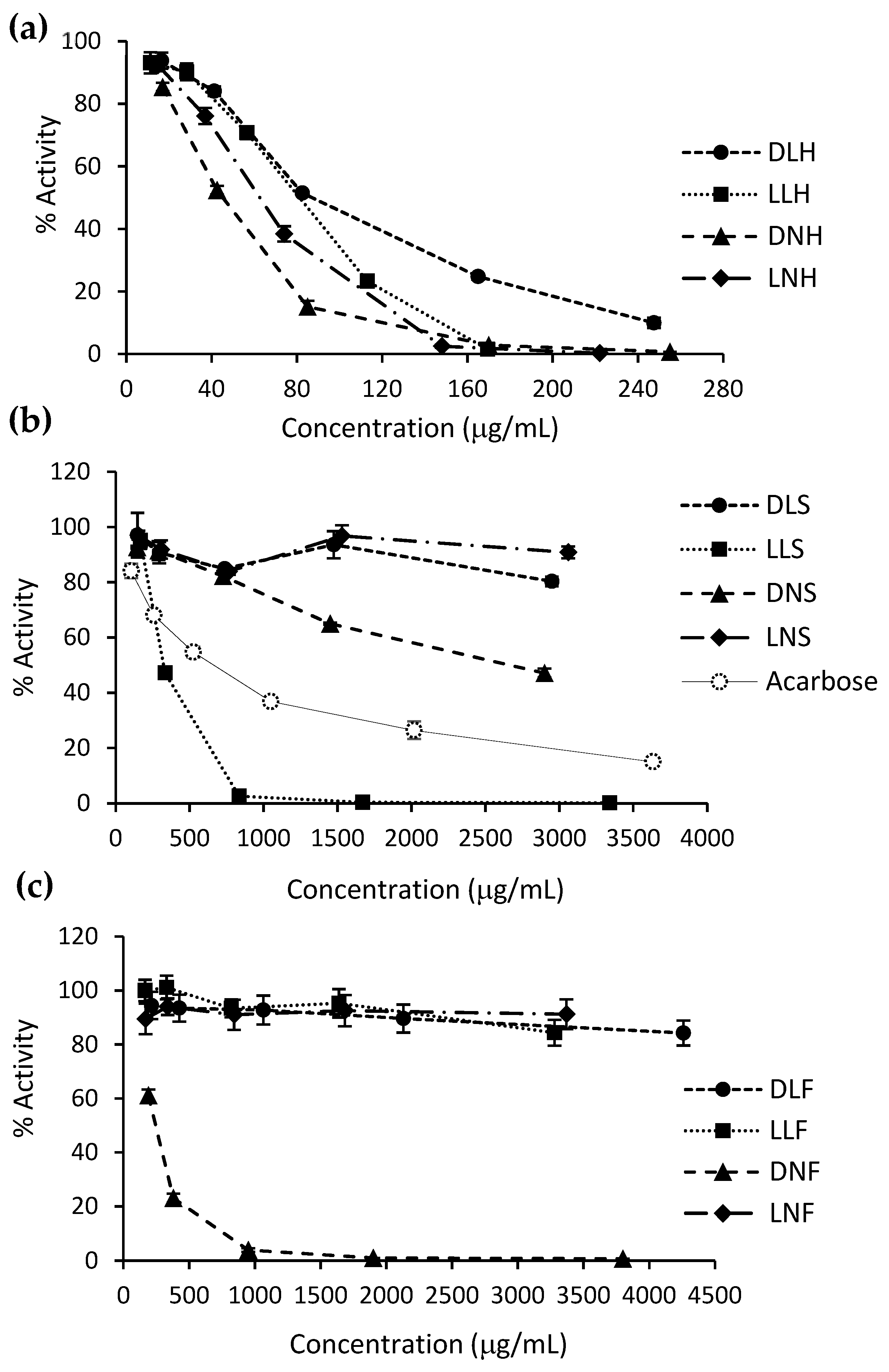
3.4. Carbohydrate-Hydrolyzing Enzymes’ Inhibitory Activity

Postprandial hyperglycemia is an essential factor in developing insulin resistance and cardiovascular events in type 2 diabetes [51]. The inhibition of carbohydrate-hydrolyzing enzymes—α-glucosidase and α-amylase—is critical in reducing postprandial glycemia and preventing diabetes or diabetes complications [52,53]. Brown seaweed extracts have shown the capacity to reduce the activity of α-glucosidase or both enzymes at the same level as, or even more than, commercial hypoglycemic drugs (e.g., acarbose and miglitol) [18,53]. The α-glucosidase and α-amylase inhibition capacity of the PHLE extracts were assessed. Holdfast extracts were analyzed in the range of 10–260 μg/mL and frond and stipe extracts from 150 to 4000 μg/mL.
For α-glucosidase, all holdfast extracts (Figure 3a) and three extracts from fronds and stipes (Figure 3b,c) displayed an inhibitory activity above 50% in the assayed ranges. LNH, LLH, and DNH extracts almost completely inhibited the enzyme from around 160 μg/mL, whereas LLS and DNF inhibited the enzyme from about 1000 μg/mL. As seen in Table 4, and according to the one-way ANOVA and Tukey post hoc tests (Tables S4 and S5), the DNH extract presented the highest α-glucosidase inhibitory capacity, with an IC50 value of 45.2 ± 1.6 μg/mL, i.e., around fifteen-fold lower than that of acarbose (659.5 ± 36.7 μg/mL). Except for DNS, the other active extracts showed IC50 values far lower than acarbose, ranging from 62.6 ± 2.4 to 324.1 ± 6.5 μg/mL. These inhibition results positively correlate with the TPC, DPPH RSA, and ORAC values of the extracts, showing correlation coefficients of 0.90, 0.93, and 0.87, respectively (not statistically different). It seems that holdfast polyphenols are more bioactive than stipe and frond polyphenols since the differences between holdfast and stipe/frond extracts are more significant in glucosidase inhibition activity than in TPC and AC.
Different α-glucosidase inhibitory capacity results have been found in other brown seaweed species. For instance, F. vesiculosus acetone-water extracts have shown IC50 values ranging from eight-fold lower than acarbose (32 ± 3 vs. 264 ± 41 μg/mL) to two thousand-fold lower than acarbose (0.34 ± 0.01 vs. 720 ± 10 μg/mL) [18,54,55]. Ethanolic and acetone-water A. nodosum extracts presented IC50 values ranging between twenty and two thousand-fold lower than acarbose [54]. Although the ORAC values found in Mexican seaweed ethanolic extracts were comparable to some of the PHLE extracts, their α-glucosidase inhibition capacities were far lower (one to four-fold lower than the acarbose IC50) [50]. Previous studies with D. antarctica and Lessonia extracts have presented lower α-glucosidase inhibitory activities than those found in this research. Pacheco et al. (2020) [34] reported IC50 values two times lower and seven times higher than acarbose, respectively, for southern Chilean D. antarctica’s and L. spicata’s PHLE ethanol-water extracts (473 ± 0.9 and 5318 ± 0.8 μg/mL vs. 798 ± 1.1 μg/mL). Yuan et al. (2018) [56] found IC50 values to be four times lower than those of acarbose for methanol-water extracts from Lessonia trabeculata and observed no α-glucosidase inhibitory activity for similar L. nigrescens extracts.
Regarding the α-amylase inhibitory capacity, all PHLE extracts proved to be inactive at the assayed concentrations (Figure 4), whereas acarbose showed an IC50 of 75.5 ± 4.7 μg/mL. These results are consistent with previous studies [34]. Even though some authors have reported brown seaweed extracts with similar inhibitory effects to those of acarbose, such as cold water extracts of A. nodosum and F. vesiculosus [53], in general, they have demonstrated low α-amylase inhibitory activity [18,50,57]. This behavior might be beneficial in terms of glycemic homeostasis. It has been observed that the initial starch digestion carried out in the mouth by α-amylase triggers a preabsorptive insulin release (cephalic phase), preparing the body for the upcoming glucose [58].
Overall, the D. incurvata and L. spicata food-grade PHLE extracts, especially those from the holdfast, constitute a promising alternative to traditional α-glucosidase inhibitors. Similar extracts from the same species demonstrated no cytotoxic effects in human colon adenocarcinoma HT-29 cells at the inhibitory concentrations reported here and up to 1000 μg/mL [34]. Although the α-glucosidase inhibitory capacity has been mainly correlated with phlorotannins, some authors have found the same capacity in fucoxanthin- and fucoidan-rich brown seaweed extracts [18,59,60]. Therefore, further characterization analyses or purification steps must be performed in D. incurvata and L. spicata PHLE extracts to verify the association between α-glucosidase inhibition and the extracts’ phlorotannin content.

3.5. Presence of Contaminants: Mannitol, HMF, and Heavy Metals

Crude extracts contain desired metabolites and co-extracted matrix components that could affect their applicability and safety as food or pharmaceutical ingredients. Significant glucose and fructose concentrations have been detected in high-polyphenol-content PHLE extracts from grape pomace [22]. Additionally, high extraction temperatures may produce potential carcinogens derived from non-enzymatic browning reactions, such as HMF, limiting the extracts’ applicability [61]. Therefore, to evaluate the relevance of seaweed PHLE extracts as potential nutraceuticals or functional food ingredients, we quantified mannitol, heavy metals, and HMF.

3.5.1. Mannitol and HMF in PHLE Extracts

Carbohydrates are the major components of brown seaweeds. They include fucoidan, laminarin, alginate, and mannitol. Given its relevance as an energy source, mannitol is one of the most abundant carbohydrates in seaweeds, accounting for 3–21%—even up to 30%—of the alga’s dry weight, depending on the species, season, and growth conditions [62]. The mannitol concentrations of the PHLE extracts, expressed as dry seaweed and dry extract, are presented in Table 5. According to a three-way ANOVA and Tukey post hoc tests (Tables S6 and S7), D. incurvata showed significantly higher mannitol contents than L. spicata (means ± SD: 11.5% ± 3.5% vs. 5.6% ± 1.3%), and the Las Cruces samples contained more mannitol than the Nieblas samples (means ± SD: 10.0% ± 4.8% vs. 7.2% ± 2.5%). In general, mannitol contents ranged from 3.6% ± 0.8% to 16.4% ± 3.0% of dry seaweed, similar to those reported previously in brown algae [63,64]. Mannitol concentrations expressed in a dry extract basis ranged from 147 ± 42 to 473 ± 59 mg/g, that is, up to 50% of the extract weight. These large concentrations are relevant, as mannitol has been found to produce abdominal symptoms in people with irritable bowel syndrome [65]. Otherwise, mannitol has been described as a free radical scavenger [66], interfering with TPC and AC measurements.
HMF has been reported as a by-product of PHLE processes performed at high temperatures (over 120 °C) in various plant materials [24]. As HMF has been recognized as a cytotoxic compound and a potential carcinogen for humans [67], its content in the PHLE extracts was assessed. No detectable concentration of HMF was found in any extract (LOD: 0.012 mg/L). The generation of HMF has been commonly described as a side reaction of the thermal acid hydrolysis of seaweeds (e.g., 121 °C, 0.5 M HCl, 15 min), a typical pretreatment for bioethanol production [68,69]. Mannitol is not a good HMF precursor, as its hydrothermal decomposition (0.5 % w/w, 10 min, 5 MPa) produced low levels of HMF (up to 5 μM) at temperatures under 220 °C [70]. The lack of HMF in the extracts could also be attributed to mannitol’s capacity to form adducts with specific amino acids [71].

3.5.2. Heavy Metals in Dry Seaweeds and PHLE Extracts

The world’s per capita intake of algae is continuously growing, raising concerns about the hazardous concentrations of toxic metals they may contain [7,72]. Concentrations of Cd and As above the maximum permitted levels have been identified in several edible seaweeds and seaweed-derived products [73,74]. In general, brown algae have a higher biosorption capacity than red and green algae; in fact, many countries recommend avoiding the consumption of the brown seaweed Hijiki (Sargassum fusiforme) due to its potentially dangerous levels of inorganic As, the toxic form of this metalloid [7]. Only a few countries regulate the maximum levels of heavy metals in algae and derived foods. The French law is the most complete in this regard, establishing limits for inorganic As (3 mg/kg dw), Cd (0.5 mg/kg dw), Pb (5 mg/kg dw), Hg (0.1 mg/kg dw), and Sn (5 mg/kg dw) for edible seaweeds [75]. The content of these heavy metals in the seaweed samples and the PHLE extracts are presented in Table 6.
The concentrations of total As in the seaweed samples ranged from 8.29 to 84.08 mg/kg dw. According to a three-way ANOVA and Tukey post hoc tests (Tables S8 and S9), holdfasts showed higher levels than stipes and fronds (means ± SD: 49.7 ± 25.4 mg/kg dw vs. 16.7 ± 9.6 and 21.2 ± 8.2 mg/kg dw), and samples of L. spicata from Niebla presented higher concentrations than their counterparts. Differences in the As content among alga parts and locations were also seen in the Phaeophyta Alaria nana (Alaska), with holdfasts showing the highest levels [76]. Arsenic values described here were in the range reported for other brown seaweeds, including species from Laminaria and Sargassum genus and Eisenia bicyclis (arame) [73,75,77,78]. However, the As concentrations in fronds of this study were smaller. The arsenic content of D. incurvata and L. spicata fronds (means ± SD: 7.9 ± 4.9 and 27.8 ± 2.1 mg/kg dw) were lower than those previously reported for D. antarctica and L. nigrescens (49.0 ± 34.5 and 57.1 ± 22.8 mg/kg dw) [79]. Although inorganic As (iAS) was not assessed in this study, it is known that the iAS content of brown seaweeds is not greater than 10% (and generally < 2%) of the total As, with the exception of S. fusiforme, in which the iAS level can reach up to 80% of the total As [74,75,76]. iAS concentrations representing 0.6% of the total As (0.31 and 0.35 mg/kg dw) were found in the abovementioned D. antarctica and L. nigrescens samples [79]. Therefore, the iAS contents of the studied species D. incurvata and L. spicata would probably not exceed the maximum limits established in some countries (France/USA: 3 mg/kg dw, Australia/New Zealand: 1 mg/kg dw) [80], although this must be further confirmed. On the other hand, the total As concentrations of derived extracts ranged between 14.88 and 258.47 mg/kg dw. These higher values mean that all or almost all the seaweed’s As content was extracted by the PHLE process. Thus, the dry PHLE extracts are more likely to have iAs levels close to or even above the maximum acceptable limits.
The cadmium concentrations in the seaweed samples ranged from 0.84 to 4.81 mg/kg dw, and no clear trend could be observed (mean ± SD: 2.2 ± 1.2 mg/kg dw), although L. spicata’s values were slightly higher than D. incurvata’s. These Cd contents were higher than those found in the Laminaria species E. byciclis and Undaria pinnatifida (wakame) from Europe; and were at the same level or even lower than those described for the Sargassum species U. pinnatifida from Asia and Fucus spiralis [72,73,75,77,78,81]. Sáez et al. (2012) [82] reported high Cd concentrations (0.5–3.5 mg/kg dw) for the different thallus parts of L. trabeculata harvested in Chile; the holdfast showed higher concentrations than the stipe and the latter showed higher values than the frond. A Cd level of 2.46 mg/kg dw was detected in a Chilean D. antarctica sample, which is higher than the mean of the D. incurvata samples in this study (1.3 ± 0.5 mg/kg dw) [7]. All samples reported here greatly exceeded the maximum permitted concentrations established by French and Australian/New Zealand regulations for edible seaweeds (0.5 and 0.2 mg/kg dw) [73]. Hence, their consumption could represent a risk for human health in terms of Cd intake. The contribution of the edible seaweed D. incurvata to the total daily intake (TDI) of Cd must be determined, especially considering that other marine products and some vegetables are also Cd accumulators [83,84]. In the other PHLE extracts, Cd concentrations ranged between 0.20 and 6.13 mg/kg dw, with only holdfast extracts showing values over 0.38 mg/kg dw. The Cd values of frond and stipe extracts were lower than those of the dry seaweed parts, indicating that the PHLE process extracted just a small amount of Cd from the source matrix. Concerning the EU regulations for seaweed-derived foods, 25% of the extracts exceeded the limit established for “food supplements consisting exclusively or mainly of seaweed, or products derived from seaweed” (3 mg/kg ww) [85]. Cd concentrations well below the EU and French limits but above the Australian regulations were found in tablets and soluble concentrates of Fucus species (0.011–0.356 mg/kg dw) [73].
The concentration of Pb was below the LOQ of the analytical method (1.0 mg/kg dw) in all seaweed samples. Pb levels in brown seaweeds are usually under 1.0 mg/kg, although higher values have been detected in Sargassum species and U. pinnatifida [73,75,77]. In contrast to As and Cd, Pb values found in samples from the Norwegian coasts were lower in brown seaweeds than in red and green seaweeds [74]. Sáez et al. (2012) [82] reported values greater than 4 mg/kg dw in fronds of L. trabeculata harvested from Pb-polluted seawaters in Chile; however, Pb concentrations were below 1 mg/kg in stipes and holdfasts. No Pb content was detected in a Chilean D. antarctica sample [73]. All seaweed samples analyzed here presented Pb levels under the French limit (5 mg/kg dw), and therefore their consumption may not pose a risk for human health in terms of Pb toxicity. The Pb levels of the PHLE extracts ranged from 0.28 to 0.81 mg/kg dw. These low values mean that the PHLE process extracted just a small part of the Pb content from the seaweed matrix. The Pb content of all extracts was under the French limit for edible seaweeds. Pb concentrations well below (<LOD–0.25 mg/kg dw) and others much higher (11.3–14 mg/kg dw) than those of this study have been found in Fucus tablets [73].
Hg was not detected in any seaweed samples (LOD: 0.001 mg/kg dw) and therefore it was not analyzed in the extracts. In general, Hg contents in red, green, and brown algae are under 0.5 mg/kg dw. In brown seaweeds, concentrations below 0.01 mg/kg dw have been systematically reported for Laminaria and Fucus species, and slightly higher levels —0.03–0.05 mg/kg dw—have been found in U. pinnatifida, S. fusiforme, and Pelvetia canaliculata from Norway [74,75,78]. Higher than usual Hg concentrations (over 0.5 mg/kg dw) were reported by Squadrone et al. (2018) [86] in seaweed species from the Italian Mediterranean coasts. The low Hg contents of brown seaweeds should not be a concern according to the French legislation (limit: 0.1 mg/kg dw); however, special care must be taken considering the EU limit for edible seaweeds (0.01 mg/kg).
Concentrations of Sn in the seaweed samples ranged from not detected (LOD: 0.01 mg/kg dw) to the LOQ of the analytical method (2.00 mg/kg dw). On the other hand, Sn was not detected in the analyzed PHLE extracts. The literature about the Sn content in seaweeds is scarce because this metal is mainly associated with canned food [87]. Brown seaweeds from coastal areas of Italy showed levels under 1.0 mg/kg dw, except for Halopteris filicina, which presented an Sn content of 1.8 ± 0.04 mg/kg dw [86]. Padina pavonica and Cystoseira mediterranea collected in Sardinian coastal areas showed Sn values below 0.05 mg/kg dw [88]. Sn concentrations between 0.11–0.17 mg/kg dw were found in species belonging to the Chlorophyta Ulva genera grown in fishpond aquaculture systems [89]. Higher Sn levels (<1–34 mg/kg dw) were found in samples from edible seaweeds purchased in the Italian market [90]. The study reported levels close to or well above the French limit for Sn (5 mg/kg dw) in A. nodosum (4.7 ± 0.7 mg/kg dw) and Laminaria digitata (34.0 ± 5.1 mg/kg dw); interestingly, the concentrations of Cd and Pb were also above the available regulations in some seaweed samples.
In summary, potentially toxic Cd levels were found in D. incurvata and L. spicata from central and southern Chile, which is of concern since D. incurvata is the most consumed algae in Chile, and new D. incurvata-based foodstuffs are continuously emerging. The high As concentrations, also reported for both seaweeds, should not constitute a health risk due to the low iAs (the toxic As species) contents systematically detected in most seaweeds, particularly in D. incurvata; however, this remains to be confirmed. Studies suggest that continuous monitoring of the heavy metal contents of seaweeds must be performed in order to protect consumers’ health, as the concentrations fluctuate according to environmental factors (e.g., anthropic activity, location, season, and pH). As, Cd, and Sn levels were even higher in some PHLE extracts than in dry seaweeds due to the concentration effect of the extraction process. Since some of those metal contents exceeded the available regulations, and mannitol levels were also high—accounting for up to 50% of the extract dry weight—further purification steps are suggested if food or pharmaceutical applications are intended for the PHLE extracts.

4. Conclusions

Through an effective, environmentally friendly, and food-grade PHLE process, Durvillaea incurvata and Lessonia spicata phlorotannin extracts with high antioxidant and antihyperglycemic potential were obtained. A glycerol-based PHLE process was used for the first time in the extraction of the bioactive compounds of seaweed. The anatomical part of the alga significantly impacted the dry seaweeds’ and extracts’ polyphenol content and antioxidant activity, with the holdfast showing the highest values. No α-amylase inhibitory activity was found. Instead, holdfast extracts presented α-glucosidase inhibitory capacities several-fold higher than that of acarbose, with D. incurvata’s holdfast from Niebla being the most active. Elevated concentrations of mannitol and potentially toxic cadmium levels were found in seaweeds and extracts. Therefore, the utilization of the whole plant, including the holdfast, and the implementation of further purification steps are recommended to use these seaweed PHLE extracts as effective and safe antihyperglycemic agents.

Supplementary Materials

The supplementary materials are available online at https://www.mdpi.com/article/10.3390/antiox10071105/s1.

Author Contributions

Conceptualization, F.E., M.S.M.-C., J.P. and J.R.P.-C.; methodology, F.E., M.S.M.-C. and J.R.P.-C.; investigation, F.E. and F.P.; validation, formal analysis, and writing—original draft, F.E.; supervision, M.S.M.-C. and J.R.P.-C.; writing—review and editing, M.S.M.-C., J.P. and J.R.P.-C.; project administration and funding acquisition, J.R.P.-C. All authors have read and agreed to the published version of the manuscript.

Funding

This research was funded by ANID-Chile through the grants FONDECYT Regular #1180571 and #1201670. It also received financial support from “Programa de Ciencia y Tecnología para el Desarrollo (CYTED), Red Iberoamericana de Alimentos Autóctonos Subutilizados (ALSUB-CYTED, #118RT0543)”.

Institutional Review Board Statement

Not applicable.

Informed Consent Statement

Not applicable.

Data Availability Statement

Data are contained within the article and supplementary material.

Acknowledgments

This study was part of the Ph.D. thesis of the first author, F.E., carried out at the “Laboratorio de Extracción de Compuestos de Alto Valor (LECAV),” School of Engineering, PUC. F.E. thanks CONICYT Doctoral scholarship (CONICYT-PFCHA/Doctorado Nacional/2017-21170535). The authors appreciate the help of Juan Hernández (Environmental Biologist, Universidad de Chile) and Ramona Pinochet (Ph.D. (c) in Marine Biology, Universidad Austral de Chile) for harvesting and identifying the seaweed samples. We are also grateful to Sandy González (Food Engineer, Universidad Austral de Chile) for technical support. English editing by Lisa Gingles is highly appreciated.

Conflicts of Interest

The authors declare no conflict of interest.

References

  1. Peñalver, R.; Lorenzo, J.M.; Ros, G.; Amarowicz, R.; Pateiro, M.; Nieto, G. Seaweeds as a Functional Ingredient for a Healthy Diet. Mar. Drugs 2020, 18, 301. [Google Scholar] [CrossRef]
  2. Dillehay, T.D.; Ramírez, C.; Pino, M.; Collins, M.B.; Rossen, J.; Pino-Navarro, J.D. Monte Verde: Seaweed, food, medicine, and the peopling of South America. Science 2008, 320, 784–786. [Google Scholar] [CrossRef] [Green Version]
  3. Fleurence, J.; Levine, I.A. Seaweed in Health and Disease Prevention; Elsevier: Amsterdam, The Netherlands, 2016; pp. 370–371. [Google Scholar]
  4. Gunathilaka, T.L.; Samarakoon, K.; Ranasinghe, P.; Peiris, L.D.C. Antidiabetic Potential of Marine Brown Algae—a Mini Review. J. Diabetes Res. 2020, 2020, 1230218. [Google Scholar] [CrossRef]
  5. Rocha, D.H.A.; Seca, A.M.L.; Pinto, D. Seaweed Secondary Metabolites In Vitro and In Vivo Anticancer Activity. Mar. Drugs 2018, 16, 410. [Google Scholar] [CrossRef] [Green Version]
  6. Pérez, M.J.; Falqué, E.; Domínguez, H. Antimicrobial Action of Compounds from Marine Seaweed. Mar. Drugs 2016, 14, 52. [Google Scholar] [CrossRef] [Green Version]
  7. Cherry, P.; O’Hara, C.; Magee, P.J.; McSorley, E.M.; Allsopp, P.J. Risks and benefits of consuming edible seaweeds. Nutr. Rev. 2019, 77, 307–329. [Google Scholar] [CrossRef] [PubMed] [Green Version]
  8. Afonso, N.C.; Catarino, M.D.; Silva, A.M.S.; Cardoso, S.M. Brown Macroalgae as Valuable Food Ingredients. Antioxidants 2019, 8, 365. [Google Scholar] [CrossRef] [PubMed] [Green Version]
  9. Sanjeewa, K.K.A.; Kim, E.A.; Son, K.T.; Jeon, Y.J. Bioactive properties and potentials cosmeceutical applications of phlorotannins isolated from brown seaweeds: A review. J. Photochem. Photobiol. B Biol. 2016, 162, 100–105. [Google Scholar] [CrossRef] [PubMed]
  10. Wang, T.; Jonsdottir, R.; Liu, H.; Gu, L.; Kristinsson, H.G.; Raghavan, S.; Olafsdottir, G. Antioxidant capacities of phlorotannins extracted from the brown algae Fucus vesiculosus. J. Agric. Food Chem. 2012, 60, 5874–5883. [Google Scholar] [CrossRef] [PubMed]
  11. Koivikko, R. Brown Algal Phlorotannins: Improving and Applying Chemical Methods; University of Turku: Turku, Finland, 2008. [Google Scholar]
  12. Schoenwaelder, M.; Clayton, M.N. Physode formation in embryos of Phyllospora comosa and Hormosira banksii (Phaeophyceae). Phycologia 2000, 39, 9. [Google Scholar] [CrossRef]
  13. Mannino, A.; Micheli, C. Ecological Function of Phenolic Compounds from Mediterranean Fucoid Algae and Seagrasses: An Overview on the Genus Cystoseira sensu lato and Posidonia oceanica (L.) Delile. J. Mar. Sci. Eng. 2020, 8, 19. [Google Scholar] [CrossRef] [Green Version]
  14. Lee, H.A.; Lee, J.H.; Han, J.S. A phlorotannin constituent of Ecklonia cava alleviates postprandial hyperglycemia in diabetic mice. Pharm. Biol. 2017, 55, 1149–1154. [Google Scholar] [CrossRef] [Green Version]
  15. You, H.N.; Lee, H.A.; Park, M.H.; Lee, J.H.; Han, J.S. Phlorofucofuroeckol A isolated from Ecklonia cava alleviates postprandial hyperglycemia in diabetic mice. Eur. J. Pharmacol. 2015, 752, 92–96. [Google Scholar] [CrossRef]
  16. Lee, S.H.; Ko, S.C.; Kang, M.C.; Lee, D.H.; Jeon, Y.J. Octaphlorethol A, a marine algae product, exhibits antidiabetic effects in type 2 diabetic mice by activating AMP-activated protein kinase and upregulating the expression of glucose transporter 4. Food Chem. Toxicol. 2016, 91, 58–64. [Google Scholar] [CrossRef] [PubMed]
  17. Moon, H.E.; Islam, N.; Ahn, B.R.; Chowdhury, S.S.; Sohn, H.S.; Jung, H.A.; Choi, J.S. Protein tyrosine phosphatase 1B and a-glucosidase inhibitory Phlorotannins from edible brown algae, Ecklonia stolonifera and Eisenia bicyclis. Biosc. Biotechnol. Biochem. 2011, 75, 1472–1480. [Google Scholar] [CrossRef] [Green Version]
  18. Catarino, M.D.; Silva, A.M.S.; Mateus, N.; Cardoso, S.M. Optimization of Phlorotannins Extraction from Fucus vesiculosus and Evaluation of Their Potential to Prevent Metabolic Disorders. Mar. Drugs 2019, 17, 162. [Google Scholar] [CrossRef] [PubMed] [Green Version]
  19. Tanniou, A.; Serrano-León, E.; Vandanjon, L.; Ibañez, E.; Mendiola, J.A.; Cerantola, S.; Kervarec, N.; LaBarre, S.; Marchal, L.; Stiger-Pouvreau, V. Green improved processes to extract bioactive phenolic compounds from brown macroalga using Sargassum muticum as model. Talanta 2013, 104, 44–52. [Google Scholar] [CrossRef]
  20. Getachew, A.T.; Jacobsen, C.; Holdt, S.L. Emerging Technologies for the Extraction of Marine Phenolics: Opportunities and Challenges. Mar. Drugs 2020, 18, 389. [Google Scholar] [CrossRef]
  21. Zakaria, S.M.; Mazlina, S.; Kamal, M. Subcritical Water Extraction of Bioactive Compounds from Plants and Algae: Applications in Pharmaceutical and Food Ingredients. Food Eng. Rev. 2016, 8, 23–34. [Google Scholar] [CrossRef]
  22. Huamán-Castilla, N.L.; Mariotti-Celis, M.S.; Martínez-Cifuentes, M.; Pérez-Correa, J.R. Glycerol as Alternative Co-Solvent for Water Extraction of Polyphenols from Carmenere Pomace: Hot Pressurized Liquid Extraction and Computational Chemistry Calculations. Biomolecules 2020, 10, 474. [Google Scholar] [CrossRef] [Green Version]
  23. Huamán-Castilla, N.L.; Martínez-Cifuentes, M.; Camilo, C.; Pedreschi, F.; Mariotti-Celis, M.; Pérez-Correa, J.R. The Impact of Temperature and Ethanol Concentration on the Global Recovery of Specific Polyphenols in an Integrated HPLE/RP Process on Carmenere Pomace Extracts. Molecules 2019, 24, 3145. [Google Scholar] [CrossRef] [Green Version]
  24. Ozkaynak Kanmaz, E. 5-Hydroxymethylfurfural (HMF) formation during subcritical water extraction. Food Sci. Biotechnol. 2018, 27, 981–986. [Google Scholar] [CrossRef] [PubMed]
  25. Makris, D.P.; Lalas, S. Glycerol and Glycerol-Based Deep Eutectic Mixtures as Emerging Green Solvents for Polyphenol Extraction: The Evidence So Far. Molecules 2020, 25, 5842. [Google Scholar] [CrossRef] [PubMed]
  26. Huamán-Castilla, N.L.; Campos, D.; García-Ríos, D.; Parada, J.; Martínez-Cifuentes, M.; Mariotti-Celis, M.S.; Pérez-Correa, J.R. Chemical Properties of Vitis Vinifera Carménère Pomace Extracts Obtained by Hot Pressurized Liquid Extraction, and Their Inhibitory Effect on Type 2 Diabetes Mellitus Related Enzymes. Antioxidants 2021, 10, 472. [Google Scholar] [CrossRef] [PubMed]
  27. Montero, L.; Sánchez-Camargo, A.P.; García-Cañas, V.; Tanniou, A.; Stiger-Pouvreau, V.; Russo, M.; Rastrelli, L.; Cifuentes, A.; Herrero, M.; Ibáñez, E. Anti-proliferative activity and chemical characterization by comprehensive two-dimensional liquid chromatography coupled to mass spectrometry of phlorotannins from the brown macroalga Sargassum muticum collected on North-Atlantic coasts. J. Chromatogr. A 2016, 1428, 115–125. [Google Scholar] [CrossRef]
  28. Fraser, C.I.; Velásquez, M.; Nelson, W.A.; Macaya, E.C.; Hay, C.H. The Biogeographic Importance of Buoyancy in Macroalgae: A Case Study of the Southern Bull-Kelp Genus Durvillaea (Phaeophyceae), Including Descriptions of Two New Species. J. Phycol. 2019, 56, 23–36. [Google Scholar] [CrossRef] [PubMed]
  29. González, A.; Beltrán, J.; Hiriart-Bertrand, L.; Flores, V.; de Reviers, B.; Correa, J.A.; Santelices, B. Identification of Cryptic Species in the Lessonia Nigrescens Complex (Phaeophyceae, Laminariales). J. Phycol. 2012, 48, 1153–1165. [Google Scholar] [CrossRef] [PubMed]
  30. Singleton, V.L.; Rossi, J.A. Colorimetry of Total Phenolics with Phosphomolybdic-Phosphotungstic Acid Reagents. Am. J. Enol. Vitic. 1965, 16, 144–158. [Google Scholar]
  31. Tierney, M.; Smyth, T.; Rai, D.; Soler-Vila, A.; Croft, A.; Brunton, N. Enrichment of polyphenol contents and antioxidant activities of Irish brown macroalgae using food-friendly techniques based on polarity and molecular size. Food Chem. 2013, 139, 753–761. [Google Scholar] [CrossRef] [PubMed]
  32. Li, Y.; Fu, X.; Duan, D.; Liu, X.; Xu, J.; Gao, X. Extraction and Identification of Phlorotannins from the Brown Alga Sargassum fusiforme (Harvey) Setchell. Mar. Drugs 2017, 15, 49. [Google Scholar] [CrossRef] [Green Version]
  33. Huang, D.; Ou, B.; Hampsch-Woodill, M.; Flanagan, J.; Prior, R. High-Throughput Assay of Oxygen Radical Absorbance Capacity (ORAC) Using a Multichannel Liquid Handling System Coupled with a Microplate Fluorescence Reader in 96-Well Format. J. Agric. Food Chem. 2002, 50, 4437–4444. [Google Scholar] [CrossRef]
  34. Pacheco, L.V.; Parada, J.; Pérez-Correa, J.R.; Mariotti-Celis, M.S.; Erpel, F.; Zambrano, A.; Palacios, M. Bioactive Polyphenols from Southern Chile Seaweed as Inhibitors of Enzymes for Starch Digestion. Mar. Drugs 2020, 18, 353. [Google Scholar] [CrossRef] [PubMed]
  35. ICH. FDA Guidance for Industry: Q2B Validation of Analytical Procedures: Methodology; Food and Drug Administration: Rockville, MD, USA, 1996. [Google Scholar]
  36. Toker, O.S.; Dogan, M.; Ersoz, N.B.; Yilmaz, M.T. Optimization of the content of 5-hydroxymethylfurfural (HMF) formed in some molasses types: HPLC-DAD analysis to determine effect of different storage time and temperature levels. Ind. Crop. Prod. 2013, 50, 137–144. [Google Scholar] [CrossRef]
  37. Leege, P.; Millner, P.; Thompson, W.; Watson, M. Test methods for the Examination of Composting and Compost; United States Department of Agriculture and Composting Council Research and Education Foundation: Raleigh, NC, USA, 2002. [Google Scholar]
  38. SISS. Determinación de Arsénico total por Método espectrofotometría de absorción atómica con generación de hidruros. In Manual de Métodos de Ensayo Para Agua Potable, 2nd ed.; Superitendencia de Servicios Sanitarios: Santiago, Chile, 2007; pp. 101–110. [Google Scholar]
  39. APHA. Standard Methods for the Examination of Water and Wastewater, 2nd ed.; American Public Health Association, American Water Works Association, Water Environment Federation: Washington, DC, USA, 2012; p. 724. [Google Scholar]
  40. Hagerman, A.E. Extraction of tannin from fresh and preserved leaves. J. Chem. Ecol. 1988, 14, 453–461. [Google Scholar] [CrossRef]
  41. Zhong, Y.; Shahidi, F. Methods for the assessment of antioxidant activity in foods. In Handbook of Antioxidants for Food Preservation; Sahidi, F., Ed.; Woodhead Publishing: Cambridge, UK, 2015. [Google Scholar]
  42. Schaich, K.; Tian, X.; XIe, J. Hurdles and pitfalls in measuring antioxidant efficacy: A critical evaluation of ABTS, DPPH, and ORAC assays. J. Funct. Foods 2015, 14, 111–125. [Google Scholar] [CrossRef]
  43. Vásquez, J. The Brown Seaweeds Fishery in Chile. In Fisheries and Aquaculture in the Modern World; Mikkola, H., Ed.; IntechOpen: London, UK, 2016; pp. 123–141. [Google Scholar]
  44. Ummat, V.; Tiwari, B.K.; Jaiswal, A.K.; Condon, K.; Garcia-Vaquero, M.; O’Doherty, J.; O’Donnell, C.; Rajauria, G. Optimisation of Ultrasound Frequency, Extraction Time and Solvent for the Recovery of Polyphenols, Phlorotannins and Associated Antioxidant Activity from Brown Seaweeds. Mar. Drugs 2020, 18, 250. [Google Scholar] [CrossRef] [PubMed]
  45. Agregán, R.; Munekata, P.E.S.; Franco, D.; Carballo, J.; Barba, F.J.; Lorenzo, J.M. Antioxidant Potential of Extracts Obtained from Macro-(Ascophyllum nodosum, Fucus vesiculosus and Bifurcaria bifurcata) and Micro-Algae (Chlorella vulgaris and Spirulina platensis) Assisted by Ultrasound. Medicines 2018, 5, 33. [Google Scholar] [CrossRef] [Green Version]
  46. Heffernan, N.; Brunton, N.P.; FitzGerald, R.J.; Smyth, T.J. Profiling of the molecular weight and structural isomer abundance of macroalgae-derived phlorotannins. Mar. Drugs 2015, 13, 509–528. [Google Scholar] [CrossRef]
  47. Olate-Gallegos, C.; Barriga, A.; Vergara, C.; Fredes, C.; García, P.; Giménez, B.; Robert, P. Identification of Polyphenols from Chilean Brown Seaweeds Extracts by LC-DAD-ESI-MS/MS. J. Aquat. Food Prod. Technol. 2019, 28, 17. [Google Scholar] [CrossRef]
  48. Beratto-Ramos, A.; Castillo-Felices, R.; Troncoso-León, N.; Agurto-Muñoz, A.; Agurto-Muñoz, C. Selection criteria for high-value biomass: Seasonal and morphological variation of polyphenolic content and antioxidant capacity in two brown macroalgae. J. Appl. Phycol. 2018, 31, 653–664. [Google Scholar] [CrossRef]
  49. Pinteus, S.; Silva, J.; Alves, C.; Horta, A.; Fino, N.; Rodrigues, A.I.; Mendes, S.; Pedrosa, R. Cytoprotective effect of seaweeds with high antioxidant activity from the Peniche coast (Portugal). Food Chem. 2017, 218, 591–599. [Google Scholar] [CrossRef] [PubMed]
  50. Múzquiz de la Garza, A.R.; Tapia-Salazar, M.; Maldonado-Muniz, M.; de la Rosa-Millán, J.; Gutiérrez-Uribe, J.A.; Santos-Zea, L.; Barba-Dávila, B.A.; Ricque-Marie, D.; Cruz-Suárez, L.E. Nutraceutical Potential of Five Mexican Brown Seaweeds. Biomed. Res. Int. 2019, 2019, 3795160. [Google Scholar] [CrossRef] [PubMed]
  51. Ceriello, A. Postprandial hyperglycemia and diabetes complications: Is it time to treat? Diabetes 2005, 54, 1–7. [Google Scholar] [CrossRef] [PubMed] [Green Version]
  52. Krentz, A.J.; Bailey, C.J. Oral antidiabetic agents: Current role in type 2 diabetes mellitus. Drugs 2005, 65, 385–411. [Google Scholar] [CrossRef] [PubMed]
  53. Lordan, S.; Smyth, T.J.; Soler-Vila, A.; Stanton, C.; Ross, R.P. The a-amylase and a-glucosidase inhibitory effects of Irish seaweed extracts. Food Chem. 2013, 141, 2170–2176. [Google Scholar] [CrossRef] [PubMed]
  54. Liu, B.; Kongstad, K.T.; Wiese, S.; Jager, A.K.; Staerk, D. Edible seaweed as future functional food: Identification of a-glucosidase inhibitors by combined use of high-resolution a-glucosidase inhibition profiling and HPLC-HRMS-SPE-NMR. Food Chem. 2016, 203, 16–22. [Google Scholar] [CrossRef] [PubMed]
  55. Neto, R.T.; Marcal, C.; Queirós, A.S.; Abreu, H.; Silva, A.M.S.; Cardoso, S.M. Screening of Ulva rigida, Gracilaria sp., Fucus vesiculosus and Saccharina latissima as Functional Ingredients. Int. J. Mol. Sci. 2018, 19, 2987. [Google Scholar] [CrossRef] [PubMed] [Green Version]
  56. Yuan, Y.; Zhang, J.; Fan, J.; Clark, J.; Shen, P.; Li, Y.; Zhang, C. Microwave assisted extraction of phenolic compounds from four economic brown macroalgae species and evaluation of their antioxidant activities and inhibitory effects on α-amylase, α-glucosidase, pancreatic lipase and tyrosinase. Food Res. Int. 2018, 113, 288–297. [Google Scholar] [CrossRef]
  57. Apostolidis, E.; Lee, C.M. In vitro potential of Ascophyllum nodosum phenolic antioxidant-mediated α-glucosidase and α-amylase inhibition. J. Food Sci. 2010, 75, H97–H102. [Google Scholar] [CrossRef]
  58. Parada, J.; Pérez-Correa, J.R.; Pérez-Jiménez, J. Design of low glycemic response foods using polyphenols from seaweed. J. Funct. Foods 2019, 56, 33–39. [Google Scholar] [CrossRef] [Green Version]
  59. Daub, C.D.; Mabate, B.; Malgas, S.; Pletschke, B.I. Fucoidan from Ecklonia maxima is a powerful inhibitor of the diabetes-related enzyme, α-glucosidase. Int. J. Biol. Macromol. 2020, 151, 412–420. [Google Scholar] [CrossRef]
  60. Zaharudin, N.; Staerk, D.; Dragsted, L.O. Inhibition of a-glucosidase activity by selected edible seaweeds and fucoxanthin. Food Chem. 2019, 270, 481–486. [Google Scholar] [CrossRef] [PubMed]
  61. Plaza, M.; Amigo-Benavente, M.; Del Castillo, M.D.; Ibáñez, E.; Herrero, M. Neoformation of antioxidants in glycation model systems treated under subcritical water extraction conditions. Food Res. Int. 2010, 43, 1123–1129. [Google Scholar] [CrossRef]
  62. Chades, T.; Scully, S.M.; Ingvadottir, E.M.; Orlygsson, J. Fermentation of Mannitol Extracts from Brown Macro Algae by Thermophilic Clostridia. Front. Microbiol. 2018, 9, 1931. [Google Scholar] [CrossRef] [PubMed]
  63. Adams, J.M.; Ross, A.B.; Anastasakis, K.; Hodgson, E.M.; Gallagher, J.A.; Jones, J.M.; Donnison, I.S. Seasonal variation in the chemical composition of the bioenergy feedstock Laminaria digitata for thermochemical conversion. Bioresour. Technol. 2011, 102, 226–234. [Google Scholar] [CrossRef] [PubMed]
  64. Hamid, S.S.; Wakayama, M.; Ichihara, K.; Sakurai, K.; Ashino, Y.; Kadowaki, R.; Soga, T.; Tomita, M. Metabolome profiling of various seaweed species discriminates between brown, red, and green algae. Planta 2019, 249, 1921–1947. [Google Scholar] [CrossRef]
  65. Lenhart, A.; Chey, W.D. A Systematic Review of the Effects of Polyols on Gastrointestinal Health and Irritable Bowel Syndrome. Adv. Nutr. 2017, 8, 587–596. [Google Scholar] [CrossRef]
  66. André, P.; Villain, F. Free radical scavenging properties of mannitol and its role as a constituent of hyaluronic acid fillers: A literature review. Int. J. Cosmet. Sci. 2017, 39, 355–360. [Google Scholar] [CrossRef] [Green Version]
  67. Abraham, K.; Gurtler, R.; Berg, K.; Heinemeyer, G.; Lampen, A.; Appel, K.E. Toxicology and risk assessment of 5-Hydroxymethylfurfural in food. Mol. Nutr. Food Res. 2011, 55, 667–678. [Google Scholar] [CrossRef]
  68. Nguyen, T.H.; Sunwoo, I.Y.; Jeong, G.T.; Kim, S.K. Detoxification of Hydrolysates of the Red Seaweed Gelidium amansii for Improved Bioethanol Production. Appl. Biochem. Biotechnol. 2019, 188, 977–990. [Google Scholar] [CrossRef]
  69. Yang, C.F.; Huang, C.R. Isolation of 5-hydroxymethylfurfural biotransforming bacteria to produce 2,5-furan dicarboxylic acid in algal acid hydrolysate. J. Biosci. Bioeng. 2018, 125, 407–412. [Google Scholar] [CrossRef]
  70. Duangkaew, P.; Inoue, S.; Aki, T.; Nakashimada, Y.; Okamura, Y.; Tajima, T.; Matsumura, Y. Quantitative In Situ Mass Spectrometry Analysis of Mannitol Decomposition Products under Hydrothermal Conditions. Energy Fuels 2017, 31, 10866–10873. [Google Scholar] [CrossRef]
  71. Ou, J.; Zheng, J.; Huang, J.; Ho, C.T.; Ou, S. Interaction of Acrylamide, Acrolein, and 5-Hydroxymethylfurfural with Amino Acids and DNA. J. Agric. Food Chem. 2020, 68, 5039–5048. [Google Scholar] [CrossRef] [PubMed]
  72. Paz, S.; Rubio, C.; Frías, I.; Gutiérrez, A.J.; González-Weller, D.; Martín, V.; Revert, C.; Hardisson, A. Toxic metals (Al, Cd, Pb and Hg) in the most consumed edible seaweeds in Europe. Chemosphere 2019, 218, 879–884. [Google Scholar] [CrossRef] [PubMed]
  73. Almela, C.; Clemente, M.J.; Vélez, D.; Montoro, R. Total arsenic, inorganic arsenic, lead and cadmium contents in edible seaweed sold in Spain. Food Chem. Toxicol. 2006, 44, 1901–1908. [Google Scholar] [CrossRef] [PubMed]
  74. Biancarosa, I.; Belghit, I.; Bruckner, C.G.; Liland, N.S.; Waagbo, R.; Amlund, H.; Heesch, S.; Lock, E.J. Chemical characterization of 21 species of marine macroalgae common in Norwegian waters: Benefits of and limitations to their potential use in food and feed. J. Sci. Food Agric. 2018, 98, 2035–2042. [Google Scholar] [CrossRef] [PubMed] [Green Version]
  75. Besada, V.; Andrade, J.M.; Schultze, F.; González, J.J. Heavy metals in edible seaweeds commercialised for human consumption. J. Mar. Syst. 2009, 75, 305–313. [Google Scholar] [CrossRef]
  76. Burger, J.; Gochfeld, M.; Jeitner, C.; Gray, M.; Shukla, T.; Shukla, S.; Burke, S. Kelp as a bioindicator: Does it matter which part of 5 m long plant is used for metal analysis? Environ. Monit. Assess. 2007, 128, 311–321. [Google Scholar] [CrossRef]
  77. Pan, Y.; Wernberg, T.; de Bettignies, T.; Holmer, M.; Li, K.; Wu, J.; Lin, F.; Yu, Y.; Xu, J.; Zhou, C.; et al. Screening of seaweeds in the East China Sea as potential bio-monitors of heavy metals. Environ. Sci. Pollut. Res. 2018, 25, 16640–16651. [Google Scholar] [CrossRef] [PubMed] [Green Version]
  78. Maehre, H.K.; Malde, M.K.; Eilertsen, K.E.; Elvevoll, E.O. Characterization of protein, lipid and mineral contents in common Norwegian seaweeds and evaluation of their potential as food and feed. J. Sci. Food Agric. 2014, 94, 3281–3290. [Google Scholar] [CrossRef] [PubMed]
  79. Díaz, O.; Tapia, Y.; Muñoz, O.; Montoro, R.; Vélez, D.; Almela, C. Total and inorganic arsenic concentrations in different species of economically important algae harvested from coastal zones of Chile. Food Chem. Toxicol. 2012, 50, 744–749. [Google Scholar] [CrossRef] [PubMed]
  80. Camurati, J.R.; Salomone, V.N. Arsenic in edible macroalgae: An integrated approach. J. Toxicol. Environ. Health Part B 2020, 23, 1–12. [Google Scholar] [CrossRef] [PubMed]
  81. Reis, P.A.; Cassiano, J.; Veiga, P.; Rubal, M.; Sousa-Pinto, I. Fucus spiralis as monitoring tool of metal contamination in the northwest coast of Portugal under the European Water Framework Directives. Environ. Monit. Assess. 2014, 186, 5447–5460. [Google Scholar] [CrossRef]
  82. Sáez, C.A.; Lobos, M.G.; Macaya, E.C.; Oliva, D.; Quiroz, W.; Brown, M.T. Variation in patterns of metal accumulation in thallus parts of Lessonia trabeculata (Laminariales; Phaeophyceae): Implications for biomonitoring. PLoS ONE 2012, 7, e50170. [Google Scholar] [CrossRef] [PubMed] [Green Version]
  83. Kim, J.H.; Lee, J.Y.; Seo, J.E.; Jeong, J.Y.; Jung, K.K.; Yoon, H.J.; Park, K.S. Lead, cadmium and mercury levels in the 2010 Korean diet. Food Addit. Contam. Part B 2012, 5, 260–264. [Google Scholar] [CrossRef] [PubMed]
  84. Sharma, A.; Nagpal, A.K. Contamination of vegetables with heavy metals across the globe: Hampering food security goal. J. Food Sci. Technol. 2020, 57, 391–403. [Google Scholar] [CrossRef] [PubMed]
  85. EC. Commission Regulation No 1881/2006 of 19 December 2006 Setting Maximum Levels for Certain Contaminants in Foodstuffs; European Comission: Brussels, Belgium, 2006; pp. 6–9. [Google Scholar]
  86. Squadrone, S.; Brizio, P.; Battuello, M.; Nurra, N.; Sartor, R.M.; Riva, A.; Staiti, M.; Benedetto, A.; Pessani, D.; Abete, M.C. Trace metal occurrence in Mediterranean seaweeds. Environ. Sci. Pollut. Res. 2018, 25, 9708–9721. [Google Scholar] [CrossRef]
  87. Filippini, T.; Tancredi, S.; Malagoli, C.; Cilloni, S.; Malavolti, M.; Violi, F.; Vescovi, L.; Bargellini, A.; Vinceti, M. Aluminum and tin: Food contamination and dietary intake in an Italian population. J. Trace Elem. Med. Biol. 2019, 52, 293–301. [Google Scholar] [CrossRef]
  88. Corrias, F.; Atzei, A.; Addis, P.; Secci, M.; Russo, M.; Angioni, A. Integrated environmental evaluation of heavy metals and metalloids bioaccumulation in invertebrates and seaweeds from different marine coastal areas of sardinia, mediterranean sea. Environ. Pollut. 2020, 266, 115048. [Google Scholar] [CrossRef] [PubMed]
  89. Afonso, C.; Cardoso, C.; Ripol, A.; Varela, J.; Quental-Ferreira, H.; Pousao-Ferreira, P.; Ventura, M.S.; Delgado, I.M.; Coelho, I.; Castanheira, I.; et al. Composition and bioaccessibility of elements in green seaweeds from fish pond aquaculture. Food Res. Int. 2018, 105, 271–277. [Google Scholar] [CrossRef]
  90. Desideri, D.; Cantaluppi, C.; Ceccotto, F.; Meli, M.A.; Roselli, C.; Feduzi, L. Essential and toxic elements in seaweeds for human consumption. J. Toxicol. Environ. Health Part A 2016, 79, 112–122. [Google Scholar] [CrossRef] [PubMed]
Figure 1. Effectivity of PHLE against 60% acetone in the extraction of phlorotannins from the main anatomical parts of D. incurvata and L. spicata: holdfast, stipe, and frond. (a) The TPC (mg PE/g dry seaweed) and (b) the antioxidant capacity, measured as DPPH RSA (μmol TE/g dry seaweed), were analyzed in PHLE and 60% acetone extracts obtained from a randomly selected sample per each species/thallus part combination. PHLE-TPC and PHLE-AC efficiencies were determined as a ratio of the 60% acetone extracts’ values. Bars represent the means ± SD of three extractions carried out per each sample and method, and dots correspond to the mean efficiencies of PHLE.
Antioxidants 10 01105 g001
Figure 2. Spatial, anatomical, and interspecies variability of the phlorotannin content and antioxidant capacity of the seaweeds D. incurvata and L. spicata harvested in central (Las Cruces) and southern (Niebla) Chile. (a) The TPC (mg PE/g dry seaweed) and (b,c) the antioxidant capacity, quantified by DPPH RSA and ORAC (μmol TE/g dry seaweed), were determined in the PHLE extract. Bars represent the means ± SD of five replicates. Means were compared using one-way ANOVA and Tukey post hoc tests (α ≤ 0.05). Different letters indicate significantly different groups for each response. <LOQ: below the limit of quantification.
Antioxidants 10 01105 g002
Figure 3. α-glucosidase inhibitory activity of D. incurvata and L. spicata PHLE extracts. The charts present the relative enzyme activity against increasing concentrations of extracts, classified by the seaweed part origin: (a) holdfast, (b) stipe, and (c) frond. The acarbose inhibitory curve is also shown. Data are means ± SD of two replicates.
Antioxidants 10 01105 g003
Figure 4. α-amylase inhibitory activity of D. incurvata and L. spicata PHLE extracts. The charts present the relative enzyme activity against increasing concentrations of the extracts, classified by the seaweed part origin: (a) holdfast, (b) stipe, and (c) frond. The acarbose inhibitory curve is also shown. Data are means ± SD of two replicates.
Antioxidants 10 01105 g004
Table 1. Identification and description of the twelve sample groups assessed in this study. Each group includes five replicates.
SpeciesAreaAlga PartGroup ID
D. incurvataLas CrucesHoldfastDLH
StipeDLS
FrondDLF
NieblaHoldfastDNH
StipeDNS
FrondDNF
L. spicataLas CrucesHoldfastLLH
StipeLLS
FrondLLF
NieblaHoldfastLNH
StipeLNS
FrondLNF
Table 2. Validation parameters for mannitol and HMF.
CompoundConcentration Range (mg/L)SlopeInterceptionCoefficient of Determination (R2)LOD (mg/L)LOQ (mg/L)RSD
Mannitol100–1000 0.0031−0.03990.9986421260.039
HMF0.025–0.250 0.0042−0.03320.99830.0120.0360.015
Table 3. Total polyphenol content, DPPH radical scavenging activity, and ORAC values of the twelve sample groups expressed on a dry extract basis. Extraction yield (% w/w, g extracted solids/100 g dry seaweed) and TPC denoted as g PE/100 g dry seaweed are also shown.
Group IDExtraction YieldTPCDPPH RSAORAC
(% Dry Seaweed)(mg PE/g Dry Extract)(g PE/100 g Dry Seaweed)(μmol TE/g Dry Extract)(μmol TE/g Dry Extract)
DLH36.8 ± 6.6 a75 ± 14 a,b2.7 ± 0.6 a238 ± 50 a,b416 ± 73 a
DLS34.7 ± 5.0 a,b17.9 ± 3.4 d0.6 ± 0.2 d39 ± 14 d,e135 ± 32 b
DLF33.6 ± 6.7 a,b21.1 ± 9.2 c,d0.7 ± 0.2 c,d47 ± 23 d,e126 ± 36 b
DNH34.7 ± 4.8 a,b90 ± 14 a3.1 ± 0.1 a392 ± 71 a,b530 ± 100 a
DNS31 ± 11 a,b20 ± 11 c,d0.5 ± 0.2 d,e49 ± 25 c-e144 ± 63 b
DNF31.5 ± 7.1 a,b41 ± 21 b,c1.2 ± 0.5 b,c160 ± 120 b,c220 ± 110 b
LLH23.8 ± 5.2 a,b85 ± 12 a2.0 ± 0.5 a,b377 ± 92 a,b559 ± 96 a
LLS34.4 ± 7.0 a,b24 ± 10 c,d0.8 ± 0.3 c,d82 ± 45 c,d152 ± 45 b
LLF30.3 ± 3.0 a,b16.3 ± 5.4 d0.5 ± 0.1 d,e35 ± 24 d,e181 ± 62 b
LNH24.9 ± 2.6 a,b129 ± 30 a3.2 ± 0.5 a580 ± 180 a740 ± 190 a
LNS27.0 ± 8.0 a,b13.6 ± 3.9 d0.3 ± 0.1 e,f22 ± 8 e212 ± 69 b
LNF22.9 ± 3.7 b11.7 ± 1.8 d0.3 ± 0.1 f<LOQ174 ± 51 b
Data are means ± SD (n = 5). Means were compared using one-way ANOVA and Tukey post hoc test (α ≤ 0.05). Different letters indicate significantly different groups for each response.
Table 4. IC50 values of the active seaweed PHLE extracts against α-glucosidase at the assayed ranges.
Group IDIC50 (μg/mL)
α-Glucosidase
DLH87.1 ± 0.8 c
DNH45.2 ± 1.6a
DNS2700 ± 100g
DNF245.1 ± 5.3d
LLH81.2 ± 0.9c
LLS324.1 ± 6.5e
LNH62.6 ± 2.4b
Acarbose659.5 ± 36.7f
Data are means ± SD of two replicates. Different letters indicate significantly different groups (α ≤ 0.05).
Table 5. Mannitol content of seaweed PHLE extracts expressed as a percentage of dry seaweed and on a dry extract basis. Data are means ± SD (n = 5).
Group IDMannitol
(% Dry Seaweed)(mg/g Dry Extract)
DLH15.3 ± 2.7 423 ± 85
DLS16.4 ± 2.9473 ± 59
DLF10.0 ± 1.8310 ± 120
DNH10.4 ± 0.9 303 ± 46
DNS9.3 ± 2.6360 ± 240
DNF7.7 ± 1.4248 ± 39
LLH5.9 ± 1.5260 ± 110
LLS7.2 ± 1.4213 ± 43
LLF5.0 ± 1.4168 ± 46
LNH3.6 ± 0.8147 ± 42
LNS6.6 ± 1.0257 ± 56
LNF5.4 ± 1.5237 ± 46
Table 6. Heavy metal content (Total As, Cd, Pb, Hg, and Sn) of seaweed samples (S) and derived PHLE extracts (E) expressed as mg/kg of dry weight. Data are means of two replicates.
Group IDTotal AsCdPbHgSn
SESESESESE
DLH23.0649.091.380.27<1.000.55NDn.a.<2.00ND
DLS10.8320.111.700.22<1.000.43NDn.a.<2.00ND
DLF17.8027.960.840.22<1.000.44NDn.a.<2.00ND
DNH48.6290.882.153.71<1.000.28NDn.a.NDn.a.
DNS8.2914.881.070.20<1.000.41NDn.a.NDn.a.
DNF11.3026.580.860.23<1.000.47NDn.a.NDn.a.
LLH43.09202.193.136.13<1.000.46NDn.a.<2.00ND
LLS17.8139.532.330.28<1.000.56NDn.a.<2.00ND
LLF26.2855.404.510.31<1.000.62NDn.a.NDn.a.
LNH84.08258.474.355.38<1.000.81NDn.a.<2.00ND
LNS29.7861.811.870.34<1.000.69NDn.a.NDn.a.
LNF29.2735.681.570.38<1.000.75NDn.a.<2.00ND
ND, not detected. n.a., not analyzed.
Publisher’s Note: MDPI stays neutral with regard to jurisdictional claims in published maps and institutional affiliations.

